Transformer Architectures for Slide-Level Representation Learning: A Comprehensive Guide for Biomedical AI
This article provides a comprehensive exploration of transformer-based models for slide-level representation learning in computational pathology.
Transformer Architectures for Slide-Level Representation Learning: A Comprehensive Guide for Biomedical AI
Abstract
This article provides a comprehensive exploration of transformer-based models for slide-level representation learning in computational pathology. It covers the foundational principles of adapting transformer architectures to analyze gigapixel Whole Slide Images (WSIs), detailing key methodological approaches from hierarchical and graph transformers to efficient end-to-end learning paradigms. The content addresses critical troubleshooting and optimization challenges, including computational bottlenecks and explainability needs, while presenting rigorous validation frameworks and performance comparisons across cancer types and tasks. Aimed at researchers, scientists, and drug development professionals, this guide synthesizes current advancements to empower the development of robust, interpretable AI systems for precision medicine.
Foundations of Transformer Architectures for Gigapixel Image Analysis
Whole Slide Images (WSIs) present a unique computational challenge in digital pathology. These gigapixel images can be as large as 100,000 × 100,000 pixels, making direct processing infeasible and necessitating specialized approaches for analysis [1] [2]. This application note explores the evolution from traditional patch-based methods to modern slide-level representation learning, with a specific focus on transformer architectures that are reshaping computational pathology. The transition from localized patch analysis to holistic slide understanding represents a paradigm shift enabled by recent advances in deep learning, particularly vision transformers adapted for ultra-long sequences. These approaches are critical for capturing both local cellular morphology and global tissue architecture—both essential for accurate diagnosis and predictive modeling in oncology and drug development.
Fundamental Challenges in Gigapixel WSI Analysis
Technical and Computational Barriers
The analysis of WSIs faces several fundamental challenges rooted in their massive scale and clinical requirements. Technically, a standard gigapixel slide may comprise tens of thousands of image tiles, creating significant memory and computational constraints [3]. From a clinical perspective, tissue morphology exhibits substantial heterogeneity across different regions and magnification levels, requiring models to capture features at multiple scales [4]. Additionally, WSIs commonly contain various artifacts including blurring, staining variability, folding marks, and scanning imperfections that can degrade model performance if not properly addressed [2] [5].
Annotation Limitations
Data annotation poses another critical challenge. Comprehensive pixel-level annotations across entire slides are prohibitively time-consuming and expensive to obtain. This has led to widespread adoption of weakly supervised approaches where only slide-level labels are available, requiring algorithms to identify relevant regions without explicit localization guidance [1]. The problem is further compounded by class imbalance, where clinically relevant findings may occupy only a small fraction of the total tissue area [4].
Evolution of Analytical Approaches
Patch-Based Methods
Early WSI analysis relied predominantly on patch-based methods, where gigapixel images were divided into smaller patches (typically 256×256 to 1024×1024 pixels) for processing [1] [2]. These approaches employed various sampling strategies to manage computational load:
- Random Sampling: Selecting random patches during each training epoch [1]
- Tumor-First Sampling: Using pathologist annotations or cancer detection algorithms to prioritize tumor regions [1]
- Cluster-Based Sampling: Grouping morphologically similar patches and sampling representatives from each cluster [1]
Table 1: Patch Sampling Strategies for WSI Analysis
| Sampling Method | Key Mechanism | Advantages | Limitations |
|---|---|---|---|
| Random Selection | Random patch selection each epoch | Simple implementation; avoids bias | May miss rare but critical regions |
| Tumor-First Sampling | Prioritizes annotated or detected tumor regions | Focuses on diagnostically relevant areas | Requires pre-annotation or tumor detection model |
| Cluster-Based Sampling | Groups patches by morphological similarity | Captures tissue diversity; representative sampling | Computationally intensive; complex implementation |
For feature aggregation, Multiple Instance Learning (MIL) became the predominant framework, with methods including:
- Max/Mean Pooling: Taking maximum or average values across patch predictions [1]
- Attention-Based Pooling: Learning weighted combinations of patches based on their diagnostic relevance [1]
- Quantile Aggregation: Characterizing the distribution of patch predictions using quantile functions [1]
Slide-Level Foundation Models
Recent approaches have shifted toward slide-level foundation models that process entire WSIs while capturing both local and global contextual information. The Prov-GigaPath model exemplifies this trend, leveraging 1.3 billion pathology image tiles from 171,189 whole slides for pretraining [3]. Key architectural innovations include:
- LongNet Adaptation: Employing dilated self-attention to handle sequences of up to 70,121 tiles per slide [3]
- Hierarchical Pretraining: Combining tile-level self-supervised learning (DINOv2) with slide-level masked autoencoder pretraining [3]
- Multi-Scale Modeling: Integrating features across different magnification levels to capture both cellular and architectural features
These models have demonstrated state-of-the-art performance on 25 out of 26 pathology tasks, including cancer subtyping and mutation prediction, showcasing the advantage of whole-slide context [3].
Transformer Architectures for Slide-Level Representation
Vision Transformer (ViT) Adaptations
Standard Vision Transformers face computational constraints when applied to WSIs due to the quadratic complexity of self-attention. Recent adaptations have addressed this limitation through innovative architectures:
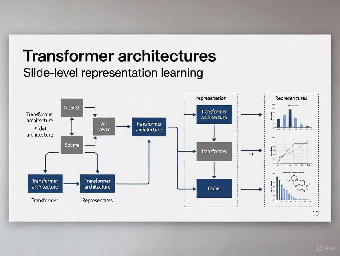
Diagram: Sequential Tokenization and Encoding Pipeline
The HoloHisto framework introduces sequential tokenization for end-to-end gigapixel WSI segmentation, using 4K resolution base patches and Vector Quantized GAN (VQGAN) to tokenize image features into discrete visual tokens [6]. This approach reduces sequence length while preserving critical morphological information, enabling efficient transformer processing.
Multi-Modal and Generative Approaches
Multi-modal transformers combine visual features with textual information from pathology reports. HistoGPT represents a breakthrough in generative pathology, employing a vision module (CTransPath or UNI) to extract image features and a language module (BioGPT) to generate comprehensive pathology reports [7]. The model integrates visual and textual domains through cross-attention mechanisms, enabling it to produce clinically accurate reports from multiple gigapixel WSIs.
Table 2: Transformer Architectures for WSI Analysis
| Architecture | Key Innovation | Application Scope | Performance Highlights |
|---|---|---|---|
| Prov-GigaPath | LongNet with dilated attention for long sequences | Cancer subtyping, mutation prediction | SOTA on 25/26 tasks; 23.5% AUROC improvement on EGFR mutation [3] |
| HoloHisto | 4K sequential tokenization with VQGAN | End-to-end WSI segmentation | Enables direct gigapixel I/O; superior segmentation accuracy [6] |
| HistoGPT | Cross-attention between vision and language modules | Automated report generation | Captures ~67% of dermatopathology keywords; human-level reports [7] |
| TransUNet | Self- and cross-attention in U-Net encoder/decoder | Medical image segmentation | 1.06-4.30% Dice improvement over nnU-Net [8] |
Experimental Protocols and Methodologies
End-to-End WSI Segmentation with HoloHisto
The HoloHisto framework enables complete WSI segmentation through the following protocol:
Sample Preparation and Preprocessing
- Obtain WSIs in standard formats (SVS, TIFF) from whole mouse kidneys
- Apply tissue detection using HSV thresholding (H: 0.5-0.65, S: >0.1, V: 0.5-0.9) to remove non-tissue regions [4]
- Implement random 4K patching (3840×2160 pixels) with foreground balancing
- Apply extensive augmentations including color variation, rotation, flipping, and elastic deformations
Model Architecture and Training
- Tokenizer Pretraining: Train VQGAN on 4K patches using perceptual and adversarial losses [6]
- Backbone Configuration: Implement two-stage ViT with ReLU linear attention to replace Softmax attention [6]
- Optimization: Use AdamW optimizer with learning rate 3e-4, batch size 8, and gradient checkpointing
- Training Schedule: Train for 100,000 steps with linear warmup and cosine decay
Evaluation Metrics
- Dice Similarity Coefficient (DSC) for segmentation accuracy
- Pixel-level precision and recall for class-wise performance
- Inference speed (minutes per WSI) for computational efficiency
Slide-Level Foundation Model Pretraining
The Prov-GigaPath protocol demonstrates large-scale foundation model training:
Data Curation and Preparation
- Collect 171,189 H&E-stained and immunohistochemistry slides from 31 tissue types [3]
- Extract 256×256 patches at 20× magnification (approximately 1.3 billion tiles)
- Implement quality control to exclude slides with severe artifacts or insufficient tissue
- Partition data by patient to prevent data leakage
Two-Stage Pretraining Approach
Diagram: Two-Stage Foundation Model Pretraining
- Tile-Level Pretraining: Train ViT using DINOv2 self-supervised learning on individual tiles [3]
- Slide-Level Pretraining:
- Form slide sequences of up to 70,121 tile embeddings
- Apply masked autoencoder with 30% masking ratio
- Use LongNet with dilated attention to capture long-range dependencies [3]
- Fine-Tuning: Adapt to downstream tasks with task-specific heads and minimal labeled data
Performance Validation
- Evaluate on 26 tasks across Providence and TCGA datasets
- Compare against HIPT, CTransPath, and REMEDIS baselines
- Report AUROC, AUPRC, and accuracy metrics with statistical significance testing
The Scientist's Toolkit: Essential Research Reagents
Table 3: Key Research Reagents and Computational Tools for WSI Analysis
| Resource Name | Type | Function/Purpose | Application Example |
|---|---|---|---|
| Prov-GigaPath Weights | Foundation Model | Pre-trained slide-level representations | Transfer learning for mutation prediction [3] |
| CTransPath | Patch Encoder | Feature extraction from image patches | Vision backbone for HistoGPT [7] |
| LongNet Architecture | Transformer Variant | Efficient long-sequence modeling | Processing >70k tiles per slide [3] |
| VQGAN Tokenizer | Image Tokenizer | Discrete visual token representation | 4K patch compression in HoloHisto [6] |
| cuCIM Library | Data Loader | Efficient WSI reading and patching | Whole slide I/O for holistic analysis [6] |
Discussion and Future Directions
The transition from patch-based analysis to slide-level understanding represents a fundamental shift in computational pathology. Transformer architectures have been instrumental in this evolution, enabling models to capture long-range dependencies and global contextual information that were previously inaccessible [3]. The demonstrated success of models like Prov-GigaPath and HistoGPT across diverse clinical tasks underscores the importance of whole-slide context for accurate pathological assessment.
Future research directions should focus on several key areas: (1) developing more efficient attention mechanisms to further reduce computational complexity, (2) improving multi-modal integration to leverage complementary information from genomics, radiomics, and clinical data, and (3) enhancing model interpretability to build clinical trust and facilitate human-AI collaboration. As these technologies mature, slide-level representation learning with transformers promises to transform pathology from a qualitative, descriptive discipline to a quantitative, predictive science—ultimately accelerating drug development and improving patient care through more precise diagnostic and prognostic tools.
The Transformer architecture, introduced in the seminal paper "Attention Is All You Need," has fundamentally redefined the landscape of sequence modeling and, more recently, visual data analysis [9] [10]. Its core innovation was to dispense with the sequential processing of Recurrent Neural Networks (RNNs) and Long Short-Term Memory (LSTM) networks, which processed data step-by-step, creating bottlenecks and struggling with long-range dependencies [11] [12]. Instead, the Transformer relies entirely on a self-attention mechanism to compute representations of its input and output, drawing global dependencies between all elements in a sequence simultaneously [10]. This architecture is not only more parallelizable—leading to significantly faster training times—but also exceptionally adept at modeling complex, long-distance relationships within data, a property that has proven invaluable for tasks ranging from machine translation to analyzing gigapixel medical images [9] [3].
In the context of visual data, and particularly for slide-level representation learning in digital pathology, these principles enable models to integrate information across vast image spaces. By treating an image as a sequence of patches (or tiles), Vision Transformers (ViTs) can contextualize local features within a global scene, moving beyond the local receptive fields of traditional Convolutional Neural Networks (CNNs) [13] [14]. This application note details the core components of the Transformer, illustrates its application in computational pathology with structured data and protocols, and provides visual and material toolkits for research implementation.
Architectural Deep Dive: Self-Attention and Encoder-Decoder
The Self-Attention Mechanism
The self-attention mechanism is the foundational operation that allows the Transformer to contextualize each element in a sequence by looking at all other elements. It maps a query and a set of key-value pairs to an output, where the queries, keys, values, and output are all vectors [10]. The operation for a single attention head is defined by the Scaled Dot-Product Attention function:
Attention(Q, K, V) = softmax( (QK^T) / √d_k ) V
Here, Q (Query), K (Key), and V (Value) are matrices formed from linearly projecting the input sequence. The dot product of the query and key matrices determines the attention scores, reflecting the relevance of other positions to the current one. Scaling by the square root of the key dimension d_k prevents the softmax function from entering regions of extremely small gradients [9] [10].
Multi-Head Attention enhances this process by employing multiple parallel attention heads. Each head learns different linear projections of the input, allowing the model to jointly attend to information from different representation subspaces. For example, in a pathology image, one head might focus on cellular textures, while another attends to structural tissue organization. The outputs of all heads are concatenated and linearly projected to form the final output [10].
Encoder-Decoder Architecture
The standard Transformer follows an encoder-decoder structure, which is highly effective for sequence transduction tasks [10].
- The Encoder is composed of a stack of identical layers (e.g., N=6 in the original paper). Each layer contains two sub-layers: a multi-head self-attention mechanism and a simple position-wise feed-forward network. A residual connection is employed around each sub-layer, followed by layer normalization. The encoder's role is to map an input sequence to a sequence of continuous representations that capture the contextual relationships within the input [10].
- The Decoder is also a stack of identical layers. In addition to the two sub-layers found in the encoder, the decoder includes a third sub-layer that performs multi-head attention over the output of the encoder stack. This allows the decoder to focus on relevant parts of the input sequence when generating each element of the output. A critical feature of the decoder is its use of masking in the self-attention sub-layer to prevent positions from attending to subsequent positions, thereby preserving the auto-regressive property of the output generation [10].
For tasks that do not require sequence generation, such as image classification, the decoder is often omitted, and the encoder's output is used directly [13] [14].
Positional Encoding
Since the self-attention mechanism is permutation-invariant and contains no inherent notion of sequence order, positional encodings are added to the input embeddings to inject information about the absolute or relative position of each token. The original Transformer uses fixed, sinusoidal functions of different frequencies for this purpose, allowing the model to generalize to sequence lengths longer than those encountered during training [9] [10]. In vision applications, the position of each image patch is encoded similarly to provide spatial context.
Diagram: The Scaled Dot-Product Attention Mechanism
Application in Computational Pathology: Quantitative Performance
The application of transformer architectures has led to state-of-the-art performance in computational pathology, enabling slide-level representation learning from gigapixel Whole-Slide Images (WSIs). The table below summarizes the quantitative performance of key transformer-based models on benchmark tasks.
Table 1: Performance Comparison of Transformer-Based Models in Computational Pathology
| Model / Framework | Task / Dataset | Key Metric | Reported Performance | Comparative Baseline Performance |
|---|---|---|---|---|
| COBRA [15] | Cancer Subtyping (4 CPTAC cohorts) | Average AUC | > +4.4% AUC (vs. previous SOTA) | Weakly-supervised MIL approaches |
| Prov-GigaPath [3] | EGFR Mutation Prediction (TCGA) | AUROC / AUPRC | 23.5% higher AUROC, 66.4% higher AUPRC (vs. REMEDIS) | REMEDIS (pretrained on TCGA) |
| Prov-GigaPath + XGBoost [16] | BRAF-V600 Mutation Prediction (TCGA-SKCM) | AUC | 0.824 (cross-validation) | Previous image-only methods |
| Medical Slice Transformer (MST) [14] | Breast Cancer Detection (Duke MRI) | AUC | 0.94 ± 0.01 | 3D ResNet-50: 0.91 ± 0.02 |
| Medical Slice Transformer (MST) [14] | Meniscus Tear Detection (Knee MRI) | AUC | 0.85 ± 0.04 | 3D ResNet-50: 0.69 ± 0.05 |
| Hybrid ViT + Perceiver IO [13] | Alzheimer's Detection (MRI) | Recall | 1.00 | Conventional CNN models |
These results demonstrate the transformative impact of transformer models, particularly their ability to leverage large-scale pretraining and whole-slide context for superior performance in disease diagnosis and mutation prediction.
Experimental Protocols for Slide-Level Representation Learning
Protocol 1: Whole-Slide Feature Representation with Prov-GigaPath
This protocol outlines the methodology for using the Prov-GigaPath foundation model to generate slide-level embeddings for downstream prediction tasks, as validated in [3] and [16].
- Input Data Preparation: Collect H&E-stained Whole-Slide Images (WSIs). Segment each gigapixel WSI into a sequence of non-overlapping 256x256 pixel image tiles at a specified magnification level (e.g., 20X).
- Tile Encoding: Process each image tile through a pretrained DINOv2 vision transformer model, which serves as the tile encoder. This self-supervised model outputs a feature vector (embedding) for every tile, capturing rich local visual patterns [3].
- Slide-Level Sequence Modeling: Assemble the sequence of tile embeddings for an entire slide. Process this long sequence through the GigaPath slide encoder, a transformer architecture that leverages LongNet's dilated self-attention to efficiently model relationships between all tiles, even for sequences of tens of thousands of elements [3].
- Slide Representation Aggregation: The output of the GigaPath encoder is a contextualized embedding for each tile. To generate a single, slide-level representation, aggregate these embeddings using a softmax attention layer, which learns to weight the importance of different tile embeddings [3].
- Downstream Task Fine-Tuning: For a specific task (e.g., cancer subtyping or mutation prediction), the pretrained Prov-GigaPath model can be fine-tuned end-to-end. Alternatively, the extracted slide-level embeddings can be used as features to train a separate classifier, such as an XGBoost model, which has been shown to be highly effective [16].
Protocol 2: Self-Supervised Slide-Level Pretraining with COBRA
The COBRA framework provides a protocol for unsupervised learning of slide representations that are compatible with multiple foundation models, as detailed in [15].
- Multi-FM Feature Extraction: For each WSI tile, extract multiple feature embeddings using different, pretrained foundation models (FMs). This creates a diverse set of feature representations for the same underlying data.
- Contrastive Pretraining: The core of COBRA is a contrastive self-supervised learning objective in the feature space. The model, built using a Mamba-2 architecture, is trained to identify "positive pairs" (different augmentations or views of the same slide) while pushing apart "negative pairs" (representations from different slides) in the latent space [15].
- Representation Output: The trained COBRA model outputs a compact, task-agnostic slide-level representation that encodes the essential characteristics of the entire WSI. These representations are highly effective for various downstream clinical tasks, even with limited labeled data [15].
Diagram: Whole-Slide Image Analysis Workflow
The Scientist's Toolkit: Research Reagent Solutions
Table 2: Essential Research Reagents and Materials for Slide-Level Transformer Research
| Item Name | Function / Description | Example in Use |
|---|---|---|
| Whole-Slide Images (WSIs) | The primary input data; high-resolution digital scans of pathology slides. | Prov-GigaPath was pretrained on 171,189 H&E-stained WSIs from the Prov-Path dataset [3]. |
| The Cancer Genome Atlas (TCGA) | A publicly available dataset containing WSIs with associated genomic and clinical data. | Used for training and benchmarking models for tasks like BRAF mutation prediction in melanoma [16]. |
| Foundation Model (FM) Feature Extractors | Pretrained models (e.g., DINOv2) used to convert image tiles into feature vector embeddings. | DINOv2 is used as a tile encoder in both Prov-GigaPath and the Medical Slice Transformer to extract high-quality local features [14] [3]. |
| LongNet / Dilated Self-Attention | A transformer architecture designed to handle ultra-long sequences efficiently, overcoming the quadratic complexity of standard self-attention. | Core to the GigaPath slide encoder, enabling it to process sequences of >70,000 tile embeddings per slide [3]. |
| Gradient Boosting Classifier (XGBoost) | A powerful machine learning algorithm often used on top of slide-level embeddings for final prediction tasks. | Used in conjunction with Prov-GigaPath embeddings to achieve SOTA in BRAF mutation prediction [16]. |
| Saliency & Attention Maps | Visualization tools that highlight regions of the input image (or tiles) most influential to the model's decision, aiding in explainability. | The Medical Slice Transformer generates more precise saliency maps than CNNs, highlighting relevant lesions [14]. |
Application Notes
Vision Transformers (ViT) in Medical Image Analysis
Vision Transformers have emerged as a powerful alternative to Convolutional Neural Networks (CNNs) for various medical imaging tasks. Their core strength lies in the self-attention mechanism, which allows them to model global relationships across an entire image, rather than being limited to local receptive fields like CNNs [17]. This capability is particularly valuable in medical imaging, where the diagnostic context can depend on interactions between distant anatomical structures [18].
- Medical Image Classification: ViTs have demonstrated state-of-the-art performance in classifying diseases from medical images. For instance, a Hierarchical Multi-Scale Attention (HMSA) ViT framework achieved 98.7% accuracy in classifying brain tumors into four categories (glioma, meningioma, pituitary adenoma, and healthy tissue) from MRI scans, outperforming traditional CNNs and other transformer variants [18]. Similarly, in osteoporosis detection from X-ray images, ViTs have shown superior outcomes compared to CNN-based approaches [19].
- Medical Image Segmentation: Architectures like TransUNet combine ViT encoders with CNN-based decoders, leveraging the ViT's ability to capture global context for more accurate segmentation of medical structures [17]. The LVM-Med framework demonstrated that ViTs can achieve a 95.75% Dice score on prostate segmentation in MRI, significantly reducing false positives in irregularly shaped lesions [20].
Comparative Performance of Vision Transformers in Medical Imaging
| Model / Architecture | Application | Dataset | Key Metric | Performance | Comparative Performance (CNN Baseline) |
|---|---|---|---|---|---|
| ViT with HMSA [18] | Brain Tumor Classification | Brain Tumor MRI Dataset (7,023 images) | Accuracy | 98.7% | EfficientNet-B0 (96.5%), ResNet-50 (95.8%) |
| LVM-Med ViT [20] | Prostate Segmentation | BMC Dataset | Dice Score | 95.75% | ~15% improvement over CNN-based methods |
| LVM-Med ViT [20] | Breast Ultrasound Segmentation | 647 training samples | Dice Score | 89.69% | ~11% improvement over CNNs in low-data scenario |
Graph Transformers in Drug Discovery
Graph Transformers are revolutionizing computational drug discovery by natively operating on molecular structures represented as graphs, where atoms are nodes and bonds are edges [21]. They enhance classic Graph Neural Networks (GNNs) by incorporating self-attention, which allows them to model complex, long-range interactions within a molecule that are crucial for predicting biological activity [22] [23].
- Molecular Property Prediction: These models accurately predict key pharmacological properties such as solubility, toxicity, and binding affinity, which are critical for prioritizing lead compounds in the early stages of drug discovery [23] [24] [21].
- Drug-Target Interaction (DTI) Prediction: Graph Transformers can model both the drug molecule and the protein target, significantly improving the prediction of how strongly a drug will bind to its intended target. This application accelerates virtual screening, reducing reliance on costly and time-consuming experimental assays [23] [21].
- De Novo Drug Design: Generative Graph Transformer models can design novel molecular structures with desired properties from scratch, exploring a vast chemical space to propose new candidate drugs [21].
A key architectural advancement is the hierarchical mask framework, which unifies various Graph Transformer designs. This framework posits that an effective model must have both a large receptive field and high label consistency. Models like M3Dphormer use this principle with multi-level masking and a Mixture-of-Experts (MoE) approach to adaptively integrate information from different levels of molecular structure, achieving state-of-the-art performance [22].
Hierarchical Models for Multi-Scale Data
Hierarchical models address the limitation of standard transformers in processing information at multiple scales. They are essential for data with a inherent hierarchical structure, such as whole-slide images (WSIs) in pathology, which contain tissue-, cellular-, and sub-cellular level information [18] [25].
- Hierarchical Multi-Scale Vision Transformers: These models process image patches at multiple resolutions (e.g., 8x8, 16x16, 32x32). Lower-resolution patches capture broader contextual information, while higher-resolution patches retain fine-grained details. This multi-scale feature extraction leads to more robust and accurate representations [18].
- Computational Efficiency: Hierarchical models, such as H-MHSA (Hierarchical Multi-Head Self-Attention), dramatically reduce computational load by first computing self-attention within local patches and then merging them to model global dependencies. This approach can reduce training duration by up to 35% compared to standard ViT implementations [18] [25].
- Swin Transformer: A prominent example that uses a shifted window approach to create hierarchical feature maps, making it highly effective for dense prediction tasks like segmentation [18].
Experimental Protocols
Protocol 1: Benchmarking ViT for Medical Image Classification
Objective: To evaluate the performance of a Hierarchical Vision Transformer model for the classification of tumors in medical images.
Materials:
- Dataset: Publicly available Brain Tumor MRI Dataset (7,023 T1-weighted contrast-enhanced images) [18].
- Model Architecture: Hierarchical Multi-Scale Attention (HMSA) Vision Transformer.
- Hardware: High-performance computing node with GPUs (e.g., NVIDIA V100 or A100).
- Software: Python, PyTorch or TensorFlow, and libraries for medical image processing (e.g., MONAI).
Methodology:
- Data Preprocessing:
- Resample all images to a uniform voxel size (e.g., 1mm³ isotropic).
- Apply intensity normalization (e.g., Z-score normalization) across the dataset.
- Perform data augmentation including random rotation (±15°), horizontal flipping, and slight contrast adjustment.
Multi-Scale Patch Embedding:
Model Training:
- Architecture: Feed the sequence of multi-scale patch embeddings into a transformer encoder with Hierarchical Multi-Head Self-Attention (H-MHSA) layers [18] [25].
- Loss Function: Use a combined loss of Cross-Entropy for classification and a regularization term (e.g., Label Smoothing).
- Optimization: Train using the AdamW optimizer with a learning rate of 1e-4, a batch size of 32, and for 100 epochs.
Model Evaluation:
- Evaluate the model on a held-out test set.
- Report standard metrics: Accuracy, Precision, Recall, F1-Score.
- Assess model calibration by calculating the Expected Calibration Error (ECE) [18].
Protocol 2: Assessing Graph Transformer for Molecular Property Prediction
Objective: To predict the binding affinity (pIC50) of small molecules to a target protein using a Graph Transformer model.
Materials:
- Dataset: Publicly available binding affinity data (e.g., KIBA or BindingDB).
- Model Architecture: M3Dphormer or similar Graph Transformer with hierarchical masking [22].
- Software: Python, Deep Graph Library (DGL) or PyTorch Geometric, RDKit.
Methodology:
- Data Preparation:
- Represent each molecule as a graph where nodes are atoms and edges are bonds.
- Initialize node features using atom properties (e.g., atom type, degree, hybridization).
- Initialize edge features using bond properties (e.g., bond type, conjugation).
Model Configuration:
- Implement a Graph Transformer layer that updates node representations using the self-attention mechanism over their neighbors [22] [24].
- Incorporate a hierarchical mask framework to manage interactions at different scales (e.g., local atom environment vs. global molecular structure) [22].
- Use a Mixture-of-Experts (MoE) module with a bi-level routing mechanism to adaptively integrate these multi-level interactions [22].
Training Procedure:
- Readout: Perform a global mean pooling on the final node embeddings to get a graph-level representation.
- Loss Function: Mean Squared Error (MSE) between predicted and experimental pIC50 values.
- Optimization: Use the Adam optimizer with an initial learning rate of 1e-3 and a learning rate scheduler.
Validation:
- Perform 10-fold cross-validation on the training set.
- Report Mean Absolute Error (MAE) and Root Mean Square Error (RMSE) on the test set.
- Compare performance against baseline GNNs (e.g., GCN, GAT) and other state-of-the-art methods.
Protocol 3: Hierarchical ViT for Slide-Level Representation Learning
Objective: To extract a holistic feature representation from a whole-slide image (WSI) by integrating features from multiple magnification levels.
Materials:
- Data: A set of WSIs in SVS or TIFF format, typically from The Cancer Genome Atlas (TCGA).
- Model: A pre-trained Hierarchical ViT (e.g., Swin Transformer) as a feature extractor [18] [25].
Methodology:
- Patch Sampling and Processing:
- For each WSI, sample tissue patches at multiple magnification levels (e.g., 5x, 10x, 20x) using a sliding window approach.
- Exclude patches with low tissue content using an Otsu thresholding algorithm.
Multi-Magnification Feature Extraction:
- Process all sampled patches from a single WSI through the Hierarchical ViT.
- The H-MHSA mechanism within the ViT will compute local self-attention within patches at a given magnification and global attention across different patches and scales [25].
Feature Aggregation:
- Extract the [CLS] token embedding from the final transformer layer for each processed patch.
- Aggregate these patch-level embeddings to form a slide-level representation using a method like mean pooling or an attention-based pooling mechanism that weights the importance of each patch.
Downstream Task Application:
- Use the resulting slide-level feature vector for tasks such as cancer sub-type classification, survival prediction, or patient stratification.
- Train a simple classifier (e.g., SVM or MLP) on these features if labels are available.
The Scientist's Toolkit: Research Reagent Solutions
Essential Materials and Tools for Transformer-Based Research
| Category | Item / Solution | Function / Explanation | Example Use Case |
|---|---|---|---|
| Computational Framework | PyTorch / TensorFlow | Deep learning frameworks for building and training custom transformer models. | Core infrastructure for all model development. |
| Graph Processing Library | Deep Graph Library (DGL) / PyTorch Geometric | Specialized libraries for efficient graph data loading and GNN/Graph Transformer operations. | Implementing M3Dphormer for molecular graphs [22]. |
| Chemical Informatics | RDKit | Open-source toolkit for cheminformatics used for molecule manipulation and feature generation. | Converting SMILES strings to molecular graphs for drug discovery tasks [23]. |
| Medical Image Processing | MONAI | A PyTorch-based framework for deep learning in healthcare imaging, providing domain-specific transforms and models. | Preprocessing and augmenting MRI data for ViT training [18]. |
| Model Architecture | Pre-trained Vision Transformer (ViT) Models | Models pre-trained on large natural image datasets (e.g., ImageNet) that can be fine-tuned for medical tasks. | Transfer learning for medical image classification with limited data [17] [19]. |
| Explainability Tool | Attention Visualization Scripts | Code to visualize the attention maps of transformer models, highlighting regions of the input that were most influential for the prediction. | Interpreting model decisions in medical diagnosis or molecular activity prediction [18] [24]. |
| Optimization | AdamW Optimizer | A variant of the Adam optimizer that correctly handles weight decay, leading to better generalization. | Standard optimizer for training transformer models [18]. |
Whole Slide Images (WSIs) present a unique computational challenge in digital pathology. These gigapixel images, which can exceed 150,000 × 150,000 pixels, are too large for direct processing by standard deep learning models [26]. The prevailing solution divides WSIs into hundreds or thousands of smaller patches, creating a Multiple Instance Learning (MIL) framework where each WSI represents a "bag" containing many patch "instances"[ccitation:1] [27]. This paradigm efficiently leverages readily available slide-level labels while avoiding prohibitive patch-level annotation costs. With the integration of advanced transformer architectures and pathology foundation models, MIL has become the cornerstone of modern computational pathology, enabling tasks ranging from cancer diagnosis and subtyping to predicting molecular markers and clinical outcomes [28] [29] [27].
Theoretical Foundations of MIL in Pathology
In standard MIL formulation for WSIs, a slide (bag) ( Xi ) comprises ( K ) patches (instances), ( Xi = \{x{i,1}, x{i,2}, ..., x{i,K}\} ), with an associated slide-level label ( Yi ). The fundamental MIL assumption states that a bag is positive if it contains at least one positive instance, and negative if all instances are negative: ( Yi = 0 ) if ( \sumk y_{i,k} = 0 ), and 1 otherwise [30]. This weakly supervised setup presents two primary challenges: accurately classifying the entire slide and identifying critical instances within positive slides that drive the classification.
Two principal MIL approaches have emerged: Instance-based (IAMIL) and Representation-based (RAMIL) methods [29]. IAMIL first classifies each instance and then aggregates these predictions for the bag-level label. While offering superior potential for spatial quantification, traditional IAMIL tends to produce highly skewed attention maps, focusing only on the most discriminative regions and missing other relevant areas. In contrast, RAMIL first aggregates instance features into a single bag-level representation, which is then classified. Although often achieving strong bag-level classification, RAMIL provides less precise spatial localization, as attention scores do not always correlate directly with clinical importance and can be misled by confounding features [29].
State-of-the-Art Architectures and Performance
Recent advances have integrated transformer architectures and specialized modules to address the limitations of traditional MIL approaches. The table below summarizes the key characteristics and reported performance of contemporary methods.
Table 1: Performance Comparison of State-of-the-Art MIL Methods
| Method | Core Innovation | Reported AUC/Accuracy | Datasets Validated | Key Advantage |
|---|---|---|---|---|
| SeLa-MIL [30] | Weakly-supervised self-training reformulating MIL as semi-supervised instance classification | Superior to existing methods in instance & bag-level classification (Exact values N/S) | Synthetic, MIL benchmarks, Public WSI datasets | Improves hard positive instance recognition |
| GTP [31] | Fusion of graph convolutional network & vision transformer | Mean Accuracy: 91.2% (internal), 82.3% (external) | CPTAC, NLST, TCGA (Lung) | Effectively captures WSI-level information |
| PATHS [26] | Hierarchical transformer with top-down patch selection | Comparable/Superior to SOTA on TCGA tasks | Five TCGA datasets (Multi-cancer) | Computational efficiency; processes <5% of slide |
| SMMILe [29] | Superpatch-based measurable MIL with custom modules | Macro AUC up to 94.11% (Ovarian), 92.75% (Gastric) | Eight datasets (Six cancer types) | Superior spatial quantification & classification |
| NPKC-MIL [32] | Integrates nuclei-level prior knowledge with patch features | Outperforms comparable deep learning models | Breast WSI | Improved interpretability via prior knowledge |
| Foundation Model + MIL [28] | Uses pathology foundation models (UNI, Prov-Gigapath) as patch encoders | AUROC >0.980 (internal); Robust external performance | KPMP, JP-AID, UT (Kidney) | Robustness to inter-institutional variability |
The adoption of pathology foundation models as patch feature extractors represents a significant leap forward. Models like UNI, Conch, and Prov-Gigapath, pre-trained on millions of pathology patches, provide markedly superior feature representations compared to ImageNet-pretrained models. When integrated with MIL frameworks, they have driven performance on tasks like kidney disease diagnosis to over 0.980 AUROC internally and maintained robustness during external validation [28] [29].
Table 2: Aggregation Method Performance with Different Encoders (Macro AUC, %) [29]
| Method | Breast (Camelyon16) | Lung (TCGA-LU) | Renal-3 (TCGA-RCC) | Ovarian (UBC-OCEAN) |
|---|---|---|---|---|
| ResNet-50 Encoder | ||||
| ABMIL | 89.14 ± 0.89 | 88.15 ± 1.03 | 94.26 ± 0.96 | 88.36 ± 1.74 |
| CLAM | 91.85 ± 0.. |
. . | 90.08 ± 1. . . | 96.15 ± 0. . . | 91.91 ± 1. . . | | SMMILe | 97.32 ± 0.41 | 93.87 ± 0.78 | 97.88 ± 0.52 | 94.11 ± 1.02 | | Conch Foundation Model Encoder | | | | | | ABMIL | 99.12 ± 0.21 | 97.45 ± 0.45 | 99.56 ± 0.18 | 97.12 ± 0.67 | | CLAM | 99.58 ± 0.11 | 98.01 ± 0.39 | 99.72 ± 0.10 | 97.95 ± 0.55 | | SMMILe | 99.75 ± 0.08 | 98.89 ± 0.31 | 99.81 ± 0.07 | 98.43 ± 0.41 |
Detailed Experimental Protocols
Protocol 1: Whole Slide Image Preprocessing and Feature Extraction
This protocol details the critical first steps for preparing WSIs for MIL analysis.
Materials:
- Whole Slide Images (WSIs) in SVS or other standard formats.
- High-performance computing workstation with substantial RAM and GPU memory.
- Software: Python with Slideflow or Libvips for WSI handling.
Procedure:
- Patch Extraction: Use a library like Slideflow to divide each WSI into non-overlapping tiles at a specified magnification (typically 20x, corresponding to 256 pixels at 0.5 microns per pixel). A patch size of 256x256 or 512x512 pixels is common [28].
- Background Filtering: Apply a multi-step filtering process to exclude non-tissue regions:
- Use Otsu's thresholding for initial segmentation.
- Apply a Gaussian blur filter to reduce noise.
- Remove patches with low tissue content (e.g., < 45% tissue) [28].
- Feature Extraction: Process each retained patch through a pre-trained neural network to extract a feature vector.
- Baseline Encoder: Use a standard model like ResNet50 pre-trained on ImageNet.
- Recommended - Foundation Model Encoder: For superior performance, use a pathology foundation model such as UNI, Conch, or Phikon. These models, pre-trained on large-scale histology datasets, yield features that are more robust to stain variations and tissue artifacts [28] [29].
- Feature Storage: Save the extracted feature vectors and their spatial coordinates within the original WSI for downstream analysis.
Diagram 1: WSI preprocessing and feature extraction workflow.
Protocol 2: Implementing a Standard Attention-Based MIL (ABMIL)
This protocol outlines the implementation of a foundational attention-based MIL model for slide-level classification.
Materials:
- Extracted feature vectors from Protocol 1.
- Deep learning framework: PyTorch or TensorFlow.
- Implementation of ABMIL [27].
Procedure:
- Model Architecture:
- Input: The set of feature vectors ( H = \{h1, ..., hK\} ) for a WSI.
- Attention Network: Implement a learnable attention mechanism ( a ) that calculates an attention score ( ak ) for each instance: ( ak = \frac{\exp\{w^T \tanh(V hk^T)\}}{\sum{j=1}^K \exp\{w^T \tanh(V hj^T)\}} ) where ( w ) and ( V ) are learnable parameters.
- Bag Embedding: Compute the slide-level representation ( z ) as a weighted sum of the instance features: ( z = \sum{k=1}^K ak hk ).
- Classifier: Feed the bag embedding ( z ) through a fully connected layer with a softmax activation to generate the slide-level prediction ( \hat{Y} ) [27].
- Training:
- Use a standard cross-entropy loss between the predicted bag label ( \hat{Y} ) and the ground-truth slide label ( Y ).
- Optimize using Adam or SGD with a suitable learning rate scheduler.
- Interpretation: After training, the attention scores ( a_k ) can be visualized as a heatmap overlaid on the original WSI, highlighting regions the model deemed most important for its decision.
Protocol 3: Advanced Training with SMMILe for Spatial Quantification
This protocol describes the setup for SMMILe, a state-of-the-art method designed for high-fidelity spatial quantification alongside accurate slide-level classification [29].
Materials:
- Extracted feature vectors and their spatial coordinates.
- Implementation of SMMILe architecture.
Procedure:
- Superpatch Construction: Organize the WSI into "superpatches" – local neighborhoods of adjacent patches. This preserves spatial context that is lost in set-based methods.
- Model Components:
- Convolutional Layer: Apply a convolutional layer to the instance embeddings to enhance the local receptive field.
- Instance Detector: A multi-stream network that identifies the significance of each instance's embedding for different categories, crucial for multilabel tasks.
- Instance Classifier: A parallel network that assigns each instance's embedding to a category.
- Specialized Modules:
- Consistency Constraint: Ensures that the detection and classification scores for an instance are consistent.
- Parameter-free Instance Dropout: Randomly drops instances during training to prevent over-reliance on a small subset.
- Delocalized Instance Sampling: Actively samples instances from across the entire slide to counteract the high skewness of instance-based attention.
- Markov Random Field (MRF) Refinement: Applies spatial smoothing to the instance-level predictions as a post-processing step to improve coherence.
- Aggregation: The final slide-level prediction is derived by aggregating the product of detection and classification scores from all instances.
Diagram 2: SMMILe architecture with core and specialized modules.
The Scientist's Toolkit: Research Reagent Solutions
Table 3: Essential Computational Tools for MIL in Digital Pathology
| Category | Item/Resource | Function | Example/Note |
|---|---|---|---|
| Data Preprocessing | Slideflow, Libvips | Efficient WSI tiling and patch extraction | Handles gigapixel images and background filtering [28]. |
| Feature Extraction | ImageNet-pretrained CNNs | Baseline patch feature encoder | ResNet50 is a common choice [29]. |
| Pathology Foundation Models | Superior patch feature encoder | UNI, Conch, Prov-Gigapath for robust, domain-specific features [28] [29]. | |
| Core MIL Models | ABMIL, CLAM | Baseline attention-based MIL | Provides interpretable attention maps [27]. |
| TransMIL | Transformer-based feature aggregation | Models inter-patch relationships [31]. | |
| SMMILe, PATHS | State-of-the-art for classification & spatial quantification | Implements advanced modules for accuracy and localization [29] [26]. | |
| Evaluation Datasets | Public Benchmarks | Model validation and benchmarking | Camelyon16, TCGA (NSCLC, RCC), CPTAC [30] [31] [29]. |
| Visualization | GraphCAM, Attention Heatmaps | Model interpretation and insight generation | Identifies regions highly associated with the class label [31] [28]. |
The effective extraction of knowledge from biomedical data, a domain characterized by complex terminology, rapid neologism, and a high density of specialized entities, is paramount for advancements in healthcare and research. A significant challenge is that over 80% of healthcare data resides in unstructured text, such as clinical notes and biomedical literature [33]. Transformer architectures have emerged as a powerful tool for processing this information, but their success is critically dependent on domain-specific pre-training strategies. This is especially true for specialized applications like slide-level representation learning in computational pathology, where models must interpret vast whole-slide images (WSIs) to identify diagnostically relevant morphological patterns. This document outlines application notes and protocols for implementing effective pre-training strategies, framing them within the context of biomedical data analysis and slide-level representation learning research.
Current Pre-training Strategies and Performance
Domain-specific adaptation of transformer models bridges the gap between general language understanding and the specialized semantics of biomedical text and images. The following strategies have proven effective:
- Domain-Adaptive Pre-training (DAPT): This involves continued pre-training of a general-purpose base model (e.g., BERT) on a large, in-domain corpus. This process helps the model internalize the vocabulary, syntax, and knowledge prevalent in biomedical sources [33] [34].
- Task-Adaptive Pre-training (TAPT): A lighter-weight adaptation where pre-training is continued on a smaller, task-specific dataset, further specializing the model for a particular objective [33].
- From-Scratch Pre-training: Some models, like PubMedBERT, are pre-trained entirely on domain-specific corpora, which can lead to superior performance on domain-specific tasks compared to models adapted from a general corpus [33].
- Parameter-Efficient Fine-Tuning (PEFT): Methods like Low-Rank Adaptation (LoRA) freeze the pre-trained model weights and inject trainable rank-decomposition matrices into transformer layers. This approach drastically reduces the number of trainable parameters and computational cost while often matching or exceeding the performance of full fine-tuning [33].
Quantitative evidence demonstrates the superiority of domain-specific strategies. The DRAGON benchmark, a comprehensive clinical NLP benchmark, found that domain-specific pre-training achieved a test score of 0.770, outperforming mixed-domain (0.756) and general-domain pre-training (0.734) [34]. Furthermore, the OpenMed NER project showcased that combining DAPT with LoRA fine-tuning established new state-of-the-art micro-F1 scores on 10 out of 12 established biomedical Named Entity Recognition (NER) benchmarks, with substantial gains on specialized gene and clinical cell line corpora [33] [35]. This performance was achieved with high efficiency, completing training in under 12 hours on a single GPU [33].
For slide-level representation learning, unsupervised methods like SAMPLER provide a rapid alternative to supervised attention models. SAMPLER generates slide-level representations by encoding the cumulative distribution functions of multiscale tile-level features, achieving AUCs comparable to state-of-the-art models (e.g., 0.911 for BRCA subtyping) while training over 100 times faster [36].
Table 1: Benchmark Performance of Domain-Adapted Models
| Model / Benchmark | Domain Adaptation Strategy | Key Performance Metric | Result |
|---|---|---|---|
| OpenMed NER [33] | DAPT + LoRA Fine-tuning | Micro-F1 on BC5CDR-Disease | New SOTA (+2.70 percentage points) |
| DRAGON Benchmark [34] | Domain-specific Pretraining | Overall Test Score | 0.770 |
| SAMPLER (BRCA) [36] | Unsupervised Statistical Learning | AUC on Tumor Subtyping | 0.911 ± 0.029 |
| SAMPLER (NSCLC) [36] | Unsupervised Statistical Learning | AUC on Tumor Subtyping | 0.940 ± 0.018 |
Table 2: Computational Efficiency Comparison
| Model / Approach | Training Resource | Training Time | Number of Trainable Parameters |
|---|---|---|---|
| OpenMed NER [33] | Single GPU | < 12 hours | < 1.5% of total (via LoRA) |
| SAMPLER [36] | Not Specified | >100x faster than attention models | Not Applicable (Non-neural) |
| Standard Full Fine-tuning | Multiple GPUs | Often days | 100% of model parameters |
Application Notes and Experimental Protocols
Protocol 1: Domain-Specific Pre-training for Biomedical NER
This protocol outlines the methodology used by OpenMed NER for achieving state-of-the-art results on biomedical named entity recognition tasks [33].
1. Rationale: To create a highly efficient and effective NER model for the biomedical domain by leveraging lightweight domain-adaptive pre-training (DAPT) combined with parameter-efficient fine-tuning (LoRA).
2. Pre-training Corpus Curation:
- Source: Compile a corpus from ethically sourced, publicly available repositories. The OpenMed NER corpus consisted of 350,000 passages from PubMed, arXiv, and de-identified clinical notes from MIMIC-III [33].
- Focus: Ensure the corpus covers a broad spectrum of biomedical literature and clinical text to foster model generalization.
3. Domain-Adaptive Pre-training (DAPT):
- Base Models: Select strong transformer backbones such as DeBERTa-v3, PubMedBERT, or BioELECTRA.
- Process: Perform continued pre-training (DAPT) on the curated corpus using the standard Masked Language Modeling (MLM) objective. This step adapts the model's parameters to the biomedical domain.
4. Task-Specific Fine-tuning with LoRA:
- Low-Rank Adaptation (LoRA): For each downstream NER task, employ LoRA instead of full fine-tuning. This involves freezing the pre-trained weights and adding low-rank matrices to the attention layers.
- Efficiency: This strategy updates less than 1.5% of the total model parameters, making it computationally efficient and reducing the risk of catastrophic forgetting [33].
- Benchmarking: Evaluate the final model on established NER benchmarks (e.g., BC5CDR, NCBI-Disease, BC2GM) and report micro-F1 scores.
Protocol 2: Unsupervised Slide-Level Representation Learning for Digital Pathology
This protocol is based on the SAMPLER method, which provides a fast, unsupervised alternative for generating representations from whole-slide images (WSIs) [36].
1. Rationale: To generate informative slide-level representations from WSIs without the need for supervised training, enabling rapid tumor subtyping and analysis.
2. Whole-Slide Image Processing:
- Tiling: Divide each WSI into a collection of smaller image patches (tiles) at multiple magnification levels.
- Feature Encoding: Use a pre-trained neural network to encode each tile into a feature vector, resulting in a set of feature vectors for the entire slide.
3. Representation Generation via Cumulative Distribution:
- Method: For each slide, model the distribution of the tile-level features by calculating their cumulative distribution functions (CDFs) at multiple scales.
- Aggregation: Encode these CDFs to form a fixed-dimensional, slide-level representation vector that captures the statistical distribution of morphological features across the slide.
4. Downstream Analysis:
- Classification: Use the generated slide-level representations to train a separate classifier (e.g., for tumor subtype distinction).
- Validation: Evaluate the classifier performance using AUC on internal and external test sets. Histopathological review should confirm that high-attention tiles identified by the model contain subtype-specific morphological features [36].
Visualizing Workflows and Architectures
Pre-training and Adaptation Workflow
The following diagram illustrates the end-to-end workflow for creating a domain-adapted model, as exemplified by OpenMed NER [33].
SAMPLER Architecture for Slide-Level Representation
This diagram outlines the unsupervised workflow of the SAMPLER method for generating representations from whole-slide images in digital pathology [36].
The Scientist's Toolkit: Essential Research Reagents and Materials
Table 3: Key Resources for Biomedical Model Development
| Item / Resource | Function / Application | Example Sources / Instances |
|---|---|---|
| Pre-trained Base Models | Foundation for domain-adaptive pre-training. Provides initial language representation. | DeBERTa-v3, PubMedBERT, BioELECTRA [33]. |
| Biomedical Text Corpora | Source data for Domain-Adaptive Pre-training (DAPT). Provides domain-specific knowledge. | PubMed, PubMed Central (PMC), MIMIC-III (clinical notes) [33]. |
| Biomedical NER Benchmarks | Standardized datasets for evaluating named entity recognition performance. | BC5CDR (Chemicals/Diseases), NCBI-Disease, BC2GM (Genes), JNLPBA [33]. |
| Parameter-Efficient Fine-Tuning (PEFT) Libraries | Software tools to implement methods like LoRA, reducing computational demands. | Hugging Face PEFT, LoRA implementations [33]. |
| Whole Slide Image (WSI) Datasets | Data for developing and validating digital pathology models. | The Cancer Genome Atlas (TCGA) [36]. |
| Computational Resources | Hardware necessary for training and fine-tuning large models. | Single or Multi-GPU setups (e.g., NVIDIA A100, V100) [33] [36]. |
Methodologies and Real-World Applications in Computational Pathology
The computational analysis of whole-slide images (WSIs) in digital pathology presents a unique challenge due to the gigapixel scale of the data, often reaching sizes of 150,000×150,000 pixels [26]. Traditional deep learning approaches typically process WSIs as large collections of patches using multiple instance learning (MIL), treating the slide as an unordered set and often losing crucial spatial context and hierarchical tissue relationships [26]. Inspired by the diagnostic workflow of human pathologists—who examine slides in a top-down manner, identifying regions of interest at low magnification before investigating these areas at higher resolutions—researchers have developed hierarchical transformer architectures that fundamentally transform how we analyze histopathological images [26]. These approaches represent a significant advancement in slide-level representation learning within transformer-based research, enabling more efficient, interpretable, and clinically relevant computational pathology.
Hierarchical Transformer Architectures for WSI Analysis
PATHS: A Top-Down Hierarchical Selection Approach
The Pathology Transformer with Hierarchical Selection (PATHS) implements a top-down processing methodology that directly mirrors a pathologist's examination strategy [26]. Unlike bottom-up hierarchical methods that process all patches at the highest magnification first, PATHS recursively filters patches at each magnification level to identify a small subset most relevant to diagnosis [26]. This approach processes patches at n magnification levels (m₁ < m₂ < ... < mₙ) forming a geometric sequence to ensure patch alignment between levels, with each processor 𝒫ᵢ dedicated to magnification mᵢ [26].
Key Innovation: PATHS dynamically selects only the most informative regions at each magnification level, substantially reducing computational burden while maintaining diagnostic accuracy by focusing on tissue regions with the highest predictive value [26].
HIPT: Bottom-Up Hierarchical Representation Learning
The Hierarchical Image Pyramid Transformer (HIPT) employs a bottom-up approach, constructing slide-level representations through successive stages of feature aggregation [26]. This method builds a hierarchical feature pyramid where:
- 256×256 patches at the highest magnification are processed with a pre-trained vision transformer
- Features are aggregated into 4096×4096 regions using another transformer
- Regional representations are finally aggregated into a slide-level embedding [26]
Key Limitation: While more expressive than standard MIL methods, HIPT requires processing all patches at full magnification, necessitating self-supervised rather than task-specific training due to computational constraints [26].
TITAN: Multimodal Whole-Slide Foundation Model
The Transformer-based pathology Image and Text Alignment Network (TITAN) represents a breakthrough in whole-slide foundation models, pretrained on 335,645 WSIs using visual self-supervised learning and vision-language alignment with corresponding pathology reports [37]. TITAN introduces a large-scale pretraining paradigm that leverages millions of high-resolution regions-of-interest (ROIs) for scalable WSI encoding, using a Vision Transformer (ViT) to create general-purpose slide representations deployable across diverse clinical scenarios [37].
Table 1: Comparative Analysis of Hierarchical Transformer Architectures
| Architecture | Processing Paradigm | Core Innovation | Training Data Scale | Computational Efficiency |
|---|---|---|---|---|
| PATHS [26] | Top-down hierarchical selection | Recursive attention-based patch filtering | Standard WSI datasets | High (processes only 1-10% of slide) |
| HIPT [26] | Bottom-up hierarchical aggregation | Multi-stage feature pyramid construction | Requires self-supervised pretraining | Medium (processes all patches) |
| TITAN [37] | Multimodal foundation model | Vision-language pretraining with synthetic captions | 335,645 WSIs + 423K synthetic captions | Variable (dependent on patch selection) |
| DT-MIL [38] | Deformable transformer MIL | Instance feature updating with position encoding | Standard WSI datasets | Medium |
Experimental Protocols and Methodologies
PATHS Implementation Protocol
Materials and Software Requirements:
- WSIs in pyramidal format with multiple magnification levels
- Pre-trained image encoder (e.g., EfficientNet, ViT)
- Transformer architecture with hierarchical processors
- GPU cluster with sufficient VRAM for processing
Step-by-Step Protocol:
- Slide Preprocessing:
- Segment WSIs into non-overlapping patches at each magnification level
- Extract features using pre-trained encoder ℐ such that ℐ(Xᵤ,ᵥᵐ) ∈ ℝᵈ for each patch [26]
Hierarchical Processing:
- Initialize with low-magnification view (e.g., 5×) to capture tissue architecture
- Compute attention scores for all patches at current magnification
- Select top-k patches based on attention weights for processing at next higher magnification [26]
Model Configuration:
- Implement n processors 𝒫₁, 𝒫₂, ..., 𝒫ₙ for n magnification levels
- Configure geometric sequence of magnifications: mᵢ₊₁ = M×mᵢ for alignment [26]
- Set patch selection hyperparameters (number of patches per level)
Training Procedure:
- Utilize weak slide-level labels
- Implement cross-entropy loss for classification tasks
- Employ gradient descent with learning rate scheduling
Interpretation and Visualization:
- Generate attention maps across magnification levels
- Visualize patch selection trajectory through WSI hierarchy [26]
TITAN Multimodal Pretraining Protocol
Materials Requirements:
- Large-scale WSI dataset (300K+ slides recommended)
- Corresponding pathology reports or synthetic caption generation pipeline
- High-performance computing infrastructure
Three-Stage Pretraining Protocol:
Stage 1: Vision-Only Unimodal Pretraining
- Input: 8,192×8,192 pixel ROIs at 20× magnification [37]
- Method: Apply iBOT framework (masked image modeling and knowledge distillation) on 2D feature grid [37]
- Feature Extraction: Divide WSI into non-overlapping 512×512 pixel patches, extract 768-dimensional features using CONCHv1.5 [37]
- View Creation: Randomly crop 16×16 feature regions, sample global (14×14) and local (6×6) crops for self-supervised learning [37]
Stage 2: ROI-Level Cross-Modal Alignment
- Input: 423,122 pairs of ROIs and synthetic captions generated via PathChat [37]
- Method: Contrastive learning to align visual features with fine-grained morphological descriptions [37]
Stage 3: WSI-Level Cross-Modal Alignment
- Input: 182,862 pairs of WSIs and clinical reports [37]
- Method: Vision-language pretraining to align slide representations with diagnostic text [37]
Table 2: Performance Comparison of Hierarchical Transformers on Slide-Level Tasks
| Model | Cancer Subtyping Accuracy | Survival Prediction C-index | Biomarker Prediction AUC | Slide Retrieval Precision | Zero-Shot Classification Accuracy |
|---|---|---|---|---|---|
| PATHS [26] | 92.3% | 0.741 | 0.891 | N/A | N/A |
| TITAN [37] | 94.8% | 0.763 | 0.912 | 0.945 | 88.7% |
| HIPT [26] | 89.7% | 0.698 | 0.865 | N/A | N/A |
| ABMIL [26] | 86.2% | 0.642 | 0.831 | N/A | N/A |
| DT-MIL [38] | 90.5% | 0.705 | 0.872 | N/A | N/A |
The Scientist's Toolkit: Essential Research Reagents
Table 3: Key Research Reagent Solutions for Hierarchical Transformer Implementation
| Reagent/Resource | Function | Implementation Example |
|---|---|---|
| Pre-trained Patch Encoder | Extracts meaningful features from individual patches | CONCHv1.5 [37], EfficientNet [38], ViT-L-16 [39] |
| Positional Encoding Scheme | Preserves spatial relationships between patches | 2D sinusoidal encoding, learnable positional embeddings [38] |
| Multi-Resolution WSI Dataset | Enables hierarchical processing across magnifications | The Cancer Genome Atlas (TCGA), in-house institutional datasets [26] |
| Synthetic Caption Generation | Provides fine-grained morphological descriptions for vision-language training | PathChat multimodal generative AI copilot [37] |
| Attention Visualization Tools | Enables model interpretation and validation | Gradient-based attribution analysis, attention map overlays [39] |
| Adversarial Multimodal Learning | Enhances complementarity between different magnification modalities | MamlFormer manifold adversarial learning framework [40] |
Technical Implementation and Visualizations
Diagram 1: Hierarchical Processing Workflow. Top-down approach for multi-magnification WSI analysis.
Diagram 2: Hierarchical Transformer Architecture. Integration of visual and linguistic modalities.
Hierarchical transformer architectures represent a paradigm shift in computational pathology, successfully translating the clinical workflow of pathologists into scalable deep learning frameworks. The emergence of multimodal foundation models like TITAN, combined with efficient hierarchical processing approaches such as PATHS, demonstrates the transformative potential of these methods for slide-level representation learning [37] [26]. Future research directions include developing more computationally efficient attention mechanisms, expanding cross-modal capabilities to incorporate genomic and clinical data, and improving few-shot learning performance for rare diseases where training data is severely limited [41]. As these models continue to evolve, they promise to enhance the precision, efficiency, and accessibility of pathological diagnosis while providing unprecedented insights into tissue microenvironment organization and its relationship to disease progression and treatment response.
The integration of spatial context with molecular profiles represents a frontier in computational biology, particularly for understanding tissue microenvironment and cellular heterogeneity. Spatial resolved transcriptomics (SRT) technologies have revolutionized this field by enabling high-throughput sequencing of mRNA while preserving crucial spatial information within tissues [42]. However, significant challenges persist in effectively integrating gene expression with spatial information to elucidate the heterogeneity of biological tissues. Traditional analytical methods often struggle to capture the complex, non-linear relationships between gene expressions and their spatial contexts, as they frequently rely on predefined graph structures that may inadequately represent actual biological interactions [42].
Graph transformer architectures have emerged as powerful solutions to these limitations, offering enhanced capability to model both local and global spatial dependencies within tissue microenvironments. Unlike conventional graph neural networks that rely on static, localized convolutional aggregation, transformer-based approaches employ global self-attention mechanisms that can iteratively evolve topological structural information and transcriptional signal representation [42]. This technological advancement enables researchers to more accurately identify spatial domains, denoise gene expression data, and uncover spatially variable genes with significant prognostic potential, particularly in cancer tissues [42].
Within the broader context of slide-level representation learning, graph transformers provide a unified framework for analyzing multi-scale biological data. Their ability to process long-range dependencies and integrate hierarchical information makes them particularly suited for digital pathology and spatial omics applications, where capturing both cellular-level details and tissue-level organization is essential for accurate representation learning.
Key Advancements in Graph Transformer Architectures
Spatially Informed Graph Transformers (SpaGT)
The SpaGT framework represents a significant advancement in SRT analysis by leveraging both node and edge channels to model spatially aware graph representations. This approach overcomes limitations of traditional transformers, which are typically restricted to feature representation training, by simultaneously evolving both transcriptional signal representations and relationship similarities between spots using a deep learning approach [42]. The core innovation of SpaGT lies in its structure-reinforced self-attention module, which effectively learns and updates the graph representation throughout the model. Additionally, SpaGT incorporates a clustering-augmented contrastive module to ensure that learned graph representations are suitable for spatial clustering tasks [42].
In comprehensive evaluations across 17 SRT datasets from multiple platforms including 10x Visium, Slide-seqV2, and Stereo-seq, SpaGT demonstrated superior performance in identifying spatial domains compared to seven state-of-the-art methods. For 12 Dorsolateral Prefrontal Cortex (DLPFC) datasets from 10x Visium, SpaGT achieved the highest median Adjusted Rand Index (ARI) of 0.572, indicating closer alignment with manual annotations than other methods [42].
Table 1: Performance Comparison of Spatial Domain Identification Methods on DLPFC Data
| Method | Median ARI | Key Features |
|---|---|---|
| SpaGT | 0.572 | Structure-reinforced self-attention, node and edge channels |
| STAGATE | 0.510 | Graph attention auto-encoder |
| SEDR | 0.524 | Deep learning with spatial information |
| GraphST | 0.485 | Graph self-supervised learning |
| SpaGCN | 0.465 | Graph convolutional networks |
| SiGra | 0.566 | Simplified graph architecture |
| DeepST | 0.463 | Deep learning for spatial transcriptomics |
| MUSE | 0.467 | Multi-modal integration |
Hierarchical Graph Transformers (HEIST)
HEIST represents another groundbreaking approach as a hierarchical graph transformer foundation model for spatial transcriptomics and proteomics. This model tissues as hierarchical graphs where the higher level is a spatial cell graph, and each cell is represented by its lower-level gene co-expression network graph [43]. Rather than using a fixed gene vocabulary, HEIST computes gene embeddings from its co-expression network and cellular context, enabling generalization to novel datatypes including spatial proteomics without retraining [43].
Pretrained on 22.3 million cells from 124 tissues across 15 organs using spatially-aware contrastive and masked autoencoding objectives, HEIST demonstrates remarkable capability in discovering spatially informed subpopulations missed by prior models. Downstream evaluations demonstrate state-of-the-art performance in clinical outcome prediction, cell type annotation, and gene imputation across multiple technologies, while being 8× faster than scGPT-spatial and 48× faster than scFoundation [43].
SGTB: Integrating Multiple Architectures
The SGTB model offers a innovative approach by combining graph convolutional networks (GCN), Transformer, and BERT language models to optimize the representation of spatial transcriptomics data [44]. This multi-scale feature fusion strategy enables SGTB to exhibit significant superiority in tasks such as cell type classification, gene regulatory network construction, and spatial heterogeneity analysis. The model employs multi-layer GCNs to iteratively aggregate local neighborhood information, capturing gene co-expression and physical adjacency patterns, while the Transformer's self-attention mechanism captures global spatial relationships, addressing the constraints of local receptive fields in conventional GNNs [44].
Experimental results demonstrate that SGTB outperforms existing methods across various biological datasets and tasks. In spatial clustering and heterogeneity analysis, SGTB achieves an Adjusted Rand Index (ARI) greater than 0.6 on the human dorsolateral prefrontal cortex (DLPFC) dataset, significantly higher than traditional methods [44].
Experimental Protocols and Methodologies
SpaGT Implementation Protocol
Data Preprocessing:
- Input Data Preparation: Convert spatial multimodal data into graph-structured data denoted as G(X₁, A), where X₁ ∈ R^(M×N) represents M genes across N spots/cells, and A ∈ R^(N×N) represents the adjacency matrix derived from spatial coordinates [42].
- Embedding Generation: Generate expression embeddings Hⁱ and edge embeddings Eⁱ that reflect pairwise structural information between spots. These embeddings serve as input data for transformers constructing the expression and edge channels [42].
Model Architecture and Training:
- Structure-Reinforced Self-Attention Module: Implement global self-attention as an aggregation mechanism to update expression embeddings Hⁱ while incorporating edge channels to capture spatially local information.
- Spatially Aware Attention Weights: Utilize attention weights Sⁱ generated by the self-attention module to iteratively update topological structure information Eⁱ of the graph representation across layers.
- Clustering-Augmented Contrastive Module: Apply contrastive learning to ensure learned graph representations are suitable for spatial clustering tasks.
- Training Parameters: Train the model for multiple epochs with early stopping based on validation loss, using Adam optimizer with learning rate of 1e-4 [42].
Downstream Analysis:
- Spatial Domain Identification: Construct a nearest-neighbor network from optimal transcriptional signal representation Hᴸ and integrate with Leiden algorithm to identify spatial domains.
- Expression Denoising: Employ topological structural information Eᴸ to denoise expression profile X̃₁ = X₁Eᴸ, enhancing spatial expression patterns and domain specificity.
- Differential Gene Expression: Utilize denoised data to identify domain-specific differential genes of biological interest [42].
HEIST Pretraining Protocol
Graph Construction:
- Data Preprocessing: Remove outliers, normalize gene expression, and retain highly variable genes. Apply MAGIC to denoise gene expression values and reduce dropout noise [43].
- Gene Co-expression Networks: Subset cells based on cell-types using provided annotations or Leiden clustering. Compute pairwise mutual information between denoised genes within each type, and connect gene pairs above threshold τ.
- Spatial Cell-Cell Graph: Compute Voronoi polygons from cell coordinates and connect cells in adjacent polygons. Connect each cell with gene co-expression network of corresponding cell-type.
Model Architecture:
- Hierarchical Processing: Perform intra-level message passing within each graph, followed by cross-level message passing to integrate multi-modal information.
- Embedding Computation: Compute cell embeddings Zc ∈ R^(|C|×d) and gene embeddings Zg ∈ R^(|C||V|×d) through the HEIST architecture.
- Pretraining Objectives: Employ combination of contrastive and auto-encoding objectives on gene expression and cell locations [43].
Performance Evaluation Protocol
Benchmark Datasets:
- DLPFC Dataset: Utilize 12 human dorsolateral prefrontal cortex sections from 10x Visium with manual annotations of six cortical layers (L1-L6) and white matter (WM) as ground truth [42].
- Single-cell Resolution Data: Include SRT data from osmFISH and Seq-Scope platforms to evaluate performance on high-resolution data.
- Cancer Data: Apply models to triple-negative breast cancer SRT data and mouse hippocampus data from Slide-seqV2 and mouse embryo data from Stereo-seq [42].
Evaluation Metrics:
- Adjusted Rand Index (ARI): Measure similarity between computational results and manual annotations for spatial domain identification.
- Statistical Testing: Perform Wilcoxon signed-rank test to confirm statistical significance of performance differences (P < 1e-10) [42].
- Ablation Studies: Systematically assess contribution of various model components by removing edge channels and enhancement matrices.
Table 2: Ablation Study Results for SpaGT Components
| Model Variant | Average ARI | Components Removed |
|---|---|---|
| Complete SpaGT | 0.607 | None |
| SpaGT^(-edge) | 0.486 | Edge channels |
| SpaGT^(-enhancement) | 0.517 | Enhancement matrix X₁ |
| SpaGT^(-edge&enhancement) | 0.467 | Edge channels and enhancement matrix |
Visualization of Architectures and Workflows
SpaGT Workflow Diagram
HEIST Hierarchical Architecture
Whole-Slide Processing Pipeline
Research Reagent Solutions and Computational Tools
Table 3: Essential Research Reagents and Computational Tools for Graph Transformer Applications
| Item | Function/Application | Specifications/Platform |
|---|---|---|
| 10x Visium Platform | Spatial transcriptomics data generation | Simultaneous mapping of gene expression and spatial location |
| Slide-seqV2 | Single-cell resolution spatial transcriptomics | Higher resolution spatial mapping |
| Stereo-seq | Spatial transcriptomics with large field of view | Mouse embryo and tissue mapping |
| osmFISH | Multiplexed FISH-based spatial transcriptomics | Single-cell resolution with high sensitivity |
| Seq-Scope | High-resolution spatial transcriptomics | Subcellular resolution mapping |
| Prov-GigaPath | Whole-slide pathology foundation model | Pretrained on 1.3B image tiles from 171K slides [3] |
| DINOv2 | Self-supervised learning for tile encoding | Vision transformer pretraining [3] |
| LongNet | Ultra long-sequence modeling | Adapted for gigapixel slide processing [3] |
| Masked Autoencoder | Self-supervised pretraining objective | Learns robust representations from unlabeled data |
| Graph Contrastive Learning | Representation learning enhancement | Maximizes similarity across augmented views |
Applications in Biomedical Research and Drug Development
Cancer Research and Biomarker Discovery
Graph transformers have demonstrated remarkable utility in cancer research, particularly in deciphering tumor heterogeneity and identifying prognostic biomarkers. When applied to triple-negative breast cancer SRT data, SpaGT excels in providing deeper biological insights into genes closely associated with cancer, with robustness further validated through survival analysis using independent clinical data [42]. Similarly, Prov-GigaPath has shown exceptional performance in mutation prediction from histopathological images, attaining significant improvements in pathomics tasks including a 23.5% improvement in AUROC and 66.4% improvement in AUPRC for EGFR mutation prediction compared to the second-best model [3].
The application of these models extends to predicting BRAF mutation status in melanoma directly from histopathological slides. Integrating Prov-GigaPath with XGBoost classifiers achieved an AUC of 0.824 during cross-validation and 0.772 on an independent test set, representing a state-of-the-art for image-only BRAF mutation prediction [16]. This approach employs a weakly supervised, data-efficient pipeline that reduces the need for extensive annotations and costly molecular assays, highlighting the potential for integrating AI-driven decision-support tools into diagnostic workflows.
Neuroscience and Brain Mapping
In neuroscience applications, graph transformers have proven invaluable for mapping complex brain structures. SpaGT's performance on human dorsolateral prefrontal cortex (DLPFC) data demonstrates superior accuracy in identifying the six cortical layers and white matter, with predictions exhibiting high congruence with manually annotated domains and achieving an ARI of 0.805 for specific slices [42]. These capabilities enable more precise characterization of neuronal organization and layering, facilitating deeper understanding of brain function and organization.
When applied to mouse hippocampus data from Slide-seqV2 and mouse embryo data from Stereo-seq, SpaGT reveals finer-grained anatomical regions that offer more detailed interpretations of tissue function [42]. This enhanced resolution in spatial domain identification provides neuroscientists with powerful tools for investigating cellular organization in neurodevelopment and disease states.
Drug Discovery and Development
In pharmaceutical applications, graph transformer architectures like DrugDAGT demonstrate significant potential for predicting drug-drug interactions (DDIs) by incorporating dual-attention mechanisms at both bond and atomic levels [45]. This framework enables integration of short and long-range dependencies within drug molecules to pinpoint key local structures essential for DDI discovery, outperforming state-of-the-art baseline models in both warm-start and cold-start scenarios.
The implementation of graph contrastive learning in these models further enhances discrimination of molecular structures by maximizing similarity of representations across different views, providing valuable insights for prescribing medications and guiding drug development while minimizing adverse drug events [45]. As these models continue to evolve, they offer promising avenues for accelerating drug discovery pipelines and improving medication safety profiles.
In computational pathology, the analysis of gigapixel whole-slide images (WSIs) presents unique computational challenges. The dominant two-stage paradigm, which decouples feature extraction from aggregation, faces performance limitations due to disjointed optimization. This application note explores the resurgence of end-to-end learning as a solution, detailing its protocols, quantitative advantages, and implementation for slide-level representation learning. We demonstrate that joint optimization of feature extraction and aggregation, facilitated by novel architectures like ABMILX and transformer-based models such as GigaPath, significantly surpasses the performance of state-of-the-art foundation models while maintaining computational efficiency.
Computational pathology involves the analysis of gigapixel WSIs for tasks such as cancer subtyping, grading, and prognosis [46]. The standard two-stage paradigm first uses a pre-trained, frozen encoder for offline feature extraction from thousands of tissue patches. These features are then aggregated using a Multiple Instance Learning model for slide-level prediction [46] [3]. While efficient, this approach suffers from a critical flaw: the encoder lacks adaptation to the specific downstream task, and the optimization of the feature extractor and aggregator is disjointed [46]. This limits performance, as even large-scale pathology foundation models (FMs) pretrained on massive datasets can exhibit unsatisfactory task-specific performance [46] [3].
End-to-end learning offers a fundamental solution. It is defined as training a single model that maps raw input data directly to the final output, automatically learning all intermediate representations [47]. In the context of computational pathology, this means jointly optimizing the image encoder and the MIL aggregator using only slide-level labels. This allows the encoder to learn features specifically discriminative for the clinical task at hand, creating a cohesive and optimally adapted system [46].
Comparative Analysis: Two-Stage vs. End-to-End Paradigms
The table below summarizes the core differences between the two paradigms.
Table 1: Comparison of Two-Stage and End-to-End Learning Paradigms in Computational Pathology
| Aspect | Two-Stage Paradigm | End-to-End Paradigm |
|---|---|---|
| Core Philosophy | Disjoint, sequential optimization of feature extraction and aggregation [46]. | Joint, unified optimization of the entire model from input to output [46] [47]. |
| Encoder Optimization | Frozen during MIL training; no adaptation to downstream task [46]. | Fine-tuned during MIL training; features become task-specific [46]. |
| Data Efficiency | Relies on features from encoders pre-trained on large general or pathology datasets [3]. | Requires sufficient downstream data for effective joint training; can be data-hungry [47]. |
| Computational Load | Lower during training, as encoder is frozen [46]. | Higher during training, but can be managed via efficient sampling [46]. |
| Representation Learning | Features are generic; performance depends heavily on pre-training quality [46]. | Features are highly specialized for the target task, improving discriminability [46]. |
| Typical Performance | Performance plateau with state-of-the-art FMs [3]. | Can surpass two-stage FM performance by addressing optimization misalignment [46]. |
Quantitative evidence underscores the advantages of end-to-end learning. As shown in Table 2, an E2E-trained ResNet with the novel ABMILX aggregator can achieve performance gains of over 20% in accuracy on challenging benchmarks like PANDA compared to two-stage methods [46]. Furthermore, whole-slide foundation models like Prov-GigaPath, which incorporate slide-level context, achieve state-of-the-art performance on a wide range of tasks, winning 25 out of 26 benchmarks in one study [3].
Table 2: Quantitative Performance Comparison on Pathology Tasks
| Model / Paradigm | Key Feature | Dataset | Performance Metric | Result |
|---|---|---|---|---|
| E2E ResNet-50 + ABMILX [46] | Joint optimization with multi-scale sampling | PANDA | Accuracy | ~20% improvement over two-stage |
| Prov-GigaPath (Two-Stage FM) [3] | Whole-slide pretraining with LongNet | TCGA (EGFR) | AUROC / AUPRC | State-of-the-art (Second-best) |
| E2E ResNet-50 + ABMILX [46] | Joint optimization | TCGA-BRCA | Accuracy | Surpasses SOTA FMs |
| Prov-GigaPath (Two-Stage FM) [3] | Large-scale real-world data | 26 various tasks | # of SOTA wins | 25 out of 26 tasks |
Experimental Protocols for End-to-End Learning
The following protocols detail the methodology for implementing and benchmarking slide-level end-to-end learning.
Protocol A: Multi-Scale Random Patch Sampling for E2E Learning
This protocol outlines an efficient sampling strategy to make E2E learning computationally feasible.
- Input: A gigapixel Whole-Slide Image (WSI).
- Partitioning: The WSI is divided into a set of non-overlapping patches at multiple magnification levels (e.g., 5x, 10x, 20x).
- Random Sampling: For each WSI in a training batch, a target number of patches,
s, is defined based on GPU memory constraints. A simple random sampler selectsspatches from the entire multi-scale pool of patches,𝑿, to create the input subset𝑳[46]. Formally:𝑳 = 𝒱(s, 𝑿). - Output: A fixed-size, multi-scale bag of patches
𝑳 = {𝒍₁, 𝒍₂, ..., 𝒍ₛ}that serves as input to the encoder. This strategy avoids complex and costly sampling mechanisms, maintaining a low computational budget (e.g., <10 GPU hours on an RTX3090 for TCGA-BRCA) while incorporating important multi-scale contextual information [46].
Protocol B: Joint Optimization with the ABMILX Aggregator
This protocol describes the core E2E training loop, which mitigates the optimization challenges of sparse attention.
- Feature Encoding: The sampled patches
𝒍ᵢare processed through a convolutional encoder (e.g., ResNet) to generate a set of feature vectors{𝒆₁, 𝒆₂, ..., 𝒆ₛ}, where𝒆ᵢ = ℱθ(𝒍ᵢ)[46]. - Attention-based Aggregation with ABMILX: The feature vectors are aggregated using the ABMILX model, an extension of the standard ABMIL [46].
- Multi-Head Attention: The model employs multiple independent attention heads to capture diverse local attention patterns from different feature subspaces, preventing extreme focus on redundant regions.
- Global Attention Plus Module: This module computes correlations between all patches to refine the local attention scores from each head, ensuring a more globally informed attention map.
- Prediction and Loss Calculation: The aggregated slide-level representation is passed through a task-specific head (e.g., a fully connected layer for classification). The loss (e.g., cross-entropy) is computed between the prediction and the slide-level ground-truth label.
- Backpropagation: The key differentiator from the two-stage paradigm: the loss gradients are backpropagated through the entire network, jointly updating the parameters of both the MIL aggregator (ABMILX) and the feature encoder (ℱθ). This unified optimization aligns the feature extraction process with the final clinical task.
Protocol C: Benchmarking Against Two-Stage Foundation Models
This protocol provides a standard for a fair comparative evaluation.
- Model Selection:
- E2E Model: A standard encoder (e.g., ResNet-50) with the ABMILX aggregator, trained as per Protocol B.
- Two-Stage Baseline: A state-of-the-art pathology foundation model such as Prov-GigaPath [3] or UNI [46]. Features are extracted offline using the FM's frozen encoder and then aggregated by a standard MIL model.
- Datasets: Use publicly available, challenging benchmarks like TCGA-BRCA, PANDA [46], and other TCGA subtyping tasks [3].
- Evaluation Metrics: Report standard metrics including Accuracy, Area Under the Receiver Operating Characteristic Curve (AUROC), and Area Under the Precision-Recall Curve (AUPRC) [3].
- Computational Budget: Record the total GPU hours required for training to ensure a fair comparison of efficiency.
Workflow Visualization
The following diagram illustrates the logical structure and data flow of the end-to-end learning paradigm described in the protocols.
The Scientist's Toolkit: Research Reagent Solutions
Table 3: Essential Computational Tools for Slide-Level Representation Learning
| Research Reagent | Type / Category | Primary Function in Research |
|---|---|---|
| ABMILX [46] | Multiple Instance Learning Aggregator | A novel MIL model that uses multi-head attention and global correlation to refine attention scores, mitigating optimization challenges in E2E learning. |
| Prov-GigaPath [3] | Pathology Foundation Model | A whole-slide vision transformer pretrained on large-scale real-world data. Serves as a powerful feature extractor in the two-stage paradigm and a benchmark for E2E models. |
| LongNet/Dilated Attention [3] | Transformer Architecture | Enables efficient processing of ultra-long sequences of image tiles (tens of thousands) from a single gigapixel slide, capturing global context. |
| Multi-Scale Random Sampler [46] | Data Sampling Strategy | An efficient method to select a subset of image patches from a WSI for training, making E2E learning computationally feasible without significant performance loss. |
| scikit-learn Pipeline [48] | Machine Learning Utility | Enables the creation of a unified pipeline for joint optimization of feature extraction and a classifier, ensuring cohesive model training and evaluation. |
The joint optimization of feature extraction and aggregation in an end-to-end paradigm represents a significant advancement in computational pathology. By directly addressing the limitations of the disjoint two-stage approach, E2E learning unlocks the potential for creating more accurate and task-adapted models. While challenges such as data requirements and computational cost persist, innovative solutions in efficient sampling and robust aggregator design, such as ABMILX, are paving the way for broader adoption. For researchers in slide-level representation learning, focusing on end-to-end methods is crucial for developing next-generation diagnostic and prognostic tools.
Whole Slide Images (WSIs) in computational pathology present a unique computational challenge, as a single gigapixel slide can comprise tens of thousands of image tiles [3]. Training slide-level representation models with transformer architectures is often constrained by hardware limitations, making efficient sampling strategies a critical research component. This Application Note details two complementary sampling methodologies—Multi-Scale Random Patch Sampling and Top-Down Attention Selection—that enhance computational feasibility while maintaining model performance in transformer-based WSI analysis.
Background and Significance
The development of whole-slide foundation models like Prov-GigaPath, pretrained on 1.3 billion pathology image tiles, demonstrates the significance of scalable processing methods [3]. Traditional multiple instance learning (MIL) approaches often subsample a small portion of tiles per slide, potentially missing critical slide-level context [3]. Graph-Transformer (GT) frameworks further highlight the need for efficient sampling by representing WSIs as graph structures where nodes correspond to image patches, requiring optimized selection strategies for memory-efficient processing [31].
Sampling Methodologies
Multi-Scale Random Patch Sampling
This strategy operates at the tile level during initial feature extraction, providing a foundation for subsequent analysis.
- Principle: Randomly select a subset of patches from across the entire WSI and at multiple magnification levels to capture both local cellular details and global tissue architecture without computational bias.
- Implementation: The Dynamic Residual Encoding with Slide-Level Contrastive Learning (DRE-SLCL) framework utilizes a memory bank to store features of tiles across all WSIs. During training, a mini-batch samples a subset of tiles per WSI, with features computed dynamically and augmented by features retrieved from the memory bank [49].
- Computational Advantage: Enables end-to-end WSI representation learning by breaking the dependency on processing all tiles simultaneously, which is prohibitive under current GPU memory constraints [49].
Top-Down Attention Selection
This strategy operates on the features extracted from the initial sampling, refining the representation for the slide-level task.
- Principle: Leverage a preliminary pass or an attention mechanism to identify and select the most informative regions or tiles for the final slide-level classification or representation.
- Implementation: In the GTP framework, a graph convolutional network is first constructed from patch features. A vision transformer then applies self-attention over these node embeddings, effectively performing a top-down selection of features most relevant for distinguishing disease grades (e.g., normal vs. LUAD vs. LSCC) [31].
- Computational Advantage: The self-attention mechanism in transformers weights the importance of each patch embedding, allowing the model to focus computational resources on the most salient parts of the slide for the specific task [31].
Experimental Protocols and Performance
Protocol for Implementing Multi-Scale Random Patch Sampling
- Data Preparation: Extract all patches from a WSI at multiple magnifications (e.g., 5x, 10x, 20x). Store their initial features in a dedicated memory bank [49].
- Mini-batch Sampling: For each WSI in a training batch, randomly sample a fixed number of patches (e.g., 100-1000).
- Feature Computation & Aggregation: Compute features for the sampled patches using the current tile encoder. Retrieve additional features for the same WSI from the memory bank. Aggregate both sets using a residual encoding technique (e.g., VLAD) to generate the preliminary WSI representation [49].
- Memory Bank Update: Update the memory bank entries for the sampled tiles with their newly computed features.
Protocol for Implementing Top-Down Attention Selection
- Feature Extraction: Generate a set of patch-level feature embeddings for the WSI, either from a pre-trained model or the previous sampling step.
- Graph Construction (Optional): Construct a graph where nodes are patch features. Edges can be based on spatial proximity or feature similarity [31].
- Transformer Application: Process the sequence of patch embeddings (or graph nodes) through a transformer encoder. The self-attention layers will generate a weighted representation of the input sequence.
- Slide-Level Prediction: Use the transformer's [CLS] token output or a pooling operation (e.g., attention pooling) on the weighted embeddings to compute the final slide-level representation for tasks like classification [31].
The table below summarizes the performance of models employing these sampling strategies on key computational pathology tasks.
Table 1: Performance of Models Utilizing Efficient Sampling Strategies
| Model / Strategy | Task | Dataset | Performance | Key Sampling Aspect |
|---|---|---|---|---|
| Prov-GigaPath [3] | Mutation Prediction (18 genes) | Providence (Pan-Cancer) | 3.3% macro-AUROC improvement vs. prior methods | Whole-slide modeling with LongNet for long sequences |
| GTP [31] | Lung Cancer Subtyping (3 classes) | CPTAC (Internal Test) | 91.2% ± 2.5% mean accuracy | Graph-transformer for inter-patch relationships |
| GTP [31] | Lung Cancer Subtyping (3 classes) | TCGA (External Test) | 82.3% ± 1.0% mean accuracy | Graph-transformer for inter-patch relationships |
| Prov-GigaPath + XGBoost [50] | BRAF-V600 Mutation Prediction | TCGA (SKCM) | AUC: 0.824 (Cross-validation) | Foundation model features for classifier |
Integrated Workflow and Research Reagents
The two sampling strategies are often deployed in a complementary, multi-stage pipeline. The diagram below illustrates a typical integrated workflow for slide-level representation learning.
Figure 1: Integrated sampling workflow for WSI analysis.
Research Reagent Solutions
Table 2: Essential Computational Tools for WSI Representation Learning
| Reagent / Resource | Type | Primary Function | Relevance to Sampling |
|---|---|---|---|
| Prov-GigaPath [3] | Foundation Model | Whole-slide feature extraction via tile & slide encoders | Provides pretrained backbone for feature extraction prior to top-down selection. |
| LongNet [3] | Neural Architecture | Scalable self-attention for ultra-long sequences | Enables top-down attention over tens of thousands of tiles. |
| DRE-SLCL Framework [49] | Training Methodology | End-to-end WSI rep. with a memory bank | Implements dynamic random sampling and residual encoding. |
| Graph-Transformer (GTP) [31] | Model Architecture | Fuses graph CNN and Vision Transformer (ViT) | Applies top-down self-attention on a graph of patch embeddings. |
| Vision Transformer (ViT) [31] | Model Architecture | Transformer for image patches | Core engine for top-down attention mechanisms. |
The combination of Multi-Scale Random Patch Sampling and Top-Down Attention Selection forms a powerful paradigm for achieving computational feasibility in slide-level representation learning. The random sampling strategy ensures diverse and unbiased coverage of slide content with manageable computational load, while the subsequent top-down attention refines this information, focusing the model's capacity on the most salient features for the task. This integrated approach, enabled by advanced transformer architectures, is a cornerstone of modern, high-performing computational pathology pipelines.
Application Note
Transformer architectures are revolutionizing computational oncology by providing a unified framework for analyzing complex, high-dimensional biomedical data. These models excel at capturing long-range dependencies and complex nonlinear relationships within datasets, from gigapixel whole-slide images (WSIs) to multimodal clinicogenomic records [3] [51]. Their application spans cancer subtyping, survival prediction, and drug-target interaction forecasting, demonstrating significant performance improvements over traditional methods.
A key advancement is the development of purpose-built transformers for specific data modalities. Prov-GigaPath, a whole-slide pathology foundation model, leverages LongNet's dilated self-attention to process tens of thousands of image tiles from a single slide, capturing both local histopathological features and global tissue architecture [3]. This approach has set new benchmarks, achieving state-of-the-art performance on 25 out of 26 pathology tasks including mutation prediction and cancer subtyping [3]. Similarly, the Clinical Transformer framework incorporates specialized strategies for clinical data challenges, including self-supervised pretraining on large datasets and transfer learning to effectively adapt to smaller clinical trial cohorts [51]. This model significantly outperformed established methods like random survival forest and tumor mutation burden (TMB) in stratifying patient risk, achieving a hazard ratio of 0.29 versus 0.34 for random forest in predicting immunotherapy response [51].
For drug discovery, DrugCell represents a paradigm shift toward interpretable artificial intelligence by embedding a visible neural network within a structured hierarchy of biological processes. This architecture maps tumor genotypes to cellular subsystem states and integrates drug structural information to predict therapeutic response while simultaneously revealing underlying biological mechanisms [52]. The interpretability of these models builds crucial trust with researchers and clinicians, facilitating the translation of computational predictions into clinically actionable insights.
Table 1: Performance Benchmarks of Transformer Models in Oncology Applications
| Model | Application | Dataset | Performance | Benchmark Comparison |
|---|---|---|---|---|
| Prov-GigaPath [3] | EGFR Mutation Prediction | TCGA | AUROC: 23.5% improvement, AUPRC: 66.4% improvement | Superior to REMEDIS, HIPT, CtransPath |
| Prov-GigaPath + XGBoost [16] | BRAF-V600 Mutation Detection | TCGA & UHE | AUC: 0.824 (cross-val), 0.772 (independent test) | State-of-the-art for image-only prediction |
| Clinical Transformer [51] | Immunotherapy Survival Prediction | Pan-cancer (Chowell et al.) | C-index: 0.73, HR: 0.29 | Outperformed random forest (C-index: 0.68, HR: 0.34) and TMB (C-index: 0.55, HR: 0.69) |
| COBRA [15] | Slide-level Representation | CPTAC Cohorts | Average AUC: +4.4% improvement | Superior to other slide encoders |
| Flexynesis [53] | Microsatellite Instability Classification | TCGA (7 cancer types) | AUC: 0.981 | High accuracy using gene expression and methylation only |
| DrugCell [52] | Drug Response Prediction | CTRPv2 & GDSC (1,235 cell lines, 684 drugs) | Accurate in clinical outcome stratification | Enabled design of synergistic drug combinations |
Table 2: Multi-Task Modeling Performance of Flexynesis on Diverse Oncology Tasks [53]
| Task Type | Cancer Type / Data | Input Modalities | Performance Metric | Result |
|---|---|---|---|---|
| Regression | CCLE & GDSC2 Cell Lines | Gene Expression, Copy Number Variation | Correlation: Predicted vs. Actual Drug Response | High correlation for Lapatinib and Selumetinib |
| Classification | TCGA (7 cancer types) | Gene Expression, Promoter Methylation | AUC | 0.981 for MSI status classification |
| Survival Modeling | LGG & GBM Patients | Multi-omics Data | Risk Stratification | Significant separation in Kaplan-Meier plot |
Experimental Protocols
Protocol: Whole-Slide Image Analysis with Prov-GigaPath for Mutation Prediction
Application: Predicting BRAF-V600 mutation status from H&E-stained whole-slide images in melanoma [16].
Background: This protocol enables cost-effective, rapid mutation screening directly from routine histopathology slides, potentially guiding targeted therapy decisions without the need for additional molecular assays.
Workflow:
Slide Preprocessing
- Obtain formalin-fixed, paraffin-embedded (FFPE) or frozen tissue H&E-stained whole-slide images.
- Segment tissue regions using automated algorithms (e.g., Otsu thresholding).
- Tile images into 256×256 pixel patches at 20× magnification, excluding artifacts and background.
Feature Extraction
- Process all tiles through the pretrained Prov-GigaPath foundation model.
- Use the tile encoder (pretrained with DINOv2) to extract local tile-level features.
- Process the sequence of tile embeddings through the slide encoder (LongNet transformer with masked autoencoder pretraining) to capture slide-level contextual information [3].
- Aggregate output slide embeddings using a softmax attention layer.
Classifier Training & Prediction
- Train an XGBoost classifier using slide-level embeddings as input features and known BRAF mutation status as labels.
- Optimize hyperparameters via cross-validation on the training set (TCGA-SKCM dataset).
- Make predictions on independent test sets (e.g., University Hospital Essen cohort) [16].
Validation
- Validate performance against gold-standard genomic sequencing results.
- Report AUC, sensitivity, specificity with confidence intervals.
Protocol: Clinical Transformer for Survival Prediction
Application: Predicting patient survival and stratifying risk across multiple cancer types using multimodal clinicogenomic data [51].
Background: This protocol addresses key clinical data challenges—small sample sizes, sparse features, and missing data—to generate robust survival predictions for treatment planning.
Workflow:
Data Preprocessing & Integration
- Collect multimodal patient data: clinical (age, stage), demographic, genomic (mutations, TMB), transcriptomic (gene expression), and treatment history.
- Handle missing data using imputation or mask tokens.
- Standardize continuous variables and encode categorical variables.
Model Pretraining (Optional but Recommended)
- Perform self-supervised pretraining on large datasets (e.g., TCGA, GENIE) using masked feature prediction.
- This step helps the model learn general biological patterns and improves performance on smaller clinical datasets [51].
Survival Model Training
- Initialize the Clinical Transformer with pretrained weights.
- Fine-tune using right-censored survival data with Cox proportional hazards loss function.
- The attention mechanism dynamically weights features based on context and inter-feature relationships.
Stratification & Interpretation
- Predict risk scores for patients and stratify into high/low-risk groups using median cutoff.
- Use model's interpretability module to identify clinical and molecular features driving predictions.
- Validate stratification using Kaplan-Meier analysis and log-rank test.
Protocol: DrugCell for Response Prediction and Combination Design
Application: Predicting cancer cell line response to therapeutic compounds and identifying synergistic drug combinations [52].
Background: This interpretable AI approach maps genetic features to biological subsystems, enabling mechanism-based drug response prediction and rational combination therapy design.
Workflow:
Input Data Preparation
- Genomic Data: Encode mutational status (binary) of frequently mutated cancer genes (e.g., top 15% most frequently mutated genes, ~3,008 genes).
- Compound Data: Encode drug structure using extended-connectivity fingerprints (ECFP), specifically Morgan fingerprints.
DrugCell Model Architecture
- Visible Neural Network (VNN) Branch: Process mutational status through a hierarchy of 2,086 biological processes from Gene Ontology. Each subsystem is represented by artificial neurons with connectivity mirroring biological hierarchy [52].
- Artificial Neural Network (ANN) Branch: Process drug Morgan fingerprints through a conventional neural network to generate drug embeddings.
- Integration: Combine outputs from both branches through a joint layer to predict integrated drug response (area under the dose-response curve).
Model Training & Validation
- Train on large-scale drug screening data (e.g., CTRP v2 and GDSC: 684 drugs, 1,235 cell lines, 509,294 cell line-drug pairs).
- Use 5-fold cross-validation, evaluate with Spearman correlation between predicted and observed AUC values.
Mechanism Interpretation & Combination Design
- Analyze subsystem activations to identify biological mechanisms underlying drug response.
- Use these insights to design synergistic drug combinations targeting complementary pathways.
- Validate predictions experimentally via combinatorial CRISPR, drug-drug screening in vitro, and patient-derived xenografts.
The Scientist's Toolkit
Table 3: Essential Research Reagents and Computational Tools
| Resource | Type | Primary Function | Application Example |
|---|---|---|---|
| Prov-GigaPath [3] [16] | Foundation Model | Whole-slide image feature extraction | BRAF mutation prediction from H&E slides |
| Clinical Transformer [51] | Deep Learning Framework | Multimodal survival analysis | Immunotherapy response prediction |
| DrugCell [52] | Interpretable Neural Network | Drug response prediction & mechanism elucidation | Synergistic drug combination design |
| Flexynesis [53] | Deep Learning Toolkit | Multi-omics data integration | Microsatellite instability classification |
| COBRA [15] | Contrastive Pretraining | Slide-level representation learning | Cancer subtyping and prognosis |
| TCGA (The Cancer Genome Atlas) [54] [3] [16] | Data Resource | Multimodal cancer genomics and pathology | Model training and validation |
| CPTAC (Clinical Proteomic Tumor Analysis Consortium) [15] | Data Resource | Proteogenomic cancer data | Slide-level representation benchmarking |
| GDSC/CTRP [52] | Data Resource | Drug sensitivity screening | Drug response model training |
Troubleshooting Computational Challenges and Optimizing Model Performance
The transformer architecture has become the prevailing backbone for a wide range of artificial intelligence applications, including the complex domain of computational pathology. However, the fundamental obstacle of quadratic complexity in the self-attention mechanism poses significant challenges for processing lengthy sequences, particularly in the context of whole slide images (WSIs) in pathology. As noted in recent literature, "the quadratic time and memory complexity of self-attention remains a fundamental obstacle to efficient long-context modeling" [55]. This computational bottleneck becomes especially problematic when dealing with gigapixel WSIs, which can contain hundreds of thousands of patches, each requiring representation as a token in a sequence.
The pursuit of efficient long-context modeling has catalyzed innovation in two principal directions: sparse attention techniques that limit computation to selected token subsets, and linear-time architectures that fundamentally alter the sequence modeling paradigm. These approaches are particularly relevant for pathology imaging, where the ability to model long-range dependencies across tissue structures can be crucial for accurate diagnosis and prognosis. This article examines these innovative architectures and their practical applications in slide-level representation learning, providing experimental protocols and implementation guidelines for researchers in the field.
Algorithmic Foundations: From Sparse Attention to Linear-Time Models
Sparse Attention Mechanisms
Sparse attention mechanisms address computational complexity by restricting the attention computation to strategically chosen subsets of tokens rather than all possible pairs. These approaches can be broadly categorized into fixed-pattern, learnable, and hierarchical methods. Fixed-pattern approaches use predetermined strategies like sliding windows or dilated windows to reduce connectivity, while learnable methods adaptively select relevant tokens based on content [56]. As one recent study notes, sparse attention offers "a promising direction for improving efficiency while maintaining model capabilities" for long-context modeling [57].
A particularly effective implementation called Native Sparse Attention (NSA) employs "a dynamic hierarchical sparse strategy, combining coarse-grained token compression with fine-grained token selection to preserve both global context awareness and local precision" [57]. This dual approach enables substantial computational savings while maintaining performance on tasks requiring both local precision and global contextual understanding.
Linear-Time Architectures
Beyond sparse attention, a more fundamental shift comes from architectures that replace attention altogether with sub-quadratic alternatives. State space models (SSMs) like Mamba have emerged as particularly promising candidates. Mamba incorporates a selective mechanism that allows it to "dynamically adjust what information to preserve or discard in memory" while maintaining linear time complexity [58]. This selective property is crucial for discrete data like language and visual tokens, where the importance of each element varies significantly.
The xLSTM architecture represents another approach to linear-time sequence modeling, extending traditional LSTMs with exponential gating and novel memory structures. Recent investigations reveal that "xLSTM's advantage widens as training and inference contexts grow," making it particularly suitable for long-sequence tasks [59].
Table 1: Comparative Analysis of Sub-Quadratic Attention Alternatives
| Architecture | Computational Complexity | Key Mechanism | Strengths | Limitations |
|---|---|---|---|---|
| Sparse Attention [55] [56] | O(n√n) to O(n) | Fixed/learnable patterns or block selection | Maintains exact attention for selected tokens; interpretable patterns | May miss long-range dependencies not captured by pattern |
| Linear Attention [55] [56] | O(n) | Kernel approximations or low-rank factorization | Theoretical linear scaling; parallelizable | Potential expressivity trade-offs; careful kernel selection needed |
| State Space Models (Mamba) [58] [56] | O(n) | Selective state space models; input-dependent parameters | Linear scaling; strong long-range performance; efficient inference | Less established ecosystem; hardware underutilization on short sequences |
| xLSTM [59] | O(n) | Extended LSTM with exponential gating and memory structures | Competitive scaling in billion-parameter regime | Newer architecture with less extensive benchmarking |
Application to Slide-Level Representation Learning
The COBRA Framework: A Case Study in Pathology
The application of linear-time architectures to computational pathology has shown promising results. The COBRA framework exemplifies this trend, employing "a contrastive pretraining strategy [that] uses multiple foundation models and an architecture based on Mamba-2" for slide-level representation learning [15]. This approach demonstrates the viability of sub-quadratic architectures for processing the long sequences inherent in WSIs.
Notably, COBRA "exceeds performance of state-of-the-art slide encoders on four different public Clinical Proteomic Tumor Analysis Consortium (CPTAC) cohorts on average by at least +4.4% AUC, despite only being pretrained on 3048 WSIs from The Cancer Genome Atlas (TCGA)" [15]. This performance advantage underscores the potential of linear-time architectures to not only improve efficiency but also enhance model capability for pathology applications.
Advantages for Whole Slide Image Analysis
The linear scaling of these emerging architectures offers particular advantages for WSI analysis. As context length increases, transformers with quadratic complexity become progressively more computationally prohibitive, whereas linear-time models maintain manageable computational requirements. This characteristic enables researchers to incorporate more context from entire slides without encountering the computational barriers associated with traditional transformers.
Furthermore, the dynamic token selection capabilities of models like Mamba align well with the analytical needs of pathology. Just as a pathologist might focus on diagnostically relevant regions while scanning a slide, selective state space models can learn to prioritize informative image patches, potentially leading to more interpretable and effective representations.
Experimental Protocols and Implementation Guidelines
Protocol 1: Implementing Block Sparse Attention
Block sparse attention approximates the full attention matrix by focusing computation on strategically selected blocks. The following protocol outlines its implementation for long-sequence processing:
Principle: Reduce computational cost by calculating attention scores only for token blocks likely to have high relevance, as determined by a similarity metric between block centroids [60].
Materials and Reagents:
- Sequence Data: Tokenized image patches from WSIs
- Model Framework: Transformer backbone with sparse attention layers
- Hardware: Modern GPU with sufficient VRAM for sequence processing
- Software: Deep learning framework (PyTorch/TensorFlow) with custom attention kernels
Procedure:
- Token Sequence Preparation: Partition input sequence into N non-overlapping blocks of size B (N = M × B).
- Centroid Calculation: Compute centroid embedding for each block: ci^K = (1/B) × ∑u ∈ Ki u.
- Similarity Scoring: For each query qj, compute similarity scores with all centroids: si = qj^T ci^K.
- Block Selection: Select top-k blocks with highest similarity scores for detailed attention computation.
- Attention Computation: Apply standard attention mechanism only within selected blocks and between selected query-block pairs.
Technical Notes: The success of block selection depends on the signal-to-noise ratio (SNR), which can be modeled as SNR = Δμ × √[d/(2B)], where Δμ is the similarity gap between relevant and irrelevant tokens, d is head dimension, and B is block size [60]. Increasing head dimension (d) or decreasing block size (B) improves block selection accuracy.
Protocol 2: Integrating Mamba for Slide-Level Representation
Principle: Replace transformer blocks with Mamba layers for linear-time sequence modeling while maintaining representational capacity [15].
Materials and Reagents:
- Feature Extractor: Pretrained foundation model for patch embedding generation
- Mamba Architecture: Mamba-2 blocks with selective state space models
- Training Framework: PyTorch with optimized Mamba implementations
- Data: Whole slide images with slide-level labels for self-supervised learning
Procedure:
- Patch Embedding Generation: Extract patch-level features from WSIs using a pretrained foundation model.
- Sequence Formulation: Arrange patch embeddings as a sequence while preserving spatial relationships.
- Mamba Processing: Process the sequence through Mamba blocks with the following sub-steps:
- Project input to hidden dimension with linear layer
- Apply selective state space model with input-dependent parameters
- Incorporate gated MLP for channel mixing
- Include skip connections for training stability
- Sequence Pooling: Aggregate sequence-level representation through attention pooling or mean pooling.
- Contrastive Pretraining: Train using slide-level contrastive learning with multiple augmentations.
Technical Notes: Mamba's selective mechanism employs input-dependent SSM parameters (B, C, Δ) that enable content-aware processing. Custom CUDA kernels with parallel scan algorithms and kernel fusion are essential for achieving theoretical performance advantages [58].
Protocol 3: Native Sparse Attention (NSA) Pretraining
Principle: Implement end-to-end training with hardware-aligned sparse attention for efficient long-context modeling [57].
Materials and Reagents:
- Model Architecture: Transformer with NSA layers
- Training Data: Large-scale dataset with long sequences (e.g., WSIs, documents)
- Optimization Framework: Standard deep learning framework with custom attention operations
Procedure:
- Hierarchical Sparsity Pattern Initialization:
- Configure coarse-grained token compression parameters
- Set fine-grained token selection ratios
- Arithmetic Intensity Balancing:
- Design operations to match modern hardware capabilities
- Optimize memory access patterns for hierarchical sparsity
- Native Training Loop:
- Forward pass with sparse attention computation
- Backward pass with gradient propagation through sparse attention
- Parameter updates with standard optimizer (AdamW)
- Validation: Evaluate on full attention tasks to ensure performance preservation
Technical Notes: NSA achieves "substantial speedups over Full Attention on 64k-length sequences across decoding, forward propagation, and backward propagation" while maintaining or exceeding performance on downstream tasks [57].
Table 2: Research Reagent Solutions for Efficient Attention Implementation
| Reagent / Tool | Type | Function | Implementation Considerations |
|---|---|---|---|
| FlashAttention [56] | Software Optimization | Accelerates attention computation via GPU memory hierarchy optimization | Reduces memory usage to linear in sequence length; provides 2-4× speedups |
| Block Sparse Attention [60] | Algorithmic Approach | Approximates full attention using selected token blocks | Performance depends on similarity gap (Δμ) and d/B ratio |
| Mamba Architecture [58] [15] | Alternative Architecture | Replaces attention with selective state space models | Provides linear-time scaling; requires custom CUDA kernels for optimal performance |
| Native Sparse Attention (NSA) [57] | Trainable Sparse Mechanism | Enables end-to-end training with hardware-aligned sparsity | Maintains performance while providing substantial speedups on long sequences |
| xLSTM [59] | Alternative Architecture | Extends LSTM with novel gating and memory mechanisms | Shows competitive scaling in billion-parameter regime with linear complexity |
Visualization of Architectures and Workflows
Diagram 1: Architecture comparison highlighting computational complexity differences.
Diagram 2: Workflow of the COBRA framework using Mamba for slide-level representation learning.
Performance Analysis and Comparative Results
Table 3: Quantitative Performance Comparison Across Architectures
| Architecture | Context Length | Performance Metrics | Inference Speed | Memory Usage |
|---|---|---|---|---|
| Standard Transformer [56] | 2K | Baseline performance on benchmarks | 1.0× (reference) | O(N²) |
| Standard Transformer [56] | 64K | Performance degradation on long-context tasks | 0.2-0.5× | Prohibitive for long sequences |
| Sparse Attention (NSA) [57] | 64K | Maintains or exceeds full attention performance | 2.8× faster decoding | ~40% reduction |
| Mamba [58] | 64K | Matches or exceeds transformer performance | 5× higher throughput | O(N) |
| Mamba [58] | 1M+ | Maintains performance on ultra-long sequences | Near-constant memory usage | O(N) |
| xLSTM [59] | 2K-8K | Competitive in billion-parameter regime | Faster than same-sized transformers | O(N) |
Empirical results demonstrate the practical advantages of sub-quadratic architectures, particularly for long-context scenarios. xLSTM models "are Pareto-dominant in terms of cross-entropy loss over Transformer models, enabling models that are both better and cheaper" according to scaling law analyses [59]. Similarly, Mamba achieves "5× higher throughput than transformers on long sequences" with "linear O(n) scaling to million-token contexts" while matching or exceeding transformer performance on language modeling benchmarks [58].
For pathology applications specifically, the COBRA framework demonstrates that linear-time architectures can not only address efficiency concerns but also enhance performance. The framework's improvement over state-of-the-art slide encoders by +4.4% AUC on average across multiple cohorts highlights the representational advantages of these architectures for complex medical imaging tasks [15].
The quadratic complexity of standard self-attention presents a fundamental limitation for long-sequence modeling in applications such as whole slide image analysis in computational pathology. Sparse attention mechanisms and linear-time architectures offer promising pathways to overcome this bottleneck while maintaining or even enhancing model performance.
The experimental protocols and implementation guidelines presented herein provide researchers with practical methodologies for integrating these efficient architectures into their slide-level representation learning pipelines. As the field continues to evolve, we anticipate further innovation in hybrid architectures that combine the strengths of attention mechanisms with the efficiency of sub-quadratic alternatives, potentially leading to more capable and scalable models for computational pathology and beyond.
Future research directions include developing more sophisticated sparse patterns adaptively tuned to histological structures, creating specialized foundation models pretrained specifically on medical imaging data using these efficient architectures, and exploring the integration of multimodal data within the linear-time modeling paradigm. As these architectures mature, they hold significant promise for enabling more comprehensive and computationally efficient analysis of whole slide images, potentially accelerating discoveries in drug development and personalized medicine.
Within slide-level representation learning for computational pathology, optimization collapse represents a significant challenge in Attention-Based Multiple Instance Learning (ABMIL) frameworks. This phenomenon occurs when models exhibit an excessive and counterproductive concentration of attention weights on a very small subset of instances within a Whole Slide Image (WSI), neglecting other morphologically informative regions. Such collapse leads to suboptimal feature representation, reduced generalization performance, and compromised interpretability as the model fails to capture the full histological diversity present in tissue samples [61].
The shift from traditional supervised approaches to more flexible aggregation methods has been driven by the need to address these limitations. Recent studies note that "Training supervised attention-based models is computationally intensive, architecture optimization of the attention module is non-trivial, and labeled data are not always available" [36]. This has spurred the development of advanced aggregators that incorporate spatial priors, probabilistic attention, and multi-head mechanisms to distribute attention more effectively across diagnostically relevant regions, thereby mitigating collapse and enhancing model robustness [61].
Advanced MIL Aggregator Architectures
Core Attention Mechanism and Extensions
The foundational ABMIL aggregator introduced by Ilse et al. computes bag-level representations as a data-dependent convex combination of instance embeddings: ( z = \sum{i=1}^N ai hi ), where attention weights ( ai ) are calculated through a gated mechanism combining tanh and sigmoid activations [61]. While this represented a significant advancement over fixed pooling operators, its susceptibility to attention collapse prompted several architectural innovations:
- AttriMIL: Introduces an explicit attribute-scoring mechanism ( si = ui \cdot (h_i c) ) that quantifies the signed effect of each instance on the final prediction, enabling differentiation between positive and negative contributions. This framework incorporates spatial coherence through a constraint loss that enforces smoothness in attribute scores across adjacent tissue regions [61].
- Probabilistic Spatial Attention MIL (PSA-MIL): Incorporates spatial decay priors into the attention mechanism using learnable parametric kernels (exponential, Gaussian), resulting in posterior-style softmax attention: ( \alpha{ij}^h = \text{softmax}j \left( qi^h{}^T kj^h / \sqrt{dk} + \log fh(d_{ij} \mid \theta^h) \right) ). This approach combines semantic and spatial affinity, regularizing attention distribution through negative-entropy loss on decay parameters [61].
- Multi-head Attention MIL (MAD-MIL): Partitions the feature space into multiple chunks processed by independent gated-attention branches, with outputs concatenated to form a composite bag-level representation: ( Z = \text{Concat}(z1, ..., zM) ). This architecture captures alternative discriminative patterns while maintaining linear complexity [61].
Hierarchical and Graph-Based Extensions
- Hierarchical Self-Attention: Replaces flat global attention with stacks of local self-attention blocks organized hierarchically, enabling the capture of both local morphological features and long-range contextual relationships without positional encodings [61].
- Dual Graph Attention (DGA-DMIL): Implements separate graph-attention networks at intra-instance (spatial) and inter-instance (bag) levels, simultaneously encouraging precise localization within instances while capturing co-dependencies among instances [61].
- Agent Aggregators (AMD-MIL): Employs learnable agent tokens as global intermediates for linear-complexity aggregation, coupled with a mask-denoise mechanism that suppresses noisy representations while recovering missed signals through residual connections [61].
Quantitative Performance Comparison
Table 1: Performance comparison of advanced MIL aggregators on benchmark datasets
| Aggregator | Core Extension | Camelyon16 (AUC) | TCGA-BRCA (AUC) | TCGA-NSCLC (AUC) | Computational Efficiency |
|---|---|---|---|---|---|
| ABMIL (Ilse et al.) | Gated attention | 0.918 | 0.883 | 0.901 | Linear (O(N)) |
| AttriMIL | Attribute scoring + spatial constraints | 0.934 | 0.911 | 0.925 | Linear (O(N)) |
| PSA-MIL | Spatial decay priors | 0.941 | 0.921 | 0.932 | Near-linear (with pruning) |
| MAD-MIL | Multi-head feature splitting | 0.928 | 0.898 | 0.917 | Linear (O(N)) |
| AMD-MIL | Agent tokens + denoising | 0.937 | 0.915 | 0.928 | Linear (O(N)) |
| SAMPLER | Unsupervised distribution encoding | N/A | 0.911 | 0.940 | >100x faster training |
Table 2: Attention refinement techniques for mitigating optimization collapse
| Technique | Mechanism | Effect on Attention Distribution | Interpretability Improvement |
|---|---|---|---|
| Stochastic Top-K Instance Masking (STKIM) | Randomly masks top-attended instances during training | Increases attention diversity | Moderate |
| Multiple Branch Attention (MBA) | Parallel attention branches with diversity regularization | Captures alternative discriminative patterns | High (multiple heatmaps) |
| Spatial Attribute Constraint | Enforces smoothness in adjacent spatial regions | Prevents attention fragmentation | High (spatially coherent heatmaps) |
| Probabilistic Attention | Models attention scores as random variables | Uncertainty-calibrated attention weights | High (with uncertainty estimates) |
| Inter-bag Ranking Constraint | Contrasts positive vs. negative bag attributes | Sharpens attention on truly discriminative instances | Moderate |
Performance data compiled from multiple sources demonstrates that advanced aggregators consistently outperform canonical ABMIL across histopathology benchmarks. AttriMIL and PSA-MIL achieve particularly strong results on TCGA classification tasks, with AUC improvements of 2.8-3.8% over the baseline [61]. The unsupervised SAMPLER approach achieves competitive performance (AUC = 0.940 for NSCLC) with dramatically reduced computational requirements, training ">100 times faster" than supervised attention models [36].
Experimental Protocols
Protocol 1: Implementing Attribute Scoring with Spatial Constraints
Purpose: To implement AttriMIL framework for improved attention distribution and localization fidelity [61].
Materials: Whole Slide Images (WSIs), pre-computed tile embeddings, computational environment with GPU acceleration.
Procedure:
- Tile Embedding Extraction: Process WSIs using a pre-trained feature extractor (e.g., ResNet50) to generate tile-level embeddings ( {hi}{i=1}^N ).
- Attribute Score Calculation: Compute unnormalized attention numerators ( ui ) and attribute scores ( si = ui \cdot (hi c) ), where ( c ) is the classification layer weight vector.
- Bag Logit Formation: Calculate bag prediction as ( \hat{Y} = b + \sum{i=1}^N si ), incorporating attribute scores directly into the classification objective.
- Spatial Constraint Application: Apply spatial attribute constraint loss: ( L{\text{spatial}} = \frac{1}{N} \sum{i,j} \sqrt{(s{i,j}-s{i+1,j})^2 + (s{i,j}-s{i,j+1})^2 } ).
- Inter-bag Ranking Optimization: Implement ranking constraint loss: ( L_{\text{rank}} = \max(0, -S^p + S^n) + \max(0, -S^p) + \max(0, S^n) ), where ( S^p ) is the top positive attribute in positive bags and ( S^n ) is the hardest negative in negative bags.
- Multi-task Training: Optimize combined objective: ( L = L{CE}(Y, \hat{Y}) + \alpha L{\text{spatial}} + \beta L_{\text{rank}} ) with ( \alpha = 0.1 ), ( \beta = 0.001 ) as empirically validated [61].
Protocol 2: Probabilistic Spatial Attention with Adaptive Pruning
Purpose: To implement PSA-MIL for integrating spatial coherence with computational efficiency [61].
Procedure:
- Spatial Prior Initialization: Initialize learnable spatial decay parameters ( \theta^h ) for exponential or Gaussian kernels.
- Spatial-Aware Attention Calculation: Compute attention scores incorporating spatial distance: ( \alpha{ij}^h = \text{softmax}j \left( qi^h{}^T kj^h / \sqrt{dk} + \log fh(d_{ij} \mid \theta^h) \right) ).
- Scale Diversity Regularization: Apply negative-entropy loss on learned decay parameters to encourage diversity in spatial attention scales.
- Adaptive Spatial Pruning: Implement spatial pruning to reduce quadratic cost of self-attention by restricting attention to spatially proximate tiles.
- Uncertainty Quantification: Model attention score uncertainty through parametric variational approaches using graph-Laplacian priors or Gaussian Processes.
Protocol 3: Unsupervised Slide Representation Learning
Purpose: To implement SAMPLER for rapid WSI analysis without labeled data [36].
Procedure:
- Multi-scale Tile Feature Extraction: Extract tile-level features at multiple magnification levels (e.g., 5x, 10x, 20x).
- Distribution Function Encoding: Compute cumulative distribution functions (CDFs) of tile-level features across the entire slide.
- Slide-Level Representation: Encode CDFs to generate compact slide-level representations capturing statistical distribution of morphological features.
- Downstream Classifier Training: Utilize slide-level representations to train lightweight classifiers for specific diagnostic tasks.
- Attention Map Generation: Generate interpretable attention maps by identifying tiles with feature values in extreme quantiles of the distribution.
Visualization Schematics
Core Attention Refinement Mechanism
Core Attention Refinement Dataflow
Multi-Head Attention with Attribute Scoring
Multi-Head Architecture with Constraints
The Scientist's Toolkit: Research Reagent Solutions
Table 3: Essential computational reagents for advanced MIL implementation
| Research Reagent | Function | Implementation Example |
|---|---|---|
| Gated Attention Mechanism | Computes data-dependent attention weights | a_i = exp{wᵀ[tanh(Vhᵢ) ⊙ σ(Uhᵢ)]} / ∑ⱼexp{wᵀ[tanh(Vhⱼ) ⊙ σ(Uhⱼ)]} |
| Attribute Scoring Module | Quantifies signed instance contributions | s_i = u_i · (h_i c) where c is classification weight vector |
| Spatial Decay Priors | Incorporates spatial coherence into attention | Exponential/Gaussian kernels: f_h(d_{ij} \mid θ^h) |
| Multi-Head Diversity Regularization | Prevents attention collapse across feature subspaces | Parallel attention branches with orthogonal constraints |
| Stochastic Top-K Masking | Encourages attention distribution diversity | Randomly mask top-attended instances during training |
| Spatial Attribute Constraint | Enforces smoothness in adjacent regions | L_spatial = 1/N ∑_{i,j} √[(s_{i,j}-s_{i+1,j})² + (s_{i,j}-s_{i,j+1})²] |
| Distribution Encoding Module | Unsupervised slide representation | Cumulative distribution functions of tile features |
Advanced MIL aggregators represent a significant evolution in slide-level representation learning, directly addressing the challenge of optimization collapse through sophisticated attention refinement mechanisms. The integration of attribute scoring, spatial constraints, probabilistic modeling, and multi-head diversity enables more robust and interpretable WSI analysis while maintaining computational efficiency. These architectures demonstrate consistent performance improvements across major histopathology benchmarks, with supervised approaches like AttriMIL and PSA-MIL achieving 2.8-3.8% AUC gains over baseline ABMIL, while unsupervised methods like SAMPLER offer competitive performance with dramatically reduced computational requirements [61] [36].
The continued refinement of attention mechanisms in MIL frameworks promises to further enhance their utility in digital pathology and drug discovery applications, particularly through improved uncertainty quantification, integration of multi-modal data, and adaptation to emerging transformer architectures. As these methodologies mature, they offer the potential to significantly accelerate histopathological analysis while providing deeper insights into morphological biomarkers across diverse therapeutic areas.
Table 1: Impact of Image Blur on AI Diagnostic Performance
| Metric / Study Focus | Value / Finding | Context & Dataset |
|---|---|---|
| WSIs Analyzed | 7,529 WSIs | 4 AI models, 2 scanners (Leica Aperio GT450, 3DHISTECH PANNORAMIC 250), 2 organs (Stomach, Colon) [5] [62]. |
| Blur Metric (High Blur Level) | Laplacian Variance: 133.14, Wavelet Score: 1667.98 | Corresponded to the top 8.6% and 12.15% of blurriness in the dataset; performance remained robust [5]. |
| Statistical Association | p > 0.05 (for 3 out of 4 organ-scanner pairs) | No significant link found between proportion of blurry regions and AI-pathologist discordance [5] [62]. |
| Embedding Stability (Z-stacks) | Cosine Similarity > 0.99 | Slide-level embeddings were preserved up to a focal shift of ±3 μm [5]. |
Table 2: Performance of Stain Normalization Methods and Foundation Models
| Category | Method / Model | Key Performance Outcome |
|---|---|---|
| Stain Normalization | Structure-preserving unified transformation | Consistently outperformed other state-of-the-art methods in experimental comparison [63]. |
| Foundation Models (FMs) | UNI, Virchow2, Prov-GigaPath | Top performers in domain generalization benchmarks, though most FMs remained susceptible to scanner bias [64]. |
| Lightweight Framework | HistoLite (Auto-Encoder) | Offered low representation shift and the lowest performance drop on out-of-domain data, with 0.5M parameters [64]. |
Experimental Protocols
Protocol: Evaluating the Impact of Blur on Slide-Level AI
Objective: To empirically assess the effect of out-of-focus whole-slide images (WSIs) on the robustness of AI-based slide-level classification in a real-world clinical setting [5] [62].
Materials:
- Datasets: A large-scale, retrospective cohort of WSIs (e.g., 261,395 initial WSIs) from multiple organs (e.g., colon, stomach) and scanners (e.g., Leica Aperio GT450, 3DHISTECH PANNORAMIC 250) [62].
- AI Models: Multiple trained slide-level classification models (e.g., DenseNet201 or EfficientNet-based) for specific organ-scanner pairs [62].
- Software: Quality control system (e.g., SeeDP) for AI prediction and WSI management; libraries for computing blur metrics (Laplacian variance, wavelet score) [5] [62].
Procedure:
- Cohort Selection: From the master dataset, define analysis cohorts. This can include a randomly sampled cohort and a diagnostically balanced cohort stratified by concordance between pathologist diagnoses and AI predictions [62].
- Blur Metric Quantification: For each WSI, compute quantitative blur metrics.
- Laplacian Variance: Calculate the variance of the Laplacian filter applied to image patches; a lower variance indicates a blurrier image.
- Wavelet Score: Apply a wavelet transform and analyze high-frequency components to generate a blur score [5].
- Performance Grouping: Categorize WSIs into "concordant" (AI prediction matches pathologist) and "discordant" groups based on diagnostic accuracy [5] [62].
- Statistical Analysis:
- Compare the average blur metrics between the concordant and discordant groups for each organ-scanner pair.
- Calculate the odds ratio to determine the association between the proportion of blurry regions in a WSI and prediction concordance.
- Assess model performance (e.g., accuracy) across different intensities of artificially induced blur [5].
- Embedding Stability Analysis (using Z-stacks): For a subset of slides, acquire images at multiple focal planes (Z-stacks). Extract and compare patch-level and slide-level embeddings across these planes using cosine similarity to determine the range of focal shifts within which representations remain stable [5].
Protocol: Implementing Stain Normalization for WSI Analysis
Objective: To standardize color appearance in histopathology images to minimize color variations caused by different staining protocols or scanners, thereby improving the robustness of downstream analysis [63].
Materials:
- Source and Target WSIs: The WSI to be normalized (source) and a reference WSI with desired stain appearance (target).
- Software: Implementation of a stain normalization algorithm (e.g., a structure-preserving unified transformation method) [63].
Procedure:
- Template Selection: Choose a target WSI or a region within a target WSI that exhibits the desired stain color intensity and contrast. This template serves as the reference for color distribution [63].
- Color Deconvolution: Separate the stain channels (typically Hematoxylin and Eosin) for both the source image and the target template. This step isolates the optical density of each stain [63].
- Stain Matrix Estimation: Calculate the stain density maps and the stain color basis vectors for both the source and target images.
- Mapping and Transformation: Apply a transformation function to map the color distribution and intensity of the source image to that of the target template. Advanced methods preserve the structural content of the source image during this process [63].
- Reconstruction: Reconstruct the normalized image using the transformed stain densities.
- Validation:
- Qualitative: Visually inspect the normalized images for color consistency and the absence of artifacts.
- Quantitative: Use metrics like the Structural Similarity Index Metric (SSIM) or Pearson Correlation Coefficient to compare the normalized images with the target in terms of structure and color distribution [63].
Protocol: Domain-Adversarial Learning for Invariant Feature Learning
Objective: To train a self-supervised model that learns slide-level representations invariant to domain-specific confounders (e.g., scanner bias, staining variations) [65] [64].
Materials:
- Dataset: A large collection of unlabeled WSIs from multiple domains (e.g., different scanners, sites). For multiplex immunofluorescence (mIF), a cohort of multi-channel WSIs (e.g., 435 slides, ~5.46 million tiles) [65].
- Model Architecture: A student-teacher Vision Transformer (ViT) framework, such as DINOv2, extended with a domain discriminator head and a Gradient Reversal Layer (GRL) [65].
Procedure:
- Data Preparation: Extract patches or feature grids from WSIs. For mIF data, ensure the model and augmentations support multi-channel inputs [65].
- Model Pretraining (Self-Supervised Learning):
- Generate multiple augmented views of each input image.
- The student and teacher networks process different augmented views.
- The student is trained to match the output of the teacher network using a self-distillation loss (e.g., cross-entropy). The teacher's weights are an exponential moving average (EMA) of the student's weights [65].
- Adversarial Training:
- Connect a domain classifier (discriminator) to the student encoder's output via a GRL.
- Forward Pass: The domain classifier tries to accurately predict the domain label (e.g., scanner type) of the input features.
- Backward Pass: The GRL reverses the gradient sign from the domain classifier before passing it to the encoder. This adversarial update trains the encoder to produce features that are indistinguishable across domains, confusing the domain classifier [65].
- Joint Optimization: The total loss is a combination of the self-supervised loss (e.g., from DINOv2) and the domain adversarial loss. This encourages the model to learn features that are both semantically meaningful and domain-invariant [65].
Workflow Visualizations
Diagram 1: Robust WSI Analysis Pipeline
Diagram 2: Domain-Adversarial Self-Supervised Learning (AdvDINO)
Diagram 3: Multimodal Whole-Slide Foundation Model Pretraining (TITAN)
The Scientist's Toolkit: Research Reagent Solutions
Table 3: Essential Tools for Robust Slide-Level Representation Learning
| Tool / Solution | Function | Example / Note |
|---|---|---|
| Pre-trained Patch Encoders | Extracts meaningful feature vectors from small image patches, forming the basis for slide-level models. | CONCH, models from DINOv2 or UNI, which provide a 768-dimensional feature vector per patch [37]. |
| Blur Quantification Metrics | Objectively measures the degree of focus in a WSI to filter or analyze the impact of blur. | Laplacian Variance, Wavelet Score. A Laplacian variance of 133.14 represented high blur in one study [5]. |
| Stain Normalization Algorithms | Standardizes color distributions across WSIs from different sources, mitigating one major source of domain shift. | Structure-preserving color normalization methods have been shown to outperform other techniques [63]. |
| Gradient Reversal Layer (GRL) | A key component for domain-adversarial training; it reverses gradient signs to encourage domain-invariant features. | Integrated between the feature encoder and domain discriminator in frameworks like AdvDINO and DANN [65] [66]. |
| Whole-Slide Transformer | Encodes the entire set of patch features into a single, context-aware slide-level representation. | TITAN uses a Vision Transformer on a 2D feature grid with attention mechanisms like ALiBi to handle long sequences [37]. |
| Synthetic Data Generators | Generates additional training data or fine-grained captions to augment limited datasets and enhance model generalization. | PathChat, a multimodal generative AI copilot, was used to generate 423k synthetic captions for vision-language pretraining [37]. |
The integration of transformer architectures into computational pathology, particularly for whole slide image (WSI) analysis, represents a significant advancement in biomedical research and drug development. These models demonstrate exceptional performance in tasks such as cancer detection and prognostic stratification. However, their complex "black-box" nature poses a substantial barrier to clinical adoption, where understanding the rationale behind a prediction is as critical as the prediction itself. Explainable AI (XAI) methods that generate visual heatmaps are essential tools for bridging this trust gap, offering insights into which regions of a gigapixel image most influenced the model's decision [67].
This document provides detailed application notes and protocols for three prominent heatmap generation methods—ViT-Shapley, Attention Rollout, and Integrated Gradients—within the context of slide-level representation learning. We frame this comparative analysis as a practical resource for researchers and scientists aiming to validate and interpret the decisions of Vision Transformers (ViTs) in histopathological applications, thereby facilitating their broader acceptance in clinical and drug development workflows.
Comparative Analysis of Heatmap Methods
A rigorous evaluation of XAI methods is necessary to determine their suitability for clinical-grade interpretations. The following analysis synthesizes findings from a study on the CAMELYON16 dataset, which comprises hematoxylin and eosin (H&E) stained WSIs of lymph node metastases from patients with breast cancer [67].
Table 1: Comparative Performance of Explainability Methods on a ViT Classifier (CAMELYON16 Dataset)
| Method | Underlying Principle | Insertion AUC (Higher is Better) | Deletion AUC (Lower is Better) | Qualitative Performance | Computational Efficiency |
|---|---|---|---|---|---|
| ViT-Shapley | Approximates Shapley values from cooperative game theory to attribute model predictions [67]. | High | Lowest | Superior - Concise heatmaps focusing on complete tumor cell regions [67]. | Faster runtime [67]. |
| Attention Rollout | Aggregates and multiplies attention weights across transformer layers to track information flow [68]. | High | Moderate | Poor - Prone to artifacts, highlights non-informative background regions [67]. | Moderate |
| Integrated Gradients | Integrates model gradients along a path from a baseline to the input image [69]. | Comparable to Attention Rollout | Higher (Worse) | Moderate - Focuses on a subset of individual tumor cells [67]. | Moderate |
| RISE | Probes the model with randomly masked input images to observe output changes [67]. | Marginally Higher | Higher (Worse) | Good - Highlights tumor areas but with more variance in background [67]. | Slower |
Table 2: Qualitative Assessment and Clinical Usability
| Method | Clinical Coherence | Advantages | Limitations for Clinical Use |
|---|---|---|---|
| ViT-Shapley | High - Strongly focuses on morphologically relevant tumor cell regions [67]. | High conciseness; computationally efficient; reliable heatmaps. | Requires model queries for approximation. |
| Attention Rollout | Low - Highlights overconfident artifacts and non-informative areas [67]. | Simple, intuitive concept based on model internals. | Unreliable explanations can undermine clinical trust. |
| Integrated Gradients | Medium - Identifies specific tumor cells but may miss larger context [67]. | Strong theoretical foundations; satisfies desirable axiomatic properties [70]. | Gradient saturation can lead to incomplete attributions [69]. |
The quantitative and qualitative evidence strongly indicates that ViT-Shapley outperforms other methods, generating the most reliable and clinically coherent explanations while also being computationally efficient [67]. This makes it a prime candidate for integration into pathology reports to enhance trust and scalability in clinical workflows.
Detailed Experimental Protocols
This section outlines the protocols for generating and evaluating explainability heatmaps, based on experiments conducted with a ViT trained on the CAMELYON16 dataset [67].
Protocol 1: Generating Heatmaps with ViT-Shapley
Application Note: This protocol is designed to produce concise and clinically relevant heatmaps that highlight the complete set of tumor cells in a WSI, which is crucial for pathologist validation.
Procedure:
- Input Preparation: Process a gigapixel WSI into a sequence of non-overlapping image patches (e.g., 16x16 pixels) at 20x magnification, following the standard preprocessing for the ViT model [67] [68].
- Model Inference: Pass the sequence of patches through the trained Vision Transformer to obtain a prediction.
- Shapley Value Approximation: For a target class prediction (e.g., "tumor"), approximate the Shapley value for each input patch. This involves: a. Define Coalitions: Create different subsets (coalitions) of the input patches. b. Probe Model: Evaluate the model's prediction when only patches in a given coalition are present (other patches are masked or set to a baseline). c. Attribute Value: Calculate the marginal contribution of each patch across all possible coalitions. In practice, an efficient approximation algorithm is used to avoid the computational burden of an exact calculation [67].
- Heatmap Construction: Map the computed Shapley values for each patch back to their corresponding spatial locations in the WSI. Normalize the values and apply a color map (e.g., red for high importance, blue for low) to generate the final attribution heatmap.
Protocol 2: Generating Heatmaps with Attention Rollout
Application Note: The vanilla Attention Rollout method is prone to noise and artifacts. The following protocol includes modifications to improve focus on relevant regions [68].
Procedure:
- Model Forward Pass: Run the input image patches through the Vision Transformer and extract the attention matrices from all layers and all attention heads.
- Modify Attention Matrices: For each layer, adjust the raw attention matrix ( A ) by adding the identity matrix ( I ) to account for residual connections: ( A' = A + I ) [68].
- Recursively Multiply Matrices: Starting from the input layer, recursively multiply the modified attention matrices up to the final layer to compute the total attention flow: ( \text{AttentionRollout}{L} = A'L \cdot \text{AttentionRollout}_{L-1} ). Normalize the rows at each step.
- Fuse Attention Heads: Instead of simply averaging attention heads (which can be noisy), empirically test different fusion strategies such as taking the minimum value across heads or using the maximum value combined with a discard ratio to filter out low-attention noise [71] [68].
- Extract and Reshape: To visualize which image patches the model's "class token" attends to, select the corresponding row from the final attention rollout matrix, exclude the class token itself, and reshape the remaining values into a 2D grid that can be overlaid on the original image.
Protocol 3: Generating Heatmaps with Integrated Gradients
Application Note: This method attributes the prediction by integrating the gradients from a baseline state (e.g., a black image) to the input image, satisfying sensitivity and completeness axioms [69].
Procedure:
- Select a Baseline: Choose a baseline input ( x' ) that represents the "absence" of features. A common choice is a black image (all pixel values set to zero) [69].
- Define a Path: Specify a straight-line path in input space from the baseline ( x' ) to the actual input image ( x ).
- Compute Integrated Gradients: The attribution for each pixel ( i ) is calculated as:
( \text{IntegratedGrads}i(x) = (xi - x'i) \times \int{\alpha=0}^{1} \frac{\partial F(x' + \alpha(x - x'))}{\partial xi} d\alpha )
Where:
- ( F ) is the model's prediction function for the class of interest.
- ( \frac{\partial F}{\partial xi} ) is the gradient of ( F ) along the ( i )-th pixel.
- ( \alpha ) is the interpolation constant.
- Numerical Approximation: In practice, the integral is approximated using a Riemann sum over a discrete number of steps ( m ): ( \text{IntegratedGrads}i(x) \approx (xi - x'i) \times \sum{k=1}^{m} \frac{\partial F(x' + \frac{k}{m}(x - x'))}{\partial x_i} \times \frac{1}{m} )
- Visualize Attributions: The computed Integrated Gradients for each pixel are aggregated (e.g., across RGB channels by taking the L2 norm) and visualized as a heatmap overlaid on the original image.
Visual Workflows and Signaling Pathways
The following diagrams, generated using Graphviz DOT language, illustrate the logical workflows and data flows for the featured explainability methods.
Diagram 1: High-level workflow for generating explanation heatmaps from a Whole Slide Image, showing the three core explanation methods.
Diagram 2: ViT-Shapley workflow, illustrating the process of approximating Shapley values by probing the model with different subsets of input patches.
Diagram 3: Integrated Gradients workflow, showing the path-based integration of gradients from a baseline to the input.
The Scientist's Toolkit: Research Reagent Solutions
Table 3: Essential Materials and Computational Tools for Explainability Research
| Item Name | Function/Application | Specifications/Notes |
|---|---|---|
| CAMELYON16 Dataset | Public benchmark dataset for evaluating WSI classification and explainability methods [67]. | Contains 399 H&E stained WSIs of sentinel lymph node sections from breast cancer patients. |
| Vision Transformer (ViT) Model | The core deep learning architecture being explained. | A standard ViT (e.g., pre-trained on ImageNet) fine-tuned on the target histopathology dataset [67] [68]. |
| ViT-Shapley Implementation | Software library for generating Shapley-based explanations for Vision Transformers. | Provides efficient approximation of Shapley values; demonstrated superior performance in comparative studies [67]. |
| Attention Rollout Code | Custom script for visualizing attention flow in transformers. | Requires modifications like head fusion (min/max) and discard ratio for optimal results on WSIs [71] [68]. |
| Integrated Gradients Library | (e.g., Captum for PyTorch, TF-Explain for TensorFlow) | Provides a scalable and efficient implementation for computing Integrated Gradients and other attribution methods [69]. |
| High-Performance Computing (HPC) Node | For processing gigapixel WSIs and computing resource-intensive explanations. | Requires significant GPU memory (e.g., NVIDIA A100/V100) and multiple CPU cores; essential for feasible computation times. |
The analysis of gigapixel Whole Slide Images (WSIs) in computational pathology is fundamental for diagnostic, prognostic, and therapeutic decision-making in oncology. A significant bottleneck in this field is the reliance on extensively labeled datasets to train supervised deep learning models, which is cumbersome and often infeasible for many research and clinical settings. This application note explores unsupervised and statistical alternatives for deriving slide-level representations, framing them within the broader thesis of slide-level representation learning with transformer architectures. These methods aim to bypass the need for pixel-level or tile-level annotations, offering a paradigm that is not only data-efficient but also highly interpretable and computationally scalable. By leveraging statistical summarization and novel self-supervised learning (SSL) strategies, the approaches detailed herein provide a robust foundation for various downstream analysis tasks in digital pathology.
Key Methodologies and Performance Comparison
This section details three prominent approaches for unsupervised slide-level representation, summarizing their core principles, architectures, and documented performance.
Table 1: Comparison of Unsupervised Methods for Slide-Level Representation
| Method Name | Core Principle | Architecture / Model | Key Performance Highlights | Computational Efficiency |
|---|---|---|---|---|
| SAMPLER [72] [36] | Encodes the empirical cumulative distribution function (CDF) of multiscale tile-level features. | Statistical framework (no neural network for aggregation). | BRCA subtyping: AUC = 0.911 ± 0.029; NSCLC subtyping: AUC = 0.940 ± 0.018; RCC subtyping: AUC = 0.987 ± 0.006 on FFPE WSIs. [72] [36] | >100 times faster training than attention-based models. [72] [36] |
| H2T (Handcrafted Histological Transformer) [73] | Unsupervised, handcrafted framework mimicking Transformer processes using deep CNN. | Handcrafted framework based on deep CNN. | Competitive performance with state-of-the-art methods on WSI-based cancer subtype classification across 10,042 WSIs. [73] | Up to 14 times faster than Transformer models. [73] |
| COBRA [15] | Contrastive pretraining in feature space by integrating tile embeddings from multiple Foundation Models (FMs) using a Mamba-2-based architecture. | Foundation Model-Agnostic; Mamba-2-based slide encoder. | Exceeds state-of-the-art slide encoders on four CPTAC cohorts by an average of at least +4.4% AUC. [15] | Pretrained on 3,048 WSIs; readily compatible with unseen feature extractors at inference. [15] |
| Prov-GigaPath [3] | Whole-slide foundation model using a vision transformer adapted with LongNet for long-sequence modelling on gigapixel slides. | Vision Transformer with LongNet-based slide encoder. | State-of-the-art on 25/26 tasks; e.g., significant improvement on TCGA for EGFR mutation prediction (+23.5% AUROC). [3] | Pretrained on 1.3 billion image tiles from 171,189 whole slides. [3] |
Detailed Experimental Protocols
Protocol for SAMPLER: Unsupervised Statistical Representation
1. Objective: To generate an effective slide-level representation from tile-level features without supervised training, enabling rapid classification and analysis. [72] [36]
2. Materials:
- Datasets: Whole Slide Images (e.g., from TCGA, CPTAC). For validation, use datasets with slide-level labels for tasks like breast carcinoma (BRCA), non-small cell lung carcinoma (NSCLC), and renal cell carcinoma (RCC) subtyping. [72]
- Software: A deep learning framework (e.g., PyTorch, TensorFlow) for tile-level feature extraction. Code for SAMPLER is available from the referenced study. [72]
3. Procedure: 1. WSI Tiling & Feature Extraction: * Automatically segment the tissue region of each WSI. [72] [36] * Divide the segmented tissue into non-overlapping tiles at multiple magnifications (e.g., 256x256 pixels at 5x, 10x, 20x). [72] [36] * Use a pre-trained Convolutional Neural Network (CNN) to encode each tile into a low-dimensional feature vector. [72] [36] 2. Statistical Aggregation with SAMPLER: * For each tile-level feature dimension and at each magnification scale, compute the empirical Cumulative Distribution Function (CDF) across all tiles in a WSI. [72] [36] * Sample quantile values from each CDF (e.g., the 1st, 2nd, ..., 99th percentiles). The number of quantiles is a hyperparameter. [72] * Concatenate the quantile values from all feature dimensions and all scales to form a comprehensive, fixed-length slide-level representation vector. [72] [36] 3. Downstream Task Application: * Use the generated slide-level representations to train a simple classifier (e.g., logistic regression) for tasks like cancer subtyping. [72] [36] 4. Generation of Attention Maps (Optional): * Given a phenotype label, identify the tile-level features that are most discriminative. [72] * Project these features back onto the WSI to highlight regions of interest (ROIs), which can be validated by a pathologist. [72] [36]
4. Validation:
- Assess classifier performance using Area Under the Curve (AUC) on internal and external test sets. [72] [36]
- Perform histopathological review of the attention maps to confirm they contain subtype-specific morphological features. [72] [36]
Protocol for COBRA: Foundation Model-Agnostic Contrastive Learning
1. Objective: To learn useful slide-level representations through self-supervised contrastive learning in the feature space, agnostic to the specific foundation model used for tile embedding. [15]
2. Materials:
- Datasets: A large collection of WSIs without slide-level labels (e.g., 3,048 WSIs from TCGA for pretraining). Downstream task datasets (e.g., CPTAC cohorts) for evaluation. [15]
- Software: PyTorch, foundation models for tile embedding (e.g., models pretrained on histopathology data).
3. Procedure: 1. Tile Embedding Generation: * Process each WSI to extract tile-level feature embeddings using one or multiple pre-trained foundation models. [15] 2. Contrastive Pretraining: * Employ a Mamba-2-based architecture as the slide encoder. [15] * Apply a contrastive learning objective (e.g., SimCLR, MoCo) to the slide-level representations. This involves creating different augmented views of the slide's set of tile embeddings and training the model to identify which views belong to the same original slide versus different slides. [15] * The COBRA method specifically performs this contrastive pretraining in the feature space, aligning the representations of augmented views of the same slide. [15] 3. Downstream Task Fine-tuning/Evaluation: * The pretrained slide encoder can be frozen, and a simple classifier can be trained on top of the slide-level embeddings for specific tasks. [15] * Alternatively, the entire model can be fine-tuned in a supervised manner if labels are available. [15]
4. Validation:
- Evaluate the learned representations by training and testing a classifier on downstream tasks such as cancer subtyping or mutation prediction, reporting metrics like AUC. [15]
- Benchmark performance against other state-of-the-art slide encoders. [15]
Workflow Visualization
The following diagram illustrates the logical workflow and key decision points for implementing the SAMPLER method.
The Scientist's Toolkit: Essential Research Reagents and Materials
Table 2: Key Research Reagent Solutions for Unsupervised Slide Representation
| Item | Function / Application in Workflow |
|---|---|
| Pre-trained CNN (e.g., on ImageNet or histopathology data) | Serves as a feature extractor to encode individual image tiles into low-dimensional feature vectors, which are the foundational inputs for aggregation methods like SAMPLER and H2T. [73] [72] [36] |
| The Cancer Genome Atlas (TCGA) | A primary source of publicly available Whole Slide Images used for training, validation, and benchmarking computational pathology models. [73] [72] [3] |
| Clinical Proteomic Tumor Analysis Consortium (CPTAC) | Provides additional cohorts of WSIs commonly used for external validation of model performance and generalizability. [15] [72] |
| Whole Slide Image (WSI) Processing Library (e.g., OpenSlide) | Essential software for handling multi-gigapixel WSIs, enabling tasks such as reading specific regions, managing multiple magnification levels, and segmenting tissue areas. [72] [74] |
| Logistic Regression Classifier | A simple, interpretable, and computationally efficient model often used on top of unsupervised slide-level representations (like those from SAMPLER) to perform final classification tasks with minimal risk of overfitting. [72] [36] |
Validation Frameworks and Comparative Performance Analysis
The advancement of computational pathology, particularly in slide-level representation learning with transformer architectures, is fundamentally reliant on standardized, large-scale benchmark datasets. These datasets provide the essential foundation for training, validating, and benchmarking deep learning models designed to analyze gigapixel Whole-Slide Images (WSIs). The transition from traditional patch-based analysis to WSI-level representation learning requires datasets that capture the complex spatial relationships and biological heterogeneity present in entire tissue sections. Among the most critical resources in this domain are The Cancer Genome Atlas (TCGA), Clinical Proteomic Tumor Analysis Consortium (CPTAC), and the CAMELYON16 challenge dataset. Each provides unique attributes that facilitate different aspects of model development, from basic tissue detection to complex diagnostic and prognostic prediction tasks. The integration of transformer architectures into computational pathology has further elevated the importance of these curated datasets, as their self-attention mechanisms require substantial, well-annotated data to effectively model long-range dependencies across massive WSIs [75] [76] [77].
Dataset Specifications and Quantitative Comparisons
Comprehensive Dataset Characteristics
Table 1: Key Benchmark Datasets for WSI Analysis with Transformer Architectures
| Dataset | Sample Size | Primary Tissue Types | Annotation Types | Key Applications in Representation Learning |
|---|---|---|---|---|
| TCGA | 3322+ WSIs (from GrandQC subset) [75] | 9 Cancer types including ACC, BRCA, CESC, CHOL, DLBC, ESCA, GBM, HNSC, LIHC [75] | Tissue-versus-background masks, slide-level labels [75] | Large-scale model pretraining, cross-cancer generalization, tissue segmentation benchmarks |
| CPTAC | Part of 4,818 WSIs multi-dataset study [77] | Lung adenocarcinoma (LUAD), Squamous cell carcinoma (LSCC), Normal lung [77] | Slide-level diagnostic labels [77] | Multi-class classification, transformer-based feature extraction, cross-institutional validation |
| CAMELYON16/17 | 399 (CAMELYON16) [78] to 1,399 original WSIs [79] | Breast cancer lymph nodes [79] [78] | Pixel-level tumor annotations, slide-level labels [78] | Metastasis detection, attention mechanism development, MIL benchmark |
| Camelyon+ (Cleaned) | 1,350 WSIs after quality control [79] | Breast cancer lymph nodes with metastasis categorization [79] | Corrected pixel annotations, 4-class labels: Negative, Micro, Macro, ITC [79] | High-quality benchmarking, subtle metastasis detection, model reliability assessment |
Performance Benchmarks Across Dataset Applications
Table 2: Representative Performance Metrics on Key Tasks and Datasets
| Benchmark Task | Dataset | Model Architecture | Performance Metric | Result |
|---|---|---|---|---|
| Tissue Detection | TCGA (3322 WSIs) [75] | Double-Pass (Annotation-free) | mIoU vs. Inference Time | 0.826 mIoU in 0.203s/slide [75] |
| Tissue Detection | TCGA (3322 WSIs) [75] | GrandQC (UNet++) | mIoU vs. Inference Time | 0.871 mIoU in 2.431s/slide [75] |
| Lung Cancer Classification | CPTAC (Multi-cohort) [77] | Graph-Transformer (GTP) | Three-label Accuracy | 91.2% (internal), 82.3% (external TCGA) [77] |
| Cancer Detection | Clinical Benchmark (Multi-site) [76] | SSL Foundation Models | AUC Across Tasks | Consistently >0.9 AUC [76] |
Experimental Protocols for WSI Analysis with Transformer Architectures
Protocol 1: Whole-Slide Tissue Detection for Data Quality Control
Purpose: To generate accurate tissue segmentation masks as a crucial preprocessing step for WSI analysis pipelines, enabling efficient computational resource allocation and improved downstream task performance [75].
Materials:
- WSI Source: TCGA whole-slide images
- Hardware: Standard CPU (GPU optional for deep learning methods)
- Software: OpenSlide for WSI reading, morphological operation libraries
Method Steps:
- Thumbnail Generation: Extract representative thumbnails from WSIs at low magnification (e.g., 5×) to reduce computational load while preserving structural information [75].
- Tissue Detection Method Selection:
- Mask Generation: Process thumbnails through selected method to generate binary tissue-background masks.
- Performance Validation: Calculate mean Intersection over Union (mIoU) against manually annotated ground truth masks when available [75].
- Downstream Integration: Use generated masks to focus subsequent high-resolution analysis only on tissue regions, dramatically reducing computational waste on background areas [75].
Validation Metrics: mIoU, inference time per slide, computational resource utilization [75].
Protocol 2: Slide-Level Classification Using Graph-Transformer Architecture
Purpose: To perform WSI-level classification for diagnostic categorization using a graph-transformer framework that captures both local features and global contextual relationships [77].
Materials:
- WSI Source: CPTAC, TCGA, or CAMELYON16 datasets
- Feature Extractor: SSL pretrained model (e.g., CTransPath, UNI, Phikon-v2) [76]
- Framework: Graph-Transformer implementation (e.g., GTP)
Method Steps:
- WSI Patching: Divide each WSI into manageable patches (e.g., 256×256 pixels) at appropriate magnification (typically 20×) [77] [80].
- Feature Extraction: Process each patch through a pretrained pathology foundation model to generate feature vectors [76] [77].
- Graph Construction: Represent the WSI as a graph where nodes correspond to patch features, and edges represent spatial relationships between patches [77].
- Transformer Processing: Apply multi-head self-attention mechanisms to model dependencies between different tissue regions, capturing long-range interactions across the slide [77].
- Graph Classification: Aggregate node representations through graph pooling and implement slide-level classification using fully connected layers [77].
- Interpretability Analysis: Apply GraphCAM or similar saliency mapping techniques to identify regions highly associated with class predictions [77].
Validation Approach: Five-fold cross-validation on internal datasets followed by external validation on completely separate cohorts (e.g., train on CPTAC, validate on TCGA) [77].
Protocol 3: Self-Supervised Pretraining for Pathology Foundation Models
Purpose: To leverage unlabeled WSI data from TCGA and other large-scale repositories for pretraining versatile feature extractors that can be adapted to various downstream tasks [76].
Materials:
- WSI Source: Large-scale unlabeled datasets (TCGA, institutional collections)
- SSL Framework: DINOv2, iBOT, or contrastive learning implementation
- Computing Infrastructure: High-performance GPU clusters
Method Steps:
- Data Curation: Collect 100,000+ WSIs spanning multiple tissue types, cancer subtypes, and staining protocols to ensure diversity [76].
- Tile Extraction: Sample millions of representative tiles from WSIs at multiple magnifications, employing strategies to ensure meaningful positive pairs for contrastive learning [76].
- Model Architecture Selection:
- SSL Training: Apply self-supervised objective function (e.g., masked image modeling, contrastive learning) without using manual annotations [76].
- Benchmark Evaluation: Validate feature quality on diverse downstream tasks including tile-level classification, segmentation, retrieval, and slide-level classification across multiple cancer types [76].
Performance Standards: Compare against ImageNet pretrained models and other public pathology foundation models on clinical benchmarking datasets [76].
Visualization of Experimental Workflows
WSI Analysis Pipeline with Transformer Architectures
This workflow illustrates the integrated pipeline for processing WSIs from major datasets through transformer architectures. The process begins with dataset-specific preprocessing, where tissue detection algorithms filter out non-informative background regions [75]. The cleaned WSIs are then partitioned into patches, and features are extracted using self-supervised foundation models [76]. These features feed into various transformer architectures tailored for different analytical tasks, ultimately producing clinically relevant outputs with interpretability mappings.
Table 3: Critical Tools and Resources for WSI Transformer Research
| Resource Category | Specific Tool/Platform | Function in WSI Analysis | Application Context |
|---|---|---|---|
| WSI Reading Libraries | OpenSlide | Reading multi-resolution WSI files | Fundamental data access for all WSI analysis pipelines [78] |
| Annotation Software | ASAP (Automated Slide Analysis Platform) | Visualizing, annotating, and analyzing WSIs | Ground truth generation, manual verification [78] |
| Feature Extractors | CTransPath, UNI, Phikon-v2, Virchow | Converting image patches to feature vectors | Foundation for graph-transformer and MIL frameworks [76] [77] |
| Transformer Architectures | Graph-Transformer (GTP), AB-MIL, CAMIL | Slide-level representation learning | WSI classification, survival analysis, biomarker prediction [77] [80] |
| Benchmark Datasets | TCGA, CPTAC, CAMELYON16/17, Camelyon+ | Model training, validation, and benchmarking | Standardized performance comparison across methods [75] [79] [77] |
The standardized benchmark datasets TCGA, CPTAC, and CAMELYON16 provide indispensable foundations for advancing slide-level representation learning with transformer architectures in computational pathology. Each dataset offers unique strengths that address different aspects of model development, from large-scale pretraining on diverse cancer types (TCGA) to focused diagnostic challenges (CAMELYON16) and multi-class classification tasks (CPTAC). The experimental protocols outlined enable researchers to implement robust workflows for tissue detection, feature extraction, and slide-level classification using state-of-the-art transformer architectures. As the field progresses, emerging trends including larger foundation models [76], more sophisticated attention mechanisms [80], and standardized clinical benchmarking [76] will further leverage these foundational datasets to bridge the gap between experimental research and clinical deployment in computational pathology.
In the field of computational pathology, the evaluation of slide-level prediction models requires specialized performance metrics that align with the unique challenges of whole slide image (WSI) analysis. Whole slide images are gigapixel in scale, often exceeding 150,000 × 150,000 pixels, presenting significant computational challenges for analysis and evaluation [26]. The emergence of transformer architectures and foundation models for slide-level representation learning has intensified the need for standardized, clinically relevant evaluation frameworks [81]. Performance metrics must not only quantify predictive accuracy but also capture clinically meaningful endpoints such as cancer diagnosis, biomarker status, and patient survival outcomes [81] [82]. These metrics enable researchers to compare models across diverse tasks including cancer subtyping, mutation prediction, and survival analysis, ultimately bridging the gap between research and clinical deployment.
The selection of appropriate metrics is particularly crucial for transformer-based architectures, which process WSIs through hierarchical aggregation of visual tokens [83] or graph-based representations [31]. These methods often employ multiple instance learning (MIL) approaches, where each slide is treated as a "bag" containing thousands of smaller image patches [29]. This paradigm necessitates metrics that can effectively evaluate model performance at the whole-slide level while accounting for the complex relationships between patch-level features and slide-level labels. As foundation models continue to evolve, with examples including UNI, Virchow, Prov-GigaPath, and Phikon, comprehensive benchmarking across multiple metrics and clinical tasks becomes essential for assessing their generalizability and clinical utility [81].
Core Performance Metrics for Slide-Level Tasks
Accuracy and Area Under the Curve (AUC)
Accuracy represents the most intuitive classification metric, calculating the proportion of correct predictions among the total predictions made. In slide-level classification tasks, such as cancer subtyping or metastasis detection, accuracy provides a straightforward measure of overall model performance. However, accuracy has significant limitations, particularly when dealing with imbalanced datasets where class distributions are unequal [29]. For example, in lymph node metastasis detection, where most slides may be negative, a model that always predicts "negative" would achieve high accuracy while failing to identify clinically crucial positive cases.
The Area Under the Receiver Operating Characteristic Curve (AUC) addresses this limitation by evaluating model performance across all possible classification thresholds. The ROC curve plots the true positive rate against the false positive rate at various threshold settings, and AUC provides an aggregate measure of performance across these thresholds [29]. This metric is especially valuable in computational pathology for several reasons: it is threshold-independent, robust to class imbalance, and provides a single number for comparing models across different tasks and datasets. Recent benchmarks using AUC have demonstrated that modern transformer architectures can achieve remarkable performance, with methods like SMMILe reaching AUC scores exceeding 90% across diverse cancer types including ovarian, prostate, and gastric cancers [29].
AUC Calculation Methods: The concept of AUC extends beyond classification performance to include methods for quantifying area under concentration-time curves in pharmacokinetics and area under survival curves in survival analysis [82] [84]. The linear trapezoidal method estimates AUC by applying linear interpolation between concentration-time data points, forming trapezoids whose areas are summed to calculate total AUC. This method is mathematically straightforward but can overestimate AUC when applied to exponentially decreasing concentrations [84]. The logarithmic trapezoidal method uses logarithmic interpolation between data points, providing more accurate estimation for decreasing concentrations that follow exponential decay patterns [84]. For biological applications involving both increasing and decreasing phases, the linear-up log-down method applies the linear trapezoidal method during rising concentrations and switches to the logarithmic method during declining concentrations, offering the most accurate overall estimation [84].
Table 1: AUC Calculation Methods and Their Applications
| Method | Calculation Approach | Strengths | Common Applications |
|---|---|---|---|
| Linear Trapezoidal | Linear interpolation between points | Simple implementation | Absorption phase, evenly spaced time points |
| Logarithmic Trapezoidal | Logarithmic interpolation between points | Accurate for exponential decline | Elimination phase, drug concentration curves |
| Linear-Up Log-Down | Linear for rising, logarithmic for falling concentrations | Most accurate for full profiles | Complete pharmacokinetic profiles, survival analysis |
| Truncated AUC | AUC from time 0 to predetermined time point | Reduces study duration/costs | Biologics with long half-lives, survival plateaus [85] |
Survival Concordance Index
The Concordance Index (C-index) serves as the primary metric for evaluating survival prediction models in computational pathology. This metric quantifies a model's ability to correctly rank patient survival times by comparing the predicted risk scores with actual observed survival data [82]. The C-index represents the proportion of all comparable patient pairs in which the model's predictions are concordant with the actual outcomes. A pair of patients is considered comparable if one patient experienced the event (e.g., death) before the other was last observed. A prediction is concordant if the patient with higher predicted risk experiences the event before the other patient [82].
The C-index ranges from 0 to 1, where 0.5 indicates random prediction and 1 represents perfect concordance. In clinical applications, models typically achieve C-index values between 0.60 and 0.75, with values above 0.7 generally considered clinically useful [82]. For example, in non-small cell lung cancer subtyping, transformer architectures like PATHS have demonstrated superior performance on survival prediction tasks compared to traditional multiple instance learning approaches [26]. The survival concordance index is particularly valuable for assessing long-term treatment effects, especially for immunotherapies that may produce durable survival benefits in a small percentage of patients, creating plateaus in the right tail of survival curves [82].
Additional Metrics for Comprehensive Evaluation
Beyond the core metrics, several supplementary measurements provide additional insights into model performance:
Balanced Accuracy is particularly useful for imbalanced datasets, as it calculates the average accuracy obtained from each class individually, preventing the majority class from dominating the performance assessment [86]. This metric is essential for tasks like cancer detection, where positive cases may be rare but clinically critical.
Restricted Area Under the Curve (rAUC) calculates the area under the survival curve from time zero to a predetermined time point, unlike unrestricted AUC which extends to infinity [82]. This approach is valuable when comparing treatments with different follow-up durations or when assessing early treatment effects.
Milestone Survival analyzes survival rates at a fixed, clinically relevant time point (e.g., 24 or 60 months) [82]. This method effectively captures long-term survivor populations that create plateaus in survival curves, which median survival statistics might miss.
Table 2: Comprehensive Metric Overview for Slide-Level Tasks
| Metric | Calculation | Interpretation | Optimal Range | Clinical Relevance |
|---|---|---|---|---|
| Accuracy | (TP+TN)/(TP+TN+FP+FN) | Overall correctness | >0.85 | General diagnostic performance |
| AUC | Area under ROC curve | Discrimination ability | >0.90 | Robustness to class imbalance |
| Concordance Index | Proportion of concordant pairs | Survival ranking accuracy | >0.70 | Prognostic capability |
| Balanced Accuracy | (Sensitivity+Specificity)/2 | Performance across imbalanced classes | >0.80 | Rare event detection |
| Milestone Survival | Survival rate at fixed time point | Long-term treatment benefit | Context-dependent | Durable response assessment |
Experimental Protocols for Metric Evaluation
Benchmarking Framework for Foundation Models
Establishing standardized benchmarking protocols is essential for fair comparison of different transformer architectures and foundation models in computational pathology. A comprehensive benchmark should encompass multiple clinically relevant tasks spanning various organs and diseases [81]. The following protocol outlines the key steps for evaluating performance metrics:
Dataset Curation:
- Collect whole slide images from multiple medical centers to ensure diversity in staining protocols, scanning equipment, and patient populations [81]
- Include slides associated with clinically relevant endpoints including cancer diagnoses, biomarkers, and survival data [81]
- Ensure datasets span multiple cancer types and disease states; for example, recent benchmarks have included lung, renal, ovarian, breast, gastric, and prostate cancers [29]
- Implement appropriate data splitting strategies at the patient or WSI level to prevent data leakage, typically using five-fold cross-validation [29]
Model Training and Evaluation:
- For each model architecture, maintain consistent training procedures and hyperparameter tuning approaches
- Extract patch embeddings using standardized encoders, such as ResNet-50 pretrained on ImageNet or pathology-specific foundation models like Conch [29]
- Evaluate all models on the same test sets using consistent performance metrics
- Perform statistical testing to assess significant differences in performance
Recent benchmarks have employed this approach to evaluate public pathology foundation models, providing insights into best practices for training and model selection [81]. These benchmarks have demonstrated that self-supervised learning (SSL) to train pathology foundation models significantly outperforms models pretrained on natural images [81].
Protocol for Survival Analysis Evaluation
Evaluating survival prediction models requires specialized methodologies to account for censored data and time-to-event outcomes:
Data Preparation:
- Collect whole slide images with associated survival data, including time-to-event and event indicator (e.g., death or recurrence)
- Preprocess images using standardized tiling procedures, typically extracting non-overlapping patches of 256×256 pixels at 20x magnification [26] [31]
- Extract features from each patch using pretrained encoders, either from ImageNet or pathology-specific foundation models
- Aggregate patch-level features to slide-level representations using transformer architectures or multiple instance learning approaches
Model Training:
- Implement Cox proportional hazards models or deep survival models using the slide-level representations as input
- Train models to predict hazard ratios or similar risk scores that correlate with survival outcomes
- Utilize appropriate loss functions for survival analysis, such as negative partial log-likelihood
Performance Assessment:
- Calculate the Concordance Index using the predicted risk scores and observed survival data
- Compare performance against established benchmarks and previous state-of-the-art methods
- Perform subgroup analysis to ensure consistent performance across different patient demographics and disease stages
The PATHS framework exemplifies this approach, achieving superior performance on survival prediction tasks across five TCGA datasets by mimicking the pathologist's workflow through hierarchical patch selection [26].
Visualization of Metric Relationships and Experimental Workflows
Performance Metric Selection Framework
Figure 1: Decision framework for selecting performance metrics based on research objectives
Whole Slide Image Analysis Workflow
Figure 2: End-to-end workflow for slide-level analysis with performance evaluation
Foundation Models and Feature Extractors
Pathology Foundation Models:
- CTransPath: Hybrid convolutional-transformer model trained using self-supervised learning on 15.6 million tiles from 32,220 slides [81]
- Phikon: Vision transformer trained with iBOT algorithm on TCGA data, demonstrating strong performance across 17 downstream tasks [81]
- UNI: ViT-large model trained on 100 million tiles from 100,000 slides using DINOv2 algorithm [81]
- Virchow: ViT-huge model trained on 2 billion tiles from 1.5 million slides, exhibiting state-of-the-art performance on tile-level and slide-level benchmarks [81]
- Conch: Foundation model pretrained on 1.17 million pathology image-caption pairs, enabling superior transfer learning performance [29]
Architectural Frameworks:
- HIPT (Hierarchical Image Pyramid Transformer): Leverages self-supervised learning to model WSIs at multiple resolutions (16×16, 256×256, 4096×4096) [83]
- PATHS (Pathology Transformer with Hierarchical Selection): Implements top-down hierarchical processing inspired by pathologist workflow, recursively filtering patches at each magnification [26]
- GTP (Graph-Transformer for Pathology): Fuses graph-based WSI representation with vision transformers for slide-level classification [31]
- SMMILe (Superpatch-based Measurable Multiple Instance Learning): Enables accurate spatial quantification alongside WSI classification [29]
Table 3: Essential Datasets for Benchmarking Slide-Level Tasks
| Dataset | Cancer Types | Slide Count | Key Annotations | Primary Use Cases |
|---|---|---|---|---|
| TCGA (The Cancer Genome Atlas) | 33 cancer types | ~20,000 slides | Diagnosis, genomics, clinical outcomes | Foundation model training, pan-cancer analysis [81] [31] |
| CPTAC (Clinical Proteomic Tumor Analysis Consortium) | Multiple types | ~2,000 slides | Proteomics, phosphoproteomics, clinical data | Multi-omics integration, classification [31] |
| NLST (National Lung Screening Trial) | Lung cancer | ~4,800 slides | Screening outcomes, longitudinal data | Early detection, survival analysis [31] |
| Camelyon16 | Breast cancer | 399 slides | Lymph node metastases | Metastasis detection, binary classification [29] |
| BRACS (BReAst Cancer Subtyping) | Breast cancer | 547 slides | Benign, atypical, malignant classes | Subtype classification, model interpretability [86] |
Computational Infrastructure:
- Hardware: High-performance GPUs (e.g., NVIDIA A100) with substantial VRAM (40-80GB) for processing gigapixel images [86]
- Processing Time: Model training typically requires hundreds of hours on single GPU setups [86]
- Memory Optimization: Hierarchical processing and efficient patch selection to manage computational complexity [26]
Software and Implementation Tools
Libraries and Frameworks:
- PyTorch and TensorFlow: Deep learning frameworks for implementing transformer architectures
- Vision Transformer Implementations: Custom ViT architectures optimized for pathology images [83]
- Multiple Instance Learning Frameworks: Specialized implementations for WSI analysis including attention-based MIL and representation-based MIL [29]
Evaluation Tools:
- Statistical Analysis: R or Python for calculating performance metrics and statistical significance testing
- Visualization Libraries: Matplotlib, Seaborn, or specialized pathology tools for generating ROC curves, survival plots, and attention maps
- Benchmarking Pipelines: Automated frameworks for consistent model evaluation across multiple datasets and tasks [81]
This comprehensive toolkit enables researchers to implement, train, and evaluate transformer architectures for slide-level tasks using standardized metrics and methodologies, facilitating reproducible research and meaningful comparisons across the rapidly evolving field of computational pathology.
The analysis of gigapixel Whole Slide Images (WSIs) in computational pathology presents a unique set of challenges, primarily due to their massive size and the critical need to model relationships across vast tissue regions. Slide-level representation learning has emerged as a pivotal approach for tasks such as cancer subtyping, biomarker prediction, and prognosis estimation. Traditionally, this field has been dominated by Convolutional Neural Networks (CNNs) combined with Multiple Instance Learning (MIL) frameworks. However, the recent advent of transformer architectures, inspired by their success in natural language processing, offers a new paradigm for capturing global context across WSIs. This application note provides a comparative analysis of these architectures, supplemented with structured experimental data and detailed protocols for researchers and drug development professionals working in digital pathology.
Performance Comparison of WSI Analysis Architectures
The quantitative performance of CNN, transformer, and hybrid models varies significantly across different computational pathology tasks. The following tables summarize key benchmarks reported in recent literature.
Table 1: Performance Comparison on Cancer Subtyping and Mutation Prediction Tasks
| Model Architecture | Task | Dataset | Performance Metric | Result | Key Advantage |
|---|---|---|---|---|---|
| Prov-GigaPath (Transformer) [3] | EGFR Mutation Prediction | TCGA | AUROC | Significant +23.5% vs. second-best | Whole-slide context modeling |
| Prov-GigaPath (Transformer) [3] | Pan-Cancer Biomarker Prediction | Providence (18 biomarkers) | Macro AUPRC | +8.9% improvement vs. second-best | Scalability to 1.3B tiles |
| Graph-Transformer (GTP) [31] | Lung Cancer Classification (Normal vs. LUAD vs. LSCC) | CPTAC | Mean Accuracy | 91.2% ± 2.5% | Graph-based WSI representation |
| Transformer-based Biomarker Prediction [87] | Microsatellite Instability (MSI) Prediction | Colorectal Cancer (13k patients) | Sensitivity / NPV | 0.99 / >0.99 | Generalizability & data efficiency |
| CNN (ResNet) + MIL [88] | Axillary Lymph Node Status Prediction | Internal Test Cohort | AUC | 0.832 | Effective with smaller datasets |
Table 2: Architectural Properties and Resource Requirements
| Characteristic | Traditional CNN + MIL | Vision Transformer (ViT) | Hybrid (CNN+Transformer) |
|---|---|---|---|
| Primary Strength | Local feature extraction [89], parameter efficiency [89] | Global context understanding [89] [90], scalability [89] | Balances local accuracy and global context [91] |
| Data Efficiency | High; performs well on smaller datasets [89] [91] | Low; requires large-scale data (e.g., 100M+ images) [89] [91] | Moderate to High [91] |
| Computational Load | Lower; efficient localized operations [89] | Higher; quadratic self-attention complexity [89] [31] | Variable; optimized for task [91] |
| Interpretability | Moderate; via feature activation maps [88] | Challenging; global attention weights [89] | High; methods like GraphCAM [31] |
| Typical WSI Handling | Patch-level analysis with late aggregation [31] | Sequence of patches with self-attention [3] | Hierarchical feature integration [91] [31] |
Experimental Protocols for Slide-Level Representation Learning
Protocol 1: Whole-Slide Representation Learning with Hierarchical Transformers
This protocol is adapted from the Prov-GigaPath foundation model for gigapixel pathology slides [3].
1. Objective: To learn slide-level representations from WSIs by modeling both local tile features and global slide context using a hierarchical transformer architecture.
2. Materials and Reagents:
- Hardware: High-performance computing node with multiple GPUs (≥ 32GB VRAM recommended).
- Software: Python 3.8+, PyTorch or TensorFlow, OpenSlide or similar WSI reader.
- Data: Dataset of H&E-stained whole slide images (WSIs). Prov-GigaPath was pretrained on 1.3 billion image tiles from 171,189 slides [3].
3. Procedure: 1. WSI Tiling: * Load WSIs at a predefined magnification level (e.g., 20x). * Segment the tissue area using automated algorithms (e.g., Otsu's thresholding). * Tile the segmented tissue into non-overlapping 256x256 pixel patches [3]. 2. Tile-Level Self-Supervised Pretraining: * Encoder: Use a standard Vision Transformer (ViT) or CNN backbone. * Method: Employ a self-supervised learning framework like DINOv2 [3] on the individual tiles. * Output: Generate a feature embedding vector for each tile. 3. Slide-Level Pretraining with LongNet: * Input: The sequence of tile embeddings from one whole slide. * Architecture: Use a transformer encoder adapted for long sequences. The Prov-GigaPath model uses LongNet's dilated attention mechanism to handle sequences of tens of thousands of tiles [3]. * Pretraining: Train using a Masked Autoencoder (MAE) objective, randomly masking tile embeddings and reconstructing them [3]. 4. Downstream Task Fine-Tuning: * Input: The contextualized tile embeddings from the slide encoder. * Aggregation: Use a simple softmax attention layer to aggregate tile-level features into a single slide-level representation [3]. * Classifier: Attach a task-specific classification head (e.g., linear layer) and fine-tune the entire model end-to-end.
4. Data Analysis:
- Evaluate model performance on held-out test sets using standard metrics (AUC, accuracy, F1-score).
- Use attention weights from the aggregation layer to identify tiles with high contribution to the prediction, providing interpretability [31].
Protocol 2: Multi-Instance Learning with a Cross-Scale Transformer
This protocol is based on the MIL-CT framework for enhanced arterial light reflex detection, adapted for pathology image analysis [92].
1. Objective: To classify WSIs by leveraging multi-instance learning and fusing features across multiple magnifications (scales).
2. Materials and Reagents:
- Hardware: GPU workstation.
- Software: As in Protocol 1.
- Data: WSIs with slide-level labels.
3. Procedure: 1. Multi-Scale Patch Extraction: * For each WSI, extract patches from multiple magnification levels (e.g., 5x, 10x, 20x) corresponding to the same tissue region. 2. Cross-Scale Feature Extraction: * Backbone: Use a Cross-Scale Vision Transformer as a feature extractor. * Process: The model uses a Multi-Head Cross-Scale Attention (MHCA) fusion module to enable interaction between feature sequences from different scales, enhancing global perception [92]. 3. Multi-Instance Learning Aggregation: * Input: The feature embeddings of all patches (instances) from a WSI. * MIL Head: The patch tokens (features) are processed by an MIL head. This module learns to weight the importance of each patch and aggregates them into a final slide-level prediction [92]. 4. Training: * Pre-train the feature extractor on a large-scale relevant dataset if possible. * Train the entire MIL-CT model end-to-end using the slide-level labels.
4. Data Analysis:
- Performance is evaluated via standard classification metrics.
- The MIL head provides inherent interpretability by highlighting which patches (and at which scale) were most influential for the prediction.
Workflow Visualization of Key Architectures
Hierarchical Whole-Slide Analysis with Transformers
The following diagram illustrates the two-stage pretraining workflow for a whole-slide foundation model like Prov-GigaPath [3].
Cross-Scale Multi-Instance Learning (MIL-CT) Framework
This diagram outlines the architecture of a cross-scale transformer model for multi-instance learning, as used in MIL-CT [92].
Table 3: Key Computational Tools and Datasets for Slide-Level Learning
| Resource Name | Type | Function / Application | Reference / Source |
|---|---|---|---|
| Prov-GigaPath | Foundation Model | Pre-trained model for whole-slide analysis; achieves SOTA on various subtyping and pathomics tasks. | [3] |
| HIPT | Model Architecture | Hierarchical Image Pyramid Transformer for modeling WSI at multiple resolutions. | [3] |
| Graph-Transformer (GTP) | Model Architecture | Combines graph representation of WSI with transformer for classification; includes GraphCAM for saliency mapping. | [31] |
| TransMIL | Model Algorithm | A MIL framework using transformers for aggregating patch-level features. | [31] |
| The Cancer Genome Atlas (TCGA) | Data Repository | Large, publicly available dataset of cancer WSIs and molecular data; a standard benchmark. | [87] [31] |
| CPTAC | Data Repository | Clinical Proteomic Tumor Analysis Consortium; provides WSIs with proteogenomic data. | [31] |
| DINOv2 | Algorithm | Self-supervised learning method for powerful image feature representation pretraining. | [3] |
| Masked Autoencoder (MAE) | Algorithm | Self-supervised pretraining objective for reconstructing masked portions of input data. | [3] |
| LongNet | Software Library | Transformer architecture designed to scale to extremely long sequences (e.g., 1B tokens). | [3] |
The evolution of slide-level representation learning is moving beyond the classic CNN-MIL paradigm towards more powerful transformer-based and hybrid architectures. As evidenced by the quantitative data, models like Prov-GigaPath and specialized graph-transformers demonstrate significant performance gains, particularly for tasks requiring a global understanding of slide context, such as biomarker prediction. The choice of architecture, however, remains context-dependent. For projects with limited data or computational resources, well-established CNNs and MIL frameworks remain a robust choice. For large-scale projects aiming for state-of-the-art performance on complex tasks, investing in transformer-based foundation models and their associated training protocols offers a compelling path forward. The future of the field lies in the continued development of scalable, interpretable, and data-efficient hybrid models that are accessible to the broader research community.
In slide-level representation learning, the ultimate test of a model's utility lies in its ability to generalize beyond the data on which it was trained. This is particularly critical in computational pathology, where models developed for diagnostic, prognostic, or therapeutic applications must perform reliably across diverse patient populations, tissue preparation protocols, and imaging systems. Generalization assessment through rigorous validation strategies separates clinically viable models from mere academic exercises [31] [93].
The transition to transformer architectures has introduced new challenges and opportunities for generalization assessment. These models, with their ability to capture long-range dependencies in gigapixel whole slide images (WSIs), have demonstrated remarkable performance on internal validation sets. However, their complex attention mechanisms and parameter-rich layers also increase susceptibility to learning dataset-specific biases [31] [93]. This protocol details comprehensive methodologies for evaluating the generalization of transformer-based models in computational pathology, with a specific focus on disentangling internal validation performance from true external validity.
Background and Significance
Whole slide images represent one of the most complex data types in medical AI, regularly containing billions of pixels with information spanning multiple spatial scales. Traditional convolutional neural networks approach WSIs through patch-based analysis, but struggle to integrate global context. Transformer architectures have emerged as powerful alternatives due to their self-attention mechanisms, which can theoretically model relationships between any two patches in an image regardless of spatial separation [31].
Two prominent architectural paradigms have emerged for slide-level learning: graph-transformers that construct graphs from WSI patches followed by graph convolutional networks and transformer layers [31] [94], and multimodal transformers that align histology with other data modalities such as genomic profiles [93]. The Graph-Transformer (GTP) framework represents WSIs as graphs where nodes correspond to patch embeddings, and edges represent spatial or feature-based relationships [31]. The TANGLE framework extends this by using transcriptomic data to guide visual representation learning through symmetric contrastive learning [93].
Despite their theoretical advantages, the generalization properties of these models must be empirically established through rigorous validation protocols that test their limits across diverse populations and conditions.
Experimental Protocols for Generalization Assessment
Dataset Curation and Partitioning Strategies
Robust generalization assessment begins with strategic dataset partitioning that realistically simulates how models will encounter variation in clinical practice.
- Multi-Cohort Sourcing: Curate WSIs from multiple independent studies with varying demographics, staining protocols, and scanning systems. The GTP validation utilized samples from CPTAC (primary training), NLST (contrastive feature learning), and TCGA (external testing) [31].
- Stratified Splitting: Perform internal train-validation splits using five-fold cross-validation with stratification by key clinical parameters (e.g., diagnosis, stage, age) to ensure representative distribution of classes in all folds [31].
- Temporal Validation: When temporal drift is a concern, enforce time-based partitioning where models trained on older samples are validated on more recently acquired cases.
- Institution-Based Holdout: Reserve entire institutions or imaging centers as external test sets to assess performance across variations in slide preparation and scanning protocols.
Internal Validation Methodology
Internal validation provides initial estimates of model performance while optimizing hyperparameters.
- Cross-Validation Protocol: Implement k-fold cross-validation (typically k=5) with fixed partitions. For each fold:
- Train model on k-1 partitions
- Tune hyperparameters on the validation partition
- Assess performance on the held-out test partition
- Aggregate metrics across all folds [31]
- Attention Consistency Analysis: Beyond prediction accuracy, compute spatial consistency of attention maps across cross-validation folds to ensure the model is focusing on biologically plausible regions [94].
- Ablation Studies: Systematically remove or modify components (e.g., graph structure, attention mechanisms, multimodal alignment) to quantify their contribution to performance [93].
External Validation Methodology
External validation provides the definitive test of generalization by evaluating performance on completely independent datasets.
- Frozen Model Evaluation: Apply the finalized model (trained on the entire internal dataset) to the external test set without any fine-tuning or parameter adjustments [31].
- Domain Shift Quantification: Measure distributional shifts between internal and external datasets using metrics such as:
- Feature distribution distances (e.g., Wasserstein distance between patch embeddings)
- Stain normalization efficacy
- Demographic and clinical characteristic disparities
- Failure Mode Analysis: Systematically analyze cases where model performance degrades significantly between internal and external validation to identify specific vulnerability patterns.
Performance Metrics and Comparison Standards
Comprehensive assessment requires multiple complementary metrics evaluated at different levels of granularity.
- Slide-Level Metrics: Primary endpoints should include accuracy, balanced accuracy, macro-AUC, and F1-score to account for class imbalance [31] [93].
- Patch-Level Analysis: When annotations are available, compute patch-level accuracy and intersection-over-union for specific morphological features.
- Statistical Testing: Perform McNemar's tests or DeLong's test for AUC comparisons to establish statistically significant differences between internal and external performance [31].
- Baseline Comparisons: Benchmark against multiple established methods, including:
- Traditional multiple instance learning (ABMIL)
- Vision-only self-supervised approaches (HIPT, INTRA)
- Simple averaging baselines [93]
Quantitative Performance Assessment
Table 1: Internal vs. External Performance of Graph-Transformer (GTP) for Lung Cancer Subtyping
| Validation Type | Dataset | Classes | Accuracy (%) | Macro-AUC | Performance Gap |
|---|---|---|---|---|---|
| Internal (5-fold CV) | CPTAC | 3 (Normal, LUAD, LSCC) | 91.2 ± 2.5 | 0.949 ± 0.02 | Reference |
| External Test | TCGA | 3 (Normal, LUAD, LSCC) | 82.3 ± 1.0 | 0.887 ± 0.03 | -8.9% Accuracy |
Table 2: Few-Shot Classification Performance of TANGLE Framework Across Multiple Test Sets
| Dataset | Method | k=1 Sample/Class | k=5 Samples/Class | k=10 Samples/Class | k=25 Samples/Class |
|---|---|---|---|---|---|
| Liver Lesions | ABMIL (Vision-only) | 0.612 | 0.683 | 0.701 | 0.734 |
| TANGLE (Multimodal) | 0.698 | 0.762 | 0.760 | 0.792 | |
| Breast Cancer Subtyping | ABMIL (Vision-only) | 0.524 | 0.601 | 0.635 | 0.682 |
| TANGLE (Multimodal) | 0.623 | 0.695 | 0.745 | 0.781 | |
| Lung Cancer Subtyping | ABMIL (Vision-only) | 0.581 | 0.642 | 0.673 | 0.714 |
| TANGLE (Multimodal) | 0.652 | 0.723 | 0.735 | 0.769 |
Table 3: Ablation Study on External Validation Performance (TCGA Lung Cancer Subtyping)
| Model Variant | Accuracy (%) | Macro-AUC | Attention Consistency |
|---|---|---|---|
| Full GTP Framework | 82.3 ± 1.0 | 0.887 ± 0.03 | 0.89 |
| Without Graph Structure | 76.2 ± 2.1 | 0.821 ± 0.04 | 0.72 |
| Without Transformer Attention | 74.8 ± 1.8 | 0.802 ± 0.05 | 0.61 |
| Without Contrastive Pretraining | 78.5 ± 1.4 | 0.843 ± 0.03 | 0.79 |
Visualization Workflows
Graph-Transformer WSI Classification
Multimodal Generalization Assessment
The Scientist's Toolkit: Research Reagent Solutions
Table 4: Essential Research Tools for Slide-Level Transformer Validation
| Research Reagent | Function | Implementation Examples |
|---|---|---|
| Graph Construction Library | Converts WSI patches into graph representations with spatial relationships | PyTorch Geometric, DGL [31] |
| Vision Transformer Backbone | Extracts features from individual image patches | CTransPath (human), iBOT-Tox (rodent) [93] |
| Multiple Instance Learning Pooling | Aggregates patch-level features into slide-level representations | Attention-based MIL (ABMIL) [93] |
| Contrastive Learning Framework | Aligns representations across modalities (image, transcriptomics) | Symmetric Contrastive Loss (InfoNCE) [93] |
| Interpretability Tools | Generates saliency maps and identifies important regions/genes | GraphCAM, Integrated Gradients [31] [93] |
| WSI Processing Library | Handles gigapixel whole slide images and patch extraction | OpenSlide, PixelView [94] |
Robust generalization assessment requires moving beyond internal validation metrics to rigorous external testing on completely independent cohorts. The protocols outlined here provide a standardized framework for evaluating slide-level transformer models in computational pathology. Key findings from recent studies indicate that while performance gaps between internal and external validation are inevitable, multimodal approaches and strategic architectural choices can substantially improve generalization. Future work should focus on developing more sophisticated domain adaptation techniques, standardized benchmarking datasets, and explicit modeling of technical and biological confounders to further bridge the generalization gap in clinical applications.
The pharmaceutical industry faces a critical challenge: human disease is incredibly diverse, but traditional development approaches often treat conditions as uniform, leading to concerning failure rates in clinical trials [95]. Patient stratification—the process of identifying patient subgroups with distinct disease patterns or treatment responses—has emerged as a fundamental strategy to address this heterogeneity. When stratification is precise, it enables targeted enrollment in clinical trials, increasing the likelihood of detecting therapeutic effects and ultimately improving success rates [96].
Transformer-based architectures are revolutionizing this domain by unlocking previously inaccessible patterns within complex medical data. These models process high-dimensional electronic health records (EHRs) and histopathology images to derive efficient patient representations that capture clinical trajectories and disease subtypes with remarkable fidelity [97] [98]. This technological advancement is not merely a computational achievement but represents a paradigm shift in how we match patients with treatments based on the complete biological signature of their disease [95].
The downstream impact on drug development return on investment (ROI) is substantial. By ensuring that only patients most likely to respond to a therapy are enrolled in trials, AI-enhanced stratification addresses the pharmaceutical industry's greatest challenge: the dismally low success rate of oncology drug development, where less than 10% of drugs progress from Phase I to approval [95]. This document outlines the protocols, applications, and economic evidence establishing transformer-based patient stratification as a cornerstone of efficient drug development.
Transformer Architectures for Patient Representation Learning
Technical Foundation and Model Architectures
Transformer architectures applied to healthcare data fundamentally reinterpret patient trajectories as sequences of clinical events. The PRISM model exemplifies this approach, framing clinical workups as tokenized sequences of events—including diagnostic tests, laboratory results, and diagnoses—and learning to predict the most probable next steps in the patient diagnostic journey [99]. This sequential modeling captures the dynamic reasoning patterns exhibited by clinicians, moving beyond static classification frameworks.
The TMAE framework demonstrates how transformers process heterogeneous medical claims data by collectively modeling inpatient, outpatient, and medication claims while handling irregular time intervals between medical events [100]. This approach alleviates the sparsity issue of rare medical codes and incorporates expenditure information, creating comprehensive patient representations. Similarly, foundation models like Virchow2 showcase strong performance in pan-cancer detection across multiple institutions, often outperforming both specialized AI models and human pathologists on external datasets [95].
Table: Key Transformer Architectures for Patient Stratification
| Architecture | Primary Data Source | Key Innovation | Stratification Application |
|---|---|---|---|
| TMAE [100] | Medical claims data | Multimodal autoencoder handling irregular time intervals | Risk stratification based on medical expenditure and service utilization |
| Patient Embedding Transformer [97] | EHR diagnosis & procedure codes | Sentence-BERT architecture for longitudinal patient vectors | Disease onset prediction and comorbidity pattern identification |
| PRISM [99] | Structured clinical event data | Tokenized sequences of diagnostic clinical actions | Diagnostic workflow prediction and clinical pathway simulation |
| Virchow2 [95] | Histopathology images | Self-supervised learning on gigapixel whole slide images | Pan-cancer detection and morphological biomarker discovery |
Experimental Protocol: Generating Patient Representations from EHR Data
Purpose: To create low-dimensional patient vectors from raw electronic health records that enable precise stratification for clinical trial enrichment.
Materials and Data Sources:
- EHR Data Warehouse: Longitudinal patient records containing diagnosis codes (ICD-9/10), procedures, medications, and laboratory results [97] [98].
- Computing Infrastructure: High-performance computing environment with GPU acceleration for transformer model training.
- Clinical Vocabularies: Standardized medical concept mappings (SNOMED-CT, LOINC, ATC) for tokenization [98].
Methodology:
- Data Preprocessing and Tokenization:
- Extract all clinical events for each patient across the observation period.
- Map diverse medical codes to a unified clinical vocabulary (e.g., 34,851 unique codes as reported in one study) [97].
- Chronologically order events into patient timelines, preserving temporal sequences.
Model Architecture Configuration:
- Implement a transformer encoder with multi-head self-attention mechanisms.
- Set embedding dimensions to capture clinical concept relationships (typically 128-512 dimensions).
- Apply positional encoding to maintain temporal relationships between clinical events.
Model Training:
- Utilize self-supervised training by masking 20% of clinical codes and training the model to predict masked elements from context [97].
- Train using the Adam optimizer with learning rate warming and linear decay.
- Validate reconstruction performance using precision and recall metrics (successful models achieve >90% precision) [97].
Patient Representation Extraction:
- Process complete patient timelines through the trained transformer.
- Extract the [CLS] token embedding or compute mean pooling across all time steps to generate fixed-dimensional patient vectors.
- These vectors serve as input for downstream stratification tasks.
Diagram: Transformer-based Patient Representation Learning Workflow
Application Notes: Patient Stratification for Clinical Trial Enrichment
Disease Subtyping and Precision Cohort Identification
Unsupervised learning on transformer-derived patient representations enables discovery of clinically meaningful disease subtypes that transcend conventional diagnostic boundaries. When applied to type 2 diabetes, Parkinson's disease, and Alzheimer's disease, these representations have revealed subtypes "largely related to comorbidities, disease progression, and symptom severity" [98]. This refined understanding of disease heterogeneity allows clinical trial designers to identify patient subgroups most likely to respond to targeted therapies.
The practical implementation involves clustering patient vectors using methods such as hierarchical clustering or Gaussian mixture models. For example, in a study of 1,608,741 patients across 57,464 clinical concepts, the ConvAE framework (which includes convolutional neural networks and autoencoders) significantly outperformed baseline methods in identifying patients with different complex conditions, achieving entropy of 2.61 and purity of 0.31 in clustering metrics [98]. These subtypes demonstrated clinical relevance when validated against outcomes and treatment response patterns.
Predictive Enrichment for Trial Recruitment
Transformer models excel at predicting future clinical events, enabling prospective identification of patients likely to develop specific conditions or treatment responses. In one implementation, patient embeddings demonstrated strong predictive performance for disease onset (median AUROC = 0.87 within one year) using simple logistic regression models without fine-tuning [97]. This capability is invaluable for designing prevention trials or identifying patients with early-stage disease who may derive maximum benefit from intervention.
Protocol: Predictive Stratification for Trial Recruitment:
Candidate Identification:
- Generate patient embeddings for the target population using the trained transformer model.
- Apply pre-validated clustering algorithms to identify patient subgroups with distinct clinical trajectories.
Predictive Validation:
- For each identified subgroup, assess time-to-event outcomes using historical data.
- Validate predicted disease progression against actual clinical trajectories.
Trial Matching:
- Map subgroup characteristics to specific trial inclusion criteria.
- Rank patient subgroups by predicted treatment response using similarity to patients with known positive outcomes.
Recruitment Optimization:
- Prioritize recruitment of patients from high-probability response subgroups.
- Monitor early endpoints to confirm stratification accuracy.
Multimodal Integration for Comprehensive Patient Profiling
The most advanced stratification approaches integrate multiple data modalities. Multimodal AI models that combine whole slide images with genomic and clinical data show better performance than single-modality approaches in patient stratification tasks [95]. For example, a 2024 breast cancer study combined histopathology images with genomic and clinical data using a multimodal AI model to enhance risk stratification, identifying distinct immune-metabolic subtypes within the tumor microenvironment [95].
Table: Multimodal Data Sources for Enhanced Patient Stratification
| Data Modality | Transformer Application | Stratification Value |
|---|---|---|
| Structured EHR [97] [98] | Sequential modeling of clinical events | Disease progression patterns, comorbidity profiles |
| Histopathology Images [95] | Vision transformers for whole slide images | Tissue microenvironment characterization, morphological biomarkers |
| Genomic Data [95] | Attention mechanisms to variant impact | Molecular subtypes, therapeutic target identification |
| Medical Claims [100] | Temporal modeling of service utilization | Healthcare resource use patterns, cost trajectories |
Quantitative Impact on Drug Development ROI
Economic Evidence and Performance Metrics
The economic case for AI-enhanced patient stratification is substantiated by quantifiable improvements in drug development efficiency and success rates. Recent industry analyses reveal that enhanced stratification increases trial success likelihood by ensuring only patients most likely to respond are enrolled, reducing wasted resources on ineffective treatment arms [95].
Table: ROI Impact of AI-Enhanced Patient Stratification in Oncology Drug Development
| Metric Category | Traditional Approach | AI-Enhanced Stratification | Impact |
|---|---|---|---|
| Diagnostic Costs [95] | Baseline | 10-13% reduction | Direct cost savings per patient |
| Time to Treatment Initiation [95] | ~12 days | <1 day | Accelerated trial timelines |
| Phase I to Approval Success Rate [95] | <10% | Increased likelihood | Reduced late-phase failure |
| Trial Recruitment Duration [95] | Weeks to months | Significant reduction | Earlier trial completion |
| Overall Development Cost | Not quantified | Substantial savings per approved drug | Improved portfolio ROI |
The economic value extends beyond direct cost reductions. By shortening development timelines, companies achieve earlier market entry and extended revenue periods under patent protection. One analysis estimated that AI-assisted strategies could yield population-level savings of approximately $400 million [95]. Furthermore, more precise patient targeting often results in demonstrated therapeutic effects, potentially supporting premium pricing strategies based on superior outcomes in biomarker-defined populations.
Case Study: Computational Pathology in Oncology Trials
The application of transformer models to histopathology images demonstrates a compelling ROI narrative in oncology drug development. Traditional pathology assessment is limited by what the human eye can detect, but AI algorithms can identify complex patterns within tissue architecture not apparent to human observers [95]. This capability is particularly valuable for rare diseases where limited training data is available, as foundation models can transfer knowledge from more common conditions.
Implementation Protocol: AI-Enhanced Pathology Assessment:
Whole Slide Image Processing:
- Digitize histopathology slides at high resolution (40x magnification).
- Apply quality control filters to exclude artifacts or poor-quality regions.
Feature Extraction:
- Process images through pre-trained vision transformer models.
- Extract feature embeddings from multiple tissue regions.
Stratification Model Training:
- Train multiple instance learning (MIL) models using patient-level outcomes as labels.
- Identify predictive morphological patterns associated with treatment response.
Biomarker Validation:
- Correlate AI-derived features with molecular biomarkers and clinical outcomes.
- Establish threshold values for patient stratification.
This approach has demonstrated particular success in non-small cell lung cancer trials, where "AI models trained with only slide-level labels can accurately predict EGFR mutation status and PD-L1 expression—important factors for matching patients to immunotherapies" [95].
Table: Key Research Reagent Solutions for Transformer-based Patient Stratification
| Resource Category | Specific Tools & Platforms | Function in Stratification Research |
|---|---|---|
| Clinical Data Models | ETHOS framework [99], OMOP CDM | Standardized representation of patient timelines across institutions |
| Transformer Architectures | PRISM [99], TMAE [100], MedAlBERT | Domain-specific model architectures for clinical sequence modeling |
| Pathology AI Platforms | Virchow2 [95], CHIEF [95] | Foundation models for histopathology image analysis |
| Stratification Validation | eMERGE Network [97], MIMIC-IV [99] | Annotated patient cohorts for model validation |
| Multimodal Integration | SETOR framework [99], MultiMedQA [96] | Tools for combining EHR, imaging, and genomic data |
Transformer-based patient stratification represents a paradigm shift in clinical trial methodology and drug development economics. By moving beyond simplistic demographic or single-biomarker approaches to embrace the complexity of human disease, these models enable precision enrollment that dramatically improves trial success probabilities while reducing costs and timelines.
Successful implementation requires addressing several practical considerations: ensuring robust model generalizability across healthcare settings, maintaining explainability through techniques like attention visualization, and navigating regulatory requirements for AI-based stratification [95]. Furthermore, organizations must invest in the data infrastructure necessary to support multimodal data integration at scale.
For drug development professionals, the strategic implication is clear: transformer-enhanced stratification is transitioning from a competitive advantage to a necessity in an increasingly challenging development landscape. The quantitative evidence demonstrates that organizations embracing these approaches stand to achieve not only improved ROI but, more importantly, an increased probability of delivering effective therapies to patients most likely to benefit.
Conclusion
Transformer architectures have fundamentally advanced slide-level representation learning, offering powerful tools for capturing complex morphological patterns in gigapixel WSIs. The progression from two-stage paradigms to optimized end-to-end learning, coupled with robust hierarchical and graph-based models, demonstrates significant improvements in diagnostic and prognostic accuracy across multiple cancer types. Critical challenges around computational efficiency, optimization stability, and model interpretability are being actively addressed through sparse attention mechanisms, novel MIL aggregators like ABMILX, and explainability methods such as ViT-Shapley. The successful integration of these models into multimodal AI systems that combine pathology images with genomic and clinical data heralds a new era in precision medicine. Future directions will likely focus on scaling foundation models for pathology, improving few-shot learning for rare diseases, enhancing cross-institutional generalization, and solidifying the role of these technologies in accelerating drug development and enabling more precise patient stratification.
References
- 1. From Patches to Slides: How to Train Deep Learning ... [https://pixelscientia.com/articles/from-patches-to-slides/]
- 2. Deep Learning for Whole Slide Image Analysis: An Overview [https://pmc.ncbi.nlm.nih.gov/articles/PMC6882930/]
- 3. A whole-slide foundation model for digital pathology from ... [https://www.nature.com/articles/s41586-024-07441-w]
- 4. Overcoming the limitations of patch-based learning to ... [https://pmc.ncbi.nlm.nih.gov/articles/PMC8076327/]
- 5. Evaluating the robustness of slide-level AI predictions on ... [https://www.sciencedirect.com/science/article/pii/S215335392500104X]
- 6. HoloHisto: End-to-end Gigapixel WSI Segmentation with ... [https://arxiv.org/html/2407.03307v1]
- 7. Generating dermatopathology reports from gigapixel whole ... [https://www.nature.com/articles/s41467-025-60014-x]
- 8. TransUNet: Rethinking the U-Net architecture design for ... [https://www.sciencedirect.com/science/article/pii/S1361841524002056]
- 9. Attention Is All You Need [https://en.wikipedia.org/wiki/Attention_Is_All_You_Need]
- 10. Attention Is All You Need [https://arxiv.org/html/1706.03762v7]
- 11. From RNNs to transformers: The evolution of attention in AI [https://www.vizuaranewsletter.com/p/from-rnns-to-transformers-the-evolution]
- 12. The Transformer Architecture and the Power of Self-Attention [https://medium.com/@lmpo/the-transformer-architecture-and-the-power-of-self-attention-6a0b59005c11]
- 13. A cutting-edge AI framework with vision transformers and ... [https://www.sciencedirect.com/science/article/pii/S1476927125002476]
- 14. Medical slice transformer for improved diagnosis and ... [https://www.nature.com/articles/s41598-025-09041-8]
- 15. Unsupervised Foundation Model-Agnostic Slide-Level ... [https://arxiv.org/abs/2411.13623]
- 16. A new approach combining a whole-slide foundation ... [https://www.sciencedirect.com/science/article/pii/S2001037025002338]
- 17. Vision transformer architecture and applications in digital health [https://pmc.ncbi.nlm.nih.gov/articles/PMC10333157/]
- 18. Hierarchical multi-scale vision transformer model for accurate ... [https://pmc.ncbi.nlm.nih.gov/articles/PMC12578905/]
- 19. Comparative analysis of vision transformers and ... [https://www.nature.com/articles/s41598-024-69119-7]
- 20. Comprehensive Guide to Vision Transformers [https://blog.roboflow.com/vision-transformers/]
- 21. Graph neural networks driven acceleration in drug discovery [https://www.sciencedirect.com/science/article/pii/S2211383525006884]
- 22. [2510.18825] Unifying and Enhancing Graph Transformers ... [https://arxiv.org/abs/2510.18825]
- 23. A review of transformer models in drug discovery and beyond [https://www.sciencedirect.com/science/article/pii/S2095177924001783]
- 24. Transformer models in biomedicine [https://bmcmedinformdecismak.biomedcentral.com/articles/10.1186/s12911-024-02600-5]
- 25. Vision Transformers with Hierarchical Attention [https://link.springer.com/article/10.1007/s11633-024-1393-8]
- 26. A Hierarchical Transformer for Efficient Whole Slide Image ... [https://arxiv.org/html/2411.18225v1]
- 27. Age-informed, attention-based weakly supervised learning ... [https://pmc.ncbi.nlm.nih.gov/articles/PMC12508298/]
- 28. Multiple instance learning using pathology foundation ... [https://www.nature.com/articles/s41598-025-19297-9]
- 29. SMMILe enables accurate spatial quantification in digital ... [https://www.nature.com/articles/s43018-025-01060-8]
- 30. SeLa-MIL: Developing an instance-level classifier via ... [https://www.sciencedirect.com/science/article/abs/pii/S0169260725000318]
- 31. A Graph-Transformer for Whole Slide Image Classification [https://pmc.ncbi.nlm.nih.gov/articles/PMC9670036/]
- 32. Nuclei-level prior knowledge constrained multiple instance ... [https://www.sciencedirect.com/science/article/pii/S2589004224010484]
- 33. OpenMed NER: Open-Source, Domain - Adapted State-of-the-Art... [https://arxiv.org/html/2508.01630v1]
- 34. The DRAGON benchmark for clinical NLP - PMC [https://pmc.ncbi.nlm.nih.gov/articles/PMC12084576/]
- 35. Paper page - OpenMed NER: Open-Source, Domain - Adapted ... [https://huggingface.co/papers/2508.01630]
- 36. SAMPLER: unsupervised representations for rapid ... [https://www.sciencedirect.com/science/article/pii/S2352396423004747]
- 37. A multimodal whole-slide foundation model for pathology [https://www.nature.com/articles/s41591-025-03982-3]
- 38. DT-MIL:用于组织病理学图像的MIL方法原创 [https://blog.csdn.net/qq_40943760/article/details/139500164]
- 39. Vision transformer-based weakly supervised histopathological ... [https://pmc.ncbi.nlm.nih.gov/articles/PMC9839963/]
- 40. MamlFormer: Priori-experience guiding transformer ... [https://www.sciencedirect.com/science/article/abs/pii/S1566253524001118]
- 41. Transformer optimization with meta learning on pathology ... [https://www.nature.com/articles/s41746-025-01833-6]
- 42. Spatially informed graph transformers for ... - Nature [https://www.nature.com/articles/s42003-025-08015-w]
- 43. A Graph Foundation Model for Spatial Transcriptomics and ... [https://arxiv.org/html/2506.11152v2]
- 44. SGTB: A graph representation learning model combining ... [https://www.sciencedirect.com/science/article/abs/pii/S1476927125001422]
- 45. DrugDAGT: a dual-attention graph transformer with contrastive ... [https://bmcbiol.biomedcentral.com/articles/10.1186/s12915-024-02030-9]
- 46. Revisiting End - to - End with Slide-level Supervision in... Learning [https://arxiv.org/html/2506.02408v2]
- 47. What Is End - to - End Deep Learning ? | Baeldung on Computer Science [https://www.baeldung.com/cs/end-to-end-deep-learning]
- 48. Joint optimization - Feature extraction and a classifier [https://stats.stackexchange.com/questions/490998/joint-optimization-feature-extraction-and-a-classifier]
- 49. Dynamic Residual Encoding with Slide-Level Contrastive ... [https://arxiv.org/html/2511.05034v1]
- 50. A new approach combining a whole-slide foundation model ... [https://pmc.ncbi.nlm.nih.gov/articles/PMC12182775/]
- 51. Pretrained transformers applied to clinical studies improve ... [https://www.nature.com/articles/s41467-025-57181-2]
- 52. Predicting Drug Response and Synergy Using a Deep ... [https://pmc.ncbi.nlm.nih.gov/articles/PMC7737474/]
- 53. Flexynesis: A deep learning toolkit for bulk multi-omics data ... [https://www.nature.com/articles/s41467-025-63688-5]
- 54. Cancer type and survival prediction based on ... [https://www.sciencedirect.com/science/article/pii/S0010482525006183]
- 55. Efficient Attention Mechanisms for Large Language Models [https://arxiv.org/abs/2507.19595]
- 56. The End of Transformers? On Challenging Attention and ... [https://arxiv.org/html/2510.05364v1]
- 57. Hardware-Aligned and Natively Trainable Sparse Attention [https://aclanthology.org/2025.acl-long.1126/]
- 58. Mamba: The Linear-Time Architecture That Could ... [https://medium.com/@danieljsmit/mamba-the-linear-time-architecture-that-could-challenge-transformers-b27cf1b0dac1]
- 59. xLSTM Scaling Laws: Competitive Performance with Linear ... [https://arxiv.org/html/2510.02228v1]
- 60. Statistics behind Block Sparse Attention [https://guangxuanx.com/blog/block-sparse-attn-stats.html]
- 61. Attention-Based MIL Aggregator [https://www.emergentmind.com/topics/attention-based-multiple-instance-learning-aggregator]
- 62. Evaluating the robustness of slide-level AI predictions on ... [https://pmc.ncbi.nlm.nih.gov/articles/PMC12554180/]
- 63. Stain normalization methods for histopathology image ... [https://www.sciencedirect.com/science/article/pii/S1566253523003135]
- 64. Lightweight self supervised learning framework for domain ... [https://pmc.ncbi.nlm.nih.gov/articles/PMC12541046/]
- 65. AdvDINO: Domain-Adversarial Self-Supervised ... [https://arxiv.org/html/2508.04955v1]
- 66. Teacher-Student Model for Detecting and Classifying ... [https://arxiv.org/html/2509.03614v1]
- 67. A comparative study of explainability methods for whole ... [https://pmc.ncbi.nlm.nih.gov/articles/PMC11999707/]
- 68. Exploring Explainability for Vision Transformers [http://jacobgil.github.io/deeplearning/vision-transformer-explainability]
- 69. Integrated gradients | TensorFlow Core [https://www.tensorflow.org/tutorials/interpretability/integrated_gradients]
- 70. Gradient based Feature Attribution in Explainable AI [https://arxiv.org/html/2403.10415v1]
- 71. jacobgil/vit-explain: Explainability for Vision Transformers [https://github.com/jacobgil/vit-explain]
- 72. unsupervised representations for rapid analysis of whole ... [https://pmc.ncbi.nlm.nih.gov/articles/PMC10733087/]
- 73. Handcrafted Histological Transformer (H2T): Unsupervised ... [https://www.sciencedirect.com/science/article/pii/S136184152300004X]
- 74. Data-efficient and weakly supervised computational pathology ... [https://pmc.ncbi.nlm.nih.gov/articles/PMC8711640/]
- 75. Efficient Tissue Detection in Whole-Slide Images Using ... [https://pmc.ncbi.nlm.nih.gov/articles/PMC12427738/]
- 76. A clinical benchmark of public self-supervised pathology ... [https://www.nature.com/articles/s41467-025-58796-1]
- 77. A graph-transformer for whole slide image classification [https://arxiv.org/abs/2205.09671]
- 78. Data - CAMELYON16 - Grand Challenge [https://camelyon16.grand-challenge.org/Data/]
- 79. Comprehensive Benchmark Dataset for Pathological ... [https://www.nature.com/articles/s41597-025-05586-5]
- 80. A Novel Framework for Whole-Slide Pathological Image ... [https://pmc.ncbi.nlm.nih.gov/articles/PMC11820013/]
- 81. A clinical benchmark of public self-supervised pathology ... [https://pmc.ncbi.nlm.nih.gov/articles/PMC12003829/]
- 82. of Analysis : Statistical Methods Accounting for the... Survival Curves [https://pmc.ncbi.nlm.nih.gov/articles/PMC6558210/]
- 83. Hierarchical Image Pyramid Transformers [https://medium.com/@ayyucedemirbas/hierarchical-image-pyramid-transformers-422427be5169]
- 84. How to calculate AUC (Linear and Log-linear) [https://www.certara.com/knowledge-base/calculating-auc-linear-and-log-linear/]
- 85. Evaluation of truncated AUC as an alternative metric to assess... | FDA [https://www.fda.gov/science-research/fda-stem-outreach-education-and-engagement/evaluation-truncated-auc-alternative-metric-assess-pharmacokinetic-comparability-biologics]
- 86. Tissue Concepts v2: a Supervised Foundation Model for ... [https://arxiv.org/html/2507.05742v1]
- 87. Transformer-based biomarker prediction from colorectal ... [https://www.sciencedirect.com/science/article/pii/S1535610823002787]
- 88. Applying a multi-task and multi-instance framework to predict ... [https://pmc.ncbi.nlm.nih.gov/articles/PMC12177086/]
- 89. Vision Transformers . Convolutional Neural Networks ( vs ) CNNs [https://www.geeksforgeeks.org/deep-learning/vision-transformers-vs-convolutional-neural-networks-cnns/]
- 90. -based image segmentation - Hugging Face Community... Transformer [https://huggingface.co/learn/computer-vision-course/unit3/vision-transformers/vision-transformers-for-image-segmentation]
- 91. Vision Transformers Convolutional Nets: The vs Verdict | Medium 2025 [https://medium.com/@pranavprakash4777/vision-transformers-vs-convolutional-nets-the-2025-verdict-536e22f10fb5]
- 92. MIL-CT: Multiple Instance Learning via a Cross-Scale ... [https://www.mdpi.com/2306-5354/10/8/971]
- 93. Transcriptomics-guided Slide in... | alphaXiv Representation Learning [https://www.alphaxiv.org/overview/2405.11618v1]
- 94. GitHub - vkola-lab/tmi2022: A graph- transformer for whole slide ... [https://github.com/vkola-lab/tmi2022]
- 95. Advanced AI Unlocks Precision Patient for Stratification ... Drug [https://pixelscientia.com/articles/advanced-ai-unlocks-precision-patient-stratification-for-drug-development/]
- 96. Artificial Clinic Intelligence (ACI): A Generative AI-Powered ... [https://pmc.ncbi.nlm.nih.gov/articles/PMC12611047/]
- 97. Transformer patient embedding using electronic health ... [https://www.nature.com/articles/s41746-025-01872-z]
- 98. Deep representation of electronic health records to unlock... learning [https://www.nature.com/articles/s41746-020-0301-z?error=cookies_not_supported&code=1dc90562-6974-44b6-8419-451ed8a6b04a]
- 99. PRISM: A Transformer-based Language Model of ... [https://arxiv.org/html/2506.11082v1]
- 100. [2106.12658] Transformer -based unsupervised patient ... [https://arxiv.org/abs/2106.12658]