A Comprehensive Guide to Doublet Removal for Single-Cell RNA-Seq Quality Control
This article provides a complete framework for understanding and implementing doublet removal in single-cell RNA sequencing data analysis.
A Comprehensive Guide to Doublet Removal for Single-Cell RNA-Seq Quality Control
Abstract
This article provides a complete framework for understanding and implementing doublet removal in single-cell RNA sequencing data analysis. Designed for researchers and bioinformaticians, it covers foundational concepts explaining how doublets confound biological interpretation, details major computational detection methods like DoubletFinder and Scrublet, and presents strategies for troubleshooting and optimization. The guide also offers a comparative analysis of tool performance based on recent benchmarking studies, enabling professionals to select appropriate methods, validate results effectively, and integrate robust doublet removal into their standard scRNA-seq pipelines to ensure data integrity for downstream applications in drug development and biomedical research.
Understanding Doublets: Why These Technical Artifacts Threaten Your Single-Cell Analysis
What Are Doublets? Defining Homotypic and Heterotypic Multiplets
What is a doublet in single-cell RNA sequencing?
In single-cell RNA sequencing (scRNA-seq), a doublet is an artifactual library generated when two cells are accidentally encapsulated together within a single droplet or reaction volume [1]. During sequencing, this droplet is processed as if it were a single cell, resulting in a gene expression profile that is a combination of the transcripts from the two original cells [2]. Doublets are a key technical confounder because they appear to be, but are not, real biological entities, and can lead to spurious interpretations of the data [3] [4].
The rate of doublet formation increases with the number of cells loaded in an experiment. In high-throughput scRNA-seq protocols, it is common for doublets to constitute between 10% to 40% of the total captured droplets [3] [4].
What is the difference between homotypic and heterotypic doublets?
Doublets are primarily categorized into two classes based on the transcriptional profiles of the cells that form them. The table below summarizes the key differences.
| Feature | Homotypic Doublets | Heterotypic Doublets |
|---|---|---|
| Formation | Formed by two cells of the same cell type or a very similar transcriptional state [4] [5]. | Formed by two cells of distinct cell types, lineages, or states [3] [4]. |
| Detectability | Relatively difficult to detect computationally due to their similarity to singlets [5]. | Easier to detect because their combined gene expression profile is distinct from any real cell type [4] [5]. |
| Impact on Analysis | Less harmful, as they appear highly similar to genuine singlets [5]. | High impact; can be mistaken for novel cell types, disrupt differential expression analysis, and obscure developmental trajectories [1] [4]. |
How do doublets affect my downstream analysis?
The presence of doublets, particularly heterotypic ones, can confound multiple aspects of scRNA-seq data analysis:
- Spurious Cell Clusters: Heterotypic doublets can form clusters that do not represent a true biological cell type, leading to incorrect cell type annotations [1] [4].
- Interference with Differential Expression (DE) Analysis: The mixed gene expression signature of a doublet can lead to the false identification of DE genes [6].
- Obscured Developmental Trajectories: In trajectory inference analysis, doublets can create artificial branching points or paths that do not exist biologically [7].
What are the main strategies for doublet detection?
There are two broad categories of strategies for handling doublets: experimental and computational.
Experimental Strategies
These techniques are used during sample preparation and library construction to label cells from different samples, allowing doublets to be identified bioinformatically after sequencing.
- Cell Hashing: Cells from different samples are labeled with unique oligonucleotide-conjugated antibodies. A droplet with more than one antibody tag is identified as a doublet [1] [4].
- Genetic Multiplexing (e.g., Demuxlet): Cells from multiple donors with different genotypes are pooled. Doublets are identified as libraries with allele combinations that cannot exist in a single cell [1] [8].
- MULTI-seq: Similar to cell hashing, this method uses lipid-constrained index oligos to label cells from different samples [4].
While powerful, these methods require special reagents and cannot detect doublets formed by cells from the same sample [4].
Computational Strategies
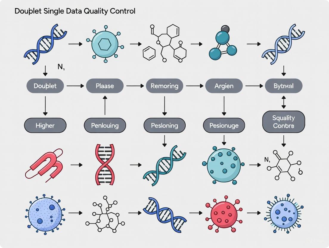
These methods use only the gene expression matrix to identify doublets and are widely applicable to existing datasets. The general workflow for the most common computational approaches is illustrated below.
Most computational tools follow a similar principle: they generate artificial doublets by combining the gene expression profiles of two randomly selected cells from the data. Then, each real cell is evaluated based on its similarity to these simulated doublets. Cells that are highly similar are flagged as potential doublets [4] [5]. The specific algorithms used for this comparison vary, employing methods such as k-nearest neighbors (kNN) graphs, gradient boosting, or neural networks [4].
Which computational doublet detection method should I use?
Numerous computational tools have been developed. A systematic benchmark study evaluating nine methods on 16 real and 112 synthetic datasets provides the following insights [4]:
| Method | Key Algorithm | Key Finding from Benchmark |
|---|---|---|
| DoubletFinder | k-nearest neighbors (kNN) on artificial doublets [4] [6] | Had the best overall detection accuracy [4]. |
| scDblFinder | Combines kNN statistics with iterative classification [5] | An independent benchmark found it to be a top performer, often outperforming alternatives [5]. |
| cxds | Co-expression of mutually exclusive gene pairs (no artificial doublets) [4] | Has the highest computational efficiency [4]. |
| Scrublet | k-nearest neighbors in PCA space [4] | Widely used; provides guidance on threshold selection [4]. |
Recommendation: Given that no single method is best in all situations, it is considered a best practice to try more than one method [5]. Furthermore, a multi-round doublet removal (MRDR) strategy, where an algorithm is run multiple times in cycles, has been shown to improve doublet removal efficiency compared to a single run [7].
What are essential research reagents and solutions for doublet detection?
The following table details key reagents and materials used in experimental doublet detection protocols.
| Reagent/Solution | Function in Doublet Detection |
|---|---|
| Antibody-Oligonucleotide Conjugates (e.g., for Cell Hashing) | Uniquely labels all cells from a single sample. A doublet is identified by the presence of two different antibodies in one droplet [1] [4]. |
| Lipid-Tagged Index Barcodes (e.g., for MULTI-seq) | Labels cell membranes with sample-specific barcodes. Droplets with more than one barcode are identified as doublets [4]. |
| Cell Lines from Different Species | Used in species-mixing experiments. Doublets are detected as cells expressing genes from both species [4]. |
A typical workflow for doublet detection and removal in a scRNA-seq analysis pipeline.
Integrating doublet removal is a standard step in single-cell quality control. The following diagram outlines a typical workflow using computational tools, which can be applied to data processed with standard packages like Seurat or Scanpy [3] [2].
Troubleshooting Guides and FAQs
Frequently Asked Questions
Q1: What are the fundamental differences between homotypic and heterotypic doublets, and why does it matter for my analysis?
Homotypic doublets are formed by two transcriptionally similar cells (e.g., of the same type), while heterotypic doublets are formed by two cells of distinct types, lineages, or states [4]. This distinction is critical because heterotypic doublets are generally easier to detect computationally due to their hybrid gene expression profiles, which appear distinct from genuine singlets [4]. Homotypic doublets are more challenging to identify and can be mistaken for novel or intermediate cell states [4].
Q2: My downstream differential expression analysis is yielding confusing markers. Could doublets be the cause?
Yes. Doublets can severely interfere with differential expression (DE) analysis [4]. A doublet formed from two distinct cell types will express genes from both parents, creating a misleading expression profile that does not correspond to any real biological state. This can result in the identification of spurious DE genes, complicating biological interpretation. Using methods like findDoubletClusters() can help identify clusters that show few uniquely expressed genes compared to potential source clusters, a classic signature of a doublet-driven artifact [1].
Q3: I am inferring cell developmental trajectories from my scRNA-seq data. How can I be sure doublets are not creating false paths?
Doublets are a known confounder in trajectory inference, as they can create artificial bridges between unrelated lineages [4]. A doublet formed from a cell at the beginning of one lineage and a cell at the end of another can be misinterpreted by trajectory analysis as a direct intermediate state or a novel transition path. To ensure robustness, it is recommended to perform doublet detection and removal before running trajectory inference. A multi-round doublet removal (MRDR) strategy has been shown to be more beneficial for cell trajectory inference than a single removal step [7].
Q4: Are all doublets just technical artifacts, or could some be biologically meaningful?
While the vast majority of doublets are technical artifacts, there is a emerging hypothesis that some doublets may represent cells that were physically interacting in the tissue (juxtacrine interactions) and did not fully dissociate [9]. Tools like CIcADA (Cell type-specific Interaction Analysis using Doublets in scRNA-seq) are being developed to identify and analyze these potentially biologically meaningful doublets, which may provide insights into intercellular communication, such as in the tumor microenvironment [9]. For standard quality control and analysis, however, the default approach is to treat doublets as artifacts and remove them.
Troubleshooting Common Problems
Problem: Spurious cell clusters appear in my UMAP/t-SNE visualization.
- Potential Cause: These clusters may be composed of heterotypic doublets. Doublets, especially heterotypic ones, often form distinct clusters because their combined gene expression profile does not match any real singlet cell [4] [1].
- Solution:
- Run a doublet detection method like
scDblFinderorDoubletFinder[4]. - Visualize the doublet scores on your dimensionality reduction plot. Clusters with high doublet scores are suspect [1].
- Use
findDoubletClusters(), which identifies clusters with expression profiles that lie between two other clusters and exhibit few unique genes [1].
- Run a doublet detection method like
Problem: My dataset has a known high doublet rate due to the experimental protocol. Standard removal seems insufficient.
- Potential Cause: A single run of a doublet detection algorithm may not capture all doublets due to the inherent randomness in the methods [7].
- Solution: Implement a Multi-Round Doublet Removal (MRDR) strategy. This involves running the doublet detection algorithm iteratively. Studies have shown that a two-round removal can improve the recall rate by 50% compared to a single round for some tools [7].
Problem: I suspect doublets are affecting my identification of rare cell populations.
- Potential Cause: Doublets can be misclassified as rare cell types because their transcriptome is unique. Conversely, real rare cells can be incorrectly flagged as doublets due to their atypical profiles.
- Solution:
- Cross-reference potential rare cell markers. A true rare cell type should have a consistent and specific marker set.
- Examine library size. Doublet libraries are typically generated from a larger initial pool of RNA and thus often have larger library sizes than genuine singlets [1]. A putative rare cell cluster with a median library size larger than its neighbors should be investigated carefully.
- Use conservative thresholds for doublet calling in conjunction with known marker genes to validate the identity of rare populations.
Table 1: Benchmarking Performance of Select Doublet Detection Methods
| Method | Key Algorithmic Approach | Reported Detection Accuracy | Computational Efficiency | Guidance on Threshold Selection |
|---|---|---|---|---|
| DoubletFinder | Generates artificial doublets; uses k-NN in PC space to score droplets [4]. | Best overall detection accuracy [4] | - | Yes [4] |
| cxds | Defines doublet score based on gene co-expression, without artificial doublets [4]. | - | Highest computational efficiency [4] | No [4] |
| bcds | Generates artificial doublets; uses gradient boosting classifier [4]. | - | - | No [4] |
| Scrublet | Generates artificial doublets; uses k-NN in PC space [4]. | - | - | Yes [4] |
| MRDR-cxds | Applies the cxds method in two iterative rounds [7]. |
Improved ROC by ~0.05 in synthetic datasets [7] | - | - |
Table 2: Impact of Multi-Round Doublet Removal (MRDR) Strategy
| Dataset Type | Recommended MRDR Method | Performance Improvement |
|---|---|---|
| Real-world datasets | DoubletFinder (two rounds) | Recall rate improved by 50% vs. single round [7] |
| Barcoded scRNA-seq datasets | cxds (two rounds) | Best results in this category [7] |
| Synthetic datasets | cxds (two rounds) | ROC improved by at least 0.05 [7] |
Experimental Protocols
Protocol 1: Identifying Doublet-Driven Clusters withfindDoubletClusters()
This protocol helps identify clusters that are likely composed of doublets by examining their relationship to other clusters [1].
- Input: A clustered SingleCellExperiment object with assigned cell labels.
- Execution: Run the
findDoubletClusters()function. The function will:- Consider every possible triplet of clusters (a query cluster and two putative "source" clusters).
- For each triplet, under the null hypothesis that the query is formed from the two sources, it computes the number of genes (
num.de) that are differentially expressed in the same direction in the query compared to both sources. A lownum.deis evidence for the doublet hypothesis. - For each query cluster, it identifies the best pair of sources (the pair with the lowest
num.de).
- Output Interpretation: The function returns a DataFrame. Clusters are ranked by
num.de; those with the fewest unique genes are more likely to be doublets. Also check thelib.size1andlib.size2fields, which should ideally be less than 1, indicating the query (doublet) cluster has a larger library size than its proposed sources [1]. - Action: Clusters flagged as potential doublets (e.g., with unusually low
num.deidentified via an outlier detection method) should be removed or investigated further before downstream analysis.
Protocol 2: Detecting Doublets by Simulation withcomputeDoubletDensity()
This method detects doublets at the individual cell level by comparing the local density of real cells to simulated doublets [1].
- Simulation: Generate thousands of artificial doublets by randomly adding together the gene expression profiles (counts) of two randomly chosen single cells from your dataset.
- Density Calculation: For each original cell in the dataset:
- Compute the density of simulated doublets in its neighborhood within a low-dimensional space (e.g., PCA).
- Compute the density of other observed cells in the same neighborhood.
- Calculate a doublet score as the ratio of the simulated doublet density to the observed cell density.
- Thresholding: Cells with high doublet scores are considered potential doublets. A threshold can be set by identifying large outliers in the score distribution, potentially on a per-sample basis [1].
- Advantage: This method is cluster-agnostic, reducing sensitivity to clustering quality.
Key Signaling Pathways and Workflows
The Scientist's Toolkit
Table 3: Essential Research Reagents and Computational Tools
| Item Name | Type | Function / Application |
|---|---|---|
| 10x Chromium | Platform | A popular droplet-based high-throughput scRNA-seq platform [10]. |
| Cell Hashing | Experimental Reagent | Uses oligo-tagged antibodies to label cells from different samples; doublets are droplets with more than one antibody tag [4]. |
| CAMML | Computational Tool | An R package for multi-label cell typing of scRNA-seq data; used in the CIcADA pipeline to score cell types and identify potential doublets [9]. |
| ChIMP | Computational Tool | An extension of CAMML that integrates CITE-seq data (protein markers) for more confident and conservative cell typing [9]. |
| CIcADA | Computational Pipeline | An R package (in development) for identifying and analyzing biologically meaningful doublets to study cell-cell interactions [9]. |
| scDblFinder | Computational Tool | A comprehensive R package that includes both cluster-based (findDoubletClusters) and simulation-based (computeDoubletDensity) doublet detection methods [1]. |
| DoubletFinder | Computational Tool | A highly accurate doublet detection method that generates artificial doublets and uses k-nearest neighbors in PCA space to identify them [4]. |
In single-cell RNA sequencing (scRNA-seq) experiments, doublets are artifactual libraries generated when two cells are encapsulated into a single reaction volume (droplet or well) instead of one. They appear as, but are not, real biological entities and represent a significant confounder in data analysis [4] [11]. During the distribution step of an scRNA-seq experiment, a droplet may encapsulate more than one cell. The doublet rate depends on the experimental protocol and throughput, with rates potentially reaching as high as 40% of all droplets [4].
There are two primary classes of doublets:
- Homotypic Doublets: Formed by two transcriptionally similar cells of the same type. These are more challenging to detect computationally.
- Heterotypic Doublets: Formed by two cells of distinct types, lineages, or states. These are generally easier to detect due to their hybrid gene expression profiles [4].
The presence of doublets, particularly heterotypic ones, can severely confound downstream analyses by forming spurious cell clusters, interfering with differential gene expression analysis, and obscuring true developmental trajectories [4] [11].
Computational Doublet Detection Methods
Realizing the limitations of experimental strategies, several computational methods have been developed to detect doublets from already-generated scRNA-seq data. These methods are based on distinct algorithm designs [4].
The table below summarizes the primary computational doublet-detection methods, their underlying algorithms, and key characteristics.
Table 1: Benchmarking of Computational Doublet-Detection Methods
| Method Name | Programming Language | Core Algorithm | Uses Artificial Doublets? | Detection Guidance |
|---|---|---|---|---|
| DoubletFinder [4] | R | k-Nearest Neighbors (kNN) classification | Yes | Provides guidance on threshold selection |
| cxds [4] | R | Gene co-expression analysis (Binomial test) | No | No threshold guidance |
| bcds [4] | R | Gradient Boosting classifier | Yes | No threshold guidance |
| hybrid [4] | R | Combination of cxds and bcds | - | No threshold guidance |
| Scrublet [4] | Python | k-Nearest Neighbors (kNN) classification | Yes | Provides guidance on threshold selection |
| doubletCells [4] | R | k-Nearest Neighbors (kNN) classification | Yes | No threshold guidance |
| DoubletDetection [4] | Python | Hypergeometric test & Louvain clustering | Yes | No threshold guidance |
| DoubletDecon [4] [12] | R | Deconvolution analysis & unique cell-state gene expression | Yes | Identifies doublets without providing per-cell scores |
| scDblFinder [11] | R | Combines simulated doublet density with iterative classification | Yes | Comprehensive and accurate method |
| COMPOSITE [8] | Python | Compound Poisson model on stable features from multiomics data | No | Statistical inference on multiplet status |
Workflow of a Typical Doublet Detection Tool
Most computational methods follow a similar high-level workflow. The following diagram illustrates the typical steps involved in doublet detection using a simulation-based approach, as employed by tools like Scrublet, DoubletFinder, and scDblFinder.
Performance Benchmarking
A systematic benchmark study of nine computational doublet-detection methods using 16 real datasets (with experimentally annotated doublets) and 112 synthetic datasets revealed diverse performance across methods [4].
Key Findings from the Benchmark Study:
- Best Detection Accuracy: The
DoubletFindermethod demonstrated the best overall detection accuracy across multiple conditions [4]. - Highest Computational Efficiency: The
cxdsmethod, which relies on gene co-expression and does not simulate artificial doublets, showed the highest computational efficiency [4]. - No Single Winner: No single method dominated in all aspects, indicating that the choice of method may depend on the specific dataset and analysis goals [4].
Experimental Detection and Prevention of Doublets
Several experimental techniques have been developed to detect and remove doublets. These typically require special preparation during library construction but provide a more direct measurement [4] [8].
Table 2: Experimental Methods for Doublet Detection
| Method Name | Underlying Principle | Key Advantage | Key Limitation |
|---|---|---|---|
| Cell Hashing [4] [8] | Cells from different samples are labeled with sample-specific oligo-tagged antibodies. Doublets have multiple tags. | Can multiplex samples; high detection accuracy. | Requires antibody staining; cannot detect homotypic doublets from same sample. |
| Species Mixing [4] | Cells from different species (e.g., human and mouse) are mixed. Doublets contain transcripts from both. | Conceptually simple and straightforward. | Limited to controlled experiments; not applicable to most clinical samples. |
| Genetic Multiplexing (e.g., Demuxlet) [4] | Cells from multiple donors are pooled. Doublets contain mutually exclusive sets of SNPs. | Leverages natural genetic variation. | Requires genotyping data; cannot detect doublets from the same individual. |
| MULTI-seq [4] | Cells are labeled with lipid-tagged barcodes. Doublets have more than one barcode. | Barcoding prior to encapsulation reduces technical artifacts. | Requires additional labeling steps. |
Troubleshooting Guides and FAQs
FAQ 1: My computational doublet detection tool did not find any doublets, but I am skeptical. What should I do?
Answer: Most tools require you to specify an expected doublet rate. If this rate is set too low, the tool may be insufficiently sensitive. Check the tool's documentation to ensure the expected doublet rate is appropriate for your platform and number of cells loaded. You can also try running a different computational method (e.g., both Scrublet and DoubletFinder) and compare the results. If available, inspect the expression of known, mutually exclusive marker genes across your clusters; co-expression of these markers in a single cluster can indicate a doublet population [11].
FAQ 2: I am working with a complex tissue with many rare cell types. How does this affect doublet detection?
Answer: Complex tissues with rare cell types present a particular challenge. Heterotypic doublets involving a rare and a common cell type can be misidentified as a novel rare population. Computational methods that rely on clustering (like findDoubletClusters) may lack the power to detect doublets in small clusters. In this scenario, methods like DoubletFinder or scDblFinder that work on a per-cell basis are often more effective. If possible, using experimental techniques like cell hashing is highly recommended for complex samples, as they do not rely on transcriptional profiles for doublet identification [8].
FAQ 3: After removing doublets, my cell count is much lower than expected. What could have gone wrong?
Answer: Overly aggressive doublet removal can occur due to:
- Incorrectly High Expected Doublet Rate: Review and adjust the expected rate in your tool's parameters.
- Poor Quality Cells Mimicking Doublets: Low-quality cells (e.g., dying cells with high mitochondrial content) can sometimes have aberrant expression profiles that resemble doublets. It is crucial to perform standard quality control (removing cells with low genes/UMIs or high mitochondrial counts) before doublet detection to avoid this confusion [13] [14].
- Overly Sensitive Thresholds: Manually inspect the distribution of doublet scores and adjust the threshold if possible, rather than relying solely on automated calls.
FAQ 4: How do I choose the best computational method for my data?
Answer: The choice depends on your data and priorities [4]:
- For highest accuracy, consider
DoubletFinderorscDblFinder. - For very large datasets where computational speed is critical,
cxdsis a fast option. - If you have pre-existing clusters and want a simple, interpretable method, the
findDoubletClustersfunction inscDblFinderis a good choice [11]. - For single-cell multiomics data (e.g., CITE-seq, DOGMA-seq), the
COMPOSITEmodel is specifically designed to leverage stable features across modalities and has been shown to outperform single-omics methods [8].
The Scientist's Toolkit: Essential Reagents and Materials
Table 3: Key Research Reagent Solutions for Doublet Management
| Item / Reagent | Function / Application | Example Use Case |
|---|---|---|
| BioLegend TotalSeq Antibodies | Antibody-derived tags (ADTs) for cell hashing and surface protein staining. | Multiplexing samples in a single channel for doublet identification via CITE-seq [8]. |
| Cell-Plex Multiplexing Kit (10x Genomics) | Commercial kit for sample multiplexing using lipid-tagged barcodes. | Similar to MULTI-seq, allows pooling of samples to identify inter-sample doublets [4]. |
| Species-Specific Cell Lines | Provide genetically distinct cells for controlled mixing experiments. | Used in species-mixing experiments to establish a ground truth for doublet detection algorithm benchmarking [4]. |
| Viability Stain (e.g., DAPI, Propidium Iodide) | Distinguish live from dead cells during cell sorting. | Prevents the encapsulation of dead cells, which can contribute to technical artifacts and be misclassified as doublets. |
| Accurate Cell Counter (e.g., Hemocytometer) | Precisely determine cell concentration and viability prior to loading. | Critical for loading the optimal cell concentration to minimize doublet formation rate [14]. |
Integrating Doublet Detection into the QC Workflow
Doublet detection is not a standalone step but an integral part of a comprehensive single-cell data preprocessing pipeline. The following diagram illustrates a recommended workflow, showing how doublet detection interacts with other quality control steps.
Summary of the Integrated Workflow:
- Initial QC: Begin by filtering out low-quality cells based on standard metrics: number of UMIs per cell, number of genes per cell, and the percentage of mitochondrial reads [13] [14].
- Normalization and Feature Selection: Normalize the data to account for technical variability and select highly variable genes for downstream analysis.
- Doublet Detection and Removal: This is the critical step where computational or information from experimental multiplexing is used to identify and remove doublets from the dataset.
- Clustering and Annotation: The cleaned dataset is then clustered, and cell types are annotated using known marker genes. The absence of doublets leads to more distinct and biologically meaningful clusters.
- Downstream Analysis: Proceed with high-confidence differential expression, trajectory inference, and other analyses on a reliable dataset.
Core Concepts: Defining Doublet-Induced Errors
What are embedded and neotypic errors, and how do they differ?
In single-cell RNA-sequencing analysis, doublets (artifactual libraries formed from two cells) cause two primary classes of errors with distinct consequences for downstream interpretation:
Embedded Errors occur when a multiplet transcriptome is grouped with a large population of singlets (true single cells) that dominate a cell state. These errors cause quantitative changes in gene expression and cell state abundance but have relatively small impact if multiplets are rare. They typically arise from multiplets formed between transcriptionally similar cells (e.g., of the same cell type) [15].
Neotypic Errors create entirely new features in the data, such as spurious cell clusters, unexpected branches from existing clusters, or bridges between distinct cell populations. These errors can lead to qualitatively incorrect biological inferences—such as falsely identifying novel cell types or transitional states—and are generated by multiplets of cells with distinct gene expression profiles (e.g., from different lineages or activation states) [15].
The table below summarizes the key differences:
| Feature | Embedded Errors | Neotypic Errors |
|---|---|---|
| Origin | Multiplets of similar cells [15] | Multiplets of distinct cell types [15] [1] |
| Impact on Data | Quantitative shifts in gene expression [15] | Creation of artifactual clusters or trajectories [15] [1] |
| Downstream Consequence | Minor distortion of existing cell states [15] | False discovery of non-existent cell types or states [15] [1] |
| Operational Classification | Dependent on multiplet rarity [15] | Dependent on data analysis choices (e.g., dimensionality reduction) [15] |
Why is it critical to identify doublets before downstream analysis?
Doublets account for several percent of transcriptomes in most scRNA-seq experiments [15]. If not removed, they confound virtually all downstream analyses:
- Cell Type Identification: Doublets can co-express marker genes of distinct cell types, leading to misannotation or the false discovery of intermediate or novel cell states [1] [16].
- Differential Expression Analysis: A doublet's hybrid transcriptome does not represent any real biological state, making its inclusion in differential expression tests between conditions misleading [15].
- Trajectory Inference: Doublets can form artificial "bridges" between unrelated lineages, suggesting incorrect differentiation paths or cellular relationships [15] [1].
Troubleshooting Guides: Identifying and Resolving Doublet-Related Issues
Symptom: A small cluster co-expresses marker genes from two distinct, well-separated cell populations.
- Potential Diagnosis: Neotypic doublet cluster [1].
- Investigation & Resolution Protocol:
- Cluster-Based Inspection: Use the
findDoubletClustersfunction from the scDblFinder R package. This function identifies clusters whose expression profiles lie between two other putative "source" clusters, a hallmark of doublets [1]. - Evaluate Key Metrics: Examine the output for clusters with a low number of unique differentially expressed genes (
num.de) and library sizes (lib.size1,lib.size2) comparable to or smaller than the proposed source clusters [1]. - Manual Verification: Check the literature to confirm whether the co-expressed markers are ever known to be expressed together in a genuine biological state [1] [17].
- Action: Remove the identified cluster and re-run downstream analyses.
- Cluster-Based Inspection: Use the
Symptom: The data contains "bridging" cells between major clusters in a UMAP/t-SNE plot, with no clear biological justification.
- Potential Diagnosis: Neotypic doublets creating artificial trajectories [15].
- Investigation & Resolution Protocol:
- Simulation-Based Scoring: Use a tool like
computeDoubletDensityfrom scDblFinder or Scrublet. These tools simulate doublets from your data and score each real cell based on its proximity to these simulated doublets [1]. - Visualize Scores: Project the doublet scores onto your embedding (e.g., UMAP). If the bridging cells have high doublet scores, they are likely artifacts [1].
- Thresholding: Convert continuous scores into doublet calls by identifying large outliers, often on a per-sample basis [1].
- Action: Filter out cells called as doublets before re-embedding and analyzing the data.
- Simulation-Based Scoring: Use a tool like
Symptom: A known cell population appears with abnormally high RNA counts and/or a widened transcriptional profile.
- Potential Diagnosis: Embedded doublets within a legitimate cell population [15].
- Investigation & Resolution Protocol:
- Library Size Check: While not always reliable alone [15], plot the distribution of total counts per cell (
total_counts). Cells with exceptionally high counts may be doublets [13]. - Computational Detection: Apply DoubletFinder, which is noted for its accuracy in downstream analyses [16] [17]. It calculates the proportion of artificial nearest neighbors (
pANN) for each cell to identify doublets even within clusters. - Parameter Optimization: When using DoubletFinder, always estimate the optimal pK parameter using the mean-variance normalized bimodality coefficient (
BCmvn). Do not rely on default values [18]. - Action: Remove the predicted doublets. The core population should appear more tightly clustered after removal.
- Library Size Check: While not always reliable alone [15], plot the distribution of total counts per cell (
Frequently Asked Questions (FAQs)
What is the expected doublet rate in my experiment?
The doublet rate is primarily determined by the platform and the number of cells loaded. For example, 10x Genomics reports that loading 10,000 cells results in a multiplet rate of about 7.6% [17]. However, note that computational tools can only detect "heterotypic" doublets (from different cell types). "Homotypic" doublets (from the same or similar cell types) are generally undetectable and form embedded errors. Therefore, your computationally detectable doublet rate will be lower than the platform's theoretical rate [18].
I have used a doublet detection tool. How do I know if it worked well?
Benchmarking studies indicate that performance varies across tools and datasets. A comprehensive benchmark found that DoubletFinder had the best overall accuracy and positive impact on downstream analyses like differential expression and clustering [17]. However, because no tool is perfect, a best practice is to use a combination of automated tools and manual inspection of results [17]. Always check if cells called as doublets show co-expression of mutually exclusive marker genes from distinct cell types.
Should I run a doublet detection tool on data that has already been integrated from multiple samples?
No, this is not recommended. If you run a tool like DoubletFinder on data aggregated from biologically distinct samples (e.g., wild-type and mutant), it will simulate artificial doublets that are biologically impossible (e.g., a WT-mutant hybrid). This will skew the results [18]. The best practice is to run doublet detection individually on each sample before integrating them for downstream analysis [18].
How does data preprocessing affect doublet detection?
Preprocessing steps like normalization can significantly impact all downstream analyses, including clustering and, by extension, cluster-based doublet detection methods [19]. For instance, the performance of the SC3 clustering algorithm is highly dependent on the choice of preprocessing method (log transformation, z-score, etc.) [19]. Since some doublet detection methods rely on clustering, ensuring optimal preprocessing for your clustering tool is an indirect but critical step for effective doublet detection.
| Tool / Resource Name | Type | Primary Function & Application Context |
|---|---|---|
| Scrublet [15] | Software Package | Identifies neotypic multiplets by simulating doublets and building a nearest-neighbor classifier. Framework for predicting multiplet impact. |
| DoubletFinder [18] [17] | Software Package | Detects doublets by generating artificial doublets and calculating the proportion of artificial nearest neighbors (pANN). Noted for high accuracy. |
| scDblFinder [1] | Software Package (R/Bioconductor) | Suite containing multiple methods, including cluster-based (findDoubletClusters) and simulation-based (computeDoubletDensity) detection. |
| findDoubletClusters [1] | Algorithm | Identifies clusters that likely represent doublets based on their intermediate gene expression profile and low number of unique marker genes. |
| computeDoubletDensity [1] | Algorithm | Scores individual cells based on the local density of simulated doublets versus real cells, independent of pre-defined clusters. |
| SoupX [16] [17] | Software Package | Corrects for ambient RNA contamination, a different but common artifact that can compound issues caused by doublets. |
| Seurat [16] [18] | Software Toolkit | A comprehensive ecosystem for single-cell analysis that is often used in conjunction with doublet detection tools for preprocessing and visualization. |
Workflow Diagrams for Doublet Error Analysis
Doublet Error Identification and Resolution Workflow
Conceptual Framework of Doublet-Induced Errors
Frequently Asked Questions (FAQs) on Doublet Rates and Cell Loading
FAQ 1: What is the expected doublet rate in a typical droplet-based scRNA-seq experiment? The doublet rate is not fixed and is primarily a function of the number of cells loaded into the instrument. Rates reported in the literature range from as low as 5% to as high as 40% of all captured droplets [20] [21]. However, commonly used heuristic estimations have been shown to systematically underestimate the true multiplet rate. Refined Poisson-based models reveal that actual rates can exceed these heuristic predictions by more than twofold [20] [21].
FAQ 2: Why is it critical to accurately estimate and account for doublets in my data? Multiplets are a pervasive confounder in scRNA-seq analysis. They are not confined to isolated clusters but are distributed throughout the transcriptional landscape, where they distort clustering and cell type annotation. They can be mistaken for novel cell types or intermediate states [20] [21]. In differential gene expression analysis, multiplets inflate artefactual signals, leading to shifts in effect sizes and the partial loss of genuinely significant genes [20] [4]. Their removal is essential for accurate biological interpretation.
FAQ 3: What is the difference between homotypic and heterotypic doublets, and why does it matter?
- Heterotypic doublets are formed from two transcriptionally distinct cells (e.g., a T cell and a monocyte). These are generally easier for computational tools to detect because their combined gene expression profile appears as a unique, hybrid cell type [4].
- Homotypic doublets are formed from two transcriptionally similar cells (e.g., two cells of the same type). Computational tools struggle to distinguish these from singlets, representing a major limitation in doublet detection [20] [21]. Most computational methods are primarily designed to identify heterotypic doublets.
FAQ 4: My experiment lacks multiplexing information (e.g., cell hashing). How can I estimate the doublet rate? In the absence of experimental demultiplexing, researchers must rely on computational predictions and general guidelines. A Poisson model is often used as a more accurate alternative to simple heuristics [20] [21]. Furthermore, computational tools like DoubletFinder and Scrublet can provide a droplet-level score indicating the likelihood of a droplet being a doublet, which can be thresholded to estimate the overall rate in the dataset [4].
Doublet Rate Data and Estimations
Table 1: Experimentally Annotated Doublet Rates from Public Datasets
The following table presents doublet rates determined via cell hashing in publicly available datasets, providing a lower-bound estimate of the true doublet rate [20] [21].
| Dataset Name | Cell Source | Number of Droplets | Annotated Multiplets | Doublet Rate |
|---|---|---|---|---|
| pbmc-ch | Human PBMCs (8 donors) | 15,272 | 2,545 | 16.66% |
| cline-ch | 4 Human Cell Lines | 7,954 | 1,465 | 18.42% |
| mkidney | Mouse Kidney Cells | 21,179 | 7,901 | 37.31% |
| Gold Standard | Human PBMCs & Bone Marrow (Healthy donors only) | 27,504 | 7,186 | 26.13% |
Table 2: Performance of Computational Doublet-Detection Methods
A systematic benchmark study of computational doublet-detection methods evaluated their performance on datasets with known doublets. The table below summarizes key findings for popular tools [4].
| Method | Key Algorithm Description | Key Finding from Benchmarking |
|---|---|---|
| DoubletFinder | Generates artificial doublets and identifies real cells with high proximity to these artificial doublets in gene expression space using k-nearest neighbors (kNN) [4] [22]. | Has the best overall detection accuracy among the methods benchmarked [4]. |
| Scrublet | Generates artificial doublets and defines a doublet score for each cell as the proportion of artificial doublets among its k-nearest neighbors in PCA space [4]. | One of the most frequently used methods in new single-cell research, alongside DoubletFinder [20] [21]. |
| cxds | Defines a doublet score based on the co-expression of genes in mutually exclusive pairs, without generating artificial doublets [4]. | Has the highest computational efficiency [4]. |
| scDblFinder | Combines simulated doublet density with an iterative classification scheme and can also identify likely doublet clusters based on intermediate gene expression [1] [4]. | Offers a cluster-based approach (findDoubletClusters) and a simulation-based approach (computeDoubletDensity) [1]. |
Detailed Experimental Protocols
Protocol: Experimental Doublet Detection Using Cell Hashing
Cell hashing uses antibody-based tagging to label cells from different samples or conditions, allowing for the identification of doublets formed after pooling samples [20] [21].
Key Research Reagent Solutions:
- Oligo-Tagged Antibodies: Antibodies targeting a ubiquitous surface protein (e.g., CD45 for human PBMCs) are conjugated to a unique oligonucleotide barcode for each sample [20] [4].
- Cell Suspension: A single-cell suspension from your biological sample(s).
- 10X Genomics Library Preparation Kits: Standard kits for 3' or 5' scRNA-seq, compatible with feature barcoding technology.
Methodology:
- Tagging: Incubate cells from each individual sample (e.g., different donors, conditions) with a unique oligo-tagged antibody.
- Pooling: After washing away unbound antibodies, pool all uniquely tagged samples into a single cell suspension.
- Library Preparation and Sequencing: Proceed with standard droplet-based scRNA-seq using a 10X Genomics platform. The hashing oligo tags and cellular transcripts will be captured together in each droplet.
- Bioinformatic Analysis: Use computational pipelines (e.g., CellRanger) to separate the transcriptome data from the hashing tag data.
- Doublet Identification: Droplets whose barcodes are associated with two or more different hashing tags are identified as multiplets originating from different samples. Note: This method only identifies inter-sample multiplets; multiplets formed within a single sample remain undetected [20] [21].
Protocol: Computational Doublet Detection Using DoubletFinder
DoubletFinder is a widely used computational tool that predicts doublets based solely on gene expression data [4] [22].
Methodology:
- Data Preprocessing: Generate a high-quality gene expression matrix (cells x genes) following standard scRNA-seq preprocessing steps (quality control, normalization, scaling).
- Parameter Estimation:
- pK Identification: The
paramSweepfunction is used to optimize the key parameterpK, which represents the proportion of artificial nearest neighbors. The optimalpKis selected based on the highest mean variance-weighted AUC (area under the curve) from the model [22]. - Expected Doublet Rate (
pN): ThepNparameter defines the number of artificial doublets generated. A common starting point is to use the overall expected doublet rate for the experiment (e.g., from a Poisson model) [22].
- pK Identification: The
- Model Execution: Run the
doubletFinderfunction using the preprocessed data and the optimizedpKandpNparameters. - Result Interpretation: The function returns doublet classifications for each cell. These classifications should be used to remove predicted doublets from the dataset before proceeding with downstream analyses like clustering and differential expression.
Visualizing Doublet Detection Strategies
Experimental vs. Computational Doublet Detection
The following diagram illustrates the logical relationship and primary focus of the two main strategies for doublet detection.
Impact of Multiplets on Downstream Analysis
This diagram summarizes how multiplets can confound key steps in scRNA-seq data analysis.
Computational Doublet Detection Methods: From Theory to Practical Implementation
In single-cell RNA sequencing (scRNA-seq) experiments, doublets are artifactual libraries generated when two cells are captured together and sequenced as a single cell [1]. These technical artifacts can constitute up to 40% of droplets in some experiments and lead to spurious biological conclusions by appearing as intermediate cell types or states that don't actually exist [22] [4]. Artificial doublet simulation has emerged as the predominant computational strategy for detecting these artifacts, providing a powerful in silico approach that doesn't require specialized experimental designs [4] [1].
The core premise is elegantly simple: by computationally creating artificial doublets that mimic how real doublets would form, we can train classifiers to distinguish these simulated doublets from genuine single cells in the actual data [4] [2]. Most doublet detection tools follow this fundamental principle, though they differ in their implementation details, classification algorithms, and how they integrate the simulation process into their detection pipelines [4].
Key Mechanism: How Artificial Doublets Are Created and Used
The Basic Simulation Process
Artificial doublet simulation typically follows a standardized workflow, regardless of the specific algorithm implementation. The process begins with the raw gene expression matrix from a scRNA-seq experiment and proceeds through several well-defined stages:
- Random Selection: Two cells are randomly selected from the expression matrix
- Profile Combination: Their gene expression profiles are mathematically combined
- Classifier Training: A machine learning model is trained to distinguish real cells from artificial doublets
- Score Assignment: Each real cell receives a doublet score based on its similarity to artificial doublets
- Thresholding: Cells exceeding a threshold are flagged as potential doublets [4] [1]
The critical variation between methods lies primarily in step 2 – how the gene expression profiles are combined. The most common approaches include:
- Expression Averaging: Used by DoubletFinder, where the transcriptional profiles of two randomly chosen cells are averaged [22] [4]
- Expression Summing: Employed by Scrublet and doubletCells, where the counts from two cells are added together [4]
- Cluster-Based Simulation: Implemented in scMODD and scIBD, where cells are first clustered, then artificial doublets are created specifically between clusters to enrich for heterotypic doublets [23] [2]
Mathematical Foundation of Profile Combination
The combination of gene expression profiles follows specific mathematical operations depending on the method. For a gene g in two cells A and B with expression counts X_{A,g} and X_{B,g}, the artificial doublet expression X_{doublet,g} is calculated as:
- Averaging Approach: ( X{doublet,g} = \frac{X{A,g} + X_{B,g}}{2} ) [22]
- Summing Approach: ( X{doublet,g} = X{A,g} + X_{B,g} ) [4]
- Normalization Variations: Some methods apply library size normalization before or after combination
Most methods operate on highly variable genes or principal components rather than the full gene set to reduce dimensionality and computational complexity [4] [2]. The number of artificial doublets generated typically ranges from thousands to tens of thousands, with the exact number often being proportional to the dataset size [4] [2].
Frequently Asked Questions (FAQs)
Core Mechanism Questions
Q1: Why can't we simply use experimental approaches instead of artificial doublet simulation?
Experimental approaches like Cell Hashing and genetic multiplexing can effectively identify some doublets but have significant limitations. They require special experimental preparation, add extra costs and time, and cannot identify all doublet types – particularly those formed within the same sample or from cells with similar genetic backgrounds [4] [23] [24]. Artificial doublet simulation provides a general computational approach that works on already-generated data without requiring specialized experimental designs [1].
Q2: What's the difference between homotypic and heterotypic doublets, and why does it matter for simulation?
Heterotypic doublets are formed by transcriptionally distinct cell types (e.g., immune cell + neuron), while homotypic doublets come from similar cells of the same type [4]. This distinction is crucial because heterotypic doublets are generally easier to detect due to their hybrid expression profiles, and they cause more significant problems in downstream analysis by creating spurious cell clusters [4] [23]. Most artificial doublet simulation methods are particularly effective at detecting heterotypic doublets, though some newer approaches specifically optimize for this by using cluster-informed simulation strategies [23] [2].
Q3: How do artificial doublet methods differ in their classification approaches after simulation?
While all major methods use artificial doublets, they employ different classification strategies:
Table 1: Classification Approaches in Doublet Detection Methods
| Method | Classification Algorithm | Key Features | Language |
|---|---|---|---|
| DoubletFinder [22] | k-nearest neighbors (kNN) | Uses artificial nearest neighbors in PCA space | R |
| Scrublet [4] | k-nearest neighbors (kNN) | Defines score as proportion of artificial doublets among neighbors | Python |
| doubletCells [4] | k-nearest neighbors (kNN) | Calculates proportion of artificial doublets in neighborhood | R |
| bcds [4] | Gradient boosting | Trains classifier on pooled droplets and artificial doublets | R |
| Solo [4] | Neural networks | Uses semi-supervised deep neural network | Python |
| DoubletDetection [4] | Hypergeometric test | Uses Louvain clustering and hypergeometric testing | Python |
| cxds [4] | Gene co-expression | Does not use artificial doublets; based on co-expression patterns | R |
Practical Implementation Questions
Q4: What are the key parameters I need to consider when using artificial doublet simulation methods?
The most critical parameters across methods include:
- Expected doublet rate: Often estimated from cell loading concentrations
- Number of artificial doublets: Typically proportional to dataset size
- Dimensionality reduction parameters: Number of PCs or highly variable genes
- Classification thresholds: Score cutoffs for calling doublets
DoubletFinder and Scrublet provide guidance on parameter selection, while other methods like cxds and doubletCells offer less guidance [4]. For DoubletFinder, the pK parameter (defines the neighborhood size) is particularly important and should be optimized [22].
Q5: My dataset has unusual cell type distributions. Will this affect artificial doublet simulation performance?
Yes, the performance of artificial doublet simulation is influenced by cell type heterogeneity and distribution. Methods that use completely random sampling for doublet simulation (like DoubletFinder and Scrublet) may generate excessive homotypic doublets in homogeneous datasets, reducing detection power for heterotypic doublets [23]. Newer approaches like scIBD address this by using cluster-informed simulation, where artificial doublets are specifically created between different cell clusters to enrich for heterotypic doublets [23].
Troubleshooting Guides
Common Issues and Solutions
Problem: Inconsistent doublet detection results across different methods
Solution: This is expected due to different algorithmic approaches. Follow this systematic troubleshooting approach:
- Benchmark on your data type: Use a consensus approach or refer to benchmarking studies. One comprehensive study showed DoubletFinder has the best detection accuracy overall, while cxds has the highest computational efficiency [4].
- Check dataset characteristics: Performance varies by data heterogeneity. For homogeneous datasets, consider cluster-informed methods like scIBD [23].
- Validate biologically: Examine marker gene expression in putative doublets - true doublets often show simultaneous expression of markers from distinct cell types [1].
Problem: Poor doublet detection in homogeneous cell populations
Solution: Standard artificial doublet simulation struggles with homotypic doublets. Consider these approaches:
- Use cluster-informed methods: Tools like scIBD perform iterative clustering and doublet detection to improve performance on homotypic doublets [23].
- Adjust simulation strategy: Some methods allow controlling the proportion of heterotypic vs. homotypic artificial doublets.
- Combine approaches: Use multiple methods or integrate with experimental techniques when possible.
Problem: Computational performance issues with large datasets
Solution: Large datasets (>50,000 cells) can challenge some methods:
- Choose efficient methods: cxds shows highest computational efficiency according to benchmarks [4].
- Subsample strategically: Some methods allow running on subsets with projection to full dataset.
- Optimize parameters: Reduce the number of artificial doublets or use fewer PCs for large datasets.
Performance Optimization Guide
Table 2: Performance Characteristics of Doublet Detection Methods
| Method | Detection Accuracy | Computational Efficiency | Ease of Use | Best For |
|---|---|---|---|---|
| DoubletFinder [4] | Best accuracy | Moderate | Moderate guidance | Standard use cases |
| Scrublet [4] | Good accuracy | Moderate | Good guidance | Standard use cases |
| cxds [4] | Moderate accuracy | Highest | Limited guidance | Large datasets |
| bcds [4] | Good accuracy | Moderate | Limited guidance | Standard use cases |
| DoubletDetection [4] | Variable | Low | Limited guidance | Specialized applications |
| scIBD [23] | High for scCAS | Moderate | Specialized | scCAS data, heterogeneous samples |
Experimental Protocols and Methodologies
Standard Artificial Doublet Simulation Protocol
This protocol outlines the general workflow for implementing artificial doublet simulation, based on the common elements across major methods:
Input Requirements:
- Raw or normalized count matrix (cells × genes)
- Optional: Pre-computed dimensionality reduction (PCA)
- Optional: Cell clustering results
Step-by-Step Procedure:
Data Preprocessing:
- Filter low-quality cells and genes
- Normalize for library size differences
- Identify highly variable genes (typically 2,000-5,000 genes)
- Perform dimensionality reduction (PCA recommended)
Artificial Doublet Generation:
- Randomly select cell pairs from the dataset
- For each pair, combine expression profiles using chosen method (sum or average)
- Generate artificial doublets (typically 2× to 5× the number of real cells)
- Combine artificial doublets with real cells into a single matrix
Dimensionality Reduction:
- Project combined data (real cells + artificial doublets) into low-dimensional space
- Use same space (e.g., PCA) for both real and artificial cells
Classification and Scoring:
- Train classifier or compute similarity metrics in low-dimensional space
- Calculate doublet scores for real cells based on similarity to artificial doublets
- Determine optimal threshold for doublet calling
Validation:
Advanced: Cluster-Informed Simulation Protocol
For methods like scIBD that use cluster-informed simulation:
- Initial Clustering: Perform clustering on the preprocessed data
- Inter-Cluster Doublet Simulation: Generate artificial doublets specifically between different clusters
- Iterative Refinement:
- Detect doublets using simulated heterotypic doublets
- Remove detected doublets
- Recluster remaining cells
- Repeat process for multiple iterations
- Ensemble Scoring: Combine scores across iterations for final doublet calls [23]
Table 3: Essential Resources for Artificial Doublet Detection
| Resource Type | Specific Tools/Reagents | Function/Purpose | Application Context |
|---|---|---|---|
| Computational Tools | DoubletFinder (R) [22] | kNN-based detection using artificial nearest neighbors | General scRNA-seq analysis |
| Scrublet (Python) [4] | kNN classifier with simulated doublets | General scRNA-seq analysis | |
| scDblFinder (R) [1] | Combines simulation with co-expression analysis | General scRNA-seq analysis | |
| scIBD (Python/R) [23] | Iterative cluster-informed detection | scCAS data, heterogeneous samples | |
| Experimental Validation | Cell Hashing [23] | Antibody-based multiplexing for experimental validation | Ground truth establishment |
| Genetic Multiplexing [24] | SNP-based doublet identification | Ground truth establishment | |
| Synthetic DNA Barcodes [24] | Introduced barcodes for ground truth singlet identification | Method benchmarking | |
| Data Types | scRNA-seq Count Matrix [13] | Primary input data for all computational methods | Essential data structure |
| Cell Cluster Labels [2] | Optional input for cluster-informed methods | Advanced applications | |
| Dimensionality Reduction [4] | PCA or other low-dimensional representations | Critical for most methods |
Workflow Integration and Quality Control
Integration with scRNA-seq Analysis Pipelines
Artificial doublet detection should be integrated into a comprehensive scRNA-seq analysis workflow:
Quality Control Metrics for Doublet Detection
After implementing artificial doublet detection, assess quality using these metrics:
- Cluster Examination: Check if putative doublets form distinct clusters between cell types [1]
- Marker Gene Expression: Verify that high-scoring cells express markers from multiple cell types simultaneously
- Library Size: Confirm that putative doublets often have higher library sizes than singlets [1]
- Downstream Impact: Assess whether doublet removal improves clustering resolution and reduces intermediate populations
Emerging Methods and Future Directions
The field of artificial doublet simulation continues to evolve with several promising directions:
Multi-modal Integration: New approaches like ImageDoubler leverage cell images captured during sequencing to provide visual confirmation of doublets, offering an orthogonal validation method [25].
Cross-Modality Applications: Methods like scIBD demonstrate how artificial doublet simulation principles can be adapted for other data types like single-cell chromatin accessibility (scCAS) data, despite additional challenges like extreme sparsity and higher dimensions [23].
Benchmarking Frameworks: New technologies like singletCode use synthetic DNA barcodes to identify ground-truth singlets, enabling more rigorous benchmarking of artificial doublet methods across diverse biological contexts [24].
Model-Driven Approaches: Alternatives like scMODD explore model-driven (as opposed to data-driven) approaches using negative binomial or zero-inflated negative binomial models, though initial results suggest consideration of zero inflation may not be necessary for doublet detection [2].
As single-cell technologies continue to advance, producing ever-larger datasets, artificial doublet simulation remains an essential tool for ensuring data quality and biological validity in single-cell genomics research.
Experimental Protocols and Workflow
Detailed Methodology forfindDoubletClusters
The findDoubletClusters function, part of the scDblFinder package in R/Bioconductor, operates on a cluster-based principle to detect potential doublet clusters in single-cell RNA sequencing (scRNA-seq) data. The core methodology can be broken down into the following steps [1] [26]:
Input Preparation: The function requires a numeric matrix-like object of count values (cells as columns, genes as rows), or more commonly, a
SingleCellExperimentobject containing such a matrix. A vector of cluster identities for all cells must be provided. ForSingleCellExperimentobjects, this is typically taken fromcolLabels(x)by default [26].Triplet Evaluation: For each cluster designated as a "query" cluster, the function examines all possible pairs of other "source" clusters. It tests the hypothesis that the query cluster consists of doublets formed from cells belonging to the two source clusters [1].
Intermediate Expression Test: Under the null hypothesis that the query is a doublet population, its gene expression profile should be strictly intermediate between the two source clusters after library size normalization. The function applies pairwise t-tests on normalized log-expression profiles to identify genes that are significantly and consistently up- or down-regulated in the query cluster compared to both sources. The number of such genes that reject the null hypothesis at a specified FDR threshold is counted (
num.de) [1] [26].Result Compilation: For each query cluster, the pair of source clusters that minimizes the number of significant genes (
num.de) is identified and reported. A lownum.desuggests the query's expression profile is consistent with an intermediate profile, supporting the doublet hypothesis. The function returns aDataFramewith statistics for each query cluster, including the best source pair,num.de, median number of DE genes across all pairs, and library size ratios [1] [26].
The following diagram illustrates the logical workflow and key relationships in the findDoubletClusters method:
Key Output Metrics and Interpretation
The findDoubletClusters function returns a DataFrame where each row corresponds to a queried cluster. Interpreting these results correctly is crucial for accurate doublet identification. The table below summarizes the key metrics and provides guidance on their interpretation [1] [26].
| Metric | Description | Interpretation Guide |
|---|---|---|
source1 & source2 |
Identities of the two putative source clusters that best explain the query cluster as a doublet. | The most likely parental populations for the doublets. |
num.de |
Number of genes that are significantly non-intermediate in the query cluster compared to both source clusters. | Primary indicator. A low value (e.g., outliers) supports the doublet hypothesis. Fewer unique genes = more likely doublets [1]. |
median.de |
Median number of significantly non-intermediate genes across all possible source cluster pairings for the query. | Provides context for the best num.de. A high value indicates the query is often very different from other clusters. |
lib.size1 & lib.size2 |
Ratio of the median library size of the source cluster to the median library size of the query cluster. | Ideally should be less than 1 for both, as doublets typically have more RNA and higher library sizes than singlets [1] [26]. |
prop |
The proportion of all cells that are in the query cluster. | Should be reasonable. Typically, doublet clusters should be a small fraction (e.g., <5%) of the total cells, depending on the protocol [1]. |
p.value |
The adjusted p-value for the gene with the lowest p-value against the doublet hypothesis (best gene). |
Of limited statistical use for final calls. Mainly for inspection, as it does not account for multiple testing across all cluster pairs [26]. |
The Scientist's Toolkit: Essential Materials and Parameters
To effectively implement the findDoubletClusters method, researchers need to be familiar with the following key reagents, parameters, and their functions within the analysis pipeline.
| Item / Parameter | Function / Role in the Experiment |
|---|---|
scDblFinder R/Bioconductor Package |
The software package that contains the findDoubletClusters function and other related doublet detection utilities [1] [26]. |
SingleCellExperiment Object |
The standard data structure in Bioconductor for storing single-cell genomics data. Serves as the primary input format for the function [1] [26]. |
Cluster Labels (clusters) |
A vector of cluster identities for every cell (e.g., from colLabels or community detection algorithms like Louvain). The quality of these labels directly impacts the method's performance [1] [26]. |
subset.row Parameter |
Allows the user to perform the analysis on a subset of features (e.g., highly variable genes) to speed up computation and reduce noise [26]. |
threshold Parameter |
A numeric scalar specifying the FDR threshold used to identify significant genes during the test for non-intermediate expression. The default is 0.05 [26]. |
| Library Size Factors | Factors used for normalizing expression profiles. findDoubletClusters uses library size normalization specifically to ensure the "intermediate expression" property of doublets holds [26]. |
Troubleshooting Guides and FAQs
FAQ: The p.value in the output seems very high for a cluster I suspect is a doublet. Is the method not working?
- Answer: The reported
p.valuehas limited utility for final statistical conclusions. As per the documentation, it is technically a Simes combined p-value against the doublet hypothesis but does not account for the multiple testing across all pairs of clusters for each query. Therefore, thenum.deand library size ratios (lib.size1/2) are far more reliable metrics for identifying potential doublet clusters. Focus on clusters with a combination of lownum.deand library size ratios below 1 [26].
FAQ: My clustering might be too coarse/too fine. How does this affect findDoubletClusters?
- Answer: Clustering quality is a known dependency of this method.
- Overly Coarse Clustering: If clusters are too broad, doublets may not be separated from true singlets, causing them to be missed.
- Overly Fine Clustering: If clusters are too granular, it complicates interpretation and may break true biological populations into multiple parts, making it harder for the algorithm to find consistent intermediate states. The method has a slight bias towards clusters with fewer cells, which is often desirable as doublets should be rare [1]. It is recommended to test different clustering resolutions as part of your analysis.
FAQ: A cluster has a very low num.de but its library size ratios are greater than 1. Should I still remove it?
- Answer: Proceed with caution. The library size ratio being greater than 1 suggests the query cluster has smaller library sizes than its putative sources, which contradicts the expectation that doublets have more RNA [1] [26]. While a low
num.deis a strong signal, this discrepancy warrants further investigation. You should:- Manually inspect the expression of known marker genes for the source and query clusters.
- Check if the cluster expresses markers for two distinct cell types simultaneously, which would be a strong indicator of a heterotypic doublet despite the library size anomaly [1].
- Consider validating the finding with an alternative, non-cluster-based doublet detection method (e.g.,
scDblFinderorDoubletFinder) [1] [4].
FAQ: How do I formally select the clusters to remove after running findDoubletClusters?
- Answer: The function itself does not provide a formal classification. A common strategy is to use an outlier-based approach on the
num.devalues. You can use theisOutlierfunction from thescuttlepackage to automatically identify clusters with unusually lownum.de[1]. For example: Additionally, you should manually enforce the condition that library size ratios are less than 1 (dbl$lib.size1 < 1 & dbl$lib.size2 < 1) to ensure the calls are biologically plausible [1] [26].
In single-cell RNA sequencing (scRNA-seq) analysis, doublets are artifacts that occur when two or more cells are encapsulated within a single reaction volume, leading to a hybrid transcriptome that can confound downstream biological interpretations. These artifacts represent a significant challenge in data quality control, as they can create spurious cell clusters, interfere with differential gene expression analysis, and obscure developmental trajectories. Density-based detection methods have emerged as powerful computational approaches for identifying these doublets directly from scRNA-seq data without requiring specialized experimental designs. This technical support guide focuses on two prominent density-based detection approaches—Scrublet and the principles underlying computeDoubletDensity—providing researchers with comprehensive troubleshooting guidance and methodological frameworks for effective doublet detection within the broader context of single-cell data quality control research.
Key Concepts and Terminology
Understanding Doublets in scRNA-seq Data
Doublets form when two cells are co-encapsulated in a single droplet or reaction volume during scRNA-seq library preparation. They can be categorized into two distinct classes:
- Homotypic doublets: Result from the combination of transcriptionally similar cells (e.g., the same cell type) [4]
- Heterotypic doublets: Formed by combining cells of distinct types, lineages, or states [4] [15]
The rate of doublet formation increases with cell concentration and can account for several percent of all transcriptomes in a typical scRNA-seq experiment, sometimes reaching as high as 40% in high-throughput protocols [4] [15]. Heterotypic doublets are generally easier to detect computationally due to their distinct gene expression profiles that differ markedly from genuine singlets [4].
Density-Based Detection Fundamentals
Density-based doublet detection methods operate on the principle that doublets occupy regions of gene expression space that are intermediate between genuine cell states. These methods typically:
- Simulate artificial doublets from observed transcriptomes by combining random pairs of cells [27] [4]
- Embed both observed cells and simulated doublets in a lower-dimensional space
- Compute density-based scores that reflect each cell's similarity to simulated doublets versus observed singlets
- Apply thresholds to classify cells as singlets or doublets [4]
Scrublet: Framework and Implementation
Scrublet (Single-Cell Remover of Doublets) employs a targeted framework for predicting doublet impact and identifying problematic multiplets in scRNA-seq data. The algorithm follows these core steps [27] [15]:
- Doublet Simulation: Synthetic doublets are generated by summing the counts of randomly sampled observed transcriptomes
- Feature Selection: Highly variable genes are identified to focus the analysis on informative dimensions
- Dimensionality Reduction: Principal Component Analysis (PCA) projects both observed and simulated cells into a lower-dimensional space
- K-Nearest Neighbor Classification: Each cell's doublet score is computed as the proportion of simulated doublets among its nearest neighbors
- Threshold Application: An automatic threshold is determined by identifying the minimum between bimodal peaks in the simulated doublet score distribution
The method specifically targets "neotypic errors"—doublets that generate new features in single-cell data such as spurious clusters or bridges between genuine clusters [15].
Key Parameters and Their Functions
Table: Essential Scrublet Parameters and Their Functions
| Parameter | Default Value | Function | Recommendation |
|---|---|---|---|
sim_doublet_ratio |
2.0 | Number of doublets to simulate relative to observed cells | Increase for smaller datasets |
expected_doublet_rate |
0.05-0.10 | Expected fraction of transcriptomes that are doublets | Base on platform-specific estimates |
min_gene_variability_pctl |
85 | Percentile threshold for highly variable gene selection | Try multiple values (80, 85, 90, 95) [28] |
n_prin_comps |
30 | Number of principal components for embedding | Adjust based on dataset complexity |
threshold |
Automatic | Doublet score cutoff for classification | Validate using histogram and UMAP [27] |
Best Practices for Scrublet Implementation
Sample-Specific Analysis: Run Scrublet separately on each sample rather than merged datasets to ensure detected doublets reflect technical artifacts rather than biological variation [27]
Parameter Optimization: Test multiple percentiles for gene variability (typically 80, 85, 90, 95) and select the value that produces the clearest bimodal distribution in the doublet score histogram [28]
Visual Validation: Always examine the doublet score histogram and UMAP visualization to verify that predicted doublets form distinct populations and that the threshold appropriately separates putative doublets from singlets [27]
Threshold Adjustment: If automatic threshold detection fails, manually set the threshold based on the histogram minima and colocalization in embedding [27] [28]
computeDoubletDensity Method
Algorithmic Approach
While the search results do not specifically detail a method named "computeDoubletDensity," the term conceptually aligns with density-based approaches used by several doublet detection tools. The fundamental principle involves:
- Artificial Doublet Creation: Generating in silico doublets by combining random pairs of observed transcriptomes [4]
- Local Density Estimation: Calculating the relative density of simulated doublets versus observed cells in a reduced-dimensional space
- Score Calculation: Assigning each cell a probability-based score reflecting its likelihood of being a doublet
This approach shares similarities with methods like DoubletFinder, which was benchmarked as having the best detection accuracy among computational doublet-detection methods [4] [29].
Comparative Performance
Table: Benchmarking Results of Doublet Detection Methods [4] [29]
| Method | Detection Accuracy | Computational Efficiency | Artificial Doublets | Key Algorithm |
|---|---|---|---|---|
| DoubletFinder | Best | Moderate | Yes | k-nearest neighbors |
| Scrublet | Moderate | Moderate | Yes | k-nearest neighbors |
| cxds | Moderate | Highest | No | Gene co-expression |
| bcds | Moderate | Low | Yes | Gradient boosting |
| DoubletDetection | Moderate | Low | Yes | Hypergeometric test |
| doubletCells | Moderate | Moderate | Yes | Neighborhood proportion |
Experimental Protocols and Workflows
Standard Scrublet Implementation Protocol
Materials Required:
- Raw count matrix from scRNA-seq data (CellRanger output format: H5 file or matrix directory with barcodes.tsv, features.tsv, and matrix.mtx) [28]
- Computational environment with Python and Scrublet installed
- Sufficient memory (approximately 15MB for 20,000 cells) and processing capacity [28]
Step-by-Step Procedure:
Data Preparation
- Load the raw count matrix without normalization
- Ensure data is in CellRanger format or convert appropriately
Parameter Initialization
- Set
expected_doublet_ratebased on platform specifications (approximately 0.008 × number of cells/1000 for 10x Genomics) [28] - Define
sim_doublet_ratioto 2.0 (default) unless working with very small datasets
- Set
Scrublet Execution
- Initialize Scrublet object with count matrix and parameters
- Run core Scrublet function to compute doublet scores and predictions
- Generate diagnostic plots (histogram and UMAP visualization)
Result Interpretation
- Examine doublet score histogram for bimodal distribution
- Verify doublet localization in UMAP embedding
- Adjust threshold manually if automatic detection appears suboptimal
Output Generation
- Export doublet scores and predictions for downstream filtering
- Record parameters used for reproducibility
Workflow Diagram
Troubleshooting Guide
Common Issues and Solutions
Problem: Poor Bimodal Separation in Doublet Score Histogram
- Potential Causes: Insufficient gene variability filtering; inappropriate number of principal components; low doublet simulation ratio
- Solutions:
- Test different
min_gene_variability_pctlvalues (80, 85, 90, 95) [28] - Adjust
n_prin_compsbased on dataset complexity - Increase
sim_doublet_ratiofor smaller datasets to improve simulation coverage - Check data quality and consider more stringent pre-filtering of low-quality cells
- Test different
Problem: Predicted Doublets Do Not Form Distinct Clusters in Embedding
- Potential Causes: Incorrect threshold selection; over-clustering of genuine cell states; high rates of homotypic doublets
- Solutions:
- Manually adjust doublet score threshold based on embedding patterns [27]
- Verify whether putative doublet clusters co-express marker genes from distinct cell types
- Run Scrublet on individual samples rather than merged datasets [27]
- Compare results with alternative doublet detection methods for consensus
Problem: Unrealistically High Doublet Prediction Rates
- Potential Causes: Incorrect expecteddoubletrate parameter; poor cell suspension quality in original experiment; data normalization issues
- Solutions:
- Verify platform-specific expected doublet rates (typically 0.5-8% depending on cell load) [15]
- Check whether the count matrix includes empty droplets or low-quality cells
- Ensure raw counts are used without normalization for Scrublet input
- Consult experimental notes regarding cell concentration and viability
Problem: Computational Performance Issues with Large Datasets
- Potential Causes: Default parameters not scaling to large cell numbers; memory constraints; inefficient neighbor search
- Solutions:
- For very large datasets (>50,000 cells), consider using approximate nearest neighbor methods [30]
- Increase computing resources—Scrublet typically requires ~1 minute and 15MB memory for 20,000 cells [28]
- For extreme scaling issues, consider the cxds method which has the highest computational efficiency [4] [29]
Diagnostic Framework for Doublet Detection
Frequently Asked Questions (FAQs)
Q1: Should I run Scrublet before or after data filtering and normalization?
Run Scrublet on raw counts before any normalization but after basic cell-level filtering to remove empty droplets and extremely low-quality cells. Scrublet includes its own gene filtering based on variability, which works best with raw count data [28] [30].
Q2: How does Scrublet performance compare to other doublet detection methods?
According to comprehensive benchmarking studies, DoubletFinder generally demonstrates the best detection accuracy, while Scrublet offers a balanced approach with moderate accuracy and computational efficiency. The cxds method has the highest computational efficiency but uses a different approach without artificial doublet simulation [4] [29].
Q3: Can Scrublet detect homotypic doublets (doublets of the same cell type)?
Scrublet is generally less effective at detecting homotypic doublets because they embed within genuine cell populations rather than forming distinct clusters. The method primarily targets heterotypic doublets that create "neotypic errors" by appearing as novel cell states [15].
Q4: What is the appropriate expecteddoubletrate for my dataset?
The expected doublet rate depends on your platform and cell loading concentration. For 10x Genomics protocols, the rate is approximately 0.8% per 1,000 cells recovered (e.g., ~0.008 × [number of cells/1000]). Consult your platform documentation for specific estimates [28].
Q5: How should I handle multiple samples in a Scrublet analysis?
Run Scrublet separately on each sample rather than on merged datasets. This ensures that detected doublets reflect technical artifacts from co-encapsulation rather than biological differences between samples [27].
Q6: What should I do if the automatic threshold detection fails?
Manually set the threshold parameter after examining the doublet score histogram and UMAP visualization. Look for the minimum between distribution modes in the histogram and verify that adjusted thresholds result in predicted doublets that co-localize in distinct regions of the embedding [27] [28].
Research Reagent Solutions
Table: Essential Computational Tools for Doublet Detection
| Tool/Resource | Function | Implementation |
|---|---|---|
| Scrublet | Python-based doublet detection using simulated doublets and KNN classification | Python package [27] [15] |
| DoubletFinder | R-based doublet detection with highest benchmarked accuracy | R package [4] [31] |
| cxds | R-based method using gene co-expression without artificial doublets | R package [4] |
| Seurat | Comprehensive scRNA-seq analysis toolkit compatible with doublet detection methods | R package [31] |
| Scanpy | Python-based single-cell analysis with integrated Scrublet implementation | Python package [30] |
| CellRanger | 10x Genomics pipeline producing count matrices compatible with doublet detection | Command line tool [28] |
Density-based doublet detection methods, particularly Scrublet and related approaches, provide essential tools for quality control in single-cell RNA sequencing experiments. By understanding their algorithmic principles, implementing best practices for parameter optimization, and applying systematic troubleshooting when issues arise, researchers can effectively identify technical artifacts that might otherwise compromise biological interpretations. As single-cell technologies continue to evolve in throughput and application, robust computational doublet detection remains a critical component of rigorous analytical workflows, enabling more accurate characterization of cellular heterogeneity and function in health and disease.
Doublets are a fundamental challenge in droplet-based single-cell RNA sequencing (scRNA-seq). They occur when two or more cells are captured within a single droplet, causing their gene expression profiles to be combined and mistakenly interpreted as a single cell. Doublets can be categorized as homotypic (formed by transcriptionally similar cells) or heterotypic (formed by cells of distinct types). Heterotypic doublets are particularly problematic as they can create the illusion of non-existent cell types or transitional states, significantly confounding downstream analyses such as clustering, differential expression, and trajectory inference [20] [4].
Computational doublet-detection methods have been developed to address this issue. These tools typically work by generating artificial doublets from the existing data and then identifying real cells that bear a strong resemblance to these simulated artifacts. The five tools overviewed here—DoubletFinder, Scrublet, cxds, bcds, and Solo—employ this core principle but differ in their specific algorithms and implementations, leading to variations in performance, accuracy, and computational demand [4].
Tool Comparison Tables
The following tables summarize the key characteristics and performance metrics of the five doublet detection tools, providing a clear, side-by-side comparison for researchers.
Table 1: Algorithm Overview and Key Features
| Tool | Programming Language | Core Algorithm | Artificial Doublets? | Key Advantage |
|---|---|---|---|---|
| DoubletFinder | R | k-Nearest Neighbors (kNN) in PC space | Yes | Highest overall detection accuracy in benchmarks [4] |
| Scrublet | Python | k-Nearest Neighbors (kNN) in PC space | Yes | Widely used, provides guidance on threshold selection [4] |
| cxds | R | Gene co-expression analysis | No | High computational efficiency [4] |
| bcds | R | Gradient Boosting classifier | Yes | Combines with cxds in the "hybrid" method [4] |
| Solo | Python | Neural Networks | Yes | Uses deep learning for classification [20] |
Table 2: Performance and Practical Considerations
| Tool | Detection Accuracy | Computational Efficiency | Ease of Use | Best For |
|---|---|---|---|---|
| DoubletFinder | Best overall accuracy [4] | Moderate | Requires parameter tuning (pK selection) | Scenarios where accuracy is the top priority [4] |
| Scrublet | Moderate | Moderate | Good, with automatic threshold suggestion | A good starting point for Python users [4] |
| cxds | Lower than DoubletFinder | Highest efficiency [4] | Simple, but no built-in threshold guidance | Very large datasets where speed is critical [4] |
| bcds | Moderate | Low | Simple, but no built-in threshold guidance | Typically used in combination with cxds as "hybrid" [4] |
| Solo | Information limited in results | Not benchmarked | Outputs a classification label and confidence score | Users interested in a deep learning approach [20] |
Experimental Protocols and Workflows
General Doublet Detection Workflow
Most computational doublet detection methods follow a common conceptual workflow, which can be visualized in the following diagram:
Detailed DoubletFinder Protocol
As a best-performing tool, DoubletFinder's application requires specific steps [18]:
Input Data Preparation: You must first create a fully processed Seurat object. This includes standard steps:
NormalizeData()FindVariableFeatures()ScaleData()RunPCA()- Determine the number of statistically significant principal components (PCs) to use.
Parameter Sweep (
paramSweep_v3): Run a parameter sweep across a range of pN (proportion of artificial doublets) and pK (neighborhood size) values. DoubletFinder performance is largely invariant to pN, so the default of 25% is often used. The critical parameter is pK.Optimal pK Selection: Use the
summarizeSweepandfind.pKfunctions to model the mean-variance normalized bimodality coefficient (BCmvn) across tested pK values. The pK value with the highest BCmvn is optimal for your dataset.Doublet Number Estimation (
nExp): Estimate the number of expected doublets. This can be derived from Poisson statistics based on your cell loading density. Alternatively, you can model the homotypic doublet rate based on known cell type abundances to "bookend" the expected number of detectable (heterotypic) doublets.Run DoubletFinder (
doubletFinder_v3): Execute the main function with the selected parameters (Seurat object, PCs, pN, optimal pK, and nExp) to predict doublets.Result Integration: The function adds metadata columns to your Seurat object with doublet/singlet classifications and scores, allowing you to remove the predicted doublets.
Frequently Asked Questions (FAQs)
Q1: Which tool is the most accurate for doublet detection? Based on a systematic benchmark study of 16 real and 112 synthetic datasets, DoubletFinder demonstrated the best overall detection accuracy among the methods tested. The same study found that cxds had the highest computational efficiency [4].
Q2: Why do different doublet detection tools give different results? Inconsistencies arise due to several factors. First, tools use different algorithms (e.g., kNN, gradient boosting, neural networks) to calculate doublet scores. Second, they may be sensitive to different types of doublets. Finally, a key limitation is that most tools can only detect heterotypic doublets (from different cell types) and are largely insensitive to homotypic doublets (from the same cell type) [20] [32]. This is a fundamental limitation of transcription-based computational methods.
Q3: My dataset is from multiple samples/lanes. Can I run DoubletFinder on the merged data? It is technically possible but not recommended unless the samples are biological replicates from the same condition. If you run DoubletFinder on aggregated data from different conditions (e.g., WT and mutant), it will generate artificial doublets by combining cells from these distinct groups, creating biologically impossible doublets that will skew the results [18].
Q4: How can I improve doublet removal in my analysis? A promising strategy is the Multi-round Doublet Removal (MRDR). Running an algorithm like DoubletFinder or cxds for two rounds has been shown to improve the recall rate and overall performance by reducing the randomness inherent in a single run [7]. Furthermore, for multiplexed datasets where cells from different donors are pooled, a consensus approach that intersects the results of multiple demultiplexing and doublet detection methods (as implemented in the Demuxafy platform) significantly improves droplet assignment [32].
Q5: Are there tools designed for multi-omics single-cell data? Yes, traditional tools like DoubletFinder are designed for single-modality data (e.g., transcriptomics). Newer methods are being developed specifically for multi-omics data. OmniDoublet integrates transcriptomic and epigenomic data to calculate a more robust, multimodal doublet score [33]. Another advanced method is COMPOSITE, a compound Poisson model-based framework that uses stable features across modalities (e.g., RNA, ADT, ATAC) for multiplet detection and has been validated on large, experimentally annotated datasets [8].
The Scientist's Toolkit: Research Reagent Solutions
Table 3: Key Experimental and Computational Materials
| Item / Resource | Type | Function / Application | Context / Note |
|---|---|---|---|
| Cell Hashing [20] | Experimental Method | Uses oligo-tagged antibodies to label cells from different samples, allowing for experimental identification of heterogenic doublets after multiplexing. | Provides a "quasi-ground-truth" for benchmarking computational tools. |
| Demuxlet [32] | Computational Tool (Demultiplexing) | Uses natural genetic variation (SNPs) to assign droplets to individual donors and identify doublets in pooled samples. | Cannot detect doublets from the same individual (homogenic doublets). |
| Demuxafy [32] | Software Platform | A framework that integrates the results of multiple demultiplexing and doublet detection methods to achieve a consensus, improving overall accuracy. | Recommended for multiplexed experiments to enhance singlet classification. |
| Seurat [18] | R Toolkit | A comprehensive toolkit for single-cell genomics. DoubletFinder and related methods are designed to interface with Seurat objects. | Essential for the preprocessing and analysis workflow in R. |
| Scanny [33] | Python Toolkit | A scalable toolkit for analyzing single-cell gene expression data. Used in the preprocessing pipelines of tools like OmniDoublet. | The Python equivalent to Seurat for many applications. |
In single-cell RNA sequencing (scRNA-seq) data analysis, doublets are a pervasive technical artifact that occurs when two or more cells are encapsulated within a single droplet. These doublets can form spurious cell clusters, interfere with differential expression analysis, and obscure the inference of accurate developmental trajectories, ultimately leading to false biological discoveries [4] [8]. Computational doublet detection methods have been developed to address this challenge, but their performance can be inconsistent due to the inherent randomness of their algorithms and their varying sensitivities to different doublet types [7] [4]. This guide explores hybrid approaches that combine multiple algorithms to create a more robust and effective doublet removal strategy, enhancing the overall quality control pipeline for single-cell data.
Key Hybrid Strategies and Methodologies
Multi-Round Doublet Removal (MRDR)
The MRDR strategy involves running a doublet detection algorithm iteratively to progressively refine the results. This approach directly counteracts the randomness inherent in single runs of these algorithms [7].
- Protocol: After an initial run of a doublet detection tool and removal of predicted doublets, the same algorithm is applied again to the purified set of cells. This process can be repeated for multiple cycles.
- Performance: In real-world datasets, applying DoubletFinder for two rounds improved the recall rate by 50% compared to a single application. Other algorithms like cxds, bcds, and hybrid showed an improvement in ROC of approximately 0.04 [7].
- Recommendation: For optimal results, use the
cxdsalgorithm with two rounds of iteration, as this combination has demonstrated superior performance across synthetic and barcoded datasets [7].
Hybrid Algorithm Score Combination
Some methods are inherently designed as hybrids, integrating scores from multiple independent algorithms to improve detection accuracy.
- The
hybridMethod: This approach, part of thescdssuite, normalizes the doublet scores from bothcxds(which uses gene co-expression patterns without artificial doublets) andbcds(which uses a gradient boosting classifier with artificial doublets) to a range between 0 and 1. The final doublet score for each droplet is the sum of these two normalized scores [4]. - Rationale: By combining two fundamentally different detection philosophies—one based on biological patterns (co-expression) and one on simulation and classification—the hybrid method leverages the complementary strengths of each approach.
Experimental Protocols for Hybrid Doublet Detection
Protocol 1: Implementing Multi-Round Doublet Removal (MRDR)
This protocol outlines the steps for implementing a two-round doublet removal process using the cxds algorithm, as validated by benchmarking studies [7].
- Data Preprocessing: Begin with a fully preprocessed SingleCellExperiment or Seurat object. Ensure quality control steps (mitochondrial content, library size filters) have been applied and that normalization and log-transformation are complete.
- First Round of Doublet Detection:
- Run the
cxdsalgorithm on the preprocessed data to calculate an initial doublet score for each cell. - Determine a score threshold for doublet classification. This can be based on the expected doublet rate from the experiment (often a function of the number of cells loaded) or by inspecting the distribution of scores.
- Create a filtered dataset by removing all cells identified as doublets in this first round.
- Run the
- Second Round of Doublet Detection:
- Re-run the
cxdsalgorithm on the filtered dataset from step 2. - Calculate new doublet scores for the remaining cells and apply a new threshold to identify residual doublets that were missed in the first round.
- Create a final, purified dataset by removing the doublets identified in the second round.
- Re-run the
- Downstream Analysis: Proceed with your standard analysis pipeline (clustering, differential expression, trajectory inference) using the final doublet-free dataset.
Protocol 2: Using the InherenthybridMethod
This protocol describes how to use the pre-defined hybrid method from the scds package [4].
- Data Input: Load your preprocessed single-cell data (as a SingleCellExperiment object) into R.
- Algorithm Execution: Run the
hybridfunction on your dataset. Internally, this function will:- Execute both
cxdsandbcdson your data. - Normalize their respective scores to a [0,1] interval.
- Sum the normalized scores to produce a final hybrid doublet score for each cell.
- Execute both
- Threshold Selection: The
hybridmethod itself does not provide automatic threshold guidance. You must select a threshold based on the expected doublet rate or by analyzing the distribution of the hybrid scores to distinguish clear outliers. - Result Interpretation: Remove cells classified as doublets based on your chosen threshold. The resulting dataset is now ready for further biological analysis.
Performance Comparison of Doublet Detection Methods
The table below summarizes the performance characteristics of key algorithms, including hybrid approaches, based on comprehensive benchmarking studies [7] [4].
| Method | Underlying Algorithm | Key Strength | Performance in Hybrid/MRDR Context |
|---|---|---|---|
| DoubletFinder | k-NN classification with artificial doublets | Best overall detection accuracy [4] | MRDR strategy improved recall by 50% over a single run [7] |
| cxds | Gene co-expression (no artificial doublets) | Highest computational efficiency [4] | Best results in MRDR for barcoded/synthetic data [7] |
| bcds | Gradient boosting with artificial doublets | - | MRDR improved ROC by ~0.04 [7] |
| hybrid | Combination of cxds and bcds scores |
Leverages two different detection principles | MRDR improved ROC by ~0.04 [7] |
| Scrublet | k-NN classification in PCA space | Provides guidance on threshold selection [4] | - |
| DoubletDetection | Hypergeometric test after clustering | - | - |
Computational Characteristics of Detection Methods
| Method | Programming Language | Artificial Doublets? | Guidance on Threshold? |
|---|---|---|---|
| DoubletFinder | R | Yes | Yes [4] |
| cxds | R | No | No [4] |
| bcds | R | Yes | No [4] |
| hybrid | R | - | No [4] |
| Scrublet | Python | Yes | Yes [4] |
| DoubletDetection | Python | Yes | No [4] |
Essential Research Reagent Solutions
The following table lists key computational tools and resources essential for implementing hybrid doublet detection workflows.
| Tool / Resource | Function in Hybrid Workflow | Description |
|---|---|---|
| cxds R Package [4] | Core detection algorithm | Executes the gene co-expression based doublet detection, often used in the MRDR strategy. |
| DoubletFinder R Package [22] | Core detection algorithm | Identifies doublets based on proximity to artificial nearest neighbors; shows strong performance in MRDR. |
| scds R Package [4] | Core detection suite | Provides the bcds and hybrid methods in addition to cxds. |
| Scrublet (Python) [4] | Core detection algorithm | A popular tool that uses k-NN in PCA space and offers threshold guidance. |
| COMPOSITE (Python) [8] | Specialized multiomics detection | A model-based framework for multiplet detection in single-cell multiomics data, using stable features. |
| Benchmarking Datasets [7] [8] | Validation | Real-world, barcoded, and synthetic datasets with known doublets for testing and validation. |
Workflow Visualization
Multi-Round Doublet Removal (MRDR) Workflow
Hybrid Algorithm Score Combination
Frequently Asked Questions (FAQs)
Q1: Why should I use a hybrid approach instead of just one good doublet detection method? Even the best individual doublet detection methods exhibit randomness and may leave a significant proportion of doublets undetected after a single application [7]. Hybrid approaches, such as MRDR or score combination, mitigate this inherent randomness. By leveraging the complementary strengths of multiple algorithms or repeated applications, they provide a more robust and thorough removal of doublets, which leads to cleaner data and more reliable downstream biological conclusions [7] [4].
Q2: How do I choose between the MRDR strategy and an inherent hybrid method like hybrid?
The choice depends on your specific goals and data. The MRDR strategy is a flexible framework that can be applied with various core algorithms (e.g., cxds, DoubletFinder) and is particularly effective at reducing false negatives through iterative purification [7]. The inherent hybrid method is a specific tool that combines two algorithmic philosophies in a single step, potentially capturing a wider variety of doublet types at once [4]. For critical analyses where maximum doublet removal is desired, one could even consider applying the MRDR framework using the hybrid method as the core algorithm.
Q3: What is the most important practical consideration when implementing these hybrid approaches?
A key challenge is threshold selection. Most of the algorithms used in these hybrid approaches (cxds, bcds, hybrid) do not provide automatic guidance on the score threshold for calling a doublet [4]. Researchers must carefully determine this threshold, often based on the expected doublet rate for their experimental protocol (which is influenced by the number of cells loaded) or by inspecting the distribution of doublet scores to identify a clear outlier population. Inconsistent threshold selection can lead to variable results.
Q4: Are hybrid approaches suitable for single-cell multiomics data?
While methods like MRDR and hybrid were developed and benchmarked primarily on scRNA-seq data, the challenge of multiplets is exacerbated in multiomics settings [8]. For multiomics data, consider specialized tools like COMPOSITE, which is the first statistical model-based framework explicitly designed for multiplet detection in single-cell multiomics data. COMPOSITE integrates signals from multiple modalities (e.g., RNA, ADT, ATAC) and uses stable features rather than highly variable genes, which enhances its detection power for both homotypic and heterotypic multiplets [8].
Frequently Asked Questions
What are doublets and why do they matter in scRNA-seq analysis?
Doublets are artifactual libraries generated when two or more cells are captured together in a single reaction volume (droplet or well) and mistakenly processed as a single cell [34]. They occur due to errors in cell sorting or capture, especially in high-throughput droplet-based protocols where the multiplet rate can reach 5-40% of all captured droplets [20]. Doublets are problematic because they can be mistaken for novel cell types, interfere with differential expression analysis, obscure developmental trajectories, and generally compromise biological interpretation of your data [4] [34].
When should I perform doublet removal in my scRNA-seq workflow?
Doublet removal should occur after initial quality control (filtering out low-quality cells based on counts, genes, and mitochondrial percentage) but before deeper biological analysis such as clustering, differential expression, or trajectory inference [35] [13]. The typical order is: (1) process FASTQ files to count matrices, (2) initial QC filtering, (3) doublet detection and removal, (4) normalization, (5) downstream analysis [35].
Which doublet detection method should I choose for my experiment?
Table 1: Comparison of Computational Doublet Detection Methods
| Method | Programming Language | Key Algorithm | Strengths | Best For |
|---|---|---|---|---|
| DoubletFinder [4] | R | k-nearest neighbors with artificial doublets | Best overall detection accuracy [4] [16] | General use when accuracy is priority |
| Scrublet [4] | Python | k-nearest neighbors with artificial doublets | Good performance, widely used [20] | Python-based workflows |
| cxds [4] | R | Gene co-expression patterns | Highest computational efficiency [4] | Large datasets (>10,000 cells) |
| scDblFinder [34] | R | Cluster-based detection | Identifies inter-cluster doublets [34] | Well-clustered data with distinct cell types |
| Multi-round Doublet Removal (MRDR) [7] | R | Multiple algorithm iterations | Reduces randomness, improves recall by 50% [7] | Critical applications requiring maximal doublet removal |
How can I improve doublet detection performance?
Recent research shows that running doublet detection algorithms in multiple rounds significantly improves performance. The Multi-round Doublet Removal (MRDR) strategy involves running the algorithm in cycles, which reduces randomness and enhances effectiveness [7]. For example, using cxds for two rounds of doublet removal yielded the best results in barcoded scRNA-seq datasets, with ROC values improving by at least 0.05 compared to single removal [7]. This approach is particularly beneficial for differential gene expression analysis and cell trajectory inference.
What are common pitfalls in doublet removal and how can I avoid them?
Over-filtering: Setting thresholds too stringently may remove genuine rare cell populations. Always visualize results and compare with biological expectations [34].
Homotypic doublets: Most computational methods cannot reliably detect doublets formed by transcriptionally similar cells (homotypic doublets) [20]. Consider experimental approaches like cell hashing for these cases.
Cluster dependence: Some methods (like findDoubletClusters) depend heavily on clustering quality [34]. Use multiple methods and compare results.
Threshold selection: Many methods don't provide clear guidance on score thresholds [4]. Examine score distributions and consider using outlier detection approaches.
Troubleshooting Guides
Problem: Poor Clustering After Doublet Removal
Symptoms: Unexpected cluster patterns, loss of expected cell populations, or artificial intermediate populations persisting after doublet removal.
Solutions:
- Verify doublet scores: Use computeDoubletDensity() to calculate doublet scores independent of clusters and examine their distribution across your clusters [34].
- Integrate before clustering: Apply batch correction methods like Seurat CCA, scVI, or Scanorama before clustering, especially when working with multiple samples [16].
- Try alternative methods: If using a cluster-based approach (like findDoubletClusters), try simulation-based methods (like DoubletFinder) instead, as they are less dependent on clustering quality [34].
Problem: Inconsistent Doublet Detection Across Samples
Symptoms: Varying doublet rates between similar samples, or the same method giving dramatically different results across datasets.
Solutions:
- Normalize library sizes: Ensure consistent normalization across samples, as doublet simulation methods often use library size as a proxy for RNA content [34].
- Use multi-round approach: Implement the MRDR strategy with 2-3 iterations of your chosen algorithm to reduce random variation [7].
- Check cell loading densities: Verify that cell concentrations were consistent across samples, as this directly affects doublet formation rates [35].
- Employ consensus approach: Run multiple doublet detection methods and only remove cells identified as doublets by multiple algorithms.
Problem: Loss of Rare Cell Populations After Doublet Filtering
Symptoms: Known rare cell types disappear from the data after doublet removal, or population diversity decreases unexpectedly.
Solutions:
- Adjust threshold sensitivity: Use more conservative thresholds, particularly for smaller clusters that might represent rare populations.
- Cluster-aware filtering: Instead of removing all high-scoring cells, examine whether high doublet scores are concentrated in specific clusters that are likely true doublets [34].
- Validate with markers: Check expression of known marker genes for your rare population before and after filtering to ensure they aren't inadvertently removed.
- Use Scrublet with caution: Scrublet tends to perform poorly on datasets with continuous cell states or multiple similar cell types—consider alternative methods in these cases [4].
Experimental Protocols and Methodologies
Standard Doublet Removal Protocol Using DoubletFinder
Workflow: DoubletFinder Implementation
Input Preparation: Begin with a quality-controlled count matrix after removing low-quality cells based on standard QC metrics (counts, genes, mitochondrial percentage) [13].
Normalization: Normalize the data using standard scRNA-seq normalization methods (e.g., scran pooling normalization) followed by log(x+1) transformation [16].
Parameter Selection:
- Run PCA on the normalized data
- Use the
paramSweep()function to select the optimal pK parameter that maximizes doublet detection variance
Doublet Detection:
- Set pN = 0.25 (the default proportion of artificial doublets to generate)
- Run
doubletFinder()with the optimal pK parameter - The algorithm will generate artificial doublets and identify real cells with similar profiles [4]
Threshold Application:
- Examine the doublet score distribution
- Remove cells identified as doublets, typically aiming for removal of the expected doublet rate based on cell loading density
Multi-Round Doublet Removal (MRDR) Protocol
Workflow: Enhanced MRDR Strategy
Initial Detection: Run your chosen doublet detection method (cxds recommended for efficiency) on the complete dataset [7].
First Removal: Remove cells identified as doublets in the first round.
Second Detection: Run the same detection method on the remaining cells. The changed cell neighborhood structure often reveals additional doublets that were previously masked.
Final Removal: Remove the additional doublets identified in the second round. Research shows this two-round approach can improve recall rates by 50% compared to single removal [7].
Quality Control Validation Protocol
Table 2: Post-Doublet Removal Quality Metrics
| Metric | Acceptable Range | Check Method | Interpretation |
|---|---|---|---|
| Cluster characteristics | No intermediate clusters between distinct cell types | UMAP visualization | Residual doublets often appear as bridges between clusters |
| Doublet score distribution | Clear separation between high-scoring and low-scoring cells | Histogram of doublet scores | Bimodal distribution suggests good detection |
| Expected vs. detected rate | Detected rate within 1.5x of expected rate | Calculation based on cell loading | Severe under-detection suggests method failure |
| Marker gene expression | No co-expression of mutually exclusive markers | Feature plots | Residual doublets may show aberrant co-expression |
| Library size distribution | Removed cells tend toward higher library sizes | Violin plots | True doublets often have larger library sizes [34] |
The Scientist's Toolkit
Research Reagent Solutions for Doublet Management
Table 3: Essential Tools for Doublet Detection and Removal
| Tool/Reagent | Function | Application Context | Considerations |
|---|---|---|---|
| Cell Hashing Antibodies [20] | Labels cells from different samples with distinct barcodes | Multiplexed experiments | Identifies inter-sample but not intra-sample doublets |
| Demuxlet [4] | Identifies doublets using natural genetic variation | Studies with multiple donors | Requires genotype information |
| MULTI-seq [4] | Lipid-tagged indexing for doublet identification | Various experimental designs | Requires specialized reagents |
| DoubletFinder [4] | Computational doublet detection | General purpose | Most accurate in benchmarks |
| Scrublet [4] | Computational doublet detection | Python workflows | Good for heterogeneous samples |
| scDblFinder [34] | Cluster-based doublet detection | Annotated datasets | Works well with clear cell types |
| SoupX [16] | Removes ambient RNA | All droplet-based protocols | Reduces background contamination |
Integrated scRNA-seq Pipeline with Doublet Removal
Advanced Troubleshooting: When Standard Approaches Fail
Problem: Persistent Doublets in Complex Samples
Context: Some samples, particularly those with continuous differentiation trajectories or multiple similar cell types, present challenges for standard doublet detection.
Advanced Solutions:
Combined Method Approach:
- Run both DoubletFinder and cxds independently
- Remove cells identified as doublets by EITHER method
- Follow with visual inspection of high-scoring cells not meeting threshold
Cluster-Specific Thresholding:
- Calculate doublet scores separately for each cluster
- Apply cluster-specific thresholds based on cluster characteristics
- This protects rare populations while aggressively filtering abundant types
Experimental Validation:
- When possible, use cell hashing or genetic multiplexing to establish ground truth
- Use this information to optimize computational parameters for your specific system
Problem: Batch Effects Confounding Doublet Detection
Context: In large studies with multiple batches, technical variation can be mistaken for biological variation, complicating doublet detection.
Solution Strategy:
- Integrate before detection: Use batch correction methods like Harmony or scVI before running doublet detection
- Within-batch detection: Run doublet detection separately on each batch, then combine results
- Batch-aware thresholds: Adjust doublet score thresholds based on batch-specific characteristics like cell loading density
By implementing these structured approaches to doublet removal, researchers can significantly improve the quality and reliability of their scRNA-seq analyses, leading to more accurate biological insights and more robust scientific conclusions.
Optimizing Doublet Removal: Strategies for Challenging Datasets and Common Pitfalls
Frequently Asked Questions (FAQs)
Q1: What is the fundamental weakness in single-run doublet detection that MRDR addresses? A1: Most doublet detection algorithms incorporate inherent randomness, particularly during the generation of artificial doublets and nearest-neighbor classification steps. This randomness can lead to inconsistent doublet identification across runs, leaving a significant proportion of true doublets undetected in any single application. The Multi-Round Doublet Removal (MRDR) strategy is specifically designed to mitigate this effect by running the detection algorithm cyclically, thereby reducing random noise and enhancing overall removal efficiency [36].
Q2: How many rounds of doublet removal are typically sufficient? A2: Evidence from benchmarking studies indicates that two rounds of removal often provide the most significant benefit. For instance, when using DoubletFinder, a two-round MRDR strategy demonstrated a 50% improvement in recall rate compared to a single round. Performance gains for other algorithms (cxds, bcds, hybrid) beyond two rounds were less substantial, making two rounds a practical and effective default for most analyses [36].
Q3: Which doublet detection method works best with the MRDR strategy? A3: The optimal method can depend on your dataset. Evaluations on real-world datasets suggest DoubletFinder integrates well with MRDR, showing strong performance improvements [36]. However, in barcoded and synthetic scRNA-seq datasets, the cxds method applied for two rounds yielded the best results, with the four tested methods showing an improvement in ROC of at least 0.05 during two rounds of removal compared to a single run [36].
Q4: Does the MRDR strategy risk removing genuine cell populations? A4: When implemented correctly, the MRDR strategy is designed to minimize the over-removal of true singlets. The core principle is that real biological cells will consistently be classified as singlets across multiple algorithm runs, whereas doublets are more randomly classified. By focusing on cells consistently identified as doublets across rounds, the method enhances specificity. Furthermore, downstream analyses like differential gene expression and cell trajectory inference have been shown to benefit from the application of MRDR, indicating that true biological signals are preserved [36].
Q5: Can I implement MRDR with my existing scRNA-seq analysis pipeline? A5: Yes. The MRDR strategy is a flexible meta-algorithm that can be incorporated into standard analysis pipelines. It utilizes the output doublet calls from existing tools like DoubletFinder or cxds, and then repeatedly applies them after removing the identified doublets from the dataset. It does not require a fundamentally new software tool but rather a workflow that orchestrates multiple runs of your chosen doublet detection method [36] [1].
Troubleshooting Guides
Issue 1: Consistently High Doublet Rates After Multiple Rounds
Problem: Even after applying 2-3 rounds of MRDR, the estimated doublet rate in your data remains unusually high.
Potential Causes and Solutions:
- Cause: Overly sensitive parameter settings in the underlying doublet detection method (e.g., too high an expected doublet rate
pNin DoubletFinder). - Solution: Re-calibrate the key parameters of the doublet detection algorithm. For methods like DoubletFinder, use the
paramSweepfunction to find the optimal parameter combination (pK) before initiating the MRDR workflow [3]. - Cause: The initial data preprocessing was insufficient, and low-quality cells or ambient RNA are being misclassified as doublets.
- Solution: Revisit quality control metrics. Apply stricter filters on the number of genes per cell (
nFeature_RNA), UMIs per cell (nCount_RNA), and mitochondrial gene percentage (percent.mito) before starting doublet detection. This ensures you are working with a high-quality set of cells [3].
Issue 2: Excessive Loss of Cells from a Specific Cluster
Problem: After MRDR, you notice a severe depletion or complete loss of a specific cell cluster, which you suspect might be a rare but genuine population.
Potential Causes and Solutions:
- Cause: The cluster may have a hybrid expression profile that the algorithm mistakes for a heterotypic doublet. This is a known challenge for transitional cell states or bi-potent progenitors.
- Solution:
- Rescue with Marker Genes: Manually inspect the cluster for unique marker genes that are not simply a combination of markers from two other major clusters. The presence of unique, non-hybrid markers is strong evidence of a real cell type [1] [37].
- Use a Rescue-Enabled Algorithm: Consider using a tool like DoubletDecon, which includes a specific "rescue" step that returns putative doublet clusters to the singlet pool if they exhibit unique gene expression not found in the proposed parent clusters [37].
- Adjust MRDR Rigor: Be less aggressive in the final round of MRDR or use a more conservative threshold for the doublet score.
Issue 3: Inconsistent Doublet Calls Between Rounds
Problem: The list of cells called as doublets changes dramatically between the first and second round of MRDR, creating uncertainty.
Potential Causes and Solutions:
- Cause: This is the expected behavior that MRDR is designed to fix. The initial randomness leads to inconsistent calls. The goal of MRDR is to find the consensus.
- Solution:
- Focus on Consensus: The final, robust doublet list should consist of cells that are identified in multiple independent rounds. A cell must be called in two consecutive rounds to be considered a high-confidence doublet.
- Increase Rounds: If inconsistency is very high, consider running a third round. Cells identified in at least 2 out of 3 rounds can be classified as reliable doublets.
- Algorithm Choice: Note that some methods are inherently less random than others. If stability is a major concern, consider using the cxds method, which does not rely on simulating artificial doublets and may offer more consistent results [4].
Performance Data and Methodology
Quantitative Performance of MRDR Strategy
The following table summarizes the performance improvements achieved by implementing a multi-round doublet removal strategy compared to a single run, as validated across diverse datasets [36].
Table 1: Enhancement in Doublet Detection Performance with MRDR
| Dataset Type | Number of Datasets | Recommended Method for MRDR | Key Performance Improvement |
|---|---|---|---|
| Real-world scRNA-seq | 14 | DoubletFinder | 50% improvement in recall rate with two rounds vs. one round. |
| Barcoded scRNA-seq | 29 | cxds | Two-round removal with cxds yielded the best results. |
| Synthetic scRNA-seq | 106 | cxds | Highest performance; all four methods showed ≥ 0.05 ROC improvement. |
Detailed Experimental Protocol: Implementing MRDR with DoubletFinder
This protocol provides a step-by-step guide for implementing a two-round MRDR strategy using DoubletFinder within a Seurat-based analysis pipeline [36] [3].
Step 1: Preprocessing and Quality Control
- Filtering: Begin with a fully pre-processed Seurat object per sample. Remove low-quality cells using thresholds for genes (
nFeature_RNA), UMIs (nCount_RNA), and mitochondrial percentage (percent.mito) [3]. - Normalization and Clustering: Normalize the data, find variable features, scale data, run PCA, and generate clusters using
FindNeighborsandFindClusters. This provides a clean, clustered dataset for doublet detection.
Step 2: Parameter Sweep (First Round)
- Execute
paramSweep_v3(seurat_obj, PCs = 1:20, sct = FALSE)to simulate artificial doublets and compute pANN values across a range of pK parameters [3]. - Use
summarizeSweepandfind.pKto identify the optimal pK value (the one with the highest BCmetric).
Step 3: First Doublet Removal Round
- With the optimal pK, run
doubletFinder_v3specifying the expected doublet rate for your experiment. This will add a metadata column classifying each cell as a singlet or doublet. - Create a new, subsetted Seurat object containing only the cells called as singlets in this first round.
Step 4: Second Doublet Removal Round
- Re-run Preprocessing: On the new singlet-only object, repeat the normalization, scaling, PCA, and clustering steps. This is critical as the removal of doublets changes the data structure.
- Re-run DoubletFinder: Perform a second parameter sweep and run
doubletFinder_v3on this refined dataset. - The doublets identified in this second round are the final high-confidence doublets to be removed.
Step 5: Finalization
- The resulting cells after the second round of removal are your high-confidence singlets, ready for downstream biological analysis.
Workflow and Conceptual Diagrams
MRDR Strategy Workflow
The following diagram illustrates the iterative process of the Multi-Round Doublet Removal strategy.
Conceptual Foundation of Doublet Detection
This diagram outlines the core computational principles that underpin the doublet detection methods used in MRDR.
The Scientist's Toolkit: Essential Research Reagents & Software
Table 2: Key Computational Tools and Resources for Doublet Removal
| Tool/Resource Name | Function/Brief Explanation | Primary Language |
|---|---|---|
| DoubletFinder | Detects doublets by generating artificial doublets and classifying real cells based on proximity to these artificial hybrids in PCA space. Often shows high accuracy in benchmarks [4] [29]. | R |
| cxds | A co-expression-based doublet scoring method. It identifies doublets by detecting the co-expression of gene pairs that are mutually exclusive in genuine singlets. Noted for its high computational efficiency [4] [29]. | R |
| scDblFinder | A comprehensive suite that includes both the findDoubletClusters (cluster-based) and computeDoubletDensity (simulation-based) methods, and an improved combined classifier [1]. |
R |
| Scrublet | Simulates artificial doublets and uses a k-nearest neighbor classifier in a low-dimensional embedding to predict doublets. Integrated into many Python-based workflows [4]. | Python |
| Chord/ChordP | An ensemble machine learning algorithm that integrates predictions from multiple doublet-detection methods (e.g., DoubletFinder, cxds, Scrublet) to improve accuracy and stability across diverse datasets [38]. | R |
| DoubletCollection | An R package that provides a unified interface to install, execute, and benchmark eight different doublet-detection methods, facilitating comparative analysis [39]. | R |
Frequently Asked Questions (FAQs) on Data-Driven Threshold Selection
1. Why is moving beyond arbitrary thresholds critical in single-cell RNA-seq quality control? Arbitrary thresholds can introduce significant bias by either over-filtering viable cell populations (e.g., metabolically active cells with naturally high mitochondrial content) or under-filtering, allowing low-quality cells and doublets to confound downstream analysis. Data-driven methods adapt to the specific distribution of your dataset, preserving biological signal while removing technical artifacts [13].
2. What are the key quality control (QC) metrics that require data-driven threshold selection? The three primary QC metrics are:
- Number of counts per barcode (count depth): Filters out empty droplets and captures cells with sufficient RNA content.
- Number of genes per barcode: Identifies potentially damaged cells or simple contaminants like red blood cells.
- Fraction of mitochondrial counts per barcode: A key indicator of cell stress or breakage [13] [35].
3. How can I automatically set thresholds for QC metrics without manual inspection? A robust, data-driven method is thresholding based on Median Absolute Deviations (MAD). This method identifies outliers for each QC metric by calculating the median and the median absolute deviation, a robust measure of variability. Cells that deviate by more than a certain number of MADs (e.g., 5 MADs) from the median are flagged as low-quality. This approach is particularly useful for large datasets where manual inspection is impractical [13].
4. Which computational doublet-detection method should I use for my data?
Benchmarking studies have evaluated methods based on detection accuracy, impact on downstream analysis, and computational efficiency. No single method dominates all aspects, but DoubletFinder has been shown to have the best overall detection accuracy, while the cxds method offers the highest computational efficiency [4]. The choice may depend on your dataset size and computational resources.
Troubleshooting Guide: Threshold Selection for Doublet Removal & Quality Control
Problem: Inconsistent Cell Population Identification After Filtering
Potential Cause: Arbitrary application of uniform thresholds (e.g., always using 10% mitochondrial threshold) across diverse samples or cell types.
Solution: Implement data-driven thresholding using the Median Absolute Deviation (MAD) method.
- Step 1: Calculate QC metrics for your dataset, including total counts, genes per cell, and the percentage of mitochondrial counts [13].
- Step 2: For each metric, compute the median and the Median Absolute Deviation (MAD), where ( MAD = median(|X_i - median(X)|) ) [13].
- Step 3: Define a threshold, typically 3 to 5 MADs away from the median. A cell is flagged as a low-quality outlier if any of its QC metrics exceed this threshold [13].
- Step 4: Visually inspect the distribution of metrics before and after filtering to ensure the thresholds are appropriate for your specific data [40].
Problem: Doublets Persist or genuine Cells are Incorrectly Removed
Potential Cause: Relying solely on fixed thresholds in UMI or gene count distributions, which may not accurately distinguish singlets from doublets.
Solution: Employ a benchmarked computational doublet detection tool.
- Step 1: Choose a method based on your needs (e.g., DoubletFinder for high accuracy or cxds for speed) [4].
- Step 2: Run the selected tool, which typically works by generating artificial doublets and then identifying real cells that closely resemble these artificial doublets in a reduced-dimensional space (e.g., PCA) [4].
- Step 3: The tool will assign a doublet score to each cell. Do not rely on the default threshold blindly.
- Step 4: Inspect the histogram of doublet scores. It should ideally show a bimodal distribution, with one mode representing singlets and the other doublets. Set the threshold at the minimum between the two modes [40].
- Step 5: Filter out cells with a doublet score above your chosen threshold.
Problem: Batch Effects Obscure Biological Variation After Integration
Potential Cause: Suboptimal feature (gene) selection during the data integration process, which is crucial for building a coherent reference atlas.
Solution: Utilize data-driven feature selection for integration.
- Step 1: Prior to integration, select a set of highly variable genes (HVGs). Benchmarking studies show this consistently leads to higher-quality integrations than using all genes or random gene sets [41].
- Step 2: For complex datasets with multiple batches, consider using batch-aware feature selection methods, which can improve integration performance by identifying genes that are variable across batches [41].
- Step 3: The number of selected features matters. While a common practice is to use 2,000-3,000 HVGs, it is recommended to test different feature set sizes as part of an optimization process [41].
Performance Comparison of Computational Doublet-Detection Methods
Table 1: A benchmark of computational doublet-detection methods based on real and synthetic datasets. Methods are evaluated on key performance indicators critical for research accuracy and efficiency [4].
| Method | Primary Algorithm | Detection Accuracy | Computational Efficiency | Guidance on Threshold Selection? |
|---|---|---|---|---|
| DoubletFinder | k-Nearest Neighbors (kNN) & artificial doublets | Best | Moderate | Yes [4] |
| cxds | Gene co-expression (no artificial doublets) | Moderate | Highest | No [4] |
| Scrublet | k-Nearest Neighbors (kNN) & artificial doublets | Moderate | High | Yes [4] |
| DoubletDetection | Hypergeometric test & Louvain clustering | Moderate | Low | No [4] |
| hybrid | Combines cxds and bcds scores |
High | Moderate | No [4] |
Data-Driven Threshold Selection Workflow
The diagram below outlines a systematic, data-driven workflow for setting thresholds in single-cell RNA-seq quality control, covering both general QC and doublet removal.
Diagram 1: A data-driven workflow for QC and doublet removal.
Essential Research Reagent Solutions
Table 2: Key experimental reagents and computational tools essential for executing robust single-cell RNA-seq protocols and subsequent data-driven quality control [42] [43] [40].
| Item | Function in scRNA-seq | Protocol Example |
|---|---|---|
| Chromium GEM-X Kits | Enables droplet-based single-cell partitioning, barcoding, and library preparation for 3' gene expression. | 10x Genomics Chromium Single Cell 3' Protocol [43] |
| Unique Molecular Identifiers (UMIs) | Molecular tags incorporated during reverse transcription to correct for PCR amplification bias and accurately quantify transcript counts. | Used in Drop-Seq, inDrop, and 10x Genomics protocols [42] |
| Cell Ranger Software | End-to-end processing pipeline for aligning reads, demultiplexing cells, generating feature-barcode matrices, and performing initial QC from FASTQ files. | 10x Genomics Best Practices Analysis Guide [43] |
| SoupX (R Package) | Computationally estimates and subtracts the profile of ambient RNA contamination from the count matrix of genuine cells. | Used in preprocessing pipelines after Cell Ranger count [40] |
| Scanpy (Python Library) | A scalable toolkit for single-cell data analysis, including calculation of QC metrics, filtering, normalization, and clustering. | Used for calculating QC metrics and generating diagnostic plots [13] |
| Scrublet (Python Library) | A computational tool for predicting doublets in scRNA-seq data by simulating artificial doublets and scoring each cell's proximity to them. | Can be integrated into Snakemake workflows for automated doublet detection [40] |
Frequently Asked Questions (FAQs)
FAQ 1.1: How does data sparsity and a high number of zeros in scRNA-seq data affect the analysis of continuous phenotypes, and what preprocessing considerations are crucial? scRNA-seq data is inherently dropout-prone, with an excessive number of zeros due to limiting mRNA. This sparsity can be confounded with biological effects, making it crucial to select preprocessing methods that do not overcorrect or remove genuine biological signals, especially subtle transitions in continuous processes like differentiation. Quality control and normalization methods must be chosen to preserve these biological dynamics [13].
FAQ 1.2: What specific challenges do rare cell types present during quality control and doublet removal? Rare cell types are vulnerable to being mistakenly filtered out during standard quality control (QC) steps if overly aggressive thresholds are used. Furthermore, they are difficult to distinguish from doublets formed by two abundant cell types, as both can appear as unique, intermediate populations. Specialized strategies are required to protect these cells [7] [13].
FAQ 1.3: Which doublet-detection methods are best suited for protecting rare cell populations?
A Multi-Round Doublet Removal (MRDR) strategy can enhance the detection of doublets that might be missed in a single run due to algorithmic randomness. In benchmark studies, DoubletFinder has demonstrated high overall detection accuracy, while the cxds method is noted for its high computational efficiency. When applied over two rounds in an MRDR strategy, cxds has been shown to yield excellent results [7] [44].
FAQ 1.4: How can I determine if my QC thresholds are too stringent and are risking the loss of rare cells? Instead of using universal, fixed thresholds, employ adaptive methods like the Median Absolute Deviation (MAD). This involves marking cells as outliers only if they deviate by more than, for example, 5 MADs from the median value of a QC metric (e.g., number of genes or mitochondrial count). This provides a more permissive and data-driven filtering approach that helps protect rare cell subpopulations from being inadvertently removed [13].
Troubleshooting Guides
Problem: Loss of Rare Cell Populations After QC and Doublet Removal
Potential Cause: Overly aggressive filtering and doublet detection parameters are misclassifying rare cells as low-quality cells or doublets.
Solution: Adopt a conservative, multi-step strategy to preserve rare cell types.
- Step 1: Implement Permissive QC Filtering. Use adaptive thresholding based on Median Absolute Deviations (MAD) rather than fixed cut-offs. A threshold of 5 MADs is a good starting point for a more permissive filter [13].
- Step 2: Apply a Multi-Round Doublet Removal (MRDR) Strategy. Run a doublet detection algorithm multiple times to reduce randomness and improve the identification of heterotypic doublets without over-removing rare cells. The following table summarizes a recommended workflow based on benchmark studies [7]:
| Step | Action | Recommended Tool/Parameter | Rationale |
|---|---|---|---|
| 1 | Initial Doublet Detection | cxds or DoubletFinder |
cxds offers high efficiency; DoubletFinder has high accuracy [7] [44]. |
| 2 | Second Doublet Removal | Run the same or a different tool again on the cleaned data | A second round improves the recall rate and removes doublets missed in the first round [7]. |
| 3 | Post-removal Analysis | Manually inspect clusters with very few cells | Verify that small, potentially rare clusters express marker genes and are not computational artifacts. |
- Step 3: Re-assess After Annotation. It is good practice to re-check the filtering strategy after initial cell type annotation. If a known rare population is missing, consider re-running QC with more lenient parameters [13].
Problem: Differentiating Continuous Phenotype from Batch Effect
Potential Cause: Technical variation between samples (batch effect) can mimic or obscure genuine continuous biological processes, such as differentiation trajectories.
Solution: Systematically distinguish technical artifacts from biological signals.
- Step 1: Visualize and Correlate with Metadata. Use dimensionality reduction (e.g., UMAP, t-SNE) to color cells by batch, sample, and QC metrics. If the continuous-looking distribution is strongly segregated by batch, a technical effect is likely.
- Step 2: Perform Controlled Integration. Use batch effect correction tools (e.g., in Seurat or Scanny) designed to remove technical variation while preserving biological continuity. Compare the trajectories before and after integration.
- Step 3: Validate with Marker Genes. The most reliable method is to overlay the expression of known marker genes for the hypothesized continuous process onto the visualization. A true biological trajectory will show a smooth gradient of expression, whereas a batch effect will show disjointed expression patterns.
Problem: Poor Clustering Resolution Masking Subtle Differences
Potential Cause: Standard clustering algorithms and parameters may not be sensitive enough to resolve finely graded states in a continuous phenotype or to cleanly separate a rare population from a larger one.
Solution: Optimize clustering specifically for high resolution.
- Step 1: Adjust Clustering Parameters. Increase the resolution parameter in tools like Seurat or Scanpy to generate a larger number of smaller, more defined clusters.
- Step 2: Experiment with Clustering Algorithms. Different algorithms have different strengths. The following table compares the properties of several common types, which can be accessed via
scikit-learnor specialized single-cell toolkits [45].
| Clustering Method | Key Parameters | Scalability | Best Use Case | Geometry |
|---|---|---|---|---|
| K-means | Number of clusters | Very large n_samples |
Even cluster size, flat geometry | Distances between points |
| DBSCAN | Neighborhood size, min samples | Very large n_samples |
Non-flat geometry, uneven cluster sizes, outlier removal | Distances between nearest points |
| Gaussian Mixture | Number of clusters, covariance type | Not scalable with n_samples |
Flat geometry, good for density estimation | Mahalanobis distances to centers |
| Affinity Propagation | Damping, preference | Not scalable with n_samples |
Many clusters, uneven cluster size, non-flat geometry | Graph distance |
- Step 3: Use Multi-level Clustering. First, perform broad clustering to identify major cell lineages. Then, sub-cluster within the lineage of interest to uncover rare subtypes or finer developmental states.
Experimental Protocols & Workflows
Detailed Methodology: Multi-Round Doublet Removal (MRDR)
This protocol is designed to maximize doublet detection efficiency while safeguarding rare cell types, as validated in [7].
- Input: A quality-controlled (permissively filtered) single-cell dataset (e.g., an
AnnDataobject in Python). - First Round of Doublet Detection:
- Tool Selection: Choose a computationally efficient method for an initial scan, such as
cxds. - Execution: Run the tool with its default parameters to obtain a first set of doublet predictions.
- Action: Remove the predicted doublets to create a preliminarily cleaned dataset.
- Tool Selection: Choose a computationally efficient method for an initial scan, such as
- Second Round of Doublet Detection:
- Tool Selection: Re-run the same tool (
cxds) or a different one with high accuracy (e.g.,DoubletFinder) on the cleaned dataset. - Rationale: This round targets doublets that were "missed" in the first round due to algorithmic randomness or that became easier to identify after the removal of the most obvious doublets.
- Tool Selection: Re-run the same tool (
- Output: A final dataset with a higher confidence of doublet removal, which can be used for downstream analysis like trajectory inference.
The following workflow diagram illustrates the key steps and decision points in a robust single-cell analysis pipeline for complex biologies.
Single-Cell Analysis Workflow for Complex Biologies
Quality Control Decision Pathway for Complex Biologies
This diagram outlines the specific quality control decisions to make when working with rare cell types or continuous phenotypes.
QC Decision Pathway for Complex Biologies
The Scientist's Toolkit: Research Reagent Solutions
The following table details key computational tools and their functions for handling complex scRNA-seq biologies, as cited in the search results.
| Tool / Resource | Function | Relevance to Complex Biologies |
|---|---|---|
| DoubletFinder [7] [44] | Computational doublet detection | High detection accuracy; beneficial in MRDR strategy to identify heterotypic doublets that might mask rare types. |
| cxds [7] [44] | Computational doublet detection | High computational efficiency; excels in two-round MRDR strategy for effective doublet removal. |
| scikit-learn [46] [45] | Machine learning library (Python) | Provides various clustering algorithms (e.g., DBSCAN) suitable for non-flat geometries and uneven cluster sizes. |
| Scanpy [13] | Single-cell analysis toolkit (Python) | Orchestrates the entire workflow, from QC and normalization to clustering and trajectory inference. |
| UMI-tools [47] [48] | UMI processing and deduplication | Corrects for amplification bias and sequencing errors in UMI-based protocols, ensuring accurate quantification. |
| FastQC [49] [48] | Raw sequence data quality control | Provides initial assessment of FASTQ files to identify issues prior to alignment that could confound analysis. |
| RNA-STAR [47] [48] | Spliced alignment of RNA-seq reads | Accurate and fast alignment of reads to a reference genome, a critical first step in generating a count matrix. |
| MAD-based Thresholding [13] | Adaptive quality control filtering | A statistical method to define outliers, crucial for applying permissive QC that protects rare cell populations. |
Frequently Asked Questions (FAQs)
Why would standard quality control (QC) thresholds filter out genuine cell populations? Standard QC applies the same thresholds for metrics like UMI counts, gene counts, and mitochondrial percentage across all cells in a dataset [50]. However, single-cell data often contain a mixture of biologically distinct cell types that have inherently different molecular characteristics [51]. For example, some viable cells, such as neutrophils, naturally have low RNA content, causing them to be mistaken for low-quality cells and filtered out [50]. Similarly, highly metabolically active cells like cardiomyocytes may exhibit elevated levels of mitochondrial genes as part of their normal biology, which could lead to their erroneous removal if a universal mitochondrial threshold is applied [17] [50].
What are the key biological signals that can be confounded with technical noise? The key signals are often related to cell size, metabolic activity, and biological state. Larger cells may have high UMI and gene counts, which can be mistaken for doublets [51]. Cells involved in respiratory processes or from certain tissue types (e.g., kidney) can have high mitochondrial gene expression without being low-quality [17]. Furthermore, quiescent or small cell populations may have low counts and few detected genes, mimicking empty droplets or damaged cells [51].
How can I identify if my dataset requires cluster-specific QC? It is recommended to begin with a permissive initial QC filter to retain a broad set of barcodes [50]. After performing dimensionality reduction and clustering on this permissively filtered dataset, you should visualize the standard QC metrics (total counts, number of genes, mitochondrial percentage) grouped by the resulting clusters [51]. If you observe that specific clusters have systematically different distributions of these metrics, it is a strong indicator that cluster-specific QC is needed. For instance, one cluster might consistently have high mitochondrial percentages while another has low gene counts.
What is the best practice for setting thresholds in cluster-specific QC? Best practices recommend using data-driven thresholding methods, such as calculating thresholds based on the Median Absolute Deviation (MAD), which can be applied on a per-cluster basis [13] [50]. A common approach is to mark cells as outliers if they are more than 5 MADs from the cluster's median for a given QC metric [13]. This robust statistic helps account for the unique distribution of each cell type or cluster. It is an iterative process where the impact of filtering should be judged based on the performance of downstream analyses [50].
Troubleshooting Guide
Problem 1: Over-filtering of Rare Cell Populations
- Symptoms: A known or suspected rare cell type is missing after annotation; clusters appear homogenized with reduced diversity.
- Solution:
- Adopt a two-step clustering and QC process. First, use very permissive QC thresholds (e.g., 5 MADs) to retain a wide range of cells [13].
- Perform an initial, broad clustering on this dataset.
- Visualize QC metrics by cluster to identify clusters with distinct molecular signatures (e.g., a cluster with universally low gene counts or high mitochondrial reads).
- Investigate before filtering. Before applying filters, check if these clusters express markers for biologically relevant cell types. Manual inspection is crucial for validating these populations [17].
- Re-assess QC thresholds per cluster. Apply MAD-based or other thresholding methods separately to each pre-identified cluster to remove only clear technical outliers within that population [13].
The following workflow outlines the iterative process of cluster-specific quality control:
Problem 2: Persistent Doublets Masquerading as Transitional States
- Symptoms: Clusters co-express well-established markers of distinct, mature cell lineages with no clear biological rationale; doublet detection tools flag cells in these clusters, but they also show continuous patterns in dimensionality reduction.
- Solution:
- Use multiple doublet-detection tools. Tools like DoubletFinder, Scrublet, and Solo have different strengths [17] [50]. Scrublet is scalable for large datasets, while DoubletFinder has been shown to outperform others in accuracy for downstream analyses like differential expression [17].
- Correlate doublet score with gene count. Cells with a high doublet score and an exceptionally high number of UMIs or genes are strong candidates for filtering [50] [51].
- Combine automated tools with manual inspection. There is no perfect tool, and even the best have limited accuracy. Carefully scrutinize cells that co-express well-known markers of distinct cell types. While these can sometimes be valid transitional states, they are often doublets and should be removed [17].
Experimental Protocols
Protocol: Implementing Cluster-Specific QC using MAD Thresholding
This protocol uses Scanpy in Python to perform data-driven, cluster-specific quality control.
Research Reagent Solutions (Computational Tools)
| Tool/Function | Purpose | Brief Explanation |
|---|---|---|
| Scanpy | ScRNA-seq Analysis Environment | An integrated Python-based platform for analyzing single-cell gene expression data, used here for calculations, filtering, and clustering [13]. |
| Calculate QC Metrics | Metric Calculation | Computes key QC covariates (count depth, gene counts, mitochondrial percentage) for each cell barcode [13]. |
| MAD (Median Absolute Deviation) | Outlier Detection | A robust statistic for calculating data-driven thresholds that is less influenced by outliers than the standard deviation [13]. |
| Leiden Algorithm | Clustering | A community detection method used to partition cells into distinct clusters based on gene expression similarity [13]. |
Methodology
- Initial Setup and Permissive Filtering:
- Begin by loading the count matrix and calculating standard QC metrics, including the percentage of mitochondrial reads.
Broad Clustering:
- Process the data with permissive settings to identify initial clusters without over-filtering. This includes normalization, highly variable gene selection, and graph-based clustering.
Visualize and Identify Affected Clusters:
- Examine the distribution of QC metrics across the initial clusters to identify groups with unusual profiles.
Apply Cluster-Specific MAD Filtering:
- Define a function to calculate MAD-based thresholds for each cluster and metric, then remove outliers.
Reference Data Tables
Table 1: Cell-Type-Specific Considerations for Common QC Metrics
| Cell Type / Population | Typical QC Characteristic | Potential Pitfall of Standard QC | Recommended Action |
|---|---|---|---|
| Neutrophils | Low UMI counts, Low number of genes [50] | Misclassification as empty droplet or dead cell | Use permissive lower thresholds or perform cluster-specific MAD filtering post-clustering. |
| Cardiomyocytes / Hepatocytes | High mitochondrial percentage [17] [50] | Misclassification as stressed or dying cell | Adjust mitochondrial threshold based on known biology or sample type (human vs. mouse). |
| Large Cells (e.g., Megakaryocytes) | High UMI counts, High number of genes [51] | Misclassification as a doublet | Use upper thresholds based on MAD or leverage dedicated doublet detection tools for verification. |
| Small Cells / Quiescent Cells | Low UMI counts, Low number of genes [51] | Misclassification as empty droplet or dead cell | Apply lenient lower thresholds and validate population with marker genes before aggressive filtering. |
Table 2: Performance Overview of Computational Doublet Detection Tools
| Tool | Primary Algorithm | Strengths | Considerations |
|---|---|---|---|
| DoubletFinder | Nearest-neighbor classifier | High accuracy impacting downstream analyses like DEG and clustering [17]. | Requires pre-clustered data; performance can be dataset-dependent. |
| Scrublet | Artificial doublet simulation | Scalable for large datasets [17]. | As with all tools, requires manual inspection of score distribution to set threshold [50]. |
| Solo | Neural network / generative model | — | Included as an example of a tool using artificial doublets [50]. |
| doubletCells | N/A | Strong statistical stability across varying cell and gene numbers [17]. | — |
Frequently Asked Questions (FAQs)
FAQ 1: What are the primary sources of false positives in single-cell RNA-seq analysis? False positives primarily arise from two key areas: (1) Incorrect Differential Expression (DE) Analysis: Using methods that treat individual cells as independent replicates (pseudoreplication) instead of aggregating data by biological sample (pseudobulk), which artificially inflates confidence and identifies highly expressed genes as differentially expressed even when they are not [52] [53]. (2) Inadequate Quality Control (QC): Failure to properly remove low-quality cells, doublets (droplets containing two cells), and ambient RNA can create artifactual cell populations that are mistaken for true biological states, such as intermediate or transitory cells [53] [1] [54].
FAQ 2: How can I distinguish a true intermediate cell state from a doublet? True intermediate states and doublets can exhibit similar mixed expression profiles. To distinguish them:
- Employ Doublet Detection Tools: Use computational methods like
scDblFinderorfindDoubletClustersto identify and remove doublets from your data before attempting to identify intermediate states [1] [54].scDblFinderhas been benchmarked to outperform other methods in accuracy and efficiency [54]. - Examine Library Size: Doublet libraries are typically generated from a larger initial pool of RNA and thus often have larger library sizes than single cells [1].
- Leverage Biological Knowledge: A cluster expressing known, mutually exclusive marker genes (e.g., a cell strongly expressing both a basal cell marker and an alveolar cell marker) is more likely to be a doublet than a genuine intermediate state [1].
FAQ 3: What is the best statistical approach for differential expression to avoid false discoveries? Pseudobulk methods are strongly recommended. These methods aggregate gene counts from all cells of the same type within a single biological replicate before performing differential expression testing. This approach accounts for the intrinsic variation between replicates and avoids the statistical pitfall of pseudoreplication, where cells from the same individual are incorrectly treated as independent [52] [53]. Benchmarks show pseudobulk methods dramatically reduce false discoveries and more accurately recapitulate ground-truth data from matching bulk RNA-seq [52].
FAQ 4: My data has many low-quality cells. Will aggressive filtering remove rare transitory cells? Overly aggressive filtering can indeed remove rare cell populations. A best practice is to initially set permissive thresholds and potentially remove more cells later during re-analysis [54]. Instead of using fixed, manual thresholds, consider using robust, data-driven methods like the Median Absolute Deviation (MAD), which identifies outliers for metrics like the number of genes per cell, total counts, and the fraction of mitochondrial reads [13] [53]. This approach helps exclude clear low-quality cells while being more protective of rare populations.
Troubleshooting Guides
Problem 1: Identification of Plausible-but-False Intermediate Cell Populations
Symptoms: Your analysis reveals a cluster of cells that appears to be an intermediate state between two known cell types, but you suspect it might be an artifact. Solution:
- Re-run Doublet Detection: Apply a high-sensitivity doublet detection method like
scDblFinder[54]. If the suspected intermediate cluster is flagged as containing doublets, it is likely an artifact. - Validate with Marker Genes: Check if the cluster strongly co-expresses established, lineage-specific marker genes for two distinct cell types. If so, it is highly suggestive of a heterotypic doublet [1].
- Check QC Metrics: Investigate the quality control metrics (number of genes, total counts, mitochondrial read percentage) for the cluster. If the cells have library sizes significantly larger than others, it supports the doublet hypothesis [1].
Problem 2: Inflated Number of Differentially Expressed Genes
Symptoms: A differential expression analysis returns an unexpectedly high number of significant genes, many of which are highly expressed but not biologically plausible. Solution:
- Switch to a Pseudobulk Framework: Re-perform your DE analysis using a pseudobulk approach. This involves:
- Aggregating the raw counts for each cell type and each biological sample (replicate).
- Using established bulk RNA-seq tools like
edgeR,DESeq2, orlimmaon the aggregated pseudobulk counts [52].
- Avoid Pseudoreplication: Ensure your statistical model does not treat individual cells as replicates. The unit of replication must be the biological sample (e.g., the individual patient or mouse) [52] [53].
- Benchmark with Spike-Ins: If available, use datasets with external RNA spike-in controls. Methods that falsely identify abundant spike-ins as differentially expressed are prone to bias and should be avoided [52].
Problem 3: Preserving True Transitory Cell States During Quality Control
Symptoms: You are studying a dynamic process like differentiation but fear that standard QC is removing the rare, low-density transitory cells you want to study. Solution:
- Use Density-Based QC: Instead of filtering based solely on thresholds, use a tool like
Mellonto estimate cell-state density. Genuine transitory states often occupy low-density regions in the transcriptional landscape [55]. You can then use this density information to inform your QC, being more cautious about filtering low-density cells. - Apply Less Stringent Mitochondrial Thresholds: For human data, a 10% mitochondrial read cutoff is a common, lenient threshold [53]. For sensitive experiments, you may adjust this or use MAD-based filtering to avoid removing unique cell states that might naturally have higher mitochondrial activity.
- Iterate and Re-assess: QC is not a one-time step. After initial clustering, re-inspect the QC metrics of clusters that represent putative transitional states to ensure they weren't overly filtered [54].
Protocol 1: A Rigorous Workflow for Doublet Removal
This protocol outlines steps for identifying and removing doublets to prevent false positive intermediate states [1] [54].
- Preprocessing: Generate a count matrix and perform initial, permissive quality control to remove obvious low-quality cells and empty droplets.
- Clustering and Simulation:
- Option A (Cluster-based): Use
findDoubletClusters()to identify clusters with expression profiles that lie between two other clusters and have few uniquely expressed genes. - Option B (Simulation-based): Use
computeDoubletDensity()orscDblFinder()to simulate doublets in silico and compute a doublet score for each cell based on the local density of simulated doublets versus real cells.
- Option A (Cluster-based): Use
- Classification: Classify cells as singlets or doublets using an outlier-based approach on the doublet scores or a thresholding method.
- Validation: Manually inspect the expression of known, mutually exclusive marker genes in the putative doublet clusters to confirm they represent artificial combinations.
- Filtering: Remove the identified doublets from the dataset before proceeding with downstream analysis.
Protocol 2: Best-Practice Pseudobulk Differential Expression Analysis
This protocol describes how to perform a DE analysis that controls false discoveries by respecting biological replication [52] [53].
- Input: A high-quality count matrix with cells annotated by both cell type and biological sample ID (e.g., patient ID).
- Aggregation: For each cell type separately, sum the raw counts for each gene across all cells belonging to the same biological sample. This creates a pseudobulk count matrix for each cell type, where rows are genes and columns are biological samples.
- Statistical Modeling: For each cell type, use the aggregated pseudobulk matrix as input to a bulk RNA-seq DE tool like
edgeRorDESeq2. The model should include the condition of interest (e.g., disease vs. control) and can include other covariates (e.g., batch, sex). - Interpretation: Analyze the list of significant DEGs from the pseudobulk analysis, which accurately reflects gene expression changes across biological replicates.
Data Presentation
Table 1: Comparison of Computational Methods for Key Tasks
| Task | Recommended Method | Key Principle | Performance Advantage |
|---|---|---|---|
| Doublet Detection | scDblFinder [54] |
Simulates artificial doublets and uses iterative classification | High accuracy and computational efficiency; outperforms other methods in benchmarks |
| Differential Expression | Pseudobulk (e.g., with edgeR/DESeq2) [52] [53] |
Aggregates counts by biological sample before testing | Avoids false positives from pseudoreplication; concordant with bulk RNA-seq ground truth |
| Cell Cycle Scoring | Tricycle [54] |
Maps data to a circular embedding representing the cell cycle | Performs well in data sets with high cell-type heterogeneity |
| Identifying Transitory States | Mellon [55] / Capybara [56] / scRCMF [57] |
Infers cell-state density or uses quadratic programming to assign hybrid identities | Identifies low-density, transitional cells and quantifies their plasticity |
Table 2: Impact of Differential Expression Methodology on False Discoveries
| Analysis Method | Number of DEGs (FDR < 0.05) | Key Issue | Resulting Artifact |
|---|---|---|---|
| Pseudoreplication (Cell-level) | 14,274 [53] | Treats cells as independent replicates | 549x more false discoveries; bias towards highly expressed genes [52] [53] |
| Pseudobulk (Sample-level) | 26 [53] | Respects biological replicates as units of variation | High-confidence, biologically plausible DEGs; avoids inflation |
Workflow Visualization
This diagram outlines a rigorous analytical workflow that integrates quality control steps to mitigate false positives while preserving true biological signals.
The Scientist's Toolkit: Essential Research Reagents & Computational Tools
| Item | Function in Research | Example/Note |
|---|---|---|
| Cell Hashtag Oligos | Labels cells from different samples with unique barcoded antibodies, enabling sample multiplexing and experimental doublet identification [1]. | BioLegend TotalSeq antibodies |
| Unique Molecular Identifiers (UMIs) | Short random barcodes that label individual mRNA molecules, allowing for accurate quantification and correction for PCR amplification bias [49]. | Standard in 10x Genomics protocols |
| spike-in RNAs | Exogenous RNA controls added in known quantities to help calibrate technical variation and absolute transcript counts [52]. | ERCC (External RNA Controls Consortium) |
| scDblFinder (R package) | Computationally detects doublets by simulating artificial doublets and comparing them to real cells [54]. | Recommended by best-practices benchmarks |
| Mellon (Python package) | Identifies rare, transitory cells by estimating cell-state density in high-dimensional space, helping to preserve them during analysis [55]. | Scalable to millions of cells |
| edgeR / DESeq2 (R packages) | Established bulk RNA-seq tools used for robust differential expression analysis on pseudobulk counts [52] [53]. | Foundation of the pseudobulk approach |
Frequently Asked Questions (FAQs)
Q1: Why is it necessary to re-check quality control metrics after doublet removal?
Doublet removal can significantly alter the composition and statistical properties of your dataset. Removing technical artifacts like doublets eliminates cells that often exhibit abnormal gene expression patterns, which may have initially skewed the distribution of key QC metrics such as total counts, detected genes, and mitochondrial proportions. Re-assessment ensures that post-doublet filtering thresholds remain appropriate and that high-quality singlets are not inadvertently discarded in subsequent analysis steps. This iterative process is recommended as best practice to avoid filtering out biologically meaningful cells and to ensure the integrity of downstream results [13] [50].
Q2: Which specific QC metrics should be re-evaluated following doublet detection and removal?
After doublet removal, you should systematically re-examine these core QC metrics:
- nUMI (total counts per cell): The distribution of total transcripts per cell will change.
- nGene (number of genes detected per cell): The range of detected genes per cell will likely shift.
- Mitochondrial RNA percentage: The proportion of mitochondrial counts across the cell population may be altered. It is critical to visually re-inspect the distributions of these metrics using violins plots, scatter plots, or histograms, and adjust any downstream filtering thresholds accordingly based on the new distributions of the purified dataset [14] [13].
Q3: What are the potential consequences of skipping the re-assessment of QC metrics?
Skipping this re-assessment can lead to two primary issues:
- Over-filtering: If thresholds (e.g., for maximum nUMI) were set based on a dataset containing doublets, applying them after doublet removal may remove valid singlets that are naturally high in RNA content.
- Under-filtering: Conversely, failing to re-assess metrics like mitochondrial percentage might leave low-quality cells in the dataset if the original thresholds were too lenient. This can confound downstream analyses like clustering and differential expression by introducing biological noise [50].
Q4: How does the choice of doublet detection method impact the need for iterative QC?
All major computational doublet detection methods (e.g., DoubletFinder, Scrublet, ScDblFinder) create a new dataset state by removing predicted doublets. Therefore, the need for iterative QC is a universal principle, independent of the specific tool used. The goal is to ensure that the final quality metrics accurately reflect the properties of a dataset enriched for true single cells [38] [18].
Troubleshooting Guides
Problem 1: Unexpected Changes in Cell Population Distributions After Doublet Removal
Symptoms:
- A previously distinct cell cluster disappears or diminishes significantly in size after doublet removal.
- Shifts in the average expression of marker genes in remaining clusters.
Diagnosis and Solutions:
- Diagnosis: This is often an expected outcome, as doublets can form artificial clusters that resemble intermediate or transitional cell states that do not biologically exist. Their removal simplifies the dataset.
- Solution:
- Validate Cell Identities: Re-run clustering and marker gene identification on the post-doublet dataset. The remaining clusters should be more distinct and align better with known biological knowledge.
- Check for Over-removal: If a biologically plausible rare population is lost, investigate the doublet scores assigned to its cells. Some tools may misclassify small, tightly clustered groups as homotypic doublets. Cross-reference with biological expectations is crucial [18].
Problem 2: Determining New Filtering Thresholds After Doublet Removal
Symptoms:
- Uncertainty about what new thresholds to apply for nUMI, nGene, and mitochondrial percentage after the doublet-containing population has been removed.
Diagnosis and Solutions:
- Diagnosis: The initial thresholds were based on a data distribution that included technical artifacts. New thresholds must be calculated from the updated distribution of the singlet-enriched data.
- Solution: Use data-driven methods to set new thresholds. A robust method is to use Median Absolute Deviation (MAD). Cells that are outliers beyond a certain number of MADs (e.g., 3 or 5) from the median of the post-doublet distribution can be flagged for removal [13].
The formula for a metric like
nGeneis:MAD = median(| nGene - median(nGene) |)A typical threshold would be:median(nGene) ± 3 * MAD
Problem 3: Integration with Downstream Analysis
Symptoms:
- Poor clustering results or confusing differential expression results persist even after doublet removal.
Diagnosis and Solutions:
- Diagnosis: Residual low-quality cells that were masked by the presence of doublets may now be the primary source of noise. Alternatively, the re-filtering steps may have been too aggressive or too lenient.
- Solution: Implement a final, gentle QC pass on the post-doublet dataset using the re-assessed metrics. Filter out clear outliers based on the new distributions before proceeding to normalization, scaling, and clustering. This iterative process ensures that only the clearest low-quality cells are removed after the major technical artifact of doublets has been addressed [13] [50].
Data Presentation: Quantitative Metrics
The following table summarizes the core QC metrics that must be re-evaluated and typical thresholds used in scRNA-seq analysis.
Table 1: Key QC Metrics for Re-assessment After Doublet Removal
| Metric | Description | Common Thresholding Method | Biological/Technical Meaning |
|---|---|---|---|
| nUMI | Total number of transcripts (counts) per cell [14]. | Absolute threshold (e.g., >500), or MAD-based outlier detection [13] [50]. | Low: Possibly empty droplet or low-quality cell. High: Possibly a doublet or large cell. |
| nGene | Number of unique genes detected per cell [14]. | Absolute threshold (e.g., >300), or MAD-based outlier detection [13] [50]. | Low: Possibly empty droplet, low-quality cell, or quiescent cell type. High: Possibly a doublet. |
| % Mitochondrial Genes | Percentage of counts originating from mitochondrial genes [14]. | Absolute threshold (e.g., <10-20%), or MAD-based outlier detection [13] [50]. | High: Indicates broken cell membrane and cell stress or death. |
Table 2: Example of Data-Driven Threshold Calculation Using MAD
| Step | Action | Example for nGene (Post-Doublet Data) |
|---|---|---|
| 1 | Calculate the median | median_genes = median(adata.obs['nGene']) |
| 2 | Calculate the MAD | MAD_genes = median_abs_deviation(adata.obs['nGene']) |
| 3 | Set upper/lower bounds | lower_bound = median_genes - 3 * MAD_genes upper_bound = median_genes + 3 * MAD_genes |
| 4 | Apply filter | adata = adata[(adata.obs['nGene'] > lower_bound) & (adata.obs['nGene'] < upper_bound)] |
Experimental Protocols
Protocol: Iterative QC and Doublet Removal Workflow
This protocol outlines the steps for a robust quality control process that includes doublet detection and subsequent re-assessment of QC metrics.
Materials:
- A raw or pre-filtered single-cell RNA-seq count matrix (e.g., from Cell Ranger).
- Computational environment with R/Python and necessary packages (e.g., Seurat, Scanpy, DoubletFinder/Scrublet).
Methodology:
- Initial Quality Control and Filtering:
- Calculate initial QC metrics:
nUMI,nGene, and percentage of mitochondrial (percent.mt) and ribosomal reads [14] [13]. - Apply a permissive initial filter to remove obvious low-quality cells and empty droplets (e.g., cells with
nGene < 200orpercent.mt > 20%). The goal is to clean the dataset enough for reliable doublet detection without being overly aggressive.
- Calculate initial QC metrics:
Doublet Detection:
- Normalization and Feature Selection: Normalize the data (e.g.,
LogNormalizein Seurat,sc.pp.normalize_totalin Scanpy) and identify highly variable features [18]. - Dimensionality Reduction: Perform PCA on the scaled data.
- Run Doublet Detection Algorithm: Use a tool like DoubletFinder [18] or Scrublet [38]. These tools simulate artificial doublets and compare them to real cells to assign a doublet score.
- Remove Predicted Doublets: Filter out cells identified as doublets based on a pre-defined score threshold.
- Normalization and Feature Selection: Normalize the data (e.g.,
Re-assessment of QC Metrics (Iterative Filtering):
- Re-calculate Metrics: On the doublet-removed dataset, re-compute the
nUMI,nGene, andpercent.mtdistributions [13]. - Visual Inspection: Generate new violin plots and scatter plots (e.g., nUMI vs nGene, colored by percent.mt) to visualize the updated distributions.
- Apply Final Filters: Set new, appropriate thresholds based on the updated distributions, preferably using a data-driven method like MAD [13]. Remove any remaining outliers.
- Re-calculate Metrics: On the doublet-removed dataset, re-compute the
Proceed with Downstream Analysis:
- With a high-quality, doublet-free dataset, you can now proceed to downstream steps like integration (if multiple samples), clustering, and cell type annotation with greater confidence.
Mandatory Visualization
Workflow for Iterative QC and Doublet Removal
The Scientist's Toolkit
Table 3: Essential Research Reagent Solutions for scRNA-seq QC & Doublet Removal
| Tool / Resource | Type | Primary Function in Workflow |
|---|---|---|
| Seurat [14] [18] | R Software Package | A comprehensive toolkit for single-cell genomics. Used for data manipulation, QC metric calculation, visualization, and integration with doublet detection tools. |
| Scanpy [13] [58] | Python Software Package | A scalable toolkit for analyzing single-cell gene expression data. Analogous to Seurat, used for the entire analysis workflow in Python. |
| DoubletFinder [18] | R Package | A model-based doublet detection method that generates artificial doublets and identifies real cells with similar profiles. |
| Scrublet [38] | Python Package | A widely used doublet detection tool that simulates doublets and uses a k-nearest neighbor classifier to identify them in the data. |
| Chord/ScDblFinder [38] | R Package | An ensemble method that combines multiple doublet detection algorithms to improve accuracy and stability across diverse datasets. |
| MAD (Median Absolute Deviation) [13] | Statistical Method | A robust, data-driven method for identifying outliers in QC metrics (nUMI, nGene, %MT) after major confounders like doublets have been removed. |
Benchmarking Doublet Detection Tools: Performance Validation and Selection Guidelines
Frequently Asked Questions
What are the main types of ground truth data used to benchmark doublet-detection tools? Benchmarking studies primarily use two types of datasets with known doublet status. Real datasets leverage experimental techniques like cell hashing or species-mixing to provide biological ground truth [4] [8]. For example, in cell hashing, antibodies with unique barcodes label cells from different samples; a droplet with more than one barcode is identified as a doublet [8]. Synthetic datasets are computationally generated by merging gene expression profiles from two randomly selected single cells to create "artificial doublets," providing a perfectly known ground truth for validation [4].
Why is it crucial to use both real and synthetic datasets for benchmarking? Each dataset type offers distinct advantages. Real datasets with biological ground truth, such as those identified by cell hashing, best represent the complexity and noise of actual experimental data, ensuring tools are evaluated in realistic conditions [24] [8]. Synthetic datasets allow for controlled, large-scale benchmarking (e.g., hundreds of datasets) where parameters like doublet rate and cell type heterogeneity can be systematically varied, which may be impractical with real data alone [4]. Using both provides a comprehensive assessment of a tool's robustness.
What are the key performance metrics when comparing doublet-detection methods? The most common metrics, derived from confusion matrix analysis (True Positives, False Positives, etc.), include [4]:
- Detection Accuracy: The overall ability to correctly identify both singlets and doublets, often summarized by the Area Under the Receiver Operating Characteristic Curve (AUROC).
- Computational Efficiency: The runtime and memory consumption required to process a dataset.
- Impact on Downstream Analyses: How the removal of predicted doublets affects subsequent analyses like differential expression, cell clustering, and trajectory inference.
Which doublet-detection methods are generally recommended?
No single method outperforms all others in every aspect, but systematic benchmarks have highlighted top performers. One extensive benchmark of nine methods found that DoubletFinder had the best overall detection accuracy, while cxds was the most computationally efficient [4]. A more recent strategy, the Multi-Round Doublet Removal (MRDR) method, which involves running an algorithm like cxds or DoubletFinder multiple times, has been shown to significantly improve doublet removal efficiency over a single run [7].
Troubleshooting Guides
Problem: Inconsistent performance of a doublet-detection tool across different datasets. Doublet detectors are sensitive to data characteristics. Homotypic doublets (from similar cells) are inherently more challenging to detect than heterotypic doublets (from distinct cell types) [4] [8].
- Solution A: Understand the method's strengths. Algorithms using artificial doublets and k-NN classification (e.g., DoubletFinder, Scrublet) often perform well on heterotypic doublets [4]. If your dataset has low cell type heterogeneity, consider methods designed to be more sensitive to homotypic doublets.
- Solution B: Validate with a multi-round strategy. If a single run of a tool leaves potential doublets, implement a Multi-Round Doublet Removal (MRDR) strategy. Research shows that two rounds of removal with tools like
cxdscan significantly improve recall rates [7]. - Solution C: Leverage multi-omics data if available. For multi-omics data (e.g., RNA + ATAC + ADT), use a specialized tool like COMPOSITE. It uses a compound Poisson model on stable features from all modalities, outperforming single-omics methods which may fail to integrate cross-modality signals [8].
Problem: Lack of a reliable ground truth for my own data to validate a doublet-detection tool. This is a common challenge. While experimental ground truth is gold-standard, there are computational strategies to build confidence in your results.
- Solution A: Use synthetic DNA barcodes. Frameworks like singletCode use synthetically introduced DNA barcodes to extract ground-truth singlets from a dataset. These true singlets can then be used to benchmark other doublet detection algorithms on your specific data [24].
- Solution B: Employ a consensus approach. Run multiple well-regarded doublet-detection methods (e.g., DoubletFinder, cxds, Scrublet) on your data. While not perfect, a high degree of consensus among different algorithms can increase confidence in the identified doublets.
- Solution C: Inspect downstream analysis. After doublet removal, check if suspected "doublet clusters" (e.g., clusters that co-express marker genes from two distinct cell types) in your UMAP/t-SNE plot have been eliminated.
Performance Comparison of Doublet-Detection Methods
The following table summarizes key findings from a systematic benchmark of nine methods using 16 real and 112 synthetic datasets [4].
| Method | Programming Language | Core Algorithm | Key Strengths | Notable Limitations |
|---|---|---|---|---|
| DoubletFinder [4] | R | Artificial doublets & k-NN classification | Best overall detection accuracy | Performance can depend on parameter selection |
| cxds [4] | R | Gene co-expression without artificial doublets | Highest computational efficiency | No built-in guidance for threshold selection |
| Scrublet [4] | Python | Artificial doublets & k-NN classification | Provides guidance on threshold selection | May struggle with homotypic doublets or highly correlated cell types |
| DoubletDetection [4] | Python | Artificial doublets & hypergeometric test | - | Can be computationally intensive; no threshold guidance |
| COMPOSITE [8] | Python | Compound Poisson model on stable features (multi-omics) | Effectively integrates multi-omics signals; robust for both homotypic and heterotypic multiplets | Designed for multi-omics data; may not be necessary for transcriptome-only data |
| MRDR Strategy [7] | - | Multiple runs of a base detector (e.g., cxds) | Improves recall and removal efficiency over a single run | Increases computational cost |
Experimental Protocols for Benchmarking
Protocol 1: Benchmarking with Real Datasets and Cell Hashing Ground Truth This protocol uses datasets where doublets are experimentally identified via cell hashing [8].
- Dataset Acquisition: Obtain a single-cell multi-omics dataset (e.g., DOGMA-seq) with cell hashing information. In this technique, cells from different samples are labeled with unique lipid-tagged barcode antibodies before pooling.
- Define Ground Truth: After sequencing, droplets are classified as singlets (one barcode) or multiplets (more than one barcode) based on their antibody-derived tag (ADT) counts. This list serves as the ground truth [8].
- Run Computational Tools: Apply the doublet-detection methods to the gene expression (RNA) data from the same dataset, without using the hashing information.
- Performance Evaluation: Compare the computationally predicted doublets against the experimental ground truth. Calculate performance metrics like precision, recall, and AUROC.
Protocol 2: Benchmarking with Synthetic Datasets This protocol evaluates a tool's performance using computationally generated doublets [4].
- Dataset Preparation: Start with a high-quality scRNA-seq dataset that is presumed to have a low natural doublet rate.
- Generate Artificial Doublets: Randomly select pairs of cells from the original data and merge their gene expression counts (either by summing or averaging) to create synthetic doublets.
- Create Benchmark Dataset: Introduce the artificial doublets into the original dataset. The known identity of all droplets (original singlets and synthetic doublets) is the ground truth.
- Tool Execution & Analysis: Run the doublet-detection method on this combined dataset. Evaluate its ability to correctly label the synthetic doublets while not falsely labeling the original singlets.
The workflow for a comprehensive benchmarking study integrating these protocols is outlined below.
| Item / Resource | Function in Doublet Detection & Benchmarking |
|---|---|
| Cell Hashing Antibodies [8] | Enables experimental identification of doublets by labeling cells from different samples with unique barcodes before pooling, providing biological ground truth. |
| Synthetic DNA Barcodes (e.g., singletCode) [24] | Provides a method to extract ground-truth singlets from any dataset, which can then be used to benchmark the performance of other doublet detection algorithms. |
| scRNA-seq Datasets with Annotated Doublets [4] [8] | Serve as essential positive controls and benchmarking standards. These are often publicly available from studies that used cell hashing or species mixing. |
| High-Performance Computing (HPC) Cluster | Essential for running large-scale benchmarking on synthetic datasets and for tools that are computationally intensive, ensuring analyses are completed in a reasonable time. |
| COMPOSITE Python Package [8] | A specialized tool for multiplet detection in single-cell multi-omics data (RNA, ADT, ATAC), integrating signals across modalities for improved performance. |
In single-cell RNA sequencing (scRNA-seq) data analysis, doublets are spurious data points that form when two cells are accidentally encapsulated into the same reaction volume. They appear to be—but are not—real cells and can significantly confound downstream biological interpretations by forming artificial cell types, interfering with differential expression analysis, and obscuring true developmental trajectories [4]. Computational doublet-detection methods have therefore become an essential part of the scRNA-seq quality control pipeline. This technical support center provides a systematic comparison of these methods, focusing on their performance metrics—accuracy, precision, recall, and computational efficiency—to help researchers and drug development professionals select and troubleshoot appropriate tools for their specific experimental contexts.
# Performance Metrics Comparison
The following tables summarize the quantitative performance of major computational doublet-detection methods based on a comprehensive benchmark study that evaluated methods on 16 real datasets with experimentally annotated doublets and 112 realistic synthetic datasets [44] [4] [29].
| Method | Best Performance Area | Detection Accuracy (AUPRC) | Computational Efficiency | Programming Language |
|---|---|---|---|---|
| DoubletFinder | Detection Accuracy | Highest | Moderate | R |
| cxds | Computational Efficiency | Moderate | Highest | R |
| scDblFinder | Overall Balanced Performance | High (Top Performer) | High | R (Bioconductor) |
| Scrublet | General Purpose | Moderate | Moderate | Python |
| DoubletDetection | General Purpose | Moderate | Low | Python |
| bcds | General Purpose | Moderate | Moderate | R |
| hybrid | Combined Approach | Moderate | Moderate | R |
| doubletCells | General Purpose | Moderate | Low | R |
Table 2: Algorithmic Approaches and Practical Considerations
| Method | Core Algorithm | Artificial Doublets | Key Strengths | Key Limitations |
|---|---|---|---|---|
| DoubletFinder | k-Nearest Neighbors (kNN) | Yes (Averaging) | Best overall detection accuracy [44] [29] | Requires parameter tuning (pK selection) |
| cxds | Gene Co-expression | No | Fastest computation; no artificial doublets needed [44] [4] | Lower sensitivity for homotypic doublets |
| scDblFinder | Iterative Classification | Yes (Mixed Strategy) | Robust across diverse datasets; iterative training [5] | Complex workflow |
| Scrublet | k-Nearest Neighbors (kNN) | Yes (Summing) | Popular; easy to use [4] | Performance varies with dataset complexity |
| DoubletDetection | Hypergeometric Test & Clustering | Yes (Summing) | - | Computationally intensive [4] |
| bcds | Gradient Boosting Classifier | Yes (Summing) | - | Requires artificial doublets |
| hybrid | Combined cxds & bcds | - | Leverages two algorithms | Scores require normalization |
| doubletCells | k-Nearest Neighbors (kNN) | Yes (Summing) | - | No guidance on threshold selection [4] |
# Frequently Asked Questions (FAQs)
Q1: Which doublet-detection method provides the best balance between accuracy and computational speed for large-scale datasets (e.g., >100,000 cells)?
For large-scale datasets, computational efficiency becomes a critical concern. The benchmarking studies indicate that while DoubletFinder excels in detection accuracy, the cxds method has the highest computational efficiency [44] [29]. However, for researchers seeking a robust and high-performing method that has shown top-tier performance in independent evaluations, scDblFinder is a strong candidate. A later independent benchmark found scDblFinder to outperform alternatives across a variety of metrics and datasets [5]. Its ability to integrate insights from multiple approaches makes it a versatile choice for large, complex datasets.
Q2: How do I determine the optimal parameters for running DoubletFinder, specifically the pK value?
DoubletFinder requires parameter tuning for optimal performance. The key parameter is pK, which defines the PC neighborhood size used to compute the proportion of artificial nearest neighbors (pANN). The recommended best practice is to use the mean-variance normalized bimodality coefficient (BCmvn) to select the optimal pK value [18].
Protocol for pK Selection:
- Run Parameter Sweep: Use
paramSweep_v3function to simulate doublets and compute pANN values across a range of pK values. - Calculate BCmvn: Use
summarizeSweepandfind.pKfunctions to compute the BCmvn for each pK. - Visualize and Select: Plot BCmvn values against pK. The pK value with the highest BCmvn peak is optimal [18].
- Spot-Check: If multiple peaks are present, visually inspect the results in gene expression (GEX) space to select the pK that makes the most biological sense.
Q3: What is the recommended strategy for estimating the number of expected doublets (nExp) in a dataset without ground truth?
Estimating the correct number of doublets (nExp) is crucial as it sets the threshold for calling doublets. Relying solely on Poisson statistical estimates derived from cell loading densities can overestimate detectable doublets because these estimates include homotypic doublets (from transcriptionally similar cells) that DoubletFinder and similar methods cannot detect [18].
Best-Practice Protocol:
- Poisson Estimate: Start with the theoretical doublet rate from your technology's user guide (e.g., 10x Genomics). This is often proportional to the number of cells captured [5].
- Adjust for Homotypic Doublets: Use literature-supported cell type annotations to model the proportion of homotypic doublets. Subtract this estimated homotypic proportion from the Poisson estimate to get a more realistic nExp for heterotypic doublet detection [18].
- Bookend Approach: Consider the Poisson estimate (upper bound) and the homotypic-adjusted estimate (lower bound) as a range for the detectable doublet rate in your data. This approach accounts for uncertainty in transcriptional divergence within annotated cell types.
Q4: Why does my dataset still contain potential doublets even after running a detection tool?
No computational method is perfect. The primary reason for residual doublets is the inherent difficulty in detecting homotypic doublets—those formed by two cells of the same or transcriptionally very similar types [4] [5]. These doublets do not exhibit a hybrid gene expression profile that is distinct enough from singlets for computational tools to distinguish them reliably. Furthermore, performance can suffer when applied to transcriptionally homogenous data in general [18]. Therefore, it is a best practice to use doublet detection as a filtering step, not an absolute guarantee, and to remain critical of unusual cell clusters that emerge in downstream analysis.
Q5: Can I run doublet-detection tools on integrated data from multiple samples or sequencing lanes?
This is not generally recommended. You should avoid running DoubletFinder on aggregated scRNA-seq data representing multiple distinct samples (e.g., WT and mutant cells from different lanes) [18]. The artificial doublets generated would include combinations of cells from different samples (e.g., WT-mutant) that cannot biologically exist in your data, skewing the results. The exception is if you are splitting a single sample across multiple lanes; in this case, it is acceptable to run the tool on the aggregated data from those technical replicates.
| Tool/Resource Name | Function/Description | Primary Use Case |
|---|---|---|
| DoubletFinder | Detects doublets using kNN classification in PCA space on artificial doublets [18] [22]. | High-accuracy doublet detection in scRNA-seq data. |
| scDblFinder (Bioconductor) | Integrates iterative classification with features from multiple neighborhood sizes for robust doublet prediction [5]. | Comprehensive doublet detection, especially in complex datasets. |
| Scrublet | Python-based tool that generates artificial doublets and scores cells based on kNN in PC space [4]. | Doublet detection for Python-based scanpy workflows. |
| Seurat | A comprehensive R toolkit for single-cell genomics that is required for running DoubletFinder [18]. | General scRNA-seq data analysis, preprocessing, and visualization. |
| Scanpy | A scalable Python-based toolkit for analyzing single-cell gene expression data [13]. | General scRNA-seq data analysis in Python, including quality control. |
| SingleCellExperiment (Bioconductor) | S4 class for storing single-cell genomics data, used as a base by many Bioconductor packages [5]. | A standardized data structure for R/Bioconductor single-cell analysis. |
| Cell Ranger | 10x Genomics' official pipeline for processing raw sequencing data into count matrices [59]. | Preprocessing raw FASTQ files from 10x experiments. |
# Experimental Protocol: Standard Workflow for Doublet Detection with DoubletFinder
This protocol outlines the key steps for identifying doublets in scRNA-seq data using the DoubletFinder algorithm within a Seurat workflow, based on the tool's documentation and benchmark studies [18] [22].
Detailed Steps:
Input Data Preparation:
Data Pre-processing:
- Follow a standard Seurat preprocessing pipeline:
NormalizeData()FindVariableFeatures()ScaleData()RunPCA()- Identify the number of statistically significant principal components (PCs) to use in subsequent steps [18].
- Follow a standard Seurat preprocessing pipeline:
Parameter Estimation (pK Selection):
- Perform a parameter sweep using
paramSweep_v3()across a range of pK values. - Summarize the sweep results and calculate the mean-variance normalized bimodality coefficient (BCmvn) for each pK using
find.pK(). - Visualize the BCmvn plot and select the pK value corresponding to the highest BCmvn peak [18].
- Perform a parameter sweep using
Artificial Doublet Generation & pANN Calculation:
- DoubletFinder internally generates artificial doublets by averaging the gene expression profiles of randomly selected cell pairs.
- These artificial doublets are merged with the real cell data, and the combined set is re-projected into the PC space.
- For each real cell, DoubletFinder calculates the proportion of artificial nearest neighbors (pANN) in the PC neighborhood defined by the selected pK [22].
Doublet Classification:
- Cells are ranked by their pANN score. The top N cells with the highest pANN scores are classified as doublets, where N is the user-defined
nExp(expected number of doublets). - Use the best practices described in FAQ #3 to estimate
nExpaccurately, adjusting the theoretical doublet rate for the anticipated proportion of homotypic doublets [18].
- Cells are ranked by their pANN score. The top N cells with the highest pANN scores are classified as doublets, where N is the user-defined
Output and Downstream Analysis:
- The output is a new column in the Seurat object metadata containing doublet/singlet classifications.
- Remove the predicted doublets before proceeding with downstream analyses such as clustering, differential expression, and trajectory inference.
Frequently Asked Questions
1. What are the core algorithmic differences between DoubletFinder and cxds? DoubletFinder and cxds employ fundamentally different strategies. DoubletFinder is an artificial-doublet-based method. It generates synthetic doublets by averaging the gene expression profiles of two randomly selected cells. It then embeds these artificial doublets alongside the real cells in a principal component (PC) space. Each real cell is assigned a pANN score, which is the proportion of artificial doublets among its nearest neighbors. Cells with high pANN scores are classified as doublets [4]. In contrast, cxds is a model-based method that does not generate artificial doublets. It operates on the principle that in single cells, certain genes are mutually exclusive in their expression. It calculates a doublet score for each cell by summing the negative log p-values of co-expressed gene pairs—genes that are not typically expressed together in a single cell [4].
2. Based on independent benchmarks, which tool is more accurate and which is faster? A systematic benchmark study of nine doublet-detection methods provides clear guidance. It concluded that DoubletFinder has the best overall detection accuracy [4] [44]. The same study found that cxds has the highest computational efficiency [4]. This establishes the primary trade-off: DoubletFinder for superior accuracy and cxds for maximum speed.
3. How can I improve the doublet removal efficiency of these tools? Research indicates that a Multi-Round Doublet Removal (MRDR) strategy can significantly enhance performance. Instead of running the algorithm once, you run it for multiple cycles. One study found that with two rounds of removal, DoubletFinder's recall rate improved by 50%, and the ROC of cxds, bcds, and hybrid improved by approximately 0.04 to 0.05 compared to a single run [7]. This strategy helps reduce the randomness inherent in the algorithms and more effectively filters out doublets.
4. Are there more recent methods that combine the strengths of different approaches? Yes, newer tools like scDblFinder have been developed that integrate insights from various methods, including DoubletFinder and cxds. scDblFinder uses an iterative, classifier-based approach that gathers statistics from multiple neighborhood sizes and has been shown in independent benchmarks to achieve top-tier performance [5]. Another method, COMPOSITE, uses a compound Poisson model framework and is specifically designed to leverage stable features in single-cell multiomics data, showing robust performance [8].
Troubleshooting Guides
DoubletFinder: A Guide to Parameter Tuning
A common challenge with DoubletFinder is selecting the correct parameters, as its performance depends heavily on their proper adjustment [18].
Problem: How to choose the optimal pK value? The parameter
pKdefines the PC neighborhood size and has no universal default value.- Solution: Use DoubletFinder's built-in parameter sweep function
paramSweepto test a range of pK values. Then, calculate the mean-variance normalized bimodality coefficient (BCmvn) for each pK. The pK value with the highest BCmvn is typically optimal [18]. - Example Code:
- Solution: Use DoubletFinder's built-in parameter sweep function
Problem: How to estimate the number of doublets (nExp) for real-world data? The
nExpparameter defines how many cells will be called as doublets. Using the raw Poisson expectation from the cell loader may overestimate detectable doublets because DoubletFinder is poor at identifying homotypic doublets (those from similar cell types) [18].- Solution: Adjust the expected doublet number based on the anticipated proportion of homotypic doublets in your data. Use your knowledge of the cell types present to model this proportion. The Poisson estimate (without adjustment) and the homotypic-adjusted estimate can be considered a realistic range [18].
cxds: Addressing Common Issues
cxds is fast and requires less parameter tuning, but it has its own limitations.
Problem: cxds does not provide a threshold for calling doublets. The method outputs a doublet score but offers no built-in threshold to classify a cell as a doublet or singlet [4].
- Solution: You must determine a threshold based on the expected number of doublets in your experiment. This can be derived from the cell loading density. Once you have the expected number (
nExp), you can rank cells by their cxds score and label the topnExpcells as doublets.
- Solution: You must determine a threshold based on the expected number of doublets in your experiment. This can be derived from the cell loading density. Once you have the expected number (
Problem: Lower accuracy on complex datasets. While cxds is fast, benchmarks show its accuracy can be lower than more sophisticated methods like DoubletFinder or scDblFinder [4] [5].
- Solution: Consider using cxds as part of a multi-round removal strategy (MRDR) to boost its performance [7], or use it as a preliminary fast filter before applying a more accurate but slower method.
Performance and Methodology Comparison
The table below summarizes the key characteristics of DoubletFinder and cxds based on benchmark studies.
Table 1: Tool Comparison Based on Independent Benchmarks
| Feature | DoubletFinder | cxds |
|---|---|---|
| Overall Strength | Best detection accuracy [4] | Highest computational efficiency [4] |
| Core Algorithm | k-Nearest Neighbors (kNN) on real cells + artificial doublets [4] | Gene co-expression analysis (no artificial doublets) [4] |
| Key Parameter | pK (neighborhood size), nExp (number of expected doublets) [18] | Requires user to set a threshold on the output score [4] |
| Typical Use Case | Final, high-accuracy doublet detection in a standard analysis pipeline | Rapid initial doublet screening on large datasets |
Experimental Protocol for Benchmarking Doublet Detection Tools
To objectively evaluate and compare doublet detection methods like DoubletFinder and cxds in a new dataset, follow this structured protocol.
- Data Preparation: Begin with a raw gene expression count matrix from a droplet-based scRNA-seq experiment (e.g., 10X Genomics).
- Quality Control & Preprocessing: Perform standard QC using tools like Seurat or Scater to filter out low-quality cells based on metrics like library size, number of detected genes, and mitochondrial gene percentage [1].
- Dataset with Ground Truth: For a rigorous benchmark, use a dataset where doublets have been experimentally annotated. This can be generated using techniques like cell hashing [5] or species mixing [4], which provide a ground truth for validation.
- Tool Execution: Run DoubletFinder and cxds on the preprocessed data, following their respective best-practice guidelines.
- For DoubletFinder, perform a pK parameter sweep and select the optimal pK before running the final detection [18].
- For cxds, run the algorithm and then threshold the scores based on the expected doublet rate.
- Performance Evaluation: Compare the predictions of each tool against the experimental ground truth. Calculate standard metrics such as the Area Under the Precision-Recall Curve (AUPRC) and Area Under the Receiver Operating Characteristic Curve (AUROC) [5].
- Downstream Analysis Impact: To assess biological impact, perform key downstream analyses (e.g., differential expression, trajectory inference) on the data after doublet removal with each tool. Compare the clarity and biological plausibility of the results [7].
Workflow Visualization
The following diagram illustrates the core logical workflows for DoubletFinder and cxds, highlighting their distinct approaches.
Research Reagent Solutions
The table below lists key computational tools and resources essential for conducting doublet detection analysis.
Table 2: Essential Resources for Computational Doublet Detection
| Resource Name | Type | Function in Analysis |
|---|---|---|
| DoubletFinder | R Package | Detects doublets by generating artificial doublets and finding their neighbors in PC space [18]. |
| scds (cxds/bcds/hybrid) | R Package | Provides the cxds algorithm for model-based doublet detection via co-expression, plus other related methods [4]. |
| scDblFinder | R/Bioconductor Package | An integrated method that combines multiple strategies and often achieves superior benchmark performance [5]. |
| Seurat | R Package | A comprehensive toolkit for single-cell analysis; often used for data preprocessing and visualization before/after applying doublet finders [18]. |
| Cell Hashing | Experimental Technique | Uses oligo-tagged antibodies to label cells from different samples, providing experimental ground truth for doublets [4] [8]. |
Frequently Asked Questions (FAQs)
Q1: Why is doublet removal a critical step before performing differential expression analysis? Doublets are artifactual libraries formed when two cells are encapsulated into one reaction volume. They can interfere with differential expression (DE) analysis by creating false cell populations that do not exist biologically. During DE analysis, these artificial hybrid profiles can be mistaken for genuine intermediate cell states or rare cell types, leading to the identification of incorrectly differentiated genes. Studies have proven that effective doublet removal is more beneficial for downstream differential gene expression analysis when using default analysis parameters [7] [4].
Q2: How do doublets disrupt trajectory inference in developmental studies? In trajectory inference, the goal is to reconstruct the continuous developmental path of cells. Doublets can obscure the inference of true cell developmental trajectories by creating artificial connections between distinct cell lineages. A doublet formed from cells in two different lineages can appear as a valid, but biologically non-existent, transitional state, thereby distorting the inferred trajectory and leading to false conclusions about developmental pathways [4] [60].
Q3: What is the difference between homotypic and heterotypic doublets, and why does it matter? Doublets are primarily classified into two types:
- Homotypic Doublets: Formed by two cells of the same or transcriptionally similar cell types. These are more challenging to detect computationally because their combined gene expression profile closely resembles a singlet.
- Heterotypic Doublets: Formed by two cells of distinct types, lineages, or states. These are generally easier to detect due to their hybrid gene expression profile, which is unlike any real singlet cell [4]. Heterotypic doublets have a more pronounced confounding effect on downstream analyses like clustering and trajectory inference, as they are more likely to be interpreted as novel or intermediate cell types [4] [38].
Q4: Are there advanced strategies to improve the performance of standard doublet removal tools? Yes, research has shown that strategies like the Multi-Round Doublet Removal (MRDR) can significantly enhance performance. Due to the inherent randomness in the algorithms of many doublet detection tools, running them for multiple cycles can effectively reduce this randomness. For instance, one study found that a two-round removal strategy improved the recall rate by 50% compared to a single round and was more beneficial for downstream analyses [7]. Furthermore, ensemble methods like Chord integrate predictions from multiple individual tools (e.g., DoubletFinder, cxds, bcds) using a machine learning model to achieve higher accuracy and stability across diverse datasets [38].
Q5: How do I handle doublet detection in single-cell multiomics data? For multiomics data, which integrates modalities like gene expression (RNA), cell surface protein (ADT), and chromatin accessibility (ATAC), specialized methods are required. Standard single-omics methods may prove inadequate. The COMPOSITE model is a statistical framework specifically tailored for multiomics data. It uses a compound Poisson distribution to model stable features across different modalities (Gamma for RNA/ATAC, Gaussian for ADT) and combines the evidence to infer multiplet status more reliably [8].
The tables below summarize benchmark findings from key studies on doublet detection methods.
Table 1: Benchmarking of Doublet Detection Method Performance [4]
| Method | Programming Language | Key Algorithm | Detection Accuracy | Computational Efficiency |
|---|---|---|---|---|
| DoubletFinder | R | k-nearest neighbors (kNN) classification with artificial doublets | Best overall accuracy | Moderate |
| cxds | R | Gene co-expression based on binomial distribution | Moderate | Highest |
| bcds | R | Gradient boosting classifier with artificial doublets | Moderate | Moderate |
| Scrublet | Python | kNN classification in PCA space | Moderate | Moderate |
| DoubletDetection | Python | Hypergeometric test after Louvain clustering | Variable | Low |
Table 2: Impact of Multi-Round Doublet Removal (MRDR) Strategy [7]
| Scenario | Performance Improvement | Recommended Tool & Rounds |
|---|---|---|
| Real-world datasets | Recall rate improved by 50% with two rounds vs one round. | DoubletFinder (2 rounds) |
| Barcoded scRNA-seq datasets | - | cxds (2 rounds) |
| Synthetic datasets | ROC improved by at least 0.05 for four methods during two rounds. | cxds (2 rounds) |
Experimental Protocols for Doublet Assessment
Protocol A: In Silico Doublet Detection and Removal via Computational Tools
This protocol outlines a standard workflow for identifying and removing doublets from scRNA-seq data using popular packages in R.
- Quality Control Preprocessing: Begin with a standard QC workflow to remove low-quality cells and empty droplets using a package like
Seurat[61]. - Doublet Score Calculation: Choose and run a doublet detection algorithm. The
scDblFinderpackage is a comprehensive tool that combines multiple approaches [1].- The
computeDoubletDensity()function simulates artificial doublets and calculates a doublet score for each cell as the ratio of simulated doublet density to observed cell density in its neighborhood [1]. - The
scDblFinder()function further refines this with an iterative classification scheme, combining simulated doublet density with co-expression analysis of mutually exclusive gene pairs [1].
- The
- Doublet Classification: Threshold the doublet scores to call doublets. This can be done by identifying large outliers or by using a threshold that best distinguishes real cells from simulated artificial doublets. The
scDblFinder()function includes an automated thresholding step [1]. - Data Filtering: Remove the cells identified as doublets from your dataset before proceeding to downstream analyses like clustering, differential expression, or trajectory inference.
Protocol B: Experimental Validation Using Cell Hashing
This protocol uses experimental techniques to ground-truth doublets, which can also be used to benchmark computational methods.
- Sample Labeling: Prior to pooling, label cell suspensions from different samples or individuals with unique oligonucleotide-tagged antibodies (cell hashing) [8] [62] or use natural genetic variations (e.g., with
demuxlet) [4]. - Library Preparation and Sequencing: Pool the labeled samples and process them through a standard droplet-based scRNA-seq workflow (e.g., 10X Genomics).
- Multiplet Identification: After sequencing, droplets whose barcodes are associated with more than one sample-specific tag (e.g., two different hashtag antibodies) are identified as multiplets [8].
- Benchmarking: This experimentally defined list of multiplets serves as a ground truth to evaluate the precision and recall of computational doublet detection methods run on the gene expression data from the same experiment [4] [38].
Workflow Visualization
The following diagram illustrates the pivotal role of doublet removal in a single-cell RNA-seq analysis pipeline and its specific impacts on downstream analyses.
The Scientist's Toolkit: Research Reagent Solutions
Table 3: Essential Tools for Doublet Detection and Removal
| Tool / Reagent | Function | Use Case |
|---|---|---|
| Cell Hashing Oligos | Antibody-derived tags (ADTs) that label cells from different samples prior to pooling, enabling experimental multiplet identification [8] [62]. | Ground-truth validation and benchmarking of computational methods. |
| scDblFinder (R package) | An all-in-one tool for doublet detection that uses a combined approach of simulation and co-expression analysis [1]. | Standardized doublet detection in scRNA-seq data. |
| DoubletFinder (R package) | A top-performing method that generates artificial doublets and uses k-NN classification to identify them in the data [7] [4]. | High-accuracy detection in real-world datasets. |
| COMPOSITE (Python package) | A unified model-based framework that uses compound Poisson distributions on stable features for multiplet detection, especially in multiomics data [8]. | Multiplet detection in single-cell multiomics (RNA+ADT+ATAC). |
| Chord (R package) | An ensemble machine learning algorithm that integrates predictions from multiple doublet detection methods (e.g., DoubletFinder, cxds) for improved accuracy and stability [38]. | Robust doublet detection across diverse datasets when no single tool is optimal. |
Frequently Asked Questions (FAQs)
Q1: What is the primary advantage of using cell hashing for doublet validation? Cell hashing provides an experimental ground truth for multiplet status by labeling cells from different samples with unique oligonucleotide-tagged antibodies before pooling. This allows for the direct identification of multiplets as droplets whose barcodes are associated with more than one antibody tag, creating a reliable benchmark for computational methods [4] [8].
Q2: My computational doublet detection predicts many doublets, but my cell hashing data shows very few. What could be the cause?
This discrepancy often arises from an overestimation of the expected doublet rate (nExp) in computational tools. Computational methods are highly sensitive to heterotypic doublets (from distinct cell types) but perform poorly with homotypic doublets (from the same cell type). The Poisson estimation used often does not account for the proportion of homotypic doublets. Use literature-supported cell type annotations to model and adjust for the homotypic doublet rate in your sample [18].
Q3: Can I run doublet detection on a Seurat object that contains data integrated from multiple samples? It is not recommended. If you run a doublet detection tool on aggregated data from biologically distinct samples (e.g., WT and mutant cells from different lanes), the algorithm will generate artificial doublets from these distinct populations which cannot exist in your actual experiment. These artificial doublets will skew the results. Doublet detection should be run on data from a single sample prior to integration [18].
Q4: Which assay should I use in my Seurat object for doublet removal: "RNA," "SCT," or "integrated"?
For tools like DoubletFinder, you should use the assay that was active when you ran RunPCA. This is typically the "RNA" assay if you followed a standard log-normalization workflow, or the "SCT" assay if you used SCTransform. Do not use the "integrated" assay for doublet detection [63] [18].
Q5: Why are some bona fide transitional cell states sometimes incorrectly flagged as doublets? Computational methods that rely solely on synthetic doublet similarity can mistake valid mixed-lineage cells or transitional states for doublets, as both can exhibit hybrid transcriptomes. To address this, some methods like DoubletDecon include a "rescue" step that identifies and preserves cells with unique gene expression patterns not found in the original clusters [37].
Key Experimental Protocols & Methodologies
Cell Hashing for Experimental Ground Truth
Cell hashing provides the experimental benchmark against which computational predictions are validated [8].
- Core Principle: Cells from different samples or conditions are labeled with unique, oligonucleotide-conjugated antibodies targeting ubiquitous surface proteins (e.g., CD45). The labeled samples are then pooled and processed together through a single-cell sequencing workflow [4].
- Multiplet Identification: During sequencing, the antibody-derived tags (ADTs) are sequenced alongside cellular transcripts. A singlet is a droplet/cell barcode associated with exactly one ADT. A multiplet is a barcode associated with two or more distinct ADTs [8].
- Workflow Diagram: The following diagram illustrates the cell hashing workflow for generating ground truth multiplet data.
Benchmarking Computational Methods with Hashing Data
Once hashing provides the ground truth, you can evaluate the performance of computational doublet-detection methods.
- Procedure:
- Generate a dataset with known multiplet status using cell hashing [8].
- Apply one or more computational doublet-detection tools (e.g., DoubletFinder, Scrublet, COMPOSITE) to the gene expression data from the same cells.
- Compare the computational predictions against the hashing-derived labels.
- Calculate performance metrics such as accuracy, precision, recall (sensitivity), and the area under the receiver operating characteristic curve (AUC-ROC) [4].
- Key Consideration: This benchmark primarily validates the detection of heterotypic doublets. Homotypic doublets (from the same sample/cell type) are largely invisible to cell hashing and thus remain a challenge to validate experimentally [4] [18].
Performance Comparison of Computational Methods
The following table summarizes key findings from benchmarking studies that used experimentally annotated datasets, including those with cell hashing.
Table 1: Benchmarking of Computational Doublet-Detection Methods
| Method | Key Algorithm Principle | Performance Highlights & Best Applications | Considerations |
|---|---|---|---|
| DoubletFinder [4] [18] | Generates artificial doublets and uses k-NN in PC space to find real cells with high proportion of artificial neighbors (pANN). | Best overall detection accuracy in benchmark studies [4]. Well-suited for standard scRNA-seq data. Performance is highly dependent on correct pK parameter selection. | Requires pre-processed Seurat object. Sensitive to heterotypic, but not homotypic, doublets [18]. |
| Scrublet [4] [15] | Simulates doublets and uses a nearest-neighbor classifier to score each cell. | Python-based. Effective at identifying neotypic errors (doublets that form novel clusters) [15]. Provides a threshold guidance [4]. | Can be applied to any pre-processed count matrix. Performance may suffer in transcriptionally homogeneous data [4]. |
| cxds [4] [7] | Defines doublet score based on the co-expression of genes, without generating artificial doublets. | Highest computational efficiency [4]. In MRDR strategy, two rounds of removal with cxds yielded the best results in barcoded datasets [7]. | Does not use artificial doublets. No built-in guidance on threshold selection [4]. |
| DoubletDecon [4] [37] | Uses deconvolution to assess the contribution of multiple cell-state gene expression programs within a single cell. | Features a "rescue" step to preserve valid transitional/mixed-lineage cells. Good performance in datasets with complex cell states [37]. | Does not provide a doublet score for each cell; identifies groups of doublets. Less standard implementation [4]. |
| COMPOSITE [8] | A compound Poisson model-based framework that uses stable features (not highly variable genes) from single-cell multiomics data. | The first method tailored for multiomics data (e.g., RNA+ADT+ATAC). Effectively eliminates multiplet clusters in complex datasets. | Requires multi-modal data for full efficacy. More complex model assumptions [8]. |
Troubleshooting Common Experimental Challenges
Problem: Low Concordance Between Computational and Experimental Doublets
- Potential Cause 1: Incorrect Parameterization. The most common issue is using the wrong
pKvalue in DoubletFinder or an incorrect expected doublet rate.- Solution: Use the
paramSweepfunction in DoubletFinder to find the optimalpKvalue that maximizes the mean-variance normalized bimodality coefficient (BCmvn). For the doublet rate, use platform-specific estimates and adjust for homotypic doublets [18].
- Solution: Use the
- Potential Cause 2: Data Quality.
- Solution: Ensure low-quality cells and empty droplets have been removed prior to doublet detection. Clusters with low RNA UMIs or high mitochondrial read percentages can confound doublet finders [18].
Problem: Computational Method Fails to Detect Doublets Known from Hashing
- Potential Cause: The doublets are homotypic. Computational methods are inherently poor at detecting doublets formed by two transcriptionally similar cells.
- Solution: This is a fundamental limitation. If homotypic doublets are a major concern, consider using a multi-sample hashing approach to make them experimentally detectable [18].
Problem: Inconsistent Results Across Multiple Runs
- Potential Cause: Randomness in algorithm initialization. Many methods rely on random sampling to generate artificial doublets.
- Solution: Implement a Multi-Round Doublet Removal (MRDR) strategy. Running the algorithm for 2-3 cycles significantly reduces randomness and improves the recall rate. For example, a two-round removal with DoubletFinder improved recall by 50% in benchmark studies [7].
Advanced Multiomics Multiplet Detection
The COMPOSITE framework represents a significant advance for multiomics data, using a model-based approach rather than synthetic doublets.
- Core Innovation: COMPOSITE uses stable features—genes or proteins with minimal variability across cell types—instead of highly variable genes. The total signal for these features in a droplet is directly proportional to the number of cells contained within it [8].
- Model Workflow: The method fits the data from each modality (RNA, ADT, ATAC) to a compound Poisson distribution to calculate the likelihood of a droplet being a singlet or multiplet, then integrates results across modalities.
The Scientist's Toolkit
Table 2: Essential Research Reagents and Computational Tools
| Item | Function in Validation | Key Notes |
|---|---|---|
| Oligo-conjugated Antibodies (Hashtags) | Labels cells from different samples for pooled sequencing, enabling experimental multiplet detection [8]. | Choose antibodies against ubiquitous surface markers (e.g., CD45, CD298) for your cell type. |
| Chromium Next GEM Kits (10x Genomics) | High-throughput single-cell partitioning technology. | The kit protocol is commonly used for generating scRNA-seq and multiomics data [64]. |
| DoubletFinder (R package) | Detects doublets in scRNA-seq data by comparing real cells to artificially generated doublets [18]. | Interfaces directly with Seurat objects. Critical to run on per-sample data, not integrated data. |
| Scrublet (Python package) | Computationally identifies doublets by simulating transcriptome mixtures and scoring cell neighbors [15]. | Platform-agnostic; works with any pre-processed count matrix. |
| COMPOSITE (Python package/Cloud App) | A model-based framework for detecting multiplets in single-cell multiomics data [8]. | Leverages stable features from RNA, ADT, and ATAC modalities for improved accuracy. |
| Seurat (R package) | A comprehensive toolkit for single-cell genomics data analysis, including QC, clustering, and visualization [61]. | Provides the standard environment for running tools like DoubletFinder and analyzing results. |
Why is doublet detection a critical step in single-cell RNA-seq data analysis?
In single-cell RNA sequencing (scRNA-seq) experiments, a doublet is an artifactual library generated when two cells are captured within a single droplet or reaction volume instead of one. These doublets appear as single cells in your data but contain a hybrid gene expression profile from two distinct cells [4] [1].
The presence of doublets can severely confound downstream analysis by:
- Creating spurious cell clusters that can be mistaken for novel or transitional cell types [4] [1].
- Interfering with differential expression analysis and the identification of true marker genes [4].
- Obscuring the inference of accurate developmental trajectories [4].
How do I choose a doublet detection method for a standard single-cell RNA-seq dataset?
The choice of method depends on your dataset's size, complexity, and your specific research goals. The table below summarizes the characteristics of several established methods to guide your selection.
| Method | Programming Language | Underlying Algorithm | Key Considerations |
|---|---|---|---|
| DoubletFinder [4] | R | k-nearest neighbors (kNN) classification of artificial doublets | Has high detection accuracy in benchmarks; provides guidance on threshold selection [4]. |
| Scrublet [4] | Python | k-nearest neighbors (kNN) classification of artificial doublets | Widely used; provides guidance on threshold selection for calling doublets [4]. |
| cxds [4] | R | Gene co-expression based on binomial testing | Has high computational efficiency; does not generate artificial doublets [4]. |
| bcds [4] | R | Gradient boosting classification of artificial doublets | Combines well with other methods in ensemble tools [4]. |
| doubletCells [4] | R | Proportion of artificial doublets in a neighborhood | A well-established method, though benchmarking shows variable performance [4]. |
| DoubletDetection [4] | Python | Hypergeometric test after Louvain clustering | Can be computationally intensive and may require multiple runs [4]. |
| scDblFinder [1] | R | Combines simulated doublet density with co-expression | A robust, modern method available in Bioconductor; can perform both cluster-based and simulation-based detection [1]. |
| findDoubletClusters [1] | R | Identifies clusters with expression profiles between two other clusters | Simple and interpretable, but highly dependent on clustering quality [1]. |
What advanced strategies exist for complex datasets, such as multiomics data or when highest accuracy is required?
For more complex scenarios, consider these advanced strategies:
Ensemble Methods: Tools like Chord and ChordP use a machine learning algorithm (Generalized Boosted Regression Modeling) to integrate the predictions of multiple doublet detection methods (e.g., DoubletFinder, bcds, cxds, Scrublet). This ensemble approach has been shown to provide higher accuracy and greater stability across diverse datasets than individual methods alone [38].
Multiomics-Specific Tools: If you are working with single-cell multiomics data (e.g., simultaneously measuring gene expression and chromatin accessibility), the COMPOSITE model is specifically designed for this purpose. Unlike methods designed only for RNA, COMPOSITE uses a compound Poisson framework to integrate stable features from multiple modalities (RNA, ADT, ATAC), which significantly enhances its detection performance for multiomics data [8].
Experimental Protocol: Implementing a Doublet Detection Workflow with scDblFinder
The following protocol outlines a standard workflow for doublet detection using the scDblFinder package in R, which is a comprehensive and well-regarded tool [1].
Title: Standard Workflow for Computational Doublet Detection
Prerequisites: Begin with a count matrix that has undergone initial quality control (QC) to remove low-quality cells and empty droplets. This matrix is typically stored in a SingleCellExperiment or Seurat object [13] [1].
Data Preprocessing: Perform basic preprocessing steps including normalization and dimensionality reduction (Principal Component Analysis - PCA). These steps are often required for the doublet detection algorithm to function correctly.
Execute Doublet Detection: Run the
scDblFinder()function on the preprocessed object. This function performs an integrated analysis, generating artificial doublets and combining multiple evidence streams to classify doublets [1].Results Interpretation: The function will add new columns to your object's colData, typically containing:
scDblFinder.score: A continuous score indicating the likelihood of a cell being a doublet.scDblFinder.class: A binary classification ("singlet" or "doublet"). Visualize the scores on a histogram or overlaid on a UMAP to inspect the distribution.
Filtering: Remove the cells classified as "doublet" from your dataset before proceeding to clustering, differential expression, and other downstream analyses.
Decision Framework: Selecting a Tool Based on Project Parameters
Use the following diagram to navigate the key decision points for selecting an appropriate doublet detection method.
Title: Doublet Detection Tool Selection Guide
The Scientist's Toolkit: Essential Research Reagent Solutions
The following table lists key computational "reagents" essential for conducting doublet detection analysis.
| Tool / Resource | Function | Application Notes |
|---|---|---|
| scDblFinder (R) [1] | A comprehensive doublet detection tool. | Recommended for its robust performance and integration with the Bioconductor ecosystem. |
| DoubletFinder (R) [4] | kNN-based doublet detection. | A strong standalone performer, especially in R-based workflows like Seurat. |
| Scrublet (Python) [4] | kNN-based doublet detection. | A standard choice in Python-based workflows like Scanpy. |
| Chord/ChordP (R) [38] | Ensemble method for doublet detection. | Use when maximum accuracy and stability across diverse datasets is the primary goal. |
| COMPOSITE (Python) [8] | Multiplet detection for single-cell multiomics data. | The go-to tool when analyzing data from technologies like CITE-seq or DOGMA-seq. |
| Scanpy (Python) [13] [65] | Single-cell analysis ecosystem. | Provides the foundational data structure (AnnData) and preprocessing steps needed for Python-based doublet detection. |
| Seurat (R) [66] | Single-cell analysis ecosystem. | Provides the foundational data structure and preprocessing steps needed for R-based doublet detection. |
Conclusion
Effective doublet removal is not merely a preprocessing step but a critical determinant of single-cell RNA-seq analysis success. By understanding doublet origins, implementing appropriate computational detection methods like DoubletFinder or Scrublet, and applying optimization strategies such as multi-round removal, researchers can significantly reduce technical artifacts that otherwise lead to biologically misleading conclusions. Future directions include developing more robust methods for complex tissues, integrating doublet detection with ambient RNA correction, and creating standardized benchmarking frameworks. As single-cell technologies advance toward clinical applications, establishing rigorous, validated doublet removal practices will be essential for generating reliable biological insights and translational discoveries in drug development and precision medicine.
References
- 1. Chapter 8 Doublet detection | Advanced Single-Cell ... [https://bioconductor.org/books/3.15/OSCA.advanced/doublet-detection.html]
- 2. A model-driven algorithm for doublet identification in single ... [https://www.frontiersin.org/journals/systems-biology/articles/10.3389/fsysb.2022.1082309/full]
- 3. Part 7: Add Doublet Detection - UC Davis Bioinformatics Core [https://ucdavis-bioinformatics-training.github.io/2022-July-Single-Cell-RNA-Seq-Analysis/data_analysis/scRNA_Workshop-PART7]
- 4. Benchmarking computational doublet-detection methods for ... [https://pmc.ncbi.nlm.nih.gov/articles/PMC7897250/]
- 5. Doublet identification in single-cell sequencing data using ... [https://pmc.ncbi.nlm.nih.gov/articles/PMC9204188/]
- 6. DoubletFinder: Doublet Detection in Single-Cell RNA ... [https://pubmed.ncbi.nlm.nih.gov/30954475/]
- 7. Improving doublet cell removal efficiency through multiple ... [https://www.sciencedirect.com/science/article/pii/S200103702500011X]
- 8. A unified model-based framework for doublet or multiplet ... [https://www.nature.com/articles/s41467-024-49448-x]
- 9. Cell type-specific interaction analysis using doublets in ... [https://pmc.ncbi.nlm.nih.gov/articles/PMC10516525/]
- 10. Guidelines for bioinformatics of single-cell sequencing data ... [https://molecularneurodegeneration.biomedcentral.com/articles/10.1186/s13024-022-00517-z]
- 11. Chapter 8 Doublet detection | Advanced Single-Cell ... [https://bioconductor.org/books/3.17/OSCA.advanced/doublet-detection.html]
- 12. DoubletDecon: Deconvoluting Doublets from Single-Cell ... [https://www.sciencedirect.com/science/article/pii/S2211124719312860]
- 13. 6. Quality Control [https://www.sc-best-practices.org/preprocessing_visualization/quality_control.html]
- 14. Single-cell RNA-seq: Quality Control Analysis - GitHub Pages [https://hbctraining.github.io/scRNA-seq/lessons/04_SC_quality_control.html]
- 15. Scrublet: Computational Identification of Cell Doublets in ... [https://pmc.ncbi.nlm.nih.gov/articles/PMC6625319/]
- 16. Unique challenges and best practices for single cell ... [https://pmc.ncbi.nlm.nih.gov/articles/PMC11027889/]
- 17. A practical handbook on single-cell RNA sequencing data quality ... [https://pmc.ncbi.nlm.nih.gov/articles/PMC11374959/]
- 18. chris-mcginnis-ucsf/DoubletFinder: R package for detecting ... [https://github.com/chris-mcginnis-ucsf/DoubletFinder]
- 19. Impact of data preprocessing on cell-type clustering based on ... [https://bmcbioinformatics.biomedcentral.com/articles/10.1186/s12859-020-03797-8]
- 20. Multiplets in scRNA-seq data: Extent of the problem and ... [https://pmc.ncbi.nlm.nih.gov/articles/PMC12574873/]
- 21. Multiplets in scRNA-seq data: Extent of the problem and ... [https://journals.plos.org/plosone/article?id=10.1371/journal.pone.0333687]
- 22. DoubletFinder: Doublet Detection in Single-Cell RNA ... [https://www.sciencedirect.com/science/article/pii/S2405471219300730]
- 23. scIBD: a self-supervised iterative-optimizing model for ... [https://genomebiology.biomedcentral.com/articles/10.1186/s13059-023-03072-y]
- 24. Synthetic DNA barcodes identify singlets in scRNA-seq ... [https://www.sciencedirect.com/science/article/pii/S2666979X24001769]
- 25. ImageDoubler: image-based doublet identification in single ... [https://pmc.ncbi.nlm.nih.gov/articles/PMC11695948/]
- 26. findDoubletClusters: Detect doublet clusters in scDblFinder [https://rdrr.io/bioc/scDblFinder/man/findDoubletClusters.html]
- 27. GitHub - swolock/ scrublet : Detect doublets in single-cell RNA-seq data [https://github.com/swolock/scrublet]
- 28. — Demuxafy 3.0.0 documentation Scrublet [https://demultiplexing-doublet-detecting-docs.readthedocs.io/en/v3.0.0/Scrublet.html]
- 29. Benchmarking Computational Doublet-Detection Methods ... [https://www.sciencedirect.com/science/article/pii/S2405471220304592]
- 30. scanpy.pp.scrublet - Read the Docs [https://scanpy.readthedocs.io/en/stable/api/generated/scanpy.pp.scrublet.html]
- 31. scRNA-seq实用分析模块| 双细胞去除篇| 基于DoubletFinder ... [https://blog.csdn.net/lijianpeng0302/article/details/147156542]
- 32. Demuxafy: improvement in droplet assignment by integrating multiple... [https://genomebiology.biomedcentral.com/articles/10.1186/s13059-024-03224-8]
- 33. OmniDoublet: a method for doublet detection in multimodal ... [https://pmc.ncbi.nlm.nih.gov/articles/PMC12516964/]
- 34. Chapter 16 Doublet detection | Orchestrating Single-Cell ... [https://bioconductor.org/books/3.12/OSCA/doublet-detection.html]
- 35. Getting Started with scRNA-seq: Post-Sequencing Data ... [https://www.parsebiosciences.com/blog/getting-started-with-scrna-seq-post-sequencing-data-analysis/]
- 36. Improving doublet cell removal efficiency through multiple ... [https://pubmed.ncbi.nlm.nih.gov/39911841]
- 37. DoubletDecon: Deconvoluting Doublets from Single-Cell ... [https://pmc.ncbi.nlm.nih.gov/articles/PMC6983270/]
- 38. Chord: an ensemble machine learning algorithm to identify ... [https://www.nature.com/articles/s42003-022-03476-9]
- 39. [2101.08860] Protocol for Executing and Benchmarking ... [https://arxiv.org/abs/2101.08860]
- 40. Steps to do preprocessing of single cell data [https://github.com/CarolinaPB/single-cell-data-processing]
- 41. Feature selection methods affect the performance of ... [https://www.nature.com/articles/s41592-025-02624-3]
- 42. Navigating single-cell RNA-sequencing: protocols, tools ... [https://pmc.ncbi.nlm.nih.gov/articles/PMC12085826/]
- 43. Best Practices for Analysis of 10x Genomics Single Cell ... [https://www.10xgenomics.com/analysis-guides/best-practices-analysis-10x-single-cell-rnaseq-data]
- 44. Benchmarking Computational Doublet-Detection Methods for ... [https://pubmed.ncbi.nlm.nih.gov/33338399/]
- 45. 2.3. Clustering [https://scikit-learn.org/stable/modules/clustering.html]
- 46. 2.7. Novelty and Outlier Detection [https://scikit-learn.org/stable/modules/outlier_detection.html]
- 47. Pre-processing of Single-Cell RNA Data [https://training.galaxyproject.org/training-material/topics/single-cell/tutorials/scrna-preprocessing/tutorial.html]
- 48. scRNA‐seq data analysis method to improve ... [https://pmc.ncbi.nlm.nih.gov/articles/PMC10190501/]
- 49. 3. Raw data processing [https://www.sc-best-practices.org/introduction/raw_data_processing.html]
- 50. Common Considerations for Quality Control Filters ... [https://www.10xgenomics.com/analysis-guides/common-considerations-for-quality-control-filters-for-single-cell-rna-seq-data]
- 51. Current best practices in single‐cell RNA‐seq analysis [https://pmc.ncbi.nlm.nih.gov/articles/PMC6582955/]
- 52. Confronting false discoveries in single-cell differential ... [https://www.nature.com/articles/s41467-021-25960-2]
- 53. Avoiding false discoveries in single-cell RNA-seq by ... [https://elifesciences.org/articles/90214]
- 54. Best practices for single-cell analysis across modalities [https://pmc.ncbi.nlm.nih.gov/articles/PMC10066026/]
- 55. Mellon, a single-cell analysis tool, identifies transitory cells ... [https://www.fredhutch.org/en/news/spotlight/2024/03/bs-otto-biorxiv.html]
- 56. Profiling intermediate cell states in high resolution - PMC [https://pmc.ncbi.nlm.nih.gov/articles/PMC9046438/]
- 57. scRCMF: Identification of cell subpopulations and transition ... [https://pmc.ncbi.nlm.nih.gov/articles/PMC7250043/]
- 58. Systematic evaluation of single-cell RNA-seq analyses ... [https://pmc.ncbi.nlm.nih.gov/articles/PMC12126699/]
- 59. Top 10 Bioinformatics Tools for scRNA-seq in 2025 [https://neovarsity.org/blogs/top-bioinformatics-tools-for-scrna-seq]
- 60. A Computational Topology Approach to Trajectory ... [https://elifesciences.org/reviewed-preprints/95861/reviews]
- 61. A practical guide for single-cell transcriptome data analysis ... [https://www.sciencedirect.com/science/article/pii/S0168010225000653]
- 62. Quantitative measures to assess the quality of cellular ... [https://www.frontiersin.org/journals/bioinformatics/articles/10.3389/fbinf.2025.1630161/full]
- 63. removing doublets? · Issue #3266 · satijalab/seurat [https://github.com/satijalab/seurat/issues/3266]
- 64. Evaluating cell-specific gene expression using single ... [https://www.nature.com/articles/s41598-025-21595-1]
- 65. 4. Analysis frameworks and tools [https://www.sc-best-practices.org/introduction/analysis_tools.html]
- 66. Navigating the Complexity of Single-Cell RNA-Seq Data ... [https://www.nygen.io/resources/blog/navigating-scrnaseq-data-analysis]