Circulating Tumor DNA: Release Mechanisms, Analytical Techniques, and Clinical Translation in Modern Oncology
This article provides a comprehensive exploration of circulating tumor DNA (ctDNA), covering its fundamental biology and the mechanisms by which it is released into the circulation.
Circulating Tumor DNA: Release Mechanisms, Analytical Techniques, and Clinical Translation in Modern Oncology
Abstract
This article provides a comprehensive exploration of circulating tumor DNA (ctDNA), covering its fundamental biology and the mechanisms by which it is released into the circulation. It delves into the advanced methodologies, including next-generation sequencing and novel biosensors, used for its detection and analysis. The content further addresses the significant challenges and optimization strategies in ctDNA analysis, such as achieving ultrasensitive detection for minimal residual disease (MRD). Finally, it examines the critical process of clinical validation and compares the performance of various assay platforms. Tailored for researchers, scientists, and drug development professionals, this review synthesizes current evidence and future directions for integrating ctDNA into precision oncology.
The Origin and Nature of ctDNA: Biological Release Mechanisms and Fundamental Characteristics
Circulating tumor DNA (ctDNA) is a tumor-derived fragmented DNA present in the bloodstream that is not associated with cells and carries tumor-specific genomic alterations [1]. It is a specific component of the broader pool of cell-free DNA (cfDNA), which describes DNA freely circulating in the bloodstream but not necessarily of tumor origin [2] [1]. The fundamental distinction lies in its origin: while cfDNA primarily originates from apoptosis of hematopoietic cells in healthy individuals, ctDNA is released specifically from tumor cells [2]. This critical difference makes ctDNA a valuable biomarker in oncology, as it reflects the genetic landscape of the tumor and provides a non-invasive means to monitor tumor dynamics, a concept often referred to as a "liquid biopsy" [3] [4].
The biological and clinical significance of ctDNA stems from its ability to provide real-time information about tumor characteristics and evolution. ctDNA fragments contain the same genetic alterations found in tumor tissue, including mutations, gene rearrangements, epigenetic changes, and microsatellite instability [3]. With a short half-life of approximately 1-2 hours, ctDNA can reflect current tumor burden and offer timely insights into tumor progression, making it superior to traditional biomarkers for dynamic monitoring [3] [5]. These properties have established ctDNA as an ideal biomarker involved in the entire process of tumor development, from early screening and diagnosis to prognosis prediction and recurrence monitoring [3].
Biological Origins and Release Mechanisms
The release of ctDNA into the circulation occurs through multiple biological processes, primarily passive mechanisms related to cell death, though active release from viable cells has also been postulated [2] [1].
Passive Release Mechanisms
Apoptosis, a form of programmed cell death, is considered a major source of ctDNA [2]. During apoptosis, caspases activate nucleases like caspase-activated DNase (CAD) that execute continual DNA fragmentation with specificity for internucleosomal regions [2]. This process results in DNA fragments of characteristic sizes, predominantly 167 base pairs, corresponding to the length of DNA wrapped around one nucleosome (147 bp) plus linker DNA (20 bp) [2] [1]. These fragments are packaged into apoptotic bodies and eventually released as soluble debris after phagocytosis and enzymatic digestion [2].
Necrosis represents another significant release pathway, particularly in the adverse tumor environment characterized by nutrient depletion, hypoxia, and metabolic stress [2]. Unlike the systematic fragmentation in apoptosis, necrosis involves organelle dysfunction and plasma membrane aberration, leading to random release of cellular components [2]. This process results in larger DNA fragments of up to many kilo-base pairs, which are then efficiently eliminated mainly by macrophages, leading to the release of digested ctDNA into circulation [2].
ctDNA Clearance and Dynamics
The concentration of ctDNA in circulation is influenced not only by release mechanisms but also by clearance efficiency. In healthy individuals, infiltrating phagocytes clear apoptotic and necrotic cellular debris, including cfDNA [1]. Higher levels of ctDNA in cancer patients potentially occur due to inefficient immune cell infiltration at tumor sites, reducing effective clearance from the bloodstream [1]. ctDNA dynamics are also affected by hepatic and renal function, immune clearance, and underlying metabolic conditions, which may vary across patient populations and influence assay sensitivity [6].
Analytical Methodologies for ctDNA Detection
The detection and analysis of ctDNA require highly sensitive techniques due to its low abundance relative to total cfDNA, especially in early-stage cancers or low-shedding tumors where ctDNA can constitute less than 0.1% of total cfDNA [7] [5]. The analytical approaches can be broadly categorized into targeted and untargeted methods.
Pre-analytical Considerations
Proper sample collection and processing are critical for reliable ctDNA detection. Key pre-analytical considerations include:
- Blood Collection Tubes: EDTA tubes require plasma separation within 2-4 hours to prevent white blood cell lysis and contamination with genomic DNA. Cell stabilization tubes (e.g., Streck BCT) can prevent cell lysis for longer periods [1].
- Sample Processing: Double centrifugation is recommended to remove cellular debris prior to DNA extraction [1].
- Sample Type: Plasma is preferred over serum for ctDNA recovery, as serum tends to have greater levels of contaminating cfDNA from lymphocytes [1].
- Sample Handling: Blood samples should never be frozen before plasma extraction, and heparinized tubes should be avoided as heparin inhibits PCR [1].
Targeted Detection Approaches
Targeted methods focus on specific mutations or genomic regions of interest and generally offer higher sensitivity for detecting low-frequency variants.
Droplet Digital PCR (ddPCR) utilizes a droplet generator to partition individual DNA molecules into thousands of nanoliter-sized droplets, creating an oil/water emulsion [4] [1]. After PCR amplification with fluorescent probes, each droplet is analyzed for fluorescence to determine the presence of mutant alleles. ddPCR allows absolute quantification of allele frequencies without standard curves and can detect mutations at frequencies as low as 0.001%-0.01% [4] [1]. The main limitation is the number of targets (up to 5) that can be simultaneously interrogated in a single assay [1].
Beads, Emulsification, Amplification, and Magnetics (BEAMing) builds upon ddPCR principles by combining emulsion PCR with flow cytometry [1]. After PCR amplification with tagged primers, DNA is mixed with streptavidin-coated magnetic beads and emulsified into droplets. Biotinylated primers bind the tags for amplification, and mutated sequences are detected using flow cytometry with allele-specific fluorescent probes [1].
Next-Generation Sequencing (NGS) Panels target specific gene panels relevant to particular cancers. These methods include tagged-amplicon deep sequencing (TAm-Seq), Safe-Sequencing System (Safe-SeqS), CAncer Personalized Profiling by deep Sequencing (CAPP-Seq), and targeted error correction sequencing (TEC-Seq) [5]. These approaches typically incorporate unique molecular identifiers (UMIs) to distinguish true mutations from sequencing artifacts [5].
Table 1: Comparison of Major ctDNA Detection Methods
| Method | Sensitivity | Multiplexing Capacity | Primary Applications | Key Limitations |
|---|---|---|---|---|
| ddPCR | 0.001%-0.01% [4] | Low (3-5-plex) [1] | Monitoring known mutations, MRD detection | Limited to known mutations |
| BEAMing | ~0.01% [1] | Moderate | Mutation quantification, therapy monitoring | Complex workflow |
| Targeted NGS | 0.1%-1% [5] | High (dozens to hundreds of genes) | Comprehensive profiling, resistance mutation detection | Higher cost, bioinformatics complexity |
| Whole-Genome Sequencing | Varies | Entire genome | Discovery applications, structural variant detection | High cost, low sensitivity for rare variants |
Untargeted and Emerging Approaches
Untargeted methods provide broader genomic coverage and are valuable for discovery applications.
Whole-Genome Sequencing (WGS) enables comprehensive analysis of the entire genome, including non-coding regions, and can recover structural properties of cfDNA such as fragment size and fragmentation patterns [1]. These fragmentation patterns have emerged as an important source of information to improve ctDNA detection and even localize the tissue of origin [1].
Digital Karyotyping uses DNA sequences of loci throughout the genome to calculate copy number variations, which are common in cancers and can indicate gene losses or amplifications [1].
DNA Methylation Analysis exploits the stable methylation patterns in regions called "CpG islands" that are frequently altered in cancer [1]. Bisulfite treatment converts unmethylated cytosines to uracils while leaving methylated cytosines unmodified, allowing sequencing-based detection of methylation patterns [1].
The following workflow diagram illustrates the key decision points in selecting appropriate ctDNA analysis methods:
Essential Research Reagents and Materials
Successful ctDNA analysis requires carefully selected reagents and materials throughout the workflow. The following table details key research reagent solutions and their specific functions in ctDNA research:
Table 2: Essential Research Reagent Solutions for ctDNA Analysis
| Reagent/Material | Function | Technical Specifications |
|---|---|---|
| Cell Stabilization Blood Collection Tubes (e.g., Streck BCT) | Prevents white blood cell lysis and genomic DNA contamination during sample transport/storage [1] | Preserves sample integrity for up to 3-7 days at room temperature |
| Plasma Preparation Tubes (EDTA) | Anticoagulant for blood collection with processing within 2-4 hours [1] | K2EDTA or K3EDTA formulations; avoid heparinized tubes |
| Nucleic Acid Extraction Kits | Isolation of high-quality ctDNA from plasma samples | Optimized for low-concentration, fragmented DNA; typically yield 5-30 ng ctDNA per 10 mL blood |
| ddPCR Supermixes | Partitioned PCR amplification for digital quantification of rare variants [4] | Contains DNA polymerase, dNTPs, buffers, and fluorescent probes (FAM/HEX) for mutation detection |
| Unique Molecular Identifiers | Molecular barcodes for error correction in NGS workflows [5] | Short random nucleotide sequences ligated to DNA fragments pre-amplification |
| Targeted Hybrid Capture Panels | Enrichment of cancer-relevant genomic regions for NGS | Panels typically cover 50-500 genes with known cancer associations |
| Bisulfite Conversion Reagents | Chemical modification of DNA for methylation analysis [1] | Converts unmethylated cytosine to uracil while preserving methylated cytosines |
Clinical Applications and Utility
ctDNA has demonstrated significant value across multiple clinical applications in oncology, particularly in treatment response monitoring and minimal residual disease detection.
Treatment Response Monitoring
The use of ctDNA for monitoring treatment response has gained substantial validation across cancer types. In the ctMoniTR Project, analysis of patients with advanced non-small cell lung cancer treated with tyrosine kinase inhibitors showed that those whose ctDNA levels dropped to undetectable levels within 10 weeks had significantly better overall survival and longer disease-free survival [4]. This study was particularly notable for pooling patient-level data from eight clinical studies across five different ctDNA assays, providing robust real-world evidence [4].
The quantitative nature of ctDNA makes it ideal for assessing molecular response through various metrics, including ctDNA clearance after treatment, percent change from baseline, and variant allele frequency dynamics [5]. Studies have shown that ctDNA can detect genomic changes reflecting resistance to targeted therapies earlier than standard CT scanning, enabling earlier treatment modification [3].
Minimal Residual Disease Detection
The high sensitivity of modern ctDNA assays enables detection of minimal residual disease after curative-intent therapy. Tumor-informed approaches, which personalize ctDNA analysis using mutations identified in a patient's tumor tissue, offer improved specificity for MRD detection [6]. In breast cancer, ctDNA has shown promise in identifying patients at high risk of recurrence after neoadjuvant chemotherapy, with significant prognostic value for survival analysis [3].
Prognostic Stratification
ctDNA levels have demonstrated strong correlation with clinical outcomes. In patients with advanced solid tumors, variant allele frequency in ctDNA correlates with overall survival [8]. Recent research has identified a maximum VAF threshold of 4% as optimal for prognostic subgrouping, with patients above this threshold showing significantly shorter overall survival (5.9 vs. 12.1 months) [8]. This has implications for optimizing patient selection for early-phase clinical trials.
Current Challenges and Technical Considerations
Despite significant advances, several challenges remain in the implementation of ctDNA analysis in both research and clinical settings.
Biological and Technical Variability
Tumor biology significantly influences ctDNA detection, as shedding rates vary by cancer type, stage, and microenvironment. Tumors with high proliferative activity, such as triple-negative breast cancer, tend to release more ctDNA, while indolent or low-burden tumors may shed minimal ctDNA [6]. Biological factors including hepatic and renal function, immune clearance, and metabolic conditions can affect ctDNA kinetics and lead to differences in assay sensitivity [6].
Technical challenges include the need for high-sensitivity detection methods, standardization of pre-analytical procedures, and establishment of validated response thresholds [4] [6]. The presence of clonal hematopoiesis of indeterminate potential can also lead to false-positive results, requiring careful interpretation of sequencing data [4].
Tumor Fraction Considerations
The ctDNA tumor fraction represents the proportion of circulating tumor DNA in the total cell-free DNA, which is critical for interpreting test results, particularly negative findings [7]. Foundation Medicine's FoundationOne Liquid CDx assay uses a 1% tumor fraction threshold to determine sample adequacy, with samples above this threshold providing higher confidence in negative results for short variants and rearrangements [7]. This metric helps clinicians determine whether a negative liquid biopsy result truly reflects the absence of targetable alterations or whether follow-up tissue biopsy might be necessary.
ctDNA represents a biologically distinct component of cell-free DNA that carries tumor-specific genomic alterations. Its release through apoptosis, necrosis, and potentially active secretion mechanisms provides a window into tumor dynamics that is both non-invasive and reflective of tumor heterogeneity. Advances in detection technologies, particularly digital PCR and targeted NGS with error correction, have enabled sensitive detection of ctDNA even at low variant allele frequencies. The clinical applications in treatment monitoring, MRD detection, and prognostic stratification continue to expand, though challenges remain in standardization and biological variability. As research into ctDNA biology and release mechanisms progresses, this biomarker is poised to play an increasingly central role in precision oncology and cancer drug development.
Within the framework of circulating tumor DNA (ctDNA) biology research, understanding the cellular mechanisms that release tumor-derived molecules into the bloodstream is paramount. CtDNA, a key analyte in liquid biopsies, originates from tumor cells through distinct release pathways: the programmed and controlled process of apoptosis, the inflammatory and disruptive process of necrosis, and the active secretion of cellular components by viable cells [9] [10]. The specific mechanism of release directly dictates the quantity, quality, and informational content of the ctDNA recovered, thereby influencing its clinical utility for cancer diagnosis, prognosis, and monitoring [9] [11]. This guide details the intricate biology of these pathways, their experimental detection, and their impact on the characteristics of circulating nucleic acids.
Core Release Mechanisms and Their Impact on ctDNA
The following table summarizes the defining characteristics of the primary ctDNA release mechanisms.
Table 1: Characteristics of Major ctDNA Release Mechanisms
| Mechanism | Primary Triggers | Key Molecular Players | Morphological Hallmarks | Resulting ctDNA Profile |
|---|---|---|---|---|
| Apoptosis | Physiological turnover, developmental cues, mild therapeutic insult [12] | Caspases, CAD, Caspase-Activated DNase (CAD) [9] [13] | Cell shrinkage, chromatin condensation, nuclear fragmentation, formation of apoptotic bodies [13] | Dominant source of cfDNA/ctDNA [9]. Mononucleosomal (~167 bp) or oligonucleosomal "ladder" pattern due to internucleosomal cleavage [9]. |
| Necroptosis | TNFα, FasL, TLR ligands, IFNs, viral infection (e.g., MCMV) [12] | RIPK1, RIPK3, MLKL (forms membrane pores) [12] | Swelling, plasma membrane rupture, release of intracellular content [12] | Larger, more heterogeneous DNA fragments from disordered digestion; highly inflammatory [9]. |
| Pyroptosis | Pathogen-associated molecular patterns (PAMPs), danger-associated molecular patterns (DAMPs) [12] | Inflammasomes, Caspase-1, Gasdermin D (forms membrane pores) [12] | Similar to necrosis (swelling, membrane rupture); occurs in immune cells upon pathogen detection [12] | Inflammatory cell death; DNA release likely similar to necroptosis with heterogeneous fragment sizes. |
| Active Secretion | Constitutive cellular processes, signaling [9] | Machinery for extracellular vesicle (EV) biogenesis (e.g., exosomes) [9] | No cell death; active packaging of DNA into vesicles [9] | DNA is protected within vesicles; fragment size profile is an active area of research and may differ from apoptotic DNA [9]. |
Detailed Signaling Pathways in Programmed Cell Death
The Necroptotic Pathway
Necroptosis represents a highly regulated form of inflammatory cell death, often initiated when death receptors are engaged but caspase-8 activity is inhibited [12].
Diagram 1: Necroptosis signaling pathway. Triggering of death receptors like TNFR1 under conditions of caspase-8 inhibition leads to RIPK1/RIPK3/MLKL activation, plasma membrane pore formation, and inflammatory cell death.
Apoptosis and the Release of Characteristic ctDNA
In contrast to necroptosis, apoptosis is a non-inflammatory, programmed cell death pathway. Its role in DNA fragmentation is critical for generating the characteristic ctDNA profile observed in plasma.
Diagram 2: Apoptotic DNA fragmentation. Apoptotic signaling activates caspases and nucleases that cleave DNA at internucleosomal regions, generating a characteristic ladder of nucleosome-sized fragments that are released into circulation as ctDNA.
Experimental Detection Methodologies
Flow Cytometry-Based Apoptosis Detection
Flow cytometry is a powerful platform for multiparameter, single-cell analysis of cell death. Below are detailed protocols for key apoptotic assays [13].
Table 2: Research Reagent Solutions for Apoptosis Detection
| Reagent / Assay | Function / Target | Key Readout | Experimental Insight |
|---|---|---|---|
| TMRM (Tetramethylrhodamine Methyl Ester) | Fluorescent cationic dye accumulating in active mitochondria | Loss of mitochondrial transmembrane potential (ΔΨm); early apoptotic event [13]. | Viable cells are TMRM+ (bright); apoptotic/necrotic cells are TMRM-. Useful for multiparameter assays [13]. |
| FLICA (Fluorochrome-Labeled Inhibitors of Caspases) | Irreversible binder to active caspase enzymes | Direct measurement of caspase activation; mid-stage apoptosis marker [13]. | FLICA+ PI- cells are in early apoptosis; FLICA+ PI+ cells are in late apoptosis/secondary necrosis [13]. |
| Annexin V-FITC/PI | Binds to phosphatidylserine (PS) exposed on the outer leaflet of the plasma membrane | Distinguishes viable (Annexin V-/PI-), early apoptotic (Annexin V+/PI-), and late apoptotic/necrotic (Annexin V+/PI+) cells [13]. | Requires calcium-containing buffer. Critical early marker of apoptosis [13]. |
| Propidium Iodide (PI) / Sub-G1 Assay | DNA intercalator that stains cells with permeable membranes; used with ethanol fixation | Quantification of cells with sub-diploid DNA content (sub-G1 peak) due to extensive DNA fragmentation; late-stage apoptosis [13]. | Fixed cells are stained with PI and RNase. The sub-G1 population indicates apoptotic cells with degraded DNA [13]. |
Protocol 1: Assessment of Mitochondrial Transmembrane Potential (ΔΨm) using TMRM
- Cell Preparation: Collect cell suspension (2.5×10⁵ – 2×10⁶ cells/mL) in a FACS tube and centrifuge at 1100 rpm for 5 minutes at room temperature (RT). Resuspend the pellet in 1–2 mL of PBS and repeat centrifugation [13].
- Staining: Discard the supernatant and add 100 µL of TMRM staining mix (prepared fresh from a 1 µM working solution in PBS). Gently agitate to resuspend the pellet [13].
- Incubation: Incubate for 20 minutes at +37°C, protected from direct light [13].
- Analysis: Add 500 µL of PBS and keep samples on ice. Analyze by flow cytometry using 488 nm excitation and 575 nm emission. Viable cells display bright TMRM fluorescence, while apoptotic cells with dissipated ΔΨm are TMRM- [13].
Protocol 2: Multiparametric Detection of Caspase Activation and Membrane Integrity using FLICA & PI
- Cell Preparation: Wash cells as described in Protocol 1. Resuspend the pellet in 100 µL of PBS [13].
- FLICA Staining: Add 3 µL of FLICA working solution (e.g., FAM-VAD-FMK, a poly-caspase inhibitor). Incubate for 60 minutes at +37°C in the dark, agitating gently every 20 minutes [13].
- Washing: Add 2 mL of PBS and centrifuge at 1100 rpm for 5 minutes at RT. Discard the supernatant and repeat the wash step to remove unbound FLICA [13].
- PI Staining: Resuspend the final pellet in 100 µL of PI staining mix. Incubate for 3–5 minutes, then add 500 µL of PBS. Keep samples on ice [13].
- Analysis: Analyze by flow cytometry. FLICA fluorescence (FITC channel) indicates caspase-active cells, while PI fluorescence distinguishes cells with compromised plasma membranes [13].
Analysis of ctDNA Fragment Patterns
The mechanism of cell death leaves a signature on the size distribution of circulating DNA, which can be analyzed bioinformatically from sequencing data.
Method: Cell-free DNA Fragmentomics Analysis
- Library Preparation and Sequencing: Generate next-generation sequencing (NGS) libraries from plasma-derived cfDNA using standard protocols [9] [11].
- Bioinformatic Processing: Align sequencing reads to the reference genome. Extract the insert size (the length of the original DNA fragment) for each uniquely mapped read pair [9].
- Size Distribution Profiling: Plot the frequency distribution of cfDNA fragment lengths across the genome or specific regions of interest (e.g., gene promoters) [9].
- Interpretation:
- A strong peak at ~167 base pairs and a smaller peak at ~335 bp (dinucleosomes) is indicative of cleavage by apoptotic nucleases and is the dominant pattern in most cfDNA samples [9].
- A shift towards a broader distribution with a greater proportion of longer fragments (>1000 bp) may suggest a contribution from necrotic cell death [9].
Clinical and Research Implications
The biological release mechanisms have direct, practical consequences for ctDNA-based liquid biopsies.
- Tumor Volume and ctDNA Detectability: In metastatic pancreatic adenocarcinoma, ctDNA is not detected in about one-third of patients. This is linked to tumor volume, with a threshold of ~90.1 mL for total tumor volume and ~3.7 mL for liver metastasis volume required for reliable ctDNA detection using methylated markers. This highlights that low tumor burden is a key biological factor for "false negative" liquid biopsies [14].
- Pre-analytical Considerations: The choice of blood collection tubes is critical. Conventional EDTA tubes require plasma separation within 2-6 hours to prevent background DNA release from white blood cells. Cell-stabilizing tubes (e.g., Streck cfDNA BCT) can preserve sample integrity for up to 7 days at room temperature, which is vital for accurate ctDNA quantification [11].
- Stimulating ctDNA Release: Approaches to transiently increase ctDNA shedding from tumors to improve assay sensitivity are being explored. For instance, irradiating tumors can cause a spike in ctDNA concentration 6–24 hours post-procedure, potentially enhancing detection for minimal residual disease [11].
The journey of ctDNA from a tumor cell to the bloodstream is governed by specific and regulated cellular mechanisms. Apoptosis, necroptosis, and active secretion pathways each impart distinct molecular features on the released nucleic acids. A deep understanding of these underlying biologics—from the signaling cascades and morphological changes to the resulting fragmentomic profiles—is essential for developing robust liquid biopsy assays. This knowledge enables researchers to better interpret ctDNA data, account for pre-analytical variables, and innovate new strategies to overcome current sensitivity limitations, thereby solidifying the role of liquid biopsy in personalized cancer management.
Circulating tumor DNA (ctDNA) refers to the fraction of cell-free DNA (cfDNA) in the bloodstream that originates from tumor cells. These fragments carry tumor-specific genetic and epigenetic alterations and have emerged as a cornerstone of liquid biopsy applications in oncology [15] [16]. The physical and chemical properties of ctDNA—specifically its short half-life and characteristic fragment size—are intrinsically linked to its biological origins and underlie its utility as a dynamic biomarker for cancer monitoring and treatment response assessment [9] [17].
The release of ctDNA into the circulation occurs through multiple mechanisms, primarily including apoptosis, necrosis, and active secretion via extracellular vesicles [16] [9]. Apoptotic cell death produces highly uniform DNA fragments of approximately 167 base pairs, corresponding to the length of DNA wrapped around a nucleosome plus linker DNA, resulting in a characteristic "ladder-like" fragmentation pattern [9]. In contrast, necrotic cell death yields more variable and generally longer DNA fragments due to uncontrolled, random DNA digestion [9]. Understanding these release mechanisms provides critical context for interpreting the physical characteristics of ctDNA and their implications for diagnostic assay development.
Quantitative Physical and Chemical Properties of ctDNA
The table below summarizes the key physical and chemical properties of ctDNA in comparison to general cell-free DNA (cfDNA).
Table 1: Comparative Properties of cfDNA and ctDNA
| Property | cfDNA (General) | ctDNA (Tumor-Derived) | References |
|---|---|---|---|
| Molecular Structure | Single- and double-stranded DNA freely circulating in bloodstream; released from various cells via apoptosis and necrosis | Single- or double-stranded DNA fragments released specifically from tumor cells; carries tumor-specific alterations | [15] [18] |
| Typical Fragment Size | 150–200 base pairs (bp); peak at ~166 bp | Shorter than non-mutant cfDNA; commonly 70-200 bp with a peak around 146-166 bp | [15] [16] [10] |
| Serum Concentration (Healthy vs. Cancer) | Healthy: 0–100 ng/mL; Cancer: 0–5 ng/mL to >1000 ng/mL | Can constitute <0.01% to >90% of total cfDNA, depending on tumor burden | [15] [17] [5] |
| Half-Life in Circulation | 5–150 minutes | Approximately 16 minutes to 2.5 hours; 23–52 minutes post-surgical resection | [15] [5] |
Biological Significance of Properties
The short half-life of ctDNA, often less than 2.5 hours, enables it to provide a near real-time "snapshot" of tumor burden and molecular characteristics [15] [5]. This rapid clearance, primarily mediated by hepatic and renal mechanisms, allows clinicians to monitor treatment response and detect emerging resistance mutations much earlier than traditional imaging methods permit [16] [17].
The fragment size profile of ctDNA is a critical differentiator from non-tumor cfDNA. ctDNA fragments are generally shorter, with studies reporting a median size of approximately 146 bp compared to the 166 bp peak characteristic of cfDNA derived from healthy cell apoptosis [15] [10]. This size difference arises from distinct fragmentation processes during malignant cell death and can be exploited bioinformatically to enhance the sensitivity of ctDNA detection assays through size selection and fragmentomic analysis [19] [20].
Experimental Methodologies for ctDNA Property Analysis
Microfluidic Separation with Superparamagnetic Beads
Recent technological advances have enabled more efficient isolation and analysis of ctDNA based on its physical properties. One prominent methodology uses a microfluidic platform combined with superparamagnetic (SPM) bead particles for size-based separation and enrichment of ctDNA from blood plasma [21].
Table 2: Key Research Reagents and Solutions for ctDNA Separation
| Reagent/Material | Function in Experiment |
|---|---|
| Superparamagnetic (SPM) Bead Particles | Magnetic manipulation for high-yield separation and concentration of ctDNA fragments from plasma samples. |
| Microfluidic Platform | Provides a controlled environment for microfiltration and magnetic separation, enabling automated processing of small volume samples. |
| Blood Plasma Samples | Source of ctDNA; obtained from cancer patients (e.g., Stage I and II) via blood draw and centrifugation. |
| COMSOL Multiphysics Software | Used for computer simulations to model fluid dynamics, particle tracing, and magnetic field effects to optimize device design and parameters. |
Experimental Workflow:
- Sample Preparation: Whole blood is collected and processed to isolate plasma, which serves as the input for the microfluidic device.
- Microfiltration: The plasma is introduced into the microfluidic channel, where initial size-based filtration removes larger impurities and cells.
- Magnetic Separation: SPM beads, functionalized to target ctDNA, bind to the fragments. An applied magnetic field then separates the bead-bound ctDNA from the solution.
- Elution and Analysis: The isolated ctDNA is eluted and quantified. Simulation data using particle tracing modules reported an average yield of 5.7 ng of ctDNA per 10 µL of plasma input, with a sensitivity of 65.57% and specificity of 95.38% for early-stage cancer samples [21].
Diagram 1: Microfluidic ctDNA Separation Workflow. This diagram visualizes the key steps in isolating ctDNA using superparamagnetic beads in a microfluidic platform, from blood draw to analysis.
Fragment Enrichment and Ultrasensitive Detection
To overcome the challenge of low ctDNA abundance, especially in early-stage cancers, methods exploiting its fragment size have been developed. Fragment enrichment protocols selectively target shorter DNA fragments (90-150 bp) during library preparation for next-generation sequencing (NGS), which can increase the fractional abundance of ctDNA and improve the detection of low-frequency variants [19].
Advanced electrochemical biosensors utilizing nanomaterials like graphene, molybdenum disulfide (MoS₂), or gold-coated magnetic nanoparticles can achieve attomolar sensitivity. These sensors transduce ctDNA hybridization events into measurable electrical signals, enabling rapid and highly sensitive detection [19]. Furthermore, structural variant (SV)-based assays and phased variant approaches (e.g., PhasED-Seq) enhance specificity by targeting complex genomic rearrangements or multiple mutations on a single DNA fragment, respectively, which are highly unique to the tumor [19].
Release Mechanisms and Their Impact on ctDNA Characteristics
The fundamental properties of ctDNA are a direct consequence of its cellular release pathways. The following diagram illustrates the primary mechanisms that contribute to the ctDNA pool in circulation.
Diagram 2: ctDNA Release Mechanisms and Fragment Origin. This diagram maps the primary cellular processes that release ctDNA into circulation and links them to the resulting DNA fragment size profiles.
Detailed Mechanism Analysis
Apoptosis: This is a major source of ctDNA, resulting from programmed, caspase-dependent cell death. During apoptosis, caspase-activated DNase (CAD) cleaves DNA at internucleosomal regions, producing the characteristic ~167 bp fragments that correspond to DNA protected by a single nucleosome core. These fragments are packaged into apoptotic bodies and subsequently digested by phagocytes before being released into circulation as soluble cfDNA [9]. This process creates the uniform, short fragment size that is a hallmark of cfDNA and ctDNA.
Necrosis: In contrast to apoptosis, necrosis is an uncontrolled form of cell death often triggered by hypoxia or metabolic stress in the tumor microenvironment. It results in the release of larger, more heterogeneous DNA fragments due to random digestion by intracellular and extracellular nucleases [9]. The relative contribution of necrosis to the total ctDNA pool may be higher in advanced and aggressive tumors [16].
Active Secretion: Growing evidence indicates that viable tumor cells can actively release DNA fragments, including ctDNA, through extracellular vesicles (EVs) such as exosomes and microvesicles [16] [9]. The size of vesicle-derived DNA can vary, with larger vesicles (e.g., apoptotic bodies, microvesicles) often enriched with smaller fragments (<200 bp). This active pathway may contribute to the complex fragmentomic patterns observed in patient plasma [16].
The precise physical and chemical properties of ctDNA—its short half-life and defined fragment size—are not merely analytical parameters but are fundamental to its biology and clinical application. These characteristics are direct readouts of the underlying cell death and release mechanisms active within the tumor ecosystem [9] [20]. The integration of advanced separation technologies, like microfluidic SPM bead-based platforms, with fragmentomics and ultrasensitive detection methods is pushing the boundaries of liquid biopsy, enabling earlier cancer detection, more precise monitoring of minimal residual disease, and real-time assessment of treatment response [21] [19].
Future research will continue to deepen our understanding of the complex relationship between tumor biology, treatment pressure, and the resulting ctDNA profile. The incorporation of artificial intelligence to analyze high-dimensional fragmentomic data and the development of standardized, cost-effective point-of-care devices represent the next frontier in harnessing the full potential of ctDNA as a transformative biomarker in precision oncology [19] [20].
The analysis of circulating tumor DNA (ctDNA) has fundamentally expanded the toolbox for cancer diagnosis and monitoring, moving beyond traditional tissue biopsies toward minimally invasive liquid biopsies. While blood plasma is the most commonly analyzed biofluid, it presents limitations, including low ctDNA abundance in early-stage cancers and the need for phlebotomy. Alternative biofluids—urine, cerebrospinal fluid (CSF), and ascites—offer unique advantages for accessing tumor-derived genetic material from specific anatomical compartments. These sources can provide a richer, more localized source of ctDNA, enabling more sensitive detection for particular malignancies and overcoming some of the constraints of blood-based assays. Framed within the broader context of ctDNA biology and release mechanisms, this whitepaper provides an in-depth technical examination of these alternative biofluids, detailing their origins, quantitative characteristics, standardized experimental protocols, and their growing importance in precision oncology.
Biological Origins and Release Mechanisms
Understanding the distinct biological pathways through which ctDNA enters different biofluids is crucial for interpreting analytical results and developing effective assays.
Urine (Trans-Renal ctDNA): Cell-free DNA fragments, including ctDNA, pass from the bloodstream through the kidney's glomerular filtration system into the urine, where they are termed trans-renal ctDNA (TR-ctDNA) [22]. This process filters DNA fragments based on size, resulting in a population of TR-ctDNA that is typically shorter than 100 base pairs (bp) and often in the range of 150-250 bp [22] [2]. For cancers of the urinary tract, tumor cells can also shed DNA directly into the urine, contributing longer fragments [2]. A key technical consideration is that urine collection and preservation methods can significantly impact DNA yield and quality, potentially leading to controversies in early study results [22].
Cerebrospinal Fluid (CSF): In malignancies involving the central nervous system (CNS), such as brain metastases or leptomeningeal carcinomatosis, tumor DNA is released directly into the CSF through the turnover of malignant cells [23] [24]. The CSF is a relatively sequestered compartment with a lower background of normal cell-free DNA compared to blood, leading to a higher relative concentration of ctDNA and a more direct reflection of the CNS tumor genomics [23] [2] [24].
Ascites: In advanced ovarian and other abdominal cancers, malignant cells shed directly into the peritoneal fluid, leading to the accumulation of ascites [25] [26]. This fluid is in direct contact with the tumor tissue, resulting in very high concentrations of tumor-derived cfDNA. Studies have shown that the proportion of ctDNA in ascites can be exceptionally high, often exceeding 75% of the total cfDNA, making it an exceptionally rich source for genomic analysis [26].
The following diagram illustrates the primary release mechanisms of ctDNA into these alternative biofluids.
Quantitative Comparison of Biofluid Characteristics
The analytical utility of a biofluid is determined by its quantitative characteristics. The table below summarizes key metrics for urine, CSF, and ascites, contextualized with data from blood plasma for comparison.
Table 1: Quantitative Characteristics of Alternative Biofluids for ctDNA Analysis
| Biofluid | Exemplary ctDNA Concentration | Typical Fragment Size | Key Advantages | Primary Clinical Contexts |
|---|---|---|---|---|
| Urine | Variable; correlated with plasma levels [22] | Short fragments (<100 bp; 150-250 bp) [22] [2] | Completely non-invasive; patient self-collection; frequent sampling [22] | Lung cancer (EGFR), colorectal cancer (KRAS), urinary tract cancers [22] |
| Cerebrospinal Fluid (CSF) | High relative concentration [2] | Not specified in results | High tumor DNA fraction; low background cfDNA [23] [2] [24] | NSCLC with brain/leptomeningeal metastases [23] |
| Ascites | Mean ~669 ng/mL [26] | Not specified in results | Very high tumor DNA fraction (e.g., >75% ctDNA) [25] [26] | Ovarian cancer, other abdominal malignancies [25] [26] |
| Blood Plasma | Mean ~75 ng/mL [26]; 0.01-100 ng/mL (ctDNA) [27] | ~166 bp (apoptotic peak); <100 bp (tumor-derived) [27] [2] | Standardized protocols; systemic disease view | Broadly applicable across cancer types |
The diagnostic performance of these biofluids is a critical metric for clinical application. The following table compiles detection rates and accuracy metrics from recent studies, particularly highlighting the superior performance of CSF ctDNA for detecting central nervous system metastases.
Table 2: Diagnostic Performance of Alternative Biofluids
| Biofluid | Cancer Type | Target | Detection Rate / Sensitivity | Specificity | Comparative Method |
|---|---|---|---|---|---|
| CSF ctDNA | NSCLC with CNS Mets | Somatic Mutations | 86% Detection Rate [23] | 93.5% [23] | Tissue Biopsy / Imaging |
| CSF Cytology | NSCLC with CNS Mets | Tumor Cells | 60% Detection Rate [23] | Not specified | - |
| Urine TR-ctDNA | Colorectal Cancer | Methylated DNA Markers | Up to 70% Detection Rate [22] | 86% [22] | Tissue Biopsy |
| Ascites ctDNA | Ovarian Cancer | Somatic Mutations (e.g., TP53) | 100% (in confirmed samples) [25] | Not specified | Tumor Tissue Genotyping |
Detailed Experimental Protocols
Robust and reproducible methodologies are the foundation of reliable ctDNA analysis. Below are detailed protocols for the processing and analysis of each alternative biofluid.
Urine Processing and TR-ctDNA Analysis
Sample Collection & Pre-processing:
- Collection: Collect 30-100 mL of voided urine into sterile containers. For optimal DNA recovery, use preservative tubes designed for urine stabilization to prevent degradation [22].
- Processing: Centrifuge the urine at a low speed (e.g., 2,000 x g for 10 minutes) to pellet cells and debris. Transfer the supernatant to a new tube and perform a second, high-speed centrifugation (e.g., 16,000 x g for 10 minutes) to remove smaller particles and extracellular vesicles [22]. The resulting supernatant contains the cell-free urine ready for DNA extraction.
cfDNA Extraction & Analysis:
- Extraction: Use commercial cfDNA extraction kits optimized for low-concentration samples. Elute the DNA in a low-volume buffer (e.g., 20-50 µL) to maximize concentration.
- Quality Control: Quantify DNA yield using fluorescence-based assays (e.g., Qubit). Analyze fragment size distribution using a Bioanalyzer or TapeStation; expect a dominant peak below 100 bp for TR-ctDNA [22].
- Downstream Analysis: For mutation detection, use highly sensitive techniques such as:
- Droplet Digital PCR (ddPCR): Ideal for absolute quantification of known hotspot mutations (e.g., EGFR T790M in NSCLC) with sensitivity down to 0.001% mutant allele frequency [27] [5].
- Next-Generation Sequencing (NGS): Targeted panels (e.g., CAPP-Seq, TEC-Seq) allow for the parallel interrogation of multiple genes. Employ unique molecular identifiers (UMIs) to correct for PCR amplification errors and enable ultra-sensitive detection [5].
CSF Collection and ctDNA Profiling
Lumbar Puncture & Sample Handling:
- Collection: Collect 3-10 mL of CSF via standard lumbar puncture or from an established ventricular access device. The first 1-2 mL should be reserved for clinical tests to avoid contamination with peripheral blood [23] [24].
- Processing: Centrifuge CSF at a low speed (e.g., 2,000 x g for 10 minutes) to pellet any cellular content. The supernatant, containing cfDNA, should be aliquoted and stored at -80°C if not processed immediately.
ctDNA Enrichment & Sequencing:
- Extraction: Extract cfDNA using high-recovery silica membrane or bead-based kits.
- Analysis: Given the typically high tumor fraction in CSF, both ddPCR and NGS are highly effective.
- For a rapid, targeted approach, ddPCR can detect and quantify specific therapeutically relevant mutations (e.g., EGFR T790M, ALK fusions) [23].
- For a comprehensive profile, targeted NGS is the preferred method. It can identify a wide range of single-nucleotide variants, indels, and copy number alterations, providing a complete molecular picture of the CNS disease [23] [24]. Studies have shown a high concordance between mutations found in CSF ctDNA and those in tumor tissue from brain metastases [23].
Ascites cfDNA Extraction and Genomic Profiling
Paracentesis and Sample Preparation:
- Collection: Ascitic fluid is obtained via therapeutic or diagnostic paracentesis. Collect a sufficient volume (e.g., 50-100 mL) for molecular studies [25] [26].
- Processing: Double-centrifuge the fresh ascites. First, centrifuge at a low speed (e.g., 1,000 x g for 10 minutes) to pellet cells. Then, transfer the supernatant and perform a high-speed centrifugation (e.g., 16,000 x g for 10 minutes) to eliminate remaining debris and vesicles [25]. The resulting cell-free supernatant is used for extraction.
High-Yield cfDNA Extraction & HRD Testing:
- Extraction: Extract cfDNA from 1-4 mL of processed ascites. Yields are typically high (median >1000 ng) [25].
- Tumor Origin Confirmation: Perform targeted NGS on the cfDNA to confirm the presence of a tumor-derived mutation (e.g., TP53 mutation), which is often found at high variant allele frequencies (>60%) [25].
- Homologous Recombination Deficiency (HRD) Scoring: For ovarian cancer, perform a single nucleotide polymorphism (SNP) array on the ascites-derived cfDNA to calculate a genomic instability score (GIS). A high GIS indicates HRD, which has therapeutic implications for PARP inhibitor use. Studies have shown that SCNA profiles and GIS derived from ascitic cfDNA are superimposable with those from tumor tissue [25].
The following workflow diagram synthesizes these protocols into a unified visual guide.
The Scientist's Toolkit: Essential Research Reagents
Successful isolation and analysis of ctDNA from these biofluids depend on a suite of specialized reagents and tools.
Table 3: Essential Reagents and Kits for Alternative Biofluid ctDNA Research
| Research Tool | Function | Application Notes |
|---|---|---|
| Urine Preservative Tubes | Stabilizes cell-free DNA at room temperature post-collection to prevent degradation. | Critical for home-based collection and transport logistics; prevents false negatives [22]. |
| High-Sensitivity DNA Extraction Kits | Isolation of short-fragment, low-concentration cfDNA from biofluids. | Select kits validated for CSF, urine, or ascites; bead-based methods often offer high recovery [25]. |
| Droplet Digital PCR (ddPCR) | Absolute quantification of known low-frequency mutations without standard curves. | Provides high sensitivity for tracking specific mutations (e.g., EGFR T790M) in all biofluids [27] [5]. |
| Unique Molecular Identifiers (UMIs) | Short DNA barcodes ligated to each DNA molecule prior to PCR amplification in NGS. | Essential for error correction in NGS; enables distinction of true low-frequency variants from sequencing artifacts [5]. |
| ZN0 Nanowires | Novel substrate for catch-and-release isolation of trace amounts of cfDNA and EVs from urine. | Emerging technology showing promise for efficient capture of biomarkers from challenging samples [28]. |
| SNP Microarray Kits | Genome-wide profiling of somatic copy number alterations (SCNA). | Used on ascites cfDNA to calculate a Genomic Instability Score (GIS) for determining HRD status [25]. |
The integration of urine, CSF, and ascites into the ctDNA research landscape represents a significant advancement in liquid biopsy. Each biofluid provides unique access to tumor DNA from different anatomical compartments, overcoming specific limitations of blood-based assays. Urine enables truly non-invasive and frequent monitoring, CSF offers a window into the central nervous system with high specificity, and ascites provides a concentrated source of tumor DNA for abdominal malignancies. As the technologies for sample processing and ultra-sensitive detection continue to mature, these alternative biofluids are poised to play an increasingly critical role in personalized oncology. Their use will enhance early detection, refine monitoring of minimal residual disease, and guide targeted therapy, ultimately contributing to more precise and effective cancer patient management. Future research should focus on standardizing pre-analytical protocols and validating these approaches in large-scale clinical trials to fully realize their potential.
Circulating tumor DNA (ctDNA) comprises fragmented DNA released from tumor cells into the bloodstream and other bodily fluids, carrying the full genetic and epigenetic signature of the tumor from which it originates [2] [29]. This fraction of cell-free DNA (cfDNA) has emerged as a powerful liquid biopsy biomarker, enabling non-invasive access to tumor-specific information for clinical applications spanning diagnosis, prognosis, and treatment monitoring [5]. The biological features of ctDNA—including its fragment size, nucleosomal patterning, and genetic alterations—provide critical insights into tumor biology and the mechanisms underlying its release into circulation.
ctDNA release occurs through multiple mechanisms, broadly categorized as passive and active processes [2] [29]. Passive release primarily results from apoptosis and necrosis of tumor cells. Apoptotic cell death produces characteristic short DNA fragments (~167 bp) corresponding to DNA wrapped around nucleosomes, resulting from caspase-activated nuclease cleavage at internucleosomal regions [2]. In contrast, necrotic cell death releases larger, more random DNA fragments due to uncontrolled cellular disintegration [2]. Active release mechanisms involve secretion of DNA through extracellular vesicles (EVs) such as exosomes and microvesicles, or direct release from living tumor cells [29]. The relative contributions of these pathways to the total ctDNA pool vary depending on tumor type, treatment exposure, and tissue microenvironment.
Following release, ctDNA has a remarkably short half-life in circulation—estimated between 16 minutes to several hours—enabling real-time monitoring of tumor dynamics [5]. ctDNA levels in plasma correlate with tumor burden, disease stage, and treatment response, with concentrations ranging from <0.1% of total cfDNA in early-stage cancers to >90% in advanced metastatic disease [5]. Beyond blood, ctDNA can be isolated from other biofluids including urine, saliva, cerebrospinal fluid, and malignant effusions, often with higher local concentrations near tumor sites [2] [29].
Table 1: Biological Properties of Circulating Tumor DNA
| Property | Characteristics | Clinical Significance |
|---|---|---|
| Size Distribution | ~40-200 bp; apoptotic peak at ~167 bp [29] | Tumor-derived fragments often shorter; informs enrichment strategies [29] |
| Half-Life | 16 minutes to 2.5 hours [5] [30] | Enables real-time monitoring of tumor dynamics |
| Release Mechanisms | Apoptosis, necrosis, active secretion [2] [29] | Impacts fragment size and integrity |
| Concentration Range | <0.1% to >90% of total cfDNA [5] | Correlates with tumor burden and stage |
| Genetic Features | Tumor-specific mutations, CNVs, rearrangements [30] | Enables molecular profiling and targeted therapy selection |
Core Analytical Targets in ctDNA
Somatic Mutations
Somatic mutations in ctDNA represent acquired genetic alterations specific to tumor cells. These include point mutations (single nucleotide variants, SNVs), small insertions and deletions (indels), and copy number variations (CNVs) that drive oncogenesis and tumor progression [31]. Common driver mutations in genes such as KRAS, BRAF, EGFR, PIK3CA, and ESR1 serve as critical biomarkers for treatment selection and resistance monitoring [5] [32]. The detection of these mutations in ctDNA provides a non-invasive alternative to tissue biopsy for molecular profiling, especially when tissue is insufficient or unobtainable [32].
The clinical utility of somatic mutation detection in ctDNA is well-established across multiple cancer types. In non-small cell lung cancer (NSCLC), ctDNA testing identifies EGFR mutations guiding tyrosine kinase inhibitor therapy [29] [32]. In metastatic breast cancer, ESR1 mutations detected in ctDNA indicate resistance to aromatase inhibitors and may prompt switching to other agents like elacestrant [32]. Similarly, PIK3CA mutations in ctDNA can identify patients likely to respond to alpelisib [32]. Colorectal cancer patients with KRAS or BRAF mutations in ctDNA are unlikely to benefit from anti-EGFR therapies [5].
Table 2: Clinically Actionable Somatic Mutations Detectable in ctDNA
| Gene | Cancer Type | Therapeutic Implication | Detection Method |
|---|---|---|---|
| EGFR | Non-small cell lung cancer | Predicts response to EGFR TKIs [29] [32] | PCR, NGS |
| KRAS | Colorectal, lung cancer | Predicts resistance to anti-EGFR therapy [5] [30] | PCR, NGS |
| BRAF | Melanoma, colorectal cancer | Indicates response to BRAF/MEK inhibitors [5] | PCR, NGS |
| PIK3CA | Breast cancer | Predicts response to PI3K inhibitors [32] | PCR, NGS |
| ESR1 | Breast cancer | Indicates resistance to aromatase inhibitors [32] | PCR, NGS |
| AR | Prostate cancer | Guides PARP inhibitor use [32] | NGS |
DNA Methylation Alterations
DNA methylation represents a key epigenetic modification involving the addition of a methyl group to the cytosine base in CpG dinucleotides, typically resulting in gene silencing when occurring in promoter regions [33]. In cancer, hypermethylation of tumor suppressor gene promoters leads to their transcriptional repression, while hypomethylation of oncogenes and repetitive elements can promote genomic instability and activation [33]. The methylation patterns in ctDNA closely reflect those of the parent tumor, making them valuable biomarkers for cancer detection, classification, and prognosis [34].
Methylation-based biomarkers offer several advantages over mutation-based approaches. DNA methylation changes occur frequently and early in carcinogenesis, often at higher frequencies than genetic mutations [34]. Additionally, methylation patterns are relatively stable and target defined genomic regions, facilitating detection even when mutation sites are heterogeneous [34]. The diagnostic performance of ctDNA methylation markers has been extensively evaluated, particularly in colorectal cancer (CRC), where meta-analyses demonstrate pooled sensitivity of 65.5% and specificity of 90.2% (AUC=0.885) [34]. When combined with traditional biomarkers like carcinoembryonic antigen (CEA), the sensitivity improves to 80.4% while maintaining high specificity (90.4%) [34].
The Epi proColon test, which detects methylation of the SEPT9 gene, represents one of the first FDA-approved ctDNA methylation tests for colorectal cancer screening [30]. Beyond SEPT9, multiple genes show diagnostic potential, with multi-gene panels demonstrating superior performance (AUC=0.9059) compared to single-gene assays [34]. Emerging applications include determining tissue of origin for cancers of unknown primary, monitoring treatment response, and detecting minimal residual disease [33].
Microsatellite Instability
Microsatellite instability (MSI) refers to the accumulation of insertion or deletion mutations in short, repetitive DNA sequences (microsatellites) due to deficient DNA mismatch repair (MMR) function [35]. MSI represents a distinct form of genetic alteration detectable in ctDNA that serves as both a prognostic and predictive biomarker [35]. Tumors with high-level MSI (MSI-H) exhibit increased mutation burden and neoantigen formation, making them particularly responsive to immune checkpoint inhibitors [35].
Traditional methods for MSI detection include immunohistochemistry (IHC) for MMR proteins (MLH1, MSH2, MSH6, PMS2) and PCR-based analysis of specific microsatellite loci [35]. However, NGS-based approaches are increasingly adopted due to their ability to analyze multiple loci simultaneously and their applicability across cancer types [35]. In a large pan-cancer study of 35,563 cases, NGS-based MSI detection revealed distinct prevalence patterns, with the highest rates observed in uterine, gastric, and colorectal cancers [35]. Notably, significant differences were found between colon (10.66%) and rectal cancers (2.19%), highlighting the tissue-specific nature of MSI [35].
The MSIDRL algorithm represents an advanced NGS-based approach that analyzes 100 carefully selected microsatellite loci not overlapping with traditional PCR panels [35]. This method calculates an "unstable locus count" (ULC) based on the number of loci showing significant deviation from stable controls, with a ULC cutoff of ≥11 indicating MSI-H status [35]. NGS-based methods demonstrate high concordance (>97%) with traditional methods in colorectal and endometrial cancers, with slightly lower concordance in other cancer types [35].
Gene Rearrangements
Gene rearrangements—including fusions, translocations, and inversions—result from chromosomal rearrangements that join previously separate DNA segments, potentially creating novel oncogenic fusion proteins [31]. These structural variants are particularly relevant in cancers such as prostate cancer (TMPRSS2-ERG), lymphomas, and lung cancers (ALK, ROS1, RET, NTRK fusions) [31]. While traditionally detected through cytogenetics, FISH, or RNA sequencing, these rearrangements can now be identified in ctDNA using NGS-based approaches [31].
The detection of gene rearrangements in ctDNA presents technical challenges due to the large genomic regions involved and the random fragmentation of ctDNA [31]. However, targeted capture methods and mate-pair sequencing strategies have enabled successful identification of clinically relevant fusions in plasma [31]. For example, ALK fusions in NSCLC can be detected in ctDNA, with potential implications for monitoring response to ALK inhibitors and detecting resistance mutations [31].
Experimental Methodologies for ctDNA Analysis
Sample Collection and Processing
Proper pre-analytical handling is critical for reliable ctDNA analysis due to the low abundance and fragility of ctDNA. Blood collection typically uses specialized tubes containing preservatives that stabilize nucleated cells and prevent lysis, reducing background cfDNA from hematopoietic cells [29]. Plasma separation through centrifugation should occur within hours of collection, followed by cfDNA extraction using silica-membrane columns or magnetic beads [29]. The cfPure extraction kit exemplifies specialized reagents designed to efficiently recover short cfDNA fragments from plasma or serum, with compatibility for automation and scalability from <1 mL to >10 mL of starting material [30].
Detection Technologies
PCR-based methods including digital PCR (dPCR), droplet digital PCR (ddPCR), and BEAMing (beads, emulsion, amplification, and magnetics) enable highly sensitive detection of known mutations with low false-positive rates [5]. These techniques partition samples into thousands of individual reactions, allowing absolute quantification of mutant alleles without the need for standard curves [5]. Digital PCR approaches typically achieve sensitivity down to 0.01% mutant allele frequency, making them suitable for monitoring known mutations during treatment and detecting emerging resistance [5].
Next-generation sequencing technologies provide comprehensive profiling of ctDNA by simultaneously assessing multiple classes of alterations across many genes [31] [5]. Key NGS approaches include:
- Targeted panels: Focus on clinically relevant genes with deep sequencing coverage (>10,000x) to detect low-frequency variants [5]
- Whole-exome sequencing (WES): Covers protein-coding regions for hypothesis-free discovery [31]
- Whole-genome sequencing (WGS): Provides the most comprehensive analysis including non-coding regions [31]
Advanced NGS error-suppression techniques incorporate unique molecular identifiers (UMIs) to distinguish true mutations from PCR and sequencing artifacts [5]. Methods such as Duplex Sequencing tag and sequence both strands of DNA duplexes, enabling extremely high accuracy (error rates <10⁻⁷) [5]. Recent innovations like CODEC (Concatenating Original Duplex for Error Correction) achieve 1000-fold higher accuracy than conventional NGS while using 100-fold fewer reads than duplex sequencing [5].
Table 3: Comparison of ctDNA Detection Methodologies
| Method | Sensitivity | Advantages | Limitations | Primary Applications |
|---|---|---|---|---|
| Digital PCR | 0.01%-0.1% [5] | High sensitivity, absolute quantification | Limited to known mutations | Treatment monitoring, MRD detection [5] |
| Targeted NGS | 0.1%-1% [5] | Multiple targets, novel variant discovery | Higher cost, complex bioinformatics | Comprehensive profiling, resistance mechanism identification [31] |
| Whole-Genome Sequencing | 1%-5% | Genome-wide coverage, structural variants | Highest cost, most complex data analysis | Discovery research, novel biomarker identification [31] |
Specialized Methodologies for Epigenetic Analysis
DNA methylation analysis in ctDNA employs several technical approaches. Bisulfite conversion represents the gold standard, where treatment with sodium bisulfite deaminates unmethylated cytosines to uracils while leaving methylated cytosines unchanged, allowing discrimination through subsequent sequencing [33]. Methylation-specific PCR (MSP) enables sensitive detection of methylation at specific loci, while bisulfite sequencing provides comprehensive genome-wide methylation maps [33]. Newer approaches like methylation-sensitive restriction enzyme digestion offer alternatives without the DNA damage associated with bisulfite treatment [33].
For MSI analysis, NGS-based methods like MSIsensor and MSIDRL compare the length distribution of microsatellite loci in tumor-derived DNA (or ctDNA) to a reference set of stable samples [35]. The MSIDRL algorithm calculates a diacritical repeat length (DRL) for each locus that maximizes the read count difference between MSI-H and microsatellite-stable samples [35]. Background noise is estimated from stable samples, and binomial testing determines whether each locus shows significant instability in a test sample [35]. The unstable locus count (ULC) across a panel of 100 loci then classifies samples as MSI-H or microsatellite-stable [35].
Research Reagent Solutions
Successful ctDNA analysis requires specialized reagents and kits optimized for working with low-abundance, fragmented DNA. Key solutions include:
- cfDNA Extraction Kits (e.g., cfPure): Magnetic bead-based systems designed specifically for short cfDNA fragments, enabling efficient recovery from small plasma volumes (<1 mL) with compatibility for automation [30]
- Preservative Blood Collection Tubes: Contain stabilizers that prevent white blood cell lysis and preserve in vivo cfDNA profiles during sample transport and storage [29]
- Library Preparation Kits: Optimized for fragmented DNA with protocols incorporating unique molecular identifiers (UMIs) for error correction and molecular counting [5]
- Target Enrichment Panels: Designed to capture cancer-relevant genomic regions with high efficiency, often including content for simultaneous mutation, methylation, and MSI analysis [35]
- Bisulfite Conversion Reagents: Enable high-conversion efficiency while minimizing DNA fragmentation for methylation analysis [33]
- Methylation Standards: Controls with defined methylation patterns to validate assay performance and bisulfite conversion efficiency [33]
- Digital PCR Reagents: Include specialized master mixes, droplet stabilizers, and fluorescent probes optimized for detecting rare mutant alleles in background of wild-type DNA [5]
Visualizing ctDNA Biology and Analytical Workflows
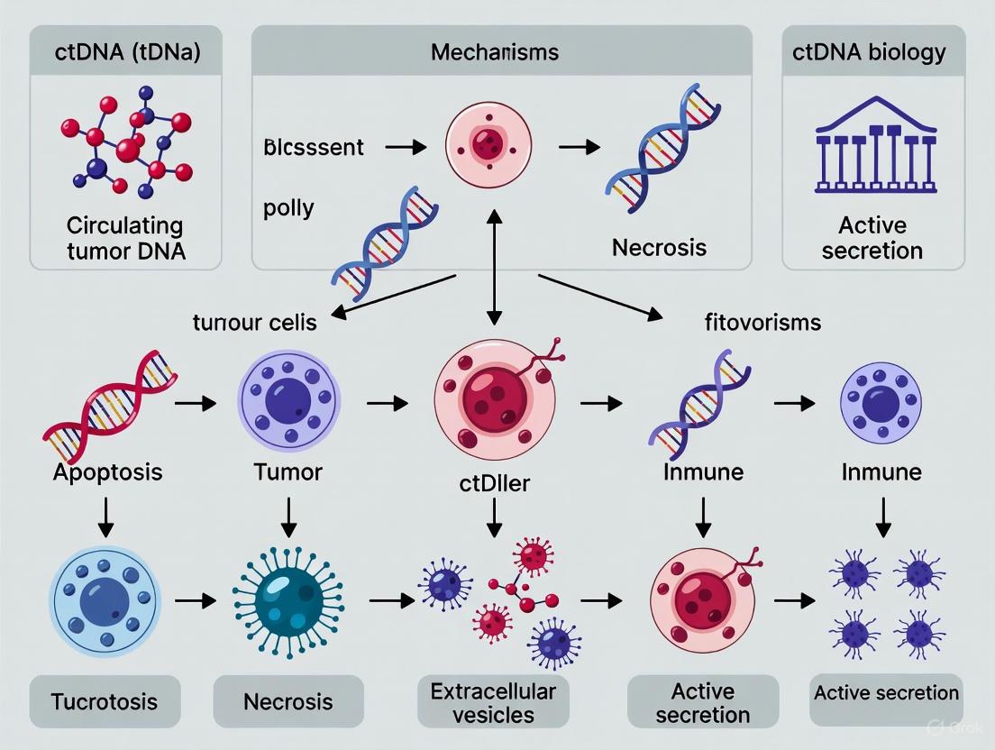
ctDNA Release Mechanisms and Clearance
Comprehensive ctDNA Analysis Workflow
Methylation Analysis Experimental Workflow
The comprehensive analysis of mutations, methylation patterns, microsatellite instability, and gene rearrangements in ctDNA provides unprecedented insights into tumor biology and enables sophisticated clinical applications in oncology. The integration of multiple analytical approaches—from highly sensitive PCR methods to comprehensive NGS panels—allows researchers and clinicians to extract maximal information from minimal invasive samples. As ctDNA analysis continues to evolve, standardization of pre-analytical procedures, validation of analytical performance, and demonstration of clinical utility will be essential for broader adoption in research and clinical practice. The ongoing development of more sensitive detection methods and multi-omic approaches promises to further enhance the value of ctDNA as a biomarker for precision oncology.
Advanced Detection Technologies and Expanding Clinical Applications of ctDNA
Circulating tumor DNA (ctDNA), a subset of cell-free DNA shed by tumors into the bloodstream, has emerged as a transformative biomarker in oncology. The analysis of ctDNA, often called liquid biopsy, provides a non-invasive means to assess tumor burden, genetic heterogeneity, and therapeutic response in real-time [19] [2]. However, a significant challenge lies in the fact that ctDNA can be present at very low concentrations, sometimes less than 0.1% of total cell-free DNA, especially in early-stage cancers or for monitoring minimal residual disease (MRD) [19]. This low abundance creates a pressing need for detection platforms with ultra-high sensitivity and specificity.
The core technologies developed to meet this challenge primarily fall into two categories: PCR-based methods (including digital PCR and BEAMing) and next-generation sequencing (NGS)-based approaches (such as CAPP-Seq, Safe-SeqS, and TEC-Seq). These platforms enable researchers and clinicians to overcome the fundamental limitation of detecting a low number of mutant molecules in a vast background of wild-type DNA, a capability that is crucial for unlocking the full clinical potential of liquid biopsies [19] [36].
PCR-Based Detection Platforms
Digital PCR (dPCR)
2.1.1 Fundamental Principles and Workflow Digital PCR (dPCR) is a method for the absolute quantification of target nucleic acids without the need for a standard curve [37]. The core principle involves partitioning a sample into many independent PCR sub-reactions—thousands to millions of individual partitions—such that each contains either zero, one, or a few target DNA molecules [37] [38]. Following end-point PCR amplification, the fraction of positive partitions is used to calculate the absolute concentration of the target sequence in the original sample based on Poisson statistics [37]. This partitioning step effectively concentrates the target sequences, reducing template competition and enabling the detection of rare mutations against a high background of wild-type sequences [37].
2.1.2 Key Experimental Protocol: dPCR for Rare Mutation Detection The standard workflow for dPCR in ctDNA analysis involves several key steps [37] [38]:
- Sample Preparation: Cell-free DNA (cfDNA) is extracted from patient plasma.
- Assay Design: Sequence-specific primers and fluorescent probes (e.g., TaqMan assays) are designed to distinguish mutant from wild-type alleles.
- Partitioning: The PCR reaction mix, containing the cfDNA sample, is partitioned into numerous individual reactions using microfluidic chambers (chip-based dPCR) or water-in-oil droplets (droplet digital PCR, ddPCR).
- Amplification: The partitioned samples undergo standard PCR cycling.
- Fluorescence Reading: Each partition is analyzed for fluorescence signal at the end of the amplification. Partitions containing the target sequence will fluoresce.
- Quantification and Analysis: The proportion of fluorescent-positive partitions is counted, and the absolute concentration of the mutant allele is calculated using Poisson correction to account for partitions containing more than one target molecule.
2.1.3 Performance and Applications dPCR offers a limit of detection (LOD) that can reach variant allele frequencies (VAF) of 0.1% or lower, making it 100-times more sensitive than conventional qPCR for rare mutation detection [38]. Its primary applications in ctDNA research include the absolute quantification of specific, known mutations, detection of rare mutations, and verification of NGS libraries [38]. A key advantage is its high tolerance to PCR inhibitors present in a sample due to the sample dilution during partitioning [37].
BEAMing (Beads, Emulsion, Amplification, and Magnetics)
2.2.1 Fundamental Principles and Workflow BEAMing is a specialized, highly sensitive technology that combines dPCR principles with flow cytometry. It transforms specific DNA sequences into detectable fluorescent beads, allowing for both quantification and isolation of mutant DNA molecules [19].
2.2.2 Key Experimental Protocol: BEAMing Workflow The BEAMing protocol consists of several stages [19]:
- Bead-Based Primer Coupling: Magnetic beads are coated with primers specific to the DNA target of interest.
- Emulsion PCR: The beads are mixed with the cfDNA sample and PCR reagents in a water-in-oil emulsion, creating millions of microreactors. Each bead acts as a separate PCR reactor. If a mutant DNA molecule is present in a microreactor, it will amplify and bind to the bead.
- Emulsion Breakage: After amplification, the emulsion is broken, and the beads are collected.
- Hybridization and Labeling: The beads are incubated with fluorescent probes designed to distinguish mutant from wild-type sequences.
- Flow Cytometry and Magnetization: The beads are analyzed by flow cytometry. Beads that carry mutant sequences will fluoresce and can be counted for quantification or isolated using a magnet for further downstream analysis.
2.2.3 Performance and Applications BEAMing is renowned for its exceptional sensitivity, capable of detecting mutations at frequencies as low as 0.01% [19]. It is particularly valuable for validating mutations discovered through NGS and for monitoring specific, known resistance mutations during targeted therapy.
Table 1: Comparison of Core PCR-Based ctDNA Detection Platforms
| Feature | Digital PCR (dPCR) | BEAMing |
|---|---|---|
| Core Principle | Sample partitioning into microreactors for absolute quantification | dPCR on primer-coated beads followed by flow cytometry |
| Key Technology | Microfluidic chips or droplets | Emulsion PCR and magnetic beads |
| Detection Method | End-point fluorescence | Fluorescence via flow cytometry |
| Limit of Detection (VAF) | ~0.001% - 0.1% [38] | ~0.01% [19] |
| Throughput | Medium to High | Medium |
| Multiplexing | Limited (typically 1-4 plex) | Limited |
| Primary Applications | Absolute quantification, rare mutation detection, NGS validation [38] | Ultra-sensitive detection and isolation of specific mutants [19] |
Diagram 1: Workflow comparison of dPCR and BEAMing technologies.
NGS-Based Detection Platforms
CAPP-Seq (Cancer Personalized Profiling by Deep Sequencing)
3.1.1 Fundamental Principles and Workflow CAPP-Seq is a targeted NGS approach that uses a selector—a set of biotinylated oligonucleotide probes—to capture and enrich a predefined set of genomic regions that are frequently mutated in a specific cancer type [39]. Its key innovation is the design of an "informative" panel that concentrates on the most relevant genomic regions, enabling highly sensitive and cost-effective detection even with a low input of ctDNA.
3.1.2 Key Experimental Protocol: CAPP-Seq Workflow The standard CAPP-Seq protocol involves [39]:
- cfDNA Extraction and Shearing: cfDNA is isolated from plasma and fragmented to an appropriate size if necessary.
- Library Preparation: Adapters containing unique molecular identifiers (UMIs) are ligated to the cfDNA fragments. UMIs are short random DNA sequences added to each original DNA molecule before amplification, allowing for the distinction of true mutations from PCR or sequencing errors.
- Hybridization and Capture: The library is hybridized with the custom CAPP-Seq biotinylated probe selector. The selector is designed to target several hundred exons in genes commonly mutated in a particular cancer.
- Pull-down and Amplification: The probe-DNA complexes are captured using streptavidin-coated magnetic beads, and the enriched target regions are amplified via PCR.
- High-Throughput Sequencing: The final library is sequenced to a high depth (often >10,000x coverage).
- Bioinformatic Analysis: Sequencing data is analyzed using a specialized bioinformatics pipeline that leverages UMIs for error suppression and variant calling, achieving a high signal-to-noise ratio.
3.1.3 Performance and Applications CAPP-Seq demonstrates a high sensitivity, with a reported limit of detection of 0.02% variant allele frequency and a specificity of 99.99% [39]. It is particularly useful for molecular profiling, treatment monitoring, and minimal residual disease (MRD) detection in a tumor-informed manner [39].
Safe-SeqS (Safe-Sequencing System)
3.2.1 Fundamental Principles and Workflow Safe-SeqS is an NGS-based method that employs a unique strategy for error correction to achieve ultra-sensitive mutation detection. Its core feature is the assignment of a unique identifier (UID) to each original DNA template before any amplification steps, enabling the accurate identification and quantification of true mutations.
3.2.2 Key Experimental Protocol: Safe-SeqS Workflow The Safe-SeqS methodology consists of the following steps [39]:
- UID Assignment: Each individual DNA molecule in the cfDNA sample is tagged with a unique, random oligonucleotide sequence during library preparation.
- Preamplification: The tagged DNA molecules are amplified to create "UID families"—groups of DNA sequences that all originate from the same initial molecule.
- Target Enrichment & Sequencing: The amplified products are subjected to further processing (e.g., targeted capture or amplicon-based enrichment) and sequenced to a high depth.
- Bioinformatic Analysis: Sequences are clustered based on their UID. A mutation is only considered real if it is present in a high percentage (e.g., >95%) of the sequences within a UID family. This stringent consensus calling effectively eliminates errors introduced during PCR or sequencing.
3.2.3 Performance and Applications Safe-SeqS is renowned for its extremely low error rate, enabling the detection of mutations at frequencies as low as 0.01% - 0.05% with high specificity (98.9%) [39]. Its primary applications include cancer detection, monitoring, and identification of targetable alterations.
TEC-Seq (Targeted Error Correction Sequencing)
3.3.1 Fundamental Principles and Workflow TEC-Seq is a comprehensive, ultra-deep sequencing method designed for the multiplexed assessment of mutations in a large panel of cancer-associated genes. It integrates targeted capture, high-depth sequencing, and a sophisticated bioinformatic error-correction model to distinguish true somatic mutations from technical artifacts.
3.3.2 Key Experimental Protocol: TEC-Seq Workflow The TEC-Seq protocol generally includes [39] [36]:
- Library Preparation with UMIs: cfDNA is used to construct sequencing libraries with the incorporation of unique molecular identifiers.
- Hybridization Capture: The libraries are enriched using a custom set of probes targeting a broad panel of cancer-related genes (e.g., 58-128 genes).
- Ultra-Deep Sequencing: The captured libraries are sequenced to a very high depth (typically >30,000x coverage) to detect low-frequency variants.
- Computational Error Suppression: A proprietary bioinformatics pipeline is applied to the sequencing data. This pipeline uses the UMI information and a background error model that accounts for context-specific sequencing artifacts to filter out noise and call low-frequency variants with high confidence.
3.3.3 Performance and Applications TEC-Seq can reliably detect mutations present at 0.1% VAF or lower. Its strength lies in its ability to screen for a wide range of mutations across many genes simultaneously, making it suitable for noninvasive genotyping, studying tumor heterogeneity, and monitoring treatment response in advanced cancers.
Table 2: Comparison of Core NGS-Based ctDNA Detection Platforms
| Feature | CAPP-Seq | Safe-SeqS | TEC-Seq |
|---|---|---|---|
| Core Principle | Targeted capture with a | UID-based error correction | Multiplexed targeted capture |
| Enrichment Method | Hybridization capture | UID assignment & pre-amplification | with bioinformatic error modeling |
| Limit of Detection (VAF) | 0.02% [39] | 0.01% - 0.05% [39] | Hybridization capture |
| Specificity | 99.99% [39] | 98.9% [39] | ~0.1% and below [39] [36] |
| Input (ng) | 32 [39] | 3 [39] | Not Specified |
| Primary Applications | MRD, treatment monitoring, molecular profiling [39] | Rare variant detection, monitoring [39] | Noninvasive genotyping, tumor heterogeneity [39] [36] |
Diagram 2: Generalized workflow for NGS-based ctDNA detection platforms.
The Scientist's Toolkit: Essential Research Reagent Solutions
The advanced ctDNA detection platforms described rely on a suite of specialized reagents and materials to achieve their high performance.
Table 3: Key Research Reagent Solutions for ctDNA Detection
| Reagent/Material | Function | Example Platforms Using |
|---|---|---|
| TaqMan Assays | Sequence-specific fluorescent probes for target detection and quantification in PCR. | dPCR [38] |
| Unique Molecular Identifiers (UMIs) | Short random nucleotide sequences used to tag individual DNA molecules prior to amplification, enabling error correction. | Safe-SeqS, CAPP-Seq, TEC-Seq [39] |
| Biotinylated Capture Probes | Oligonucleotides designed to hybridize with and enrich specific genomic regions of interest for targeted sequencing. | CAPP-Seq [39] |
| Microfluidic Array Plates (MAP) | Chips containing thousands of miniature wells for precise sample partitioning in dPCR. | Chip-based dPCR [38] |
| Streptavidin-Coated Magnetic Beads | Used to pull down and isolate biotinylated probe-DNA complexes during hybrid capture steps. | CAPP-Seq, BEAMing [19] [39] |
| DNA Polymerases for High-Fidelity PCR | Enzymes with low error rates essential for accurate amplification in both PCR and NGS library prep. | All platforms (implicit) |
The landscape of ctDNA detection is defined by a trade-off between the highly sensitive, quantitative, but low-plex capability of PCR-based methods (dPCR, BEAMing) and the broader, multiplexed discovery power of NGS-based approaches (CAPP-Seq, Safe-SeqS, TEC-Seq). The choice of platform is dictated by the specific clinical or research question—whether it is tracking a known mutation with utmost sensitivity or conducting a comprehensive search for heterogeneous genetic alterations. The ongoing development of these technologies, including the integration of fragmentomic analysis, methylation profiling, and AI-based error suppression, promises to further enhance the sensitivity and specificity of liquid biopsies, solidifying their role in precision oncology [19] [39].
Circulating tumor DNA (ctDNA) consists of fragmented, tumor-derived DNA present in the bloodstream, released through passive mechanisms such as apoptosis and necrosis and active secretion via extracellular vesicles [2] [29]. This fraction of cell-free DNA (cfDNA) carries tumor-specific genomic alterations, making it a powerful, non-invasive biomarker for cancer detection, monitoring, and management. The biology of ctDNA release is intrinsically linked to tumor dynamics; apoptosis produces shorter DNA fragments (~167 bp) protected by nucleosomes, while necrosis can release larger, more random fragments [2] [29]. However, a significant challenge in leveraging ctDNA, especially for minimal residual disease (MRD) detection and early cancer diagnosis, is its extremely low abundance in plasma, often falling below the detection limit of conventional sequencing methods [40] [41]. This limitation creates a pressing need for ultrasensitive detection technologies. This guide details two advanced approaches—Phased Variant Detection (PhasED-Seq) and Structural Variant (SV) Assays—that are designed to overcome these sensitivity barriers by exploiting distinct features of the cancer genome, thereby providing researchers and drug development professionals with robust tools for precision oncology.
Phased Variant Detection (PhasED-Seq)
Core Principle and Technological Basis
Phased Variant Enrichment and Detection Sequencing (PhasED-Seq) is a groundbreaking technology that enhances ctDNA detection sensitivity by tracking phased variants (PVs). PVs are defined as two or more single nucleotide variants (SNVs) occurring in close proximity (in cis) on the same DNA molecule [40] [41]. The core innovation of PhasED-Seq lies in leveraging these multi-mutation haplotypes to drastically reduce the background error rate that plagues conventional, single-SNV detection methods. Because the spontaneous, coincidental occurrence of two specific errors on a single DNA strand is exceedingly rare, the requirement for multiple mutation calls per molecule confers a high degree of specificity, enabling the detection of ctDNA at concentrations below one part per million (ppm) [40] [42] [43]. This approach outperforms first-generation ctDNA assays, including those using duplex barcoding, by achieving a lower limit of detection without the inefficient molecular recovery associated with duplex sequencing [41].
Genomic Distribution and Biological Origins
Phased variants are not uniformly distributed across cancers. Whole-genome sequencing analyses of 2,538 tumors reveal significant enrichment of PVs in B-cell lymphomas, such as diffuse large B-cell lymphoma (DLBCL) and follicular lymphoma (FL) [41]. This enrichment is mechanistically driven by the activity of the activation-induced cytidine deaminase (AID/AICDA) enzyme during physiological and aberrant somatic hypermutation (aSHM) [41]. PVs in these malignancies are frequently clustered in stereotyped genomic regions, including known targets of SHM like the BCL2, BCL6, and MYC oncogenes, as well as the immunoglobulin loci (IGH, IGK, IGL) [41]. In solid tumors, PVs are often associated with other mutational processes, such as APOBEC-mediated kataegis and tobacco-related mutational signatures [41]. The recurrent and predictable nature of PVs in certain cancers, particularly lymphomas, makes them ideal targets for designing highly sensitive and specific capture panels.
Experimental Protocol and Workflow
The following diagram illustrates the core conceptual workflow of the PhasED-Seq method for detecting phased variants from ctDNA.
The experimental protocol for PhasED-Seq can be broken down into key steps:
- Sample Collection and cfDNA Extraction: Collect patient blood in cell-stabilizing tubes. Isolate plasma via centrifugation and extract cfDNA using commercially available kits [43]. The typical input for the PhasED-Seq MRD assay is 120 ng of cfDNA.
- Library Preparation and Target Enrichment: Prepare sequencing libraries from cfDNA. Subsequently, use a custom hybrid-capture panel to enrich for genomic regions known to be rich in PVs. The validated panel for B-cell malignancies targets approximately 115 kb of PV-dense space and an additional 200 kb of recurrently mutated genes in B-cell non-Hodgkin lymphomas, providing a ~7500-fold enrichment over whole-genome sequencing [41].
- Sequencing: Perform deep sequencing on an Illumina platform to achieve a high median depth of coverage (typically >20,000x) for sensitive mutation detection [43].
- Bioinformatic Analysis:
- Variant Calling: Identify single nucleotide variants from the sequencing data.
- Phasing Analysis: Analyze sequencing reads to detect molecules harboring two or more SNVs within a short distance (less than ~170 bp, the typical length of a mononucleosomal cfDNA fragment).
- MRD Calling: A personalized set of PVs (a "PV list") is generated for each patient from a baseline tumor sample or high ctDNA fraction plasma sample. This patient-specific PV list is then used to interrogate subsequent samples for MRD detection [41] [43].
Performance and Validation Data
Robust analytical validation studies demonstrate the exceptional performance of PhasED-Seq. The table below summarizes key performance metrics from a recent validation study in B-cell malignancies [43].
Table 1: Analytical Performance Metrics of a PhasED-Seq-based MRD Assay
| Performance Metric | Result | Experimental Detail |
|---|---|---|
| Limit of Detection (LoD) at 95% | 0.7 parts per million (PVAF of 6.61E-07) | 120 ng input cfDNA |
| Background Error Rate | 1.95E-08 (1.95 mutant molecules per 100 million) | Measured using blank plasma samples |
| Analytical Specificity (False Positive Rate) | 0.24% | Across 4,200 technical replicates of blank samples |
| Assay Precision | >96% | Repeatability and reproducibility across operators, reagent lots, and time |
| Positive Percent Agreement (PPA) | 90.62% vs. an SNV-based method | Clinical plasma samples from DLBCL patients |
| Negative Percent Agreement (NPA) | 77.78% vs. an SNV-based method | Clinical plasma samples from DLBCL patients |
In a clinical study of DLBCL patients, PhasED-Seq identified an additional 25% of patients with detectable ctDNA after two cycles of therapy who had been classified as negative by CAPP-Seq. These patients had worse outcomes, underscoring the clinical prognostic value of this enhanced sensitivity [41].
Structural Variant (SV) Assays
Core Principle and Biological Significance
Structural variants (SVs) are large-scale genomic rearrangements—including deletions, duplications, inversions, translocations, and copy-number variants—that affect a substantial number of base pairs. They are a major driver of oncogenesis, with at least 30% of cancers harboring a pathogenic SV used in diagnosis or treatment stratification [44]. SVs can create novel fusion genes, disrupt tumor suppressors, and amplify oncogenes, making them highly specific biomarkers for liquid biopsy assays. However, their detection in ctDNA is complicated by biological and computational challenges, including intratumor heterogeneity, polyploidy, and the difficulty of distinguishing tumor-specific somatic SVs from germline variants present in healthy cells [44].
Detection Methodologies and Computational Workflow
Detecting SVs from short-read whole-genome sequencing (WGS) data relies on identifying specific patterns in aligned sequencing reads. The following diagram outlines the primary signals and the integrated computational approach required for robust SV detection.
No single algorithm performs optimally across all SV types and sizes. Therefore, a combinatorial approach integrating multiple algorithms is considered best practice for full-spectrum SV detection with high recall and precision [44]. The general workflow involves:
- Sequencing and Alignment: Perform deep WGS (often 75-90x coverage for tumor samples) on paired tumor and normal DNA. Align reads using an aligner like BWA-MEM to a reference genome (preferably GRCh38) [44].
- SV Calling with Multiple Tools: Run several specialized SV-calling algorithms (e.g., DELLY, LUMPY, Manta, SvABA, GRIDSS) on the aligned data. Each tool integrates different combinations of read-depth, discordant read-pair, and split-read evidence to call SVs [44].
- Callset Integration: Merge the results from the individual callers. To prioritize precision for clinical applications, an intersection strategy is often employed, though this can reduce recall. More advanced methods merge SVs based on breakpoint proximity and read-evidence support [44].
- Somatic Filtering: Compare the integrated tumor SV callset against the matched normal sample to identify tumor-specific SVs (TSSVs). This can be done by tools that probabilistically compare evidence between samples (e.g., Varlociraptor, Lancet) or by filtering out SVs with any supporting reads in the normal sample [44].
Performance Considerations and Challenges
The reliable detection of SVs, particularly subclonal variants with low allele frequency, is constrained by several factors. A minimum allele frequency of 20% is often required for reliable detection from tumor-normal WGS data, though this threshold can be lowered by increasing sequencing depth [44]. Distinguishing true somatic SVs from germline variants remains a primary challenge, necessitating the use of paired normal samples or large panels-of-normals for effective filtering. Furthermore, complex genomic architectures and the limitations of short-read sequencing in resolving repetitive or low-complexity regions can lead to false positives and missed variants. The integration of long-read sequencing technologies is emerging as a powerful method to overcome these limitations and validate SVs detected by short-read data [44].
The Scientist's Toolkit: Essential Research Reagents and Materials
Implementing PhasED-Seq and SV assays requires a suite of specialized reagents, computational tools, and sample types. The following table details the key components of the research toolkit for these ultrasensitive approaches.
Table 2: Key Research Reagent Solutions for Ultrasensitive ctDNA Detection
| Item | Function/Description | Example/Note |
|---|---|---|
| Cell-free DNA Collection Tubes | Preserves blood sample integrity and prevents white blood cell lysis during transport and storage. | Streck Cell-Free DNA BCT tubes are commonly used. |
| Hybrid-Capture Panels | Enriches sequencing libraries for genomic regions of interest, maximizing on-target reads. | Custom PhasED-Seq panel (~115 kb for PVs, ~200 kb for genes) [41]. |
| Molecular Barcoding Adapters | Tags individual DNA molecules pre-PCR to correct for amplification errors and deduplicate reads. | Not strictly required for PhasED-Seq but used in many ultrasensitive protocols. |
| High-Fidelity DNA Polymerase | Reduces errors introduced during PCR amplification in library preparation. | Enzymes like Q5 High-Fidelity DNA Polymerase. |
| SV Detection Algorithms | Software that identifies SVs from WGS data based on specific read signatures. | DELLY, Manta, GRIDSS, LUMPY, SvABA [44]. |
| Somatic Variant Filtering Tools | Differentiates tumor-specific variants from germline polymorphisms in sequencing data. | Varlociraptor, Lancet; also used with PhasED-Seq data [44]. |
| Reference Control Samples | Validates assay performance, used for determining limit of detection and error rates. | Commercial cfDNA reference standards or clinical-contrived samples from patient-derived material [43]. |
The advent of ultrasensitive detection technologies like PhasED-Seq and sophisticated SV assays marks a significant leap forward in the field of liquid biopsy. By moving beyond single SNV detection to exploit the rich information contained in phased mutation haplotypes and large-scale genomic rearrangements, these methods push the limits of ctDNA detection into the parts-per-million range. This enhanced sensitivity is critical for applications with low tumor burden, such as minimal residual disease monitoring and early cancer detection [45] [42]. A deep understanding of ctDNA biology—including its release mechanisms, fragmentomic profile, and clearance—is fundamental to the continued refinement of these assays [2] [29]. As these technologies mature and are standardized, they hold immense promise for integrating into clinical trials and routine practice, ultimately enabling earlier intervention and more personalized cancer therapy.
Circulating tumor DNA (ctDNA) has emerged as a transformative biomarker in oncology, representing small fragments of DNA (typically 90-150 base pairs) released into the bloodstream through apoptosis, necrosis, and active secretion from tumor cells [2]. These fragments carry tumor-specific genetic and epigenetic alterations, providing a non-invasive window into tumor dynamics, heterogeneity, and treatment response [5] [19]. The half-life of ctDNA is remarkably short, estimated between 16 minutes and several hours, enabling real-time monitoring of disease progression and therapeutic efficacy [5] [2]. However, the clinical application of ctDNA analysis faces significant challenges due to its ultralow abundance in early-stage cancers and minimal residual disease, where it can constitute less than 0.01% of total cell-free DNA [19] [46].
The convergence of ctDNA biology with advanced biosensing technologies has created unprecedented opportunities for cancer detection and monitoring. Conventional detection methods, including polymerase chain reaction (PCR) and next-generation sequencing (NGS), while valuable, face limitations in sensitivity, cost, turnaround time, and potential for point-of-care application [47] [19]. In response to these challenges, nanomaterial-based electrochemical biosensors and magnetic nano-electrode systems have emerged as next-generation platforms offering attomolar sensitivity, rapid analysis, and compatibility with miniaturized diagnostic devices [19] [46] [48]. These technologies leverage the unique electrical, magnetic, and structural properties of nanomaterials to overcome the fundamental limitations of conventional ctDNA detection methods, potentially enabling routine clinical implementation for personalized cancer management.
ctDNA Biology and Release Mechanisms: Implications for Detection
Understanding ctDNA biology is paramount for designing effective detection technologies. CtDNA release occurs primarily through two mechanisms: passive release from dying cells (apoptosis and necrosis) and active secretion from viable tumor cells [2]. Apoptosis produces characteristic ctDNA fragments of approximately 167 base pairs, corresponding to DNA wrapped around nucleosomes with linker regions, yielding a ladder-like pattern on gel electrophoresis [2]. In contrast, necrotic cells release larger, more random DNA fragments due to uncontrolled cellular disintegration [2]. The relative contributions of these release mechanisms influence ctDNA characteristics, including fragment size distribution and methylation patterns, which can be exploited for enhanced detection specificity [19] [2].
The concentration of ctDNA in circulation correlates with tumor burden, disease stage, and cancer type, presenting a moving target for detection technologies [5]. In early-stage cancers, ctDNA may be present at frequencies as low as 0.001% of total cell-free DNA, creating a significant signal-to-noise challenge [19]. Additionally, pre-analytical variables including sample collection, processing, and DNA extraction can significantly impact detection efficacy [19]. These biological and technical considerations directly inform the design requirements for biosensing platforms, necessitating exceptional sensitivity, specificity, and robustness against complex biological matrices.
Nanomaterial-Based Electrochemical Biosensors for ctDNA Detection
Fundamental Principles and Mechanisms
Electrochemical biosensors transduce biochemical events into measurable electrical signals (current, potential, impedance) through specific interactions between target ctDNA and recognition elements (probes) immobilized on the sensor surface [49] [48]. The exceptional sensitivity of recent platforms derives from nanomaterial integration, which increases electrode surface area, enhances mass transport, improves catalytic activity, and facilitates signal amplification [49] [48]. These nanomaterials include gold nanoparticles, carbon nanotubes, graphene, quantum dots, and metal oxides, each contributing unique electrochemical properties to the sensing interface [49].
A particularly advanced platform utilizes gold-coated magnetic nanoparticles (Au@MNPs) functionalized with methylene blue-labeled DNA probes complementary to target ctDNA sequences [46]. This system employs a "dispersible electrode" concept where nanoparticles distributed throughout the sample solution drastically reduce diffusional pathlengths, enabling rapid target capture. Subsequent magnetic concentration of these nanoparticle complexes onto a working electrode facilitates highly sensitive electrochemical measurement [46]. The platform incorporates a redox amplification cycle where methylene blue is reduced to leucomethylene blue, which is then re-oxidized by ferricyanide in solution, generating significantly enhanced signals [46].
Performance Metrics and Analytical Characteristics
The table below summarizes the performance characteristics of representative nanomaterial-based electrochemical biosensors for ctDNA detection:
Table 1: Performance Metrics of Nanomaterial-Based Electrochemical Biosensors for ctDNA Detection
| Nanomaterial Platform | Detection Mechanism | Linear Range | Limit of Detection | Response Time | Target Sequence |
|---|---|---|---|---|---|
| DNA-Au@MNP Network [46] | Square Wave Voltammetry with redox amplification | 2 aM - 20 nM | 3.3 aM (short strand), 5 fM (101-nt ctDNA) | 20 minutes | 22-nt DNA; 101-nt NSCLC-associated ctDNA |
| Magnetic Bead-Assisted System [50] | Differential Pulse Voltammetry (DPV) | 100 pM - 500 nM | 100 pM | Not specified | PIK3CA gene mutations |
| Graphene/MoS₂-based sensors [19] | Impedance spectroscopy | Not specified | Attomolar range | <30 minutes | Various ctDNA sequences |
| Magnetic nano-electrode systems [19] | Electrochemical detection post-PCR amplification | Not specified | Attomolar range | 7 minutes post-PCR | Various ctDNA sequences |
These platforms demonstrate significant advantages over conventional detection methods, achieving detection limits up to six orders of magnitude lower than traditional techniques while substantially reducing analysis time [19] [46]. The exceptional sensitivity enables detection of ctDNA at clinically relevant concentrations for early-stage cancers, while the rapid response facilitates near-real-time monitoring of disease dynamics.
Experimental Protocol: Au@MNP-Based ctDNA Detection
The following protocol details the experimental procedure for ctDNA detection using the gold-coated magnetic nanoparticle platform [46]:
Synthesis and Functionalization of Au@MNPs:
- Synthesize gold-coated magnetic nanoparticles (approximately 20-30 nm diameter) via reduction of gold salts onto magnetic nanoparticle cores.
- Functionalize Au@MNPs with thiolated DNA probes (complementary to target ctDNA) labeled with methylene blue at the 3' end through self-assembled monolayer formation. Use 6-mercapto-1-hexanol (MCH) as a passivating agent to minimize non-specific adsorption.
Sample Preparation and Hybridization:
- Extract cell-free DNA from plasma samples using silica-based membrane columns or magnetic bead-based methods.
- Incubate 10 μL of functionalized Au@MNP solution (1.56 × 10^10 particles/mL) with extracted ctDNA sample for 30 minutes at controlled temperature (specific temperature depends on probe-target melting characteristics).
Magnetic Separation and Washing:
- Separate nanoparticle complexes from solution using a magnetic rack.
- Remove supernatant and wash twice with phosphate-buffered saline (pH 7.4) to remove unbound contaminants.
Electrochemical Measurement:
- Resuspend washed nanoparticle complexes in 0.5 mM potassium ferricyanide in PBS.
- Transfer to custom electrochemical cell and concentrate onto gold electrode surface using a magnet.
- Perform Square Wave Voltammetry with parameters: potential range -0.2 to -0.5 V (vs. Ag/AgCl), frequency 15 Hz, amplitude 25 mV.
- Measure reduction current from methylene blue labels.
Data Analysis:
- Quantify ctDNA concentration based on decrease in peak current relative to negative controls.
- Generate calibration curve using synthetic DNA standards of known concentration.
Magnetic Nano-Electrode Systems and Integrated Platforms
Magnetic nano-electrode systems represent a synergistic integration of magnetic nanoparticle technology with electrochemical sensing platforms [19]. These systems typically employ superparamagnetic Fe₃O₄–Au core–shell particles that serve dual functions: as solid supports for ctDNA capture/enrichment and as electrochemical transducers [19]. The fundamental operating principle involves specific capture of target ctDNA sequences using functionalized magnetic nanoparticles, followed by magnetic concentration onto electrode surfaces for highly sensitive electrochemical detection [50] [19].
A notable implementation combines magnetic bead-assisted capture with ligase chain reaction (LCR) for enhanced specificity in detecting single-nucleotide polymorphisms (SNPs) [50]. This approach uses two adjacent oligonucleotides that hybridize to target DNA at the mutation site; a thermostable DNA ligase covalently joins them only if a perfect match is present, providing exceptional specificity for point mutation detection [50]. The ligation products are then captured using streptavidin-coated magnetic beads and detected via anodic differential pulse voltammetry, achieving sensitive detection of cancer-associated mutations in the PIK3CA gene [50].
Performance Advantages and Clinical Utility
Magnetic nano-electrode systems achieve remarkable sensitivity, with some platforms detecting ctDNA at attomolar concentrations (10^-18 M) within 7 minutes of PCR amplification [19]. This exceptional performance stems from several factors: the high surface area-to-volume ratio of nanomaterials, efficient magnetic enrichment of targets from complex matrices, and the synergistic combination of nucleic acid amplification with electrochemical detection [19]. These systems effectively address key challenges in ctDNA analysis, including low abundance, high background from wild-type DNA, and sequence heterogeneity [50] [19].
The clinical utility of these platforms has been demonstrated across multiple cancer types. In non-small cell lung cancer (NSCLC), magnetic nano-electrode systems have enabled detection of EGFR mutations associated with treatment response and resistance [19]. Similarly, in breast cancer, these platforms have successfully identified PIK3CA mutations with high specificity in commercial human serum samples, confirming their robustness in clinically relevant matrices [50]. The compatibility of these systems with miniaturization and point-of-care testing positions them as promising technologies for decentralized cancer diagnostics and monitoring.
Experimental Protocol: Magnetic Bead-Assisted LCR-Electrochemical Detection
The following protocol details the integrated ligase chain reaction and electrochemical detection method for ctDNA mutation analysis [50]:
Probe Design and Ligation:
- Design two probe sets: one with ferrocene label at 5' end (electrochemical reporter) and another with biotin at 3' end (for magnetic capture).
- Perform Ligase Chain Reaction with the following cycling conditions: 95°C denaturation (30 sec), 60°C annealing/ligation (2 min) for 30 cycles using thermostable DNA ligase.
- Include appropriate controls (wild-type sequences, no-template controls) to validate reaction specificity.
Magnetic Capture and Separation:
- Incubate LCR products with streptavidin-coated magnetic beads (e.g., Dynabeads) for 15 minutes at room temperature with gentle mixing.
- Apply magnetic field to separate bead-bound complexes from solution.
- Wash three times with appropriate buffer to remove non-specifically bound materials.
Electrochemical Detection:
- Transfer beads with captured ctDNA to electrochemical cell containing appropriate electrolyte.
- Perform Anodic Differential Pulse Voltammetry with parameters: potential range 0 to +0.6 V (vs. reference electrode), pulse amplitude 50 mV, pulse width 50 ms.
- Measure ferrocene oxidation current as detection signal.
Data Interpretation:
- Identify positive signals based on oxidation current exceeding threshold (typically 3× standard deviation of negative controls).
- Quantify mutant allele frequency based on calibration curves generated from synthetic standards.
Research Reagent Solutions and Essential Materials
Successful implementation of nanomaterial-based ctDNA detection platforms requires specific reagents and materials optimized for these applications. The following table details essential components and their functions:
Table 2: Essential Research Reagents for Nanomaterial-Based ctDNA Biosensing
| Reagent/Material | Function | Specific Examples | Key Considerations |
|---|---|---|---|
| Gold-coated Magnetic Nanoparticles | Dispersible electrode platform; enables magnetic concentration | Fe₃O₄–Au core-shell nanoparticles [46] | Size uniformity, magnetic responsiveness, surface functionalization capacity |
| Specific DNA Probes | Target recognition and hybridization | Thiol-modified, methylene blue-labeled DNA probes [46] | Specificity, melting temperature, secondary structure minimization |
| Signal Amplification Reagents | Enhanced electrochemical detection | Potassium ferricyanide [46] | Compatibility with redox labels, stability in solution |
| Magnetic Beads | Target capture and separation | Streptavidin-coated magnetic beads [50] | Binding capacity, minimal non-specific adsorption, size uniformity |
| Surface Passivation Agents | Reduce non-specific binding | 6-mercapto-1-hexanol (MCH) [46] | Complete coverage without disrupting probe functionality |
| Thermostable DNA Ligase | Specific mutation detection in LCR | Thermophilic DNA ligase [50] | Fidelity, temperature stability, compatibility with reaction buffers |
| Electrochemical Redox Markers | Signal generation | Ferrocene derivatives [50] | Reversible electrochemistry, stable signal generation |
Technology Integration and Future Perspectives
The convergence of nanomaterial-based biosensing with ctDNA biology represents a paradigm shift in cancer diagnostics. Future developments will likely focus on multiplexed detection platforms capable of simultaneously monitoring multiple cancer-associated mutations, integration with microfluidic systems for automated sample processing, and implementation of artificial intelligence for enhanced data analysis and error correction [51] [19]. Additionally, the incorporation of epigenetic markers such as DNA methylation patterns alongside mutational analysis may further improve cancer detection specificity and tissue-of-origin determination [19].
The path toward clinical validation and commercialization requires addressing several challenges, including standardization of assay protocols, demonstration of reproducibility across multiple sites, and validation in large-scale clinical trials [19]. As these technologies mature, they hold immense potential to transform cancer management through non-invasive, real-time monitoring of tumor dynamics, enabling earlier detection of treatment resistance and disease recurrence than currently possible with conventional imaging or tissue biopsy.
Visualizations
ctDNA Biosensing Mechanism
Magnetic Nano-Electrode Workflow
Circulating tumor DNA (ctDNA) refers to small fragments of DNA that are released by tumor cells into the bloodstream through various mechanisms, including apoptosis, necrosis, and active secretion [27] [5]. As a subset of total cell-free DNA (cfDNA), ctDNA carries tumor-specific genetic features such as somatic mutations, copy number alterations, and distinct fragmentation patterns that distinguish it from cfDNA derived from healthy cells [27]. The fragmentomic landscape of ctDNA has emerged as a critical area of investigation, as tumor-derived fragments exhibit characteristic patterns related to their cellular origins. Notably, ctDNA fragments tend to be shorter than non-cancer cfDNA fragments, with a significant portion falling below 100 base pairs in length [27] [52]. This fundamental difference in fragment size distribution forms the biological basis for leveraging size selection in library preparation to enhance the sensitivity of ctDNA detection.
The clinical significance of fragmentomics extends across the cancer care continuum, from early detection to therapy monitoring. As tumor cells undergo apoptosis, their DNA is cleaved in a pattern that reflects the underlying nucleosomal architecture, resulting in a predominant fragment size of approximately 166-167 bp, corresponding to DNA wrapped around a single histone complex [53]. However, ctDNA fragments often deviate from this pattern, exhibiting not only shorter lengths but also distinct end motifs and preferences for cleavage at specific genomic locations [54] [5]. These fragmentomic signatures provide a rich source of biological information that can be exploited for clinical applications, particularly in cases where traditional mutation-based detection reaches its sensitivity limits due to low tumor fraction [52] [53].
Library Preparation Fundamentals for ctDNA Analysis
Core Principles of NGS Library Construction
Next-generation sequencing library preparation is a critical process that converts native nucleic acids into a format compatible with sequencing platforms. Conventional library construction protocols consist of four fundamental steps: fragmentation of DNA, end repair of fragmented DNA, ligation of adapter sequences, and optional library amplification [55]. The process begins with fragmentation of input DNA to achieve desired fragment sizes, typically ranging from 300-600 bp for general applications, though ctDNA analysis often focuses on much shorter fragments [56]. Following fragmentation, DNA ends must be repaired to generate blunt-ended, 5'-phosphorylated fragments compatible with adapter ligation strategies. This end repair process exploits the 5'→3' polymerase and 3'→5' exonuclease activities of T4 DNA polymerase, while T4 polynucleotide kinase ensures 5'-phosphorylation of the blunt-ended DNA fragments [55] [56].
The prepared fragments then undergo adapter ligation, where double-stranded adapters are attached to the end-repaired library fragments using T4 DNA ligase [55]. These adapters serve dual purposes: they enable selective clonal amplification of library molecules and provide docking sites for platform-specific sequencing primers [55]. A critical consideration at this stage is the prevention of adapter dimer formation, where excess adapters ligate to each other, creating small fragments that compete with library fragments during sequencing and reduce overall data quality [55] [57]. Following ligation, reaction cleanup and DNA size selection are performed to remove free library adapters and adapter dimers, typically using magnetic beads, column-based purification methods, or agarose gel isolation [55].
Adaptation of Library Protocols for ctDNA Analysis
Standard library preparation protocols require significant adaptation to address the unique challenges of ctDNA analysis. The vanishingly low abundance of ctDNA in circulation—often constituting less than 1-10% of total cfDNA in early-stage cancers—demands exceptional sensitivity and minimal loss throughout the library preparation process [58] [27]. Input material limitations necessitate optimized protocols that maintain library complexity while minimizing amplification biases that can obscure true biological signals [57] [59]. Additionally, the naturally short length of ctDNA fragments requires modified size selection strategies that specifically enrich for these fragments while excluding the longer background cfDNA from healthy cells [52].
The need for enhanced sensitivity in ctDNA detection has driven the development of specialized library preparation methods that incorporate unique molecular identifiers (UMIs) to distinguish true low-frequency variants from PCR amplification artifacts and sequencing errors [5]. These molecular barcodes are tagged onto DNA fragments before PCR amplification, enabling bioinformatic correction of errors that arise during library amplification [5]. More advanced techniques such as Duplex Sequencing tag and sequence both strands of a DNA duplex, providing even greater accuracy by requiring that true mutations appear in the same position on both strands [5]. Recent innovations like Concatenating Original Duplex for Error Correction (CODEC) have demonstrated 1000-fold higher accuracy than conventional NGS while using up to 100-fold fewer reads than duplex sequencing [5].
Size Selection Methodologies and Experimental Protocols
In Vitro Size Selection Techniques
In vitro size selection represents a powerful strategy for enhancing the detection of ctDNA by leveraging its characteristic fragmentation pattern. This approach involves physical separation of DNA fragments based on length following library construction but prior to sequencing. Recent studies have demonstrated that targeted isolation of short cfDNA fragments can significantly enrich the mutant allele fraction (MAF) of tumor-derived sequences [52]. In a comprehensive study involving 35 stage III and IV lung cancer patients, researchers found that ctDNA fragments harboring tumor mutations exhibited distinct length profiles compared to cfDNA fragments with clonal hematopoiesis or germline mutations [52]. Implementation of in vitro size selection resulted in a median 1.36-fold enrichment of tumor mutation MAF, while CH/germline mutations showed no enrichment (median 0.95-fold) [52]. This selective enrichment translated to improved detection of key oncogenic drivers, with KRAS and EGFR mutations more likely to exhibit MAF increases following size selection.
The experimental workflow for in vitro size selection typically begins with plasma separation from peripheral blood collected in specialized blood collection tubes containing cell-stabilizing preservatives (e.g., Streck Cell-Free DNA BCT) [54] [58]. A two-step centrifugation protocol is employed: first, tubes are centrifuged at 1600× g for 10 minutes at 15°C, followed by transfer of plasma to a new tube for a second centrifugation at 16,000× g for 10 minutes at room temperature [54]. cfDNA is then extracted from plasma using silica membrane-based kits (e.g., QIAamp Circulating Nucleic Acid Kit), which have been shown to yield more ctDNA than methods utilizing magnetic beads [54] [58]. Following extraction, libraries are prepared using standard NGS library preparation kits with modified size selection steps.
Table 1: Comparison of Size Selection Performance in Lung Cancer Study
| Parameter | Without Size Selection | With Size Selection | Improvement |
|---|---|---|---|
| Median MAF Enrichment (Tumor Mutations) | Reference | 1.36-fold | 36% increase |
| Median MAF Enrichment (CH/Germline Mutations) | Reference | 0.95-fold | No enrichment |
| Aneuploidy-Positive Samples | 8/35 (23%) | 20/35 (57%) | 150% increase |
| Oncogenic Driver Detection (KRAS, EGFR) | Baseline | Enhanced MAF | Key drivers preferentially enriched |
Detailed Size Selection Protocol
A standardized protocol for in vitro size selection in ctDNA analysis involves the following steps:
Library Preparation: Convert extracted cfDNA into sequencing libraries using a preferred NGS library preparation kit, following manufacturer's instructions through the adapter ligation step [55] [56].
Post-Ligation Cleanup: Perform initial cleanup using magnetic beads (e.g., AMPure XP beads) with a bead-to-sample ratio of 0.8:1 to remove excess adapters and reagents while retaining fragments >100 bp [57].
Size Selection Setup: Prepare a 2-4% agarose gel or use a precast gradient gel system. As an alternative, use automated size selection systems (e.g., Pippin Prep, BluePippin) for higher reproducibility [57].
Gel Electrophoresis: Load the purified library onto the gel alongside an appropriate DNA size ladder. Run electrophoresis at 100V for 45-60 minutes until adequate separation is achieved between the adapter dimer band (~120-140 bp) and the library insert band (>150 bp) [57].
Target Band Excision: Visualize the gel using SYBR-safe staining and UV transillumination. Excise the region corresponding to 140-220 bp, which contains the short ctDNA-enriched fragments [52]. For early-stage cancers or low-shedding tumors, a narrower size range (150-180 bp) may provide greater enrichment.
DNA Extraction from Gel: Recover DNA from the excised gel slice using a gel extraction kit according to manufacturer's instructions. Elute in an appropriate buffer volume (typically 15-25 μL) [57].
Library Amplification (Optional): If necessary, perform limited-cycle PCR amplification (typically 4-8 cycles) to increase library concentration using high-fidelity DNA polymerases with minimal GC bias [55].
Final Purification: Clean up the amplified library using magnetic beads with a bead-to-sample ratio of 0.9:1 to remove primer dimers and concentrate the library for sequencing [57].
Quality Control: Quantify the final library using fluorometric methods (e.g., Qubit) and assess size distribution using a bioanalyzer or tape station before sequencing [55] [59].
Fragmentomics Analysis and Computational Approaches
Key Fragmentomics Metrics
Fragmentomics analysis extends beyond simple fragment length measurement to encompass multiple metrics that collectively provide a comprehensive view of ctDNA characteristics. These metrics can be calculated from sequencing data and leveraged to distinguish cancer-derived fragments from background cfDNA. Research comparing various fragmentomics approaches has identified several key metrics with strong predictive power for cancer detection and classification [53]:
Normalized Fragment Read Depth: This metric involves counting fragments overlapping specific genomic regions and normalizing for both sequencing depth and region size. In comparative analyses, normalized depth across all exons provided the best overall performance for predicting cancer types and subtypes, with an average AUROC of 0.943 in one cohort and 0.964 in another [53].
Fragment Length Proportions: This includes the fraction of small fragments (<150 bp) and the distribution of fragments across various size bins. Cancer-derived cfDNA typically shows an increased proportion of shorter fragments compared to healthy cfDNA [53].
Shannon Entropy of Fragment Sizes: This diversity metric quantifies the spread of fragment sizes in a genomic region, with lower entropy indicating more uniform fragment sizes. Shannon entropy calculated at the first exon of genes has demonstrated utility in cancer phenotyping [53].
End Motif Diversity Score (MDS): This metric quantifies the variation in 4-mer end motifs among fragments. In some cancer types, particularly small cell lung cancer, MDS across all exons emerged as the top-performing metric with an AUROC of 0.888 [53].
Transcription Factor Binding Site (TFBS) Entropy: This approach analyzes fragment size diversity overlapping transcription factor binding sites, leveraging the protective effect of DNA-binding proteins on fragmentation patterns [53].
Table 2: Performance Comparison of Fragmentomics Metrics in Cancer Detection
| Fragmentomics Metric | Average AUROC (UW Cohort) | Average AUROC (GRAIL Cohort) | Best Performing Cancer Type |
|---|---|---|---|
| Normalized Depth (All Exons) | 0.943 | 0.964 | Multiple |
| Normalized Depth (First Exon) | 0.930 | 0.956 | Healthy samples |
| Shannon Entropy (All Exons) | 0.867 | 0.922 | Renal Cell Carcinoma |
| End Motif Diversity (All Exons) | 0.841 | 0.893 | Small Cell Lung Cancer |
| Small Fragment Proportion | 0.855 | 0.905 | Prostate Adenocarcinoma |
| TFBS Entropy | 0.812 | 0.874 | Breast Cancer (ER-negative) |
Analytical Workflow for Fragmentomics
The computational analysis of fragmentomics data follows a structured workflow that begins with raw sequencing data and culminates in clinical interpretations. The standard analytical pipeline includes:
Sequencing Data Processing: Raw sequencing reads are demultiplexed and aligned to a reference genome using standard alignment tools (e.g., BWA, Bowtie2), with careful retention of fragment size information [53].
Duplicate Marking: PCR duplicates are identified and marked using tools like Picard MarkDuplicates, though some fragmentomics analyses may retain these for pattern analysis [59].
Fragment Size Distribution Analysis: The size of each aligned fragment is calculated and aggregated across the genome or specific regions of interest to generate global and local size distributions [53].
Metric Calculation: Various fragmentomics metrics are computed for predefined genomic regions, such as individual exons, transcription factor binding sites, or open chromatin regions [53].
Pattern Recognition: Machine learning models (e.g., GLMnet elastic net models) are trained on fragmentomics features to distinguish cancer samples from non-cancer controls and classify cancer types [53].
Visualization and Interpretation: Results are visualized through fragment size distribution plots, heatmaps of regional coverage, and dimensionality reduction plots to identify patterns and outliers [53].
Diagram 1: Fragmentomics Analysis Workflow
The Scientist's Toolkit: Essential Reagents and Technologies
Successful implementation of fragmentomics analysis with size selection requires specific research reagents and technologies optimized for ctDNA work. The following table outlines essential components of the fragmentomics toolkit:
Table 3: Research Reagent Solutions for Fragmentomics and Size Selection
| Category | Specific Products/Technologies | Function in Workflow | Key Considerations |
|---|---|---|---|
| Blood Collection Tubes | Streck Cell-Free DNA BCT, PAXgene Blood ccfDNA Tube | Stabilize nucleated blood cells during transport | Enable room temperature storage for up to 7 days; prevent gDNA contamination [58] |
| cfDNA Extraction Kits | QIAamp Circulating Nucleic Acid Kit, Circulating Nucleic Acid Kit | Isolate cfDNA from plasma | Silica membrane-based kits yield more ctDNA than magnetic bead methods [54] [58] |
| Library Preparation Kits | Illumina DNA Prep, KAPA HyperPrep Kit | Convert cfDNA to sequencing libraries | Optimize for low input; include UMI adapters for error correction [55] [5] |
| Size Selection Systems | Pippin HT, BluePippin, SageELF | Precisely select fragment size ranges | Automated systems improve reproducibility over manual gel extraction [57] |
| Target Enrichment Panels | FoundationOne Liquid CDx, Guardant360 CDx | Sequence cancer-relevant genes | Commercial panels contain sufficient genes for fragmentomics (55-309 genes) [53] |
| DNA Shearing Systems | Covaris AFA, Bioruptor | Fragment DNA to desired sizes | Focused acoustic shearing provides uniform fragmentation with minimal bias [56] |
| QC Instruments | Agilent Bioanalyzer, Qubit Fluorometer | Assess library quality and quantity | Essential for determining size distribution and concentration before sequencing [59] |
Clinical Applications and Performance Validation
Therapy Response Monitoring
Fragmentomics approaches have demonstrated significant utility in monitoring treatment response across various cancer types. The quantitative analysis of cfDNA fragmentation patterns can provide early indications of treatment efficacy, often before radiographic changes become apparent [54] [5]. In a prospective study of 128 patients with metastatic lung, breast, or colorectal cancer, a cfDNA fragmentomic assay successfully predicted radiographic progression at first imaging with an area under the ROC curve of 0.93 [54]. The assay measured cfDNA fragments of different sizes targeting multi-copy retrotransposon elements and integrated these measurements into a Progression Score (PS) ranging from 0-100 [54]. The scores showed strong bimodal distribution, with 92% of patients with PS > 90 experiencing disease progression, while 95% with PS < 10 did not progress [54]. This approach enabled detection of treatment failure as early as 12-21 days after therapy initiation, significantly sooner than conventional imaging assessment at 8-12 weeks [54].
The application of size selection in this context further enhances the sensitivity of therapy response monitoring. By enriching for tumor-derived fragments, size selection improves the detection of molecular response signals that might otherwise be obscured by background wild-type cfDNA [52]. This is particularly valuable for assessing minimal residual disease (MRD) after curative-intent therapy, where ctDNA levels are exceptionally low and demand ultra-sensitive detection methods [5]. Studies have shown that ctDNA-based MRD detection significantly precedes clinical recurrence, creating a window for early therapeutic intervention [27] [5].
Cancer Detection and Classification
Beyond therapy monitoring, fragmentomics analysis enables cancer detection and classification through distinct fragmentation patterns observed in different cancer types. Research has demonstrated that various cancer types exhibit unique fragmentomic signatures that can be discerned through appropriate analytical approaches [53]. In a comprehensive analysis of two independent cohorts, fragmentomics metrics applied to targeted sequencing panels successfully discriminated between cancer types and subtypes with high accuracy [53]. Notably, this approach maintained strong performance even when analysis was restricted to gene sets present on commercially available panels, with the FoundationOne Liquid CDx panel (309 genes) yielding the best performance among commercial options tested [53].
The integration of fragmentomics with mutation-based detection creates a powerful multi-modal approach to liquid biopsy. While mutation detection provides absolute specificity when variants are identified, fragmentomics offers an additional layer of sensitivity that can detect cancers lacking sufficient mutations for conventional detection [53]. This is particularly relevant for cancers with low mutation burden or in early stages where tumor fraction is minimal. As fragmentomics databases expand and machine learning models become more sophisticated, the accuracy of cancer detection and tissue-of-origin determination continues to improve, moving closer to clinical implementation for cancer screening programs.
Diagram 2: Clinical Applications of Fragmentomics
Fragmentomics analysis enhanced by strategic size selection represents a transformative approach in liquid biopsy that leverages the fundamental biological characteristics of ctDNA. The integration of these methodologies addresses critical sensitivity limitations in conventional ctDNA detection, particularly in early-stage cancers and minimal residual disease settings where tumor fraction is exceptionally low. As demonstrated across multiple studies, the deliberate enrichment of short DNA fragments through in vitro size selection techniques can significantly enhance mutant allele fractions of tumor-derived sequences while minimizing background noise from wild-type DNA [52] [53]. This enhancement translates directly to improved detection of oncogenic drivers and aneuploidies, potentially expanding the clinical utility of liquid biopsy across diverse applications.
The future of fragmentomics in clinical practice will likely involve increased standardization of analytical approaches and integration with other liquid biopsy modalities. While current research has identified normalized fragment read depth as a consistently high-performing metric across cancer types [53], further refinement of cancer-specific fragmentomic signatures will enhance classification accuracy. Additionally, the demonstration that commercial targeted sequencing panels contain sufficient information for robust fragmentomics analysis [53] facilitates more rapid clinical translation, as these panels are already established in diagnostic laboratories. As fragmentomics continues to evolve alongside advances in library preparation methodologies and computational analytics, it promises to unlock new dimensions of biological insight from circulating DNA, ultimately advancing precision oncology through enhanced non-invasive cancer detection, monitoring, and characterization.
Circulating tumor DNA (ctDNA) refers to a specific subset of cell-free DNA (cfDNA) that originates from primary tumors and metastatic lesions, carrying genomic alterations identical to those found in the tumor tissue [60]. The analysis of ctDNA via liquid biopsy has emerged as a transformative paradigm in precision oncology, enabling non-invasive, real-time assessment of tumor dynamics [19] [5]. The half-life of ctDNA is estimated between 16 minutes and several hours, facilitating almost real-time monitoring of tumor burden and therapeutic response [5]. This dynamic biomarker captures spatial and temporal tumor heterogeneity, providing a comprehensive view of the molecular landscape that single-site tissue biopsies may miss [61]. Within the context of ctDNA biology and release mechanisms research, this technical guide examines three pivotal clinical applications: minimal residual disease (MRD) detection, therapy response monitoring, and resistance mutation identification. These applications leverage the fundamental properties of ctDNA—its tumor-specific genetic features, short half-life, and representation of systemic disease—to address critical challenges in cancer management [19] [5] [60].
Minimal Residual Disease Detection
Minimal residual disease refers to the presence of subclinical tumor burden after curative-intent treatment that precedes clinical recurrence [19]. ctDNA-based MRD detection represents a paradigm shift from traditional imaging-based follow-up, offering unprecedented sensitivity for identifying molecular relapse months to years before clinical manifestation [19] [62].
Technical Approaches and Performance Characteristics
Ultrasensitive detection technologies are essential for MRD assessment due to the extremely low variant allele frequencies (VAFs) often below 0.01% [19]. Next-generation sequencing (NGS) methodologies employing structural variant (SV)-based assays, phased variant approaches, and personalized hybrid-capture probes can achieve parts-per-million sensitivity by targeting tumor-specific rearrangements or multiple single-nucleotide variants on the same DNA fragment [19]. In early-stage breast cancer, SV-based ctDNA assays detected ctDNA in 96% of patients at baseline with a median VAF of 0.15%, with 10% of patients having VAF <0.01% [19]. Fragmentomic approaches leverage the distinct fragmentation patterns of tumor-derived DNA (90-150 base pairs) versus non-tumor DNA, enabling enrichment of ctDNA through size selection to increase the detection yield of low-frequency variants [19].
Table 1: Key Performance Metrics of ctDNA-Based MRD Detection Across Cancer Types
| Cancer Type | Detection Sensitivity | Lead Time to Clinical Recurrence | Key Genetic Targets |
|---|---|---|---|
| Peripheral T-cell Lymphoma | 25.9% MRD negativity rate post-therapy | MRD negativity associated with superior PFS and OS [62] | TET2, DNMT3A, RHOA, TP53 [62] |
| Colorectal Cancer | Significantly faster than CEA and imaging | Molecular relapse detected months earlier [19] | SV-based personalized markers [19] |
| Breast Cancer | 96% detection at baseline (median VAF 0.15%) | >1 year earlier than clinical evidence [19] | Tumor-informed SV markers [19] |
Clinical Validation and Prognostic Significance
In peripheral T-cell lymphoma (PTCL), a study demonstrated that although 46.9% of patients achieved complete response by imaging at end-of-therapy, only 25.9% achieved MRD negativity (MRD), and this subset demonstrated superior progression-free and overall survival [62]. The presence of MRD was identified as a critical factor in disease progression and recurrence, highlighting the limitation of conventional response assessment methods [62]. Similarly, in colorectal cancer, longitudinal ctDNA monitoring during and after adjuvant chemotherapy has been shown to detect molecular relapse significantly faster than carcinoembryonic antigen (CEA) and imaging assessment [19].
Therapy Response Monitoring
The dynamic quantification of ctDNA levels provides a real-time pharmacodynamic measure of treatment efficacy, enabling early response assessment often weeks to months before radiographic evaluation [5] [63].
Molecular Response Assessment
ctDNA monitoring during therapy allows for the definition of molecular response, characterized by ctDNA clearance or significant reduction in variant allele frequency [5]. In a real-world pan-cancer cohort of patients with advanced solid tumors treated with chemotherapy, a methylation-based ctDNA tumor fraction (TF) decrease was strongly correlated with improved outcomes [63]. Patients achieving ≥98% decrease in TF at any timepoint had superior real-world time to next treatment and overall survival [63]. A key advantage of ctDNA monitoring is the lead time benefit; increasing TF was detected with a median lead time to clinical progression of 2.27 months, providing a critical window for therapeutic intervention [63].
Comparative Performance with Standard Modalities
Multiple studies have demonstrated the superior sensitivity of ctDNA dynamics compared to traditional response assessment methods. In non-small cell lung cancer (NSCLC), a decline in ctDNA levels predicted radiographic response more accurately than follow-up imaging in patients treated with anticancer drugs [19]. Similarly, in aggressive B-cell lymphoma, ctDNA-based MRD assays have proven more sensitive and informative than standard PET or CT imaging for predicting outcomes and guiding immunochemotherapy [19].
Table 2: ctDNA Dynamics as Predictive Biomarkers Across Tumor Types
| Cancer Type | Therapeutic Context | ctDNA Response Metric | Clinical Correlation |
|---|---|---|---|
| Multiple Solid Tumors | Chemotherapy [63] | Methylation-based TF decrease ≥98% | Improved rwTTNT (aHR 0.40) and rwOS (aHR 0.54) [63] |
| NSCLC [19] | Targeted therapy/chemotherapy | Early decline in ctDNA levels | More accurate prediction of radiographic response than imaging [19] |
| B-cell Lymphoma [19] | Immunochemotherapy | MRD detection post-therapy | More sensitive than PET/CT for prognosis [19] |
Resistance Mutation Identification
ctDNA analysis enables non-invasive genotyping for the emergence of resistance mutations during targeted therapy, capturing tumor clonal evolution in real-time [19] [5].
Mechanisms and Detection Timelines
In EGFR-mutant NSCLC, monitoring for the T790M resistance mutation allows for timely switching to third-generation EGFR inhibitors without repeated tissue sampling [19]. Resistance mutations can be detected in plasma weeks before clinical or radiographic evidence of disease progression [19] [5]. The ultrasensitive ctDNA workflows enable comprehensive assessment of actionable driver mutations, copy number alterations, and resistance variants from a simple blood sample, overcoming the limitations of tissue biopsies in advanced disease [19].
Technical Considerations for Resistance Detection
The detection of low-frequency resistance mutations requires highly sensitive technologies. Unique molecular identifiers (UMIs) are critical for distinguishing true low-frequency variants from sequencing artifacts introduced during PCR amplification [5]. Advanced error suppression methods such as Duplex Sequencing, SaferSeqS, NanoSeq, and CODEC have been developed to enhance detection accuracy [5]. These approaches tag and sequence both strands of DNA duplexes, enabling error correction by requiring mutation confirmation on both strands [5].
Experimental Protocols and Methodologies
Pre-analytical Considerations
Standardized pre-analytical protocols are essential for reliable ctDNA analysis [60]. Plasma is preferred over serum for ctDNA analysis due to reduced background from leukocyte lysis during coagulation [60]. Specialized blood collection tubes with stabilizing agents (e.g., Streck, Roche) extend ctDNA stability for up to 48 hours or longer, facilitating delayed processing [60]. A two-step centrifugation protocol (initial low-speed at 800-1,900 g for 10 minutes, followed by high-speed at 14,000-16,000 g for 10 minutes) optimizes cfDNA quality by removing cellular debris [60]. For long-term storage, plasma should be frozen at -80°C in small aliquots to minimize freeze-thaw cycles [60].
ctDNA Extraction and Analysis Workflow
Efficient ctDNA extraction is critical for downstream applications. Magnetic bead-based systems offer advantages for recovering smaller DNA fragments with lower cost, shorter processing times, and automation capability [60]. Recent advancements include magnetic ionic liquid (MIL)-based extraction and magnetic nanowire networks that demonstrate superior enrichment factors compared to conventional methods [60]. For detection, targeted NGS approaches provide the sensitivity required for low VAF variants, with hybridization-capture-based methods offering comprehensive genomic coverage [64].
Essential Research Reagents and Platforms
Table 3: Essential Research Reagent Solutions for ctDNA Analysis
| Reagent/Platform | Primary Function | Key Features |
|---|---|---|
| Streck Cell-Free DNA BCTs [60] [62] | Blood collection and stabilization | Preserves ctDNA integrity for up to 5 days at room temperature |
| Magnetic bead-based extraction kits [60] | ctDNA isolation from plasma | High recovery of short DNA fragments; automation compatible |
| Unique Molecular Identifiers (UMIs) [5] | Error correction in NGS | Molecular barcoding to distinguish true mutations from artifacts |
| Hybrid-capture target enrichment [64] | Library preparation for NGS | Comprehensive coverage of genomic regions of interest |
| Sophia DDM Software [64] | Variant analysis and interpretation | Machine learning-based variant classification and visualization |
The integration of ctDNA analysis into clinical research and practice represents a fundamental advancement in precision oncology. MRD detection, therapy response monitoring, and resistance mutation identification constitute three pillars of ctDNA application that leverage the unique biology of circulating tumor DNA. As detection technologies continue to evolve toward attomolar sensitivity and novel approaches such as methylation profiling and fragmentomics mature, the clinical utility of ctDNA will further expand. Future research directions include the development of standardized protocols, validation in large-scale prospective trials, and integration of multi-omic approaches to fully realize the potential of liquid biopsy in cancer management.
Overcoming Technical Hurdles: Pre-analytical Variables, Sensitivity Limits, and Standardization
The analysis of circulating tumor DNA (ctDNA) has ushered in a new diagnostic era in modern oncology, offering a minimally invasive method to obtain a real-time genomic snapshot of heterogeneous tumors [65]. This tumor-derived DNA, released into the bloodstream through passive mechanisms such as apoptosis and necrosis as well as active secretion, carries the genetic and epigenetic alterations of the originating tumor cells [2]. A critical challenge, however, lies in the inherent low abundance of ctDNA, which often constitutes less than 0.1% of the total cell-free DNA (cfDNA) in a patient's blood, especially in early-stage cancers or minimal residual disease (MRD) [47]. At such low variant allele frequencies (VAFs), distinguishing true tumor-derived mutations from errors introduced during sequencing and library preparation becomes a formidable task [66]. This technical whitepaper, framed within the broader context of ctDNA biology and release mechanisms, elucidates the advanced strategies and methodologies enabling robust detection of ctDNA at VAFs below 0.1%, a capability paramount for early cancer detection, therapy monitoring, and advancing drug development.
Core Technical Hurdles in Ultra-Low VAF Detection
Successfully identifying a variant with a VAF of <0.1% is constrained by several interconnected technical factors. Understanding these limitations is the first step in developing effective countermeasures.
The Sequencing Depth and Input DNA Dilemma: The probability of detecting a low-frequency variant is a direct function of sequencing depth. Achieving a 99% detection probability for a variant with a 0.1% VAF requires an effective sequencing depth of approximately 10,000x after bioinformatic processing [65]. Furthermore, the absolute number of mutant DNA molecules in a sample is the ultimate constraint. For instance, a 10 mL blood draw from a lung cancer patient might yield only ~8000 haploid genome equivalents (GEs). With a ctDNA fraction of 0.1%, this provides a mere eight mutant GEs for the entire analysis, making detection statistically improbable [65].
The Dominance of Technical Artifacts: Standard next-generation sequencing (NGS) protocols have a background error rate on the order of ~5x10⁻³ per nucleotide, which is substantially higher than the true biological signal of a <0.1% VAF mutation [66]. These errors are predominantly introduced during DNA polymerase activity in PCR amplification and from DNA damage present on the original template strands. Without sophisticated error-correction methods, these artifactual "mutations" are indistinguishable from true ctDNA-derived variants, leading to false positives.
Table 1: Key Technical Challenges in Detecting ctDNA at <0.1% VAF
| Challenge | Description | Impact on Sensitivity/Specificity |
|---|---|---|
| Background Sequencing Error | Errors from DNA damage and PCR amplification. | Obscures true low-VAF signal, increasing false positives. |
| Limited Input Material | Low number of mutant DNA fragments in a sample. | Limits absolute detection capability, increasing false negatives. |
| Insufficient Sequencing Depth | Inadequate number of reads covering a genomic position. | Reduces probability of sampling rare mutant molecules. |
| PCR Duplication Noise | Redundant sequencing of the same original molecule. | Inflates coverage metrics without adding new information. |
Strategic Methodological Improvements
To overcome the challenges outlined above, the field has evolved beyond standard NGS workflows, incorporating both wet-lab and computational enhancements.
Wet-Lab Innovations: Error-Corrected NGS
The most significant advances have come from library preparation techniques that tag individual DNA molecules to facilitate downstream error correction.
Unique Molecular Identifiers (UMIs): Short nucleotide barcodes are ligated to each original DNA fragment prior to any PCR amplification. After sequencing, bioinformatic pipelines can group reads originating from the same molecule, generating a consensus sequence that cancels out random errors introduced in later PCR cycles [65] [5]. The use of UMIs typically requires a high raw sequencing depth (~15,000x) to achieve a sufficient effective depth (~2000x) after deduplication [65].
Duplex Sequencing: This gold-standard method provides the highest accuracy by tagging and sequencing each of the two complementary strands of a DNA duplex independently. A true mutation must be present in the same position on both strands, effectively reducing the error rate by several orders of magnitude to as low as <10⁻⁷ [66] [5]. A limitation of traditional duplex sequencing is its inefficiency, requiring a very high number of reads. Newer methods like SaferSeqS, NanoSeq, and CODEC (Concatenating Original Duplex for Error Correction) have been developed to address this shortcoming, with CODEC achieving a 1000-fold higher accuracy than standard NGS while using up to 100-fold fewer reads [5].
Bioinformatic and Analytical Refinements
- Dynamic Limit of Detection (LoD): Instead of a fixed LoD, employing a dynamic LoD calibrated to the effective sequencing depth and the specific bioinformatic pipeline enhances the reliability of variant calling and clinical interpretation [65].
- Strategic Variant Filtering: Implementing bioinformatics pipelines with "allowed" and "blocked" lists based on known sequencing artifacts, germline polymorphisms, and recurrent error-prone sites can minimize false positives while preserving true variant calls [65].
Table 2: Comparison of Advanced Ultrasensitive NGS Methods
| Method | Core Principle | Reported Sensitivity (VAF) | Key Advantage |
|---|---|---|---|
| Digital PCR (dPCR) | Target molecule partitioning and amplification. | ≤ 0.1% [47] | High sensitivity for known targets; rapid turnaround. |
| UMI-Based NGS | Consensus sequencing from single-stranded tags. | ~0.5% [65] (commercial panels) | Effective error suppression; suitable for panel sequencing. |
| Duplex Sequencing | Consensus sequencing from both complementary strands. | < 0.001% (10⁻⁵) [66] [5] | Highest possible accuracy; gold standard for low-frequency variants. |
| CODEC | Concatenates both strands for single read-pair. | VAF ~10⁻⁵; MF <10⁻⁷ per nt [5] | Extreme accuracy with vastly improved efficiency over duplex seq. |
Detailed Experimental Protocol for Ultrasensitive ctDNA Detection
The following protocol outlines a generic workflow for ultrasensitive ctDNA detection using a UMI-based, targeted NGS approach, incorporating best practices from recent literature.
Pre-Analytical Phase: Sample Collection to DNA Extraction
- Blood Collection and Plasma Separation: Collect patient blood into cell-stabilizing tubes (e.g., Streck Cell-Free DNA BCT). Process samples within 4-6 hours of collection to prevent lysis of blood cells and dilution of ctDNA signal.
- Centrifuge at 1600-2000 x g for 10-20 minutes at 4°C to separate plasma from cellular components.
- Transfer the supernatant plasma to a new tube and perform a second, high-speed centrifugation at 16,000 x g for 10 minutes to remove any remaining cells and debris.
- cfDNA Extraction: Extract cfDNA from 2-5 mL of plasma using a silica-membrane or magnetic bead-based kit optimized for low-abundance DNA. Elute in a low-EDTA buffer to facilitate downstream enzymatic steps. Quantify cfDNA using a fluorescence-based assay (e.g., Qubit dsDNA HS Assay).
- DNA Quality Assessment: Analyze DNA fragment size distribution using a high-sensitivity automated electrophoresis system (e.g., Agilent Bioanalyzer or TapeStation). The characteristic peak for cfDNA should be ~167 bp, corresponding to DNA wrapped around a single nucleosome [2].
Library Preparation and Target Enrichment
- Library Preparation with UMI Integration: Construct sequencing libraries from 20-60 ng of input cfDNA using a commercial kit that incorporates UMIs during the initial adapter ligation step. Using UMIs is non-negotiable for distinguishing true low-frequency variants from technical artifacts [65] [5].
- Target Enrichment: Perform target capture via hybrid capture or multiplex PCR amplification. Hybrid capture panels (e.g., 100-200 genes) are preferred for their flexibility and ability to cover a broad genomic region, including intronic sections for detecting rearrangements. The panel should be designed to target known driver mutations (e.g., EGFR, KRAS, TP53, PIK3CA, BRAF) relevant to the cancer type under investigation [67].
- Library Amplification and Quantification: Amplify the enriched libraries with a low-number of PCR cycles (e.g., 10-12 cycles) to minimize the introduction of additional errors. Precisely quantify the final libraries via qPCR.
Sequencing and Data Analysis
- Ultra-Deep Sequencing: Sequence the libraries on a high-throughput sequencer (e.g., Illumina NovaSeq) to achieve a raw on-target mean coverage of ≥15,000x. This high depth is required to yield a sufficient effective depth after UMI deduplication for detecting sub-0.1% VAFs [65].
- Bioinformatic Processing:
- Demultiplexing and UMI Processing: Demultiplex sequenced reads by sample index and group them by their unique molecular identifier and genomic coordinates.
- Consensus Building and Deduplication: Generate a consensus sequence for each unique original DNA molecule. This step collapses PCR duplicates and corrects for random sequencing errors.
- Variant Calling: Call variants against the reference genome from the deduplicated consensus reads. Use a caller optimized for low-frequency variants and apply stringent filters based on UMI family size, strand bias, and mapping quality.
- Annotation and Reporting: Annotate called variants using standard guidelines (e.g., ACMG/AMP) [67] and report them with their calculated VAFs.
The following diagram illustrates the core workflow and logical decision points in this UMI-based error-correction method.
The Scientist's Toolkit: Essential Reagents and Materials
Table 3: Key Research Reagent Solutions for Ultrasensitive ctDNA Detection
| Reagent/Material | Function | Example/Note |
|---|---|---|
| Cell-Stabilizing Blood Collection Tubes | Preserves blood sample integrity post-draw, preventing leukocyte lysis and background cfDNA release. | Streck Cell-Free DNA BCT tubes. |
| cfDNA Extraction Kit | Isolates and purifies short-fragment cfDNA from plasma with high efficiency and low contamination. | Kits from QIAGEN, Roche, or Norgen Biotek. |
| UMI-Adapter Library Prep Kit | Prepares sequencing libraries with integrated unique molecular identifiers for error correction. | KAPA HyperPrep, Swift Biosciences Accel-NGS. |
| Targeted Hybrid-Capture Panel | Enriches for a predefined set of cancer-related genes from the whole-genome library. | Panels from IDT (xGen), Agilent (SureSelect), or Sophia Genetics. |
| High-Fidelity DNA Polymerase | Amplifies libraries with minimal introduction of errors during PCR. | KAPA HiFi HotStart ReadyMix. |
| Sequencing Platform | Provides the ultra-deep coverage required for low-VAF detection. | Illumina NovaSeq 6000. |
The reliable detection of ctDNA at variant allele frequencies below 0.1% is no longer an insurmountable challenge but a achievable goal through integrated technological advancements. Pushing the detection limit from 0.5% to 0.1% can increase the alteration detection rate from 50% to approximately 80%, significantly impacting early cancer diagnosis and MRD monitoring [65]. This whitepaper has detailed the core strategies—centered on error-corrected NGS methodologies like UMI-based and Duplex Sequencing—that are essential for this pursuit. As these protocols become more standardized and cost-effective, their systematic implementation in research and clinical trials will be crucial for validating their utility, ultimately accelerating the integration of robust, liquid biopsy-based biomarkers into personalized oncology and drug development pathways [68] [69]. Future efforts will focus on refining the efficiency of these ultrasensitive assays and expanding their application across diverse cancer types and clinical scenarios.
Cell-free DNA (cfDNA) analysis has emerged as a cornerstone of liquid biopsy, providing a non-invasive window into physiological and pathological processes for research and clinical diagnostics. Circulating tumor DNA (ctDNA), a subset of cfDNA originating from tumor cells, carries tumor-specific genetic alterations, making it a particularly valuable biomarker for oncology research [27]. The analysis of ctDNA offers immense potential for understanding tumor biology, monitoring treatment response, and detecting minimal residual disease [5]. However, the reliable detection and accurate quantification of ctDNA are technically challenging, primarily due to its low abundance in the bloodstream, often constituting less than 0.1% of total cfDNA in early-stage cancers [27] [5].
The journey of a liquid biopsy sample from blood draw to downstream analysis is fraught with pre-analytical variables that can significantly compromise the integrity and yield of the extracted cfDNA. These variables interact in complex ways, making the pre-analytical phase a critical determinant of the success and reproducibility of any cfDNA-based study. This technical guide examines the impact of three fundamental pre-analytical stages—blood collection, plasma processing, and cfDNA extraction—on the final cfDNA yield. Framed within the broader context of ctDNA biology and release mechanisms, this review synthesizes current evidence to provide researchers and drug development professionals with standardized methodologies and data-driven recommendations to mitigate pre-analytical variability and enhance the robustness of their cfDNA research.
ctDNA Biology and Release Mechanisms
To appreciate the vulnerabilities introduced during the pre-analytical phase, one must first understand the nature and origins of ctDNA. ctDNA consists of fragmented DNA released into the circulation by tumor cells through several mechanisms, primarily apoptosis, necrosis, and active secretion [27] [70].
- Apoptosis (Programmed Cell Death): This is a major source of cfDNA in both healthy individuals and cancer patients. During apoptosis, caspase-activated DNase (CAD) cleaves chromosomal DNA into short, nucleosomal-sized fragments of approximately 160-180 base pairs (bp), which is the characteristic laddering pattern observed in plasma DNA [70]. The nucleosomal structure protects DNA wrapped around histone octamers, resulting in a peak at ~166 bp, corresponding to a nucleosome plus a linker DNA segment [71].
- Necrosis (Accidental Cell Death): In contrast to apoptosis, necrosis results from severe external damage and leads to the uncontrolled release of cellular contents. This process generates longer, more variable DNA fragments, often around 10,000 bp or larger, due to non-specific chromatin digestion [27] [70].
- Active Secretion: Growing evidence suggests that living cells, including cancer cells, can actively release DNA fragments through extracellular vesicles (EVs), such as exosomes and microvesicles, or in protein-bound forms [27] [70]. The DNA associated with EVs can range from 150 to 6,000 bp in length and is protected from degradation by the lipid bilayer [71] [70].
A key differentiator of ctDNA from non-malignant cfDNA is its high degree of fragmentation. A significant portion of tumor-derived DNA is highly fragmented, with sizes often below 100 bp, which can complicate its isolation against a background of longer, non-malignant cfDNA [27] [5]. The concentration of ctDNA in plasma is positively correlated with tumor burden, and its short half-life—estimated between 16 minutes and several hours—allows for real-time monitoring of disease dynamics [5]. These biological characteristics underscore the importance of pre-analytical procedures that can preserve the integrity and representativeness of these fragile and often scarce analyte molecules.
Impact of Blood Collection Tubes
The choice of blood collection tube (BCT) is the first and one of the most critical pre-analytical decisions, as it determines the stability of the sample from the moment of venipuncture until processing.
Comparison of Common Blood Collection Tubes
Different BCTs contain specific additives that can significantly influence cfDNA parameters. A systematic comparison is essential for selecting the appropriate tube for a given research context.
Table 1: Impact of Blood Collection Tubes on cfDNA Analysis
| Tube Type | Additive | Impact on cfDNA Concentration | Impact on cfDNA Fragment Size | Key Considerations |
|---|---|---|---|---|
| EDTA | Ethylenediaminetetraacetic acid | Baseline concentration [72] | Preserves typical fragment profile [73] | Requires rapid processing (within 1-6 hours); prevents coagulation by chelating calcium [71] [73]. |
| Lithium-Heparin (LH) | Lithium Heparin | Comparable to EDTA [72] | Comparable to EDTA [72] | Historically thought to inhibit PCR, but modern studies show suitability for cfDNA analysis with timely processing [72]. |
| Serum Tubes | Clot activator | Significantly higher concentrations than EDTA and LH [72] | Altered fragment profile due to leukocyte lysis during clotting [72] | Not recommended for cfDNA/ctDNA studies due to in vitro contamination with genomic DNA from white blood cells [72] [71]. |
| Cell-free DNA BCTs | Cell-stabilizing preservative | No significant difference in LMW cfDNA yield vs. EDTA processed within 1 hour [73] | No significant difference in LMW fraction vs. EDTA processed within 1 hour [73] | Allows delayed processing (at least 72 hours at room temperature) without increasing background wild-type DNA [73]. |
Experimental Protocol: Comparing BCTs and Processing Delays
A standardized protocol for evaluating the effect of BCTs and processing delays on cfDNA quality and quantity is outlined below [73].
Objective: To assess the impact of different blood collection tubes and processing delays on cfDNA yield, fragment size, and background noise. Materials:
- Blood collection tubes (e.g., K2EDTA tubes, Cell-free DNA BCTs like Streck BCT or PAXgene Blood ccfDNA Tubes)
- Centrifuge capable of cooled centrifugation at 1600-2500 RCF
- Transfer pipettes
- 2 mL cryovials for plasma storage
Method:
- Blood Collection: Draw blood from each consented healthy volunteer or patient into one EDTA tube and two cell-free DNA BCTs.
- Sample Processing:
- Process the EDTA tube within 1 hour of venipuncture.
- Process one cell-free DNA BCT after 24 hours of storage at room temperature.
- Process the second cell-free DNA BCT after 72 hours of storage at room temperature.
- Plasma Separation: Centrifuge all tubes at 1600-2500 RCF for 10-20 minutes at room temperature. Carefully transfer the supernatant (plasma) into a new tube without disturbing the buffy coat, and perform a second centrifugation at 1600-2500 RCF for 10-15 minutes to remove residual cells.
- cfDNA Extraction & Analysis: Extract cfDNA from all plasma samples using a consistent method (e.g., QIAamp Circulating Nucleic Acid Kit). Quantify total cfDNA yield and determine the low-molecular-weight (LMW) fraction using a multiplexed droplet digital PCR (ddPCR) assay with short (~70 bp) and long (~470 bp) amplicons or capillary electrophoresis.
Expected Outcome: Studies following this protocol have found no significant differences in LMW cfDNA yield or fragment size between immediately processed EDTA tubes and cell-free DNA BCTs processed up to 72 hours later, demonstrating the efficacy of stabilized tubes [73].
Figure 1: Workflow and outcomes for different blood collection tubes. EDTA and specialized cell-free DNA BCTs are recommended for cfDNA analysis, while serum tubes are not due to in vitro genomic DNA contamination.
Plasma Processing Parameters
After blood collection, the processing of plasma is a critical step to prevent the introduction of background genomic DNA from the lysis of peripheral blood cells, which can dilute the rare ctDNA signal.
The Critical Role of Double Centrifugation
A single centrifugation step is sufficient to separate plasma from whole blood cells. However, a second centrifugation step of the initial plasma supernatant is universally recommended to pellet any remaining cells or platelets that could lyse during sample storage and release high-molecular-weight (HMW) genomic DNA [71] [73]. The presence of HMW DNA can severely impact downstream analyses; for instance, it can bias PCR and sequencing results towards wild-type alleles, increasing the false-negative rate for somatic mutation detection [73].
Processing Time and Temperature
The stability of blood samples before processing is highly dependent on the type of tube used:
- EDTA Tubes: Blood collected in EDTA tubes should be processed ideally within 1-2 hours, and certainly within 6 hours of collection, to prevent significant cell lysis and the release of background DNA [71] [73].
- Cell-free DNA BCTs: These specialized tubes contain preservatives that stabilize nucleated blood cells, preventing their lysis and the release of genomic DNA for several days at room temperature. This allows for standardized processing workflows and the transportation of samples from remote collection sites to central processing laboratories [73].
Variability in cfDNA Extraction Efficiency
The cfDNA extraction method is another major source of pre-analytical variability, influencing not only the total yield but also the size profile of the recovered DNA, which is crucial for ctDNA analysis.
Comparison of Extraction Methods and Their Performance
Different extraction technologies exhibit varying efficiencies and size selectivities. The following table summarizes key findings from comparative studies.
Table 2: Performance Comparison of Selected cfDNA Extraction Methods
| Extraction Method | Technology | Reported Extraction Efficiency | Size Selectivity / Yield Notes | Reference |
|---|---|---|---|---|
| QIAamp Circulating Nucleic Acid Kit (Manual) | Silica-membrane spin column | 84.1% (± 8.17) [74] | High yield of LMW DNA; high median LMW fraction (~89%) [73] [75]. | |
| QIAamp Circulating Nucleic Acid Kit (QIAcube) | Semi-automated spin column | Comparable to manual version [75] | High recovery rate and cfDNA quantity; reproducible [75]. | |
| Q Sepharose Protocol | Anion-exchange resin | 30.2% (± 13.2) [74] | Higher proportion of fragments < 90 bp; better recovery of short fragments [74]. | |
| Zymo Quick-DNA Urine Kit | Silica-based membrane | 58.7% (± 11.1) [74] | Higher total yield from urine vs. Q Sepharose, but less efficient for fragments < 90 bp [74]. | |
| Kit E (Magnetic Beads) | Magnetic beads | Not specified | Lower LMW yield than top spin columns, but comparable LMW fraction (~90%) [73]. |
Experimental Protocol: Assessing Extraction Efficiency and Size Selectivity
A robust protocol for evaluating the performance of different cfDNA extraction methods involves using standardized samples and precise quantification techniques.
Objective: To compare the extraction efficiency and size selectivity of different cfDNA extraction kits. Materials:
- Pooled human plasma from healthy donors or patients (to minimize biological variation)
- Multiple cfDNA extraction kits for comparison (e.g., spin-column and magnetic-bead based)
- Instruments for quantification: droplet digital PCR (ddPCR) and/or TapeStation/Fragment Analyzer
Method:
- Sample Pooling: Create a large, homogeneous pool of plasma from multiple donors. Aliquot into 1 mL samples for extraction.
- DNA Extraction: Perform cfDNA extraction from multiple aliquots (e.g., n=10 per kit) using each kit according to the manufacturer's instructions. Include both manual and automated versions if applicable.
- Quantification and Quality Control:
- Droplet Digital PCR (ddPCR): Use a multiplexed ddPCR assay with short amplicons (e.g., ~70 bp) and long amplicons (e.g., ~470 bp) to quantify the concentration of total amplifiable DNA and the fraction of LMW DNA [73]. The difference in concentration between short and long amplicons represents the LMW cfDNA concentration.
- Capillary Electrophoresis: Use systems like the Agilent TapeStation to visualize the fragment size distribution of the extracted cfDNA, confirming the presence of the characteristic ~166 bp peak and the absence of HMW DNA smearing.
- Data Analysis: Calculate the mean yield (in copies/mL plasma or ng/mL) and the LMW fraction for each kit. Perform statistical analyses (e.g., ANOVA, t-test) to identify significant differences in yield and fragment size distribution between kits.
Expected Outcome: Significant variability in cfDNA yield and LMW fraction is typically observed across different extraction kits and technologies, underscoring the importance of method selection and consistency within a study [73].
Figure 2: Workflow for comparing cfDNA extraction methods. Different methodologies yield different quantities and size profiles of cfDNA, which must be characterized using precise quality control measures.
The Scientist's Toolkit: Key Research Reagent Solutions
Selecting the right tools is paramount for robust cfDNA analysis. The following table catalogues essential reagents and their functions in the pre-analytical workflow.
Table 3: Essential Research Reagents for cfDNA Pre-analytical Workflow
| Category | Product Example | Specific Function in cfDNA Workflow |
|---|---|---|
| Blood Collection Tubes | K2EDTA Tubes | Standard tubes for plasma collection; require immediate processing to prevent cell lysis and gDNA release [72] [73]. |
| Cell-free DNA BCTs (e.g., Streck) | Contain preservatives to stabilize blood cells; enable room temperature storage for up to 14 days, standardizing processing timelines [73]. | |
| cfDNA Extraction Kits | QIAamp Circulating Nucleic Acid Kit | Silica-membrane-based manual or automated extraction; consistently shows high cfDNA recovery rates and purity [74] [73] [75]. |
| MagMAX Cell-Free DNA Isolation Kit | Magnetic beads-based extraction; suitable for automation, offering high-throughput potential with good recovery [73]. | |
| Quantification & QC Assays | Multiplexed ddPCR Assay (Short & Long Amplicons) | Enables absolute quantification of cfDNA concentration and assessment of fragment size distribution (LMW fraction) [74] [73]. |
| Agilent High Sensitivity DNA Kit (TapeStation/Fragment Analyzer) | Capillary electrophoresis for visualizing cfDNA fragment size profile and detecting high-molecular-weight gDNA contamination [74] [73] [75]. | |
| Spike-in Controls | CEREBIS (CER180bp, CER89bp) | Synthetic, non-human DNA spike-in controls to evaluate and normalize for extraction efficiency differences across samples and methods, particularly for short fragments [74]. |
The path to reliable cfDNA and ctDNA analysis is paved with pre-analytical challenges that, if unaddressed, can lead to irreproducible data and erroneous conclusions. This guide has highlighted that the choice of blood collection tube, the rigor of plasma processing protocols, and the selection of a cfDNA extraction method are not mere technical details but are foundational to research quality. The consistent use of stabilized blood collection tubes, the implementation of double centrifugation, and the selection of an extraction kit with high and reproducible efficiency for the desired fragment sizes are key strategies to mitigate variability.
For the field to advance, particularly in the context of sensitive ctDNA biology research, it is imperative that researchers not only adopt these best practices but also report all pre-analytical conditions with meticulous detail in their publications. Such transparency will enable meaningful comparisons across studies and accelerate the translation of research findings into clinical applications. Future efforts should focus on the development and widespread adoption of international standards for cfDNA handling, further automation of pre-analytical workflows to reduce human error, and the continued refinement of methods to specifically enrich for the short, fragmented ctDNA that holds the most definitive information about the tumor genome.
The analysis of circulating tumor DNA (ctDNA) has emerged as a cornerstone of precision oncology, enabling minimally invasive tumor profiling, therapy monitoring, and detection of molecular residual disease (MRD). This tumor-derived DNA, shed into the bloodstream through processes such as apoptosis and necrosis, carries the specific genetic alterations of the cancer from which it originated [2]. However, a significant technical challenge impedes its reliable detection: the often extremely low abundance of ctDNA against a large background of wild-type cell-free DNA (cfDNA) derived from normal cell turnover. In early-stage cancers or following surgery, variant allele frequencies (VAFs) can fall to 0.1% or lower, a range that overlaps with the error rates of conventional next-generation sequencing (NGS) [65] [5].
The intrinsic error rate of NGS, stemming from base misincorporation during amplification and sequencing, creates a background "noise" that can obscure true low-frequency variants. Distinguishing a true somatic mutation present at 0.1% VAF from a sequencing artefact is a formidable task. This challenge necessitates the use of sophisticated error-suppression techniques. Among these, Unique Molecular Identifiers (UMIs) have become a critical tool. When combined with robust bioinformatic pipelines for consensus building and variant calling, UMI-based approaches can drastically reduce error rates, enabling the sensitive and specific detection of ctDNA required for clinical applications [76] [65].
The Core Technology: Unique Molecular Identifiers (UMIs)
Definition and Principle
Unique Molecular Identifiers, also known as molecular barcodes, are short, random sequences of nucleotides that are ligated to each individual DNA fragment in a library preparation prior to any PCR amplification steps [65]. The fundamental principle is that every original cfDNA molecule is tagged with a unique, random sequence. During subsequent PCR amplification, all copies derived from a single original molecule will share the same UMI, forming a "UMI family" or "read group."
Mechanism of Action
The power of UMIs lies in their ability to enable the bioinformatic reconstruction of the original DNA molecules. After sequencing, reads that map to the same genomic location and share an identical UMI are grouped together. A consensus sequence is then generated for each UMI family. PCR or sequencing errors that occur randomly in only a subset of the reads within a family are filtered out during this consensus calling. Only mutations that appear in the majority of reads within a UMI family—indicating they were present in the original tagged fragment—are considered true variants [76] [77]. This process effectively suppresses errors introduced during library preparation and sequencing.
Table 1: Impact of UMI-Based Consensus Calling on Sequencing Error Rates
| Consensus Read Type | Reported Error Rate | Key Context |
|---|---|---|
| Duplex Reads (≥4x UMI-family size) | 7.4×10⁻⁷ to 7.5×10⁻⁵ | Requires reads from both DNA strands; highest accuracy [76] |
| Mixed Consensus (Duplex & Simplex) | 6.1×10⁻⁶ to 9×10⁻⁵ | Less efficient but more practical; still very high accuracy [76] |
| Conventional NGS (no UMIs) | ~1×10⁻³ (0.1%) | Standard NGS error rate, insufficient for low-VAF ctDNA [65] |
The implementation of UMIs is not without its challenges. The process of UMI-based deduplication can significantly reduce the final number of unique reads available for variant calling. For instance, a raw depth of coverage of 20,000x might yield only approximately 2,000x of deduplicated reads, a critical factor to consider when planning sequencing depth to achieve a desired sensitivity [65].
Advanced Bioinformatic Filters and Error Models
While UMIs provide the foundation for error suppression, their full potential is realized only through sophisticated bioinformatic pipelines that implement additional filtering strategies and statistical models. These tools go beyond simple consensus calling to further distinguish signal from noise.
The umiVar Pipeline: A Case Study
The umiVar pipeline, developed in conjunction with the GeneBits method, exemplifies the next generation of UMI-aware bioinformatics tools [76]. It employs a multi-faceted approach to error correction:
- UMI Barcode Correction: It first corrects for errors within the UMI sequences themselves, ensuring that molecules with small errors in their barcodes are still grouped correctly.
- Statistical Error Modeling:
umiVaruses a statistical model that incorporates UMI family information to calculate the probability that an observed variant is a true positive versus a technical artefact. This model accounts for factors like family size and sequencing quality. - Duplex Sequencing Support: The pipeline is designed to leverage the high accuracy of duplex sequencing, where consensus sequences are generated independently for both strands of the original DNA duplex. A true variant is only called if it is supported by both strands [76] [5].
This integrated approach allows umiVar to achieve a limit of detection (LoD) as low as 0.0017% with no false positives in mutation-free controls, demonstrating exceptional performance for MRD detection [76].
Strategic Bioinformatic Filtering
Complementary to advanced pipelines, strategic filtering is essential. This includes the use of "allowed" and "blocked" lists to enhance accuracy. An "allowed" list can focus the analysis on known, patient-specific somatic variants (in a tumor-informed approach), while a "blocked" list can exclude genomic regions prone to technical artefacts or recurrent sequencing errors, thereby minimizing false positives [65].
Table 2: Key Bioinformatic Filters for ctDNA Analysis
| Filter Category | Function | Example Implementation |
|---|---|---|
| UMI Family Size | Filters variants supported by insufficient numbers of original molecules. | Require ≥3 reads per UMI family for variant calling [65]. |
| Strand Bias | Excludes variants appearing on only one DNA strand, which are likely artefacts. | A core feature of duplex sequencing methods [76] [5]. |
| Allowed/Blocked Lists | Focuses analysis on relevant mutations and excludes problematic genomic regions. | Using a patient-specific SNV list from tumor sequencing; excluding regions with high GC-content or repeats [76] [65]. |
| Background Error Model | Statistically models local and run-specific error rates to assess variant confidence. | Implemented in pipelines like umiVar to calculate posterior probabilities for variants [76]. |
Experimental Protocols for UMI-Based ctDNA Analysis
The GeneBits Workflow Protocol
The following protocol, derived from the GeneBits method, outlines a complete workflow for tumor-informed ctDNA analysis utilizing UMIs and hybrid capture [76].
Step 1: Sample Collection and Processing
- Collect longitudinal blood samples in EDTA or CellSave tubes from cancer patients (e.g., at baseline, every 2-6 weeks during treatment, and during follow-up).
- Isolate plasma via a double-centrifugation protocol (e.g., 1,600 × g for 10 min, followed by 16,000 × g for 10 min) to remove cells and debris.
- Extract cfDNA from plasma using a commercial kit (e.g., QIAamp Circulating Nucleic Acid Kit). Quantify yield using a fluorometer.
Step 2: Tumor-Normal Sequencing and Panel Design
- Perform Whole-Exome Sequencing (WES) or comprehensive cancer panel sequencing on matched tumor tissue and germline (normal) DNA.
- Use a somatic variant caller (e.g., part of the megSAP pipeline) to identify tumor-specific mutations.
- Select 20-100 high-confidence somatic SNVs for the custom panel, prioritizing exonic variants and avoiding repetitive regions or clustered SNPs.
Step 3: Library Preparation and UMI Tagging
- Using 10-60 ng of input cfDNA, perform library preparation with a kit that supports UMI adapter ligation (e.g., xGen cfDNA & FFPE DNA Library Prep Kit from IDT).
- During library prep, end-repair the cfDNA and ligate UMI adapters containing a fixed 8-bp random molecular barcode.
- Amplify the library with a limited number of PCR cycles.
Step 4: Hybridization Capture and Sequencing
- Synthesize a custom, biotinylated oligonucleotide probe panel (e.g., from IDT or Twist) targeting the selected 20-100 SNVs.
- Perform hybridization capture according to the manufacturer's protocol. Use 1x, 2x, or 3x tiling density for probes to ensure uniform coverage.
- Sequence the captured libraries on an Illumina platform (e.g., NovaSeq) in paired-end mode (e.g., 2x150 bp) to an ultra-deep raw coverage (e.g., >20,000x).
Step 5: Bioinformatic Analysis with umiVar
- Demultiplex raw sequencing data and perform quality control (e.g., using FastQC).
- Use the
umiVarpipeline for:- Alignment: Map reads to a reference genome (e.g., using BWA-MEM).
- UMI Grouping: Correct UMI sequences and group reads by their genomic coordinate and UMI.
- Consensus Calling: Generate a single consensus sequence for each UMI family.
- Variant Calling: Apply a statistical model to call low-frequency variants from the consensus reads.
- Calculate VAFs for all monitored variants across timepoints and report VAF kinetics.
Workflow Visualization
The diagram below illustrates the key steps in the UMI-based ctDNA analysis workflow.
The Scientist's Toolkit: Essential Reagents and Materials
Table 3: Key Research Reagent Solutions for UMI-based ctDNA Analysis
| Reagent/Material | Specific Example | Function in Workflow |
|---|---|---|
| cfDNA Extraction Kit | QIAamp Circulating Nucleic Acid Kit (Qiagen) | Isolation of high-quality, short-fragment cfDNA from plasma samples. |
| UMI Library Prep Kit | xGen cfDNA & FFPE DNA Library Prep Kit (IDT) | Prepares NGS libraries from low-input cfDNA and ligates UMI adapters to original molecules. |
| Custom Probe Panels | xGen Lockdown Panels (IDT); Twist Custom Panels | Biotinylated oligonucleotides for hybrid capture enrichment of patient-specific or cancer-specific genomic targets. |
| Hybridization Reagents | Twist Standard Hybridization Reagent Kit v2 | Facilitates the specific binding of target DNA to biotinylated capture probes. |
| Streptavidin Beads | Dynabeads MyOne Streptavidin C1 | Magnetic beads used to capture and wash the probe-bound DNA libraries post-hybridization. |
| NGS Platform | Illumina NovaSeq 6000 | Provides the ultra-high sequencing depth required for low-VAF variant detection. |
The integration of Unique Molecular Identifiers with advanced bioinformatic pipelines like umiVar represents a paradigm shift in the sensitivity and specificity of ctDNA analysis. By enabling error rates that drop to as low as one per million base calls, these techniques effectively suppress the background noise that has historically limited NGS [76]. This technological leap is crucial for realizing the full clinical potential of liquid biopsies, particularly in the demanding contexts of molecular residual disease (MRD) detection and early relapse identification, where ctDNA levels are minimal. As these methods continue to be refined and standardized, they pave the way for ctDNA analysis to become an indispensable, robust tool in precision oncology, enabling earlier therapeutic interventions and more dynamic monitoring of cancer patients.
Circulating tumor DNA (ctDNA) consists of small fragments of DNA released into the bloodstream through processes such as apoptosis, necrosis, and active secretion from tumor cells [5]. As a biomarker, ctDNA carries tumor-specific genomic alterations, including single nucleotide variants (SNVs), structural variants (SVs), copy number alterations (CNAs), and epigenetic modifications. Its half-life in circulation is short, estimated between 16 minutes and several hours, enabling real-time monitoring of tumor dynamics and treatment response [5]. This characteristic makes ctDNA particularly valuable for detecting minimal residual disease (MRD) after curative-intent surgery and for monitoring tumor evolution during therapy.
The fundamental challenge in ctDNA analysis lies in its low abundance, especially in early-stage disease or low-shedding tumors, where it can constitute less than 0.01% of total cell-free DNA [78] [5]. This necessitates highly sensitive detection methods. Two primary methodological paradigms have emerged to address this challenge: the tumor-informed (or personalized) approach and the tumor-naïve (also called tumor-agnostic or plasma-only) approach. The core difference between them lies in whether the assay design is customized for an individual patient based on prior knowledge of their tumor genome, or whether it uses a fixed, pre-designed panel to analyze plasma cell-free DNA without requiring tumor tissue sequencing.
Core Comparative Analysis: Technical and Performance Metrics
Direct Performance Comparison Table
Table 1: Comprehensive comparison of tumor-informed versus tumor-naïve ctDNA assay characteristics.
| Parameter | Tumor-Informed Assays | Tumor-Naïve Assays |
|---|---|---|
| Fundamental Principle | Customized based on prior sequencing of patient's tumor tissue to identify patient-specific mutations [79] [80] | Uses fixed panels targeting recurrent mutations in specific cancer types without prior tumor tissue analysis [79] [80] |
| Analytical Sensitivity (VAF) | Can detect ctDNA down to 0.00024% VAF (2.4 parts per million) [78] [81] | Typically requires VAF > 0.1% for reliable detection, though some advanced methods report lower limits [79] [82] |
| Sensitivity in Clinical Studies | Higher sensitivity, particularly in low-disease-burden settings [78] [80] | Variable sensitivity; lower in low-shedding tumors (e.g., 54.5% in breast cancer), better in high-shedding tumors (e.g., 80.0% in colorectal cancer) [83] [84] |
| Specificity | High (e.g., >98%), as CHIP mutations can be filtered out during personalized panel design [83] [79] [80] | Medium; requires separate steps to exclude CHIP mutations, which are a common confounder [83] [79] |
| Tissue Requirement | Requires high-quality tumor tissue (FFPE or frozen) and matched germline DNA [83] [79] | No tumor tissue required; relies solely on plasma analysis [83] [80] |
| Turnaround Time (Initial) | Longer (weeks) due to tumor sequencing, bioinformatic analysis, and personalized assay design [79] [80] | Shorter (days) as it uses a ready-to-use panel [79] [80] |
| Turnaround Time (Subsequent) | Comparable to tumor-naïve tests once the personalized panel is established [80] | Consistently short for all testing timepoints [80] |
| Cost Structure | Higher initial cost due to tumor sequencing and personalized design [79] | Lower initial cost; single design for all patients [79] |
| Handling of CHIP | Highly unlikely to confound, as CHIP variants are identified and excluded during panel design [79] [80] | Requires separate WBC sequencing or bioinformatic filtering to avoid false positives [83] [84] |
| Ideal Use Context | MRD detection in early-stage cancer, clinical trials requiring high sensitivity, heterogeneous tumors [78] [80] | Situations with tissue unavailability, rapid turnaround need, monitoring high-shedding or advanced cancers [83] [84] |
Clinical Validation and Performance Gaps
The performance gap between tumor-informed and tumor-naïve assays is not absolute but varies significantly based on cancer type, stage, and technological approach. A 2022 meta-analysis in colorectal cancer demonstrated a pooled hazard ratio (HR) for recurrence prediction of 8.66 for tumor-informed methods versus 3.76 for tumor-naïve approaches, indicating a stronger prognostic value for the tumor-informed paradigm [80]. Similarly, in breast cancer, tumor-informed assays consistently show superior sensitivity for detecting low ctDNA concentrations [78] [81].
However, emerging multimodal tumor-naïve assays that integrate various analytical approaches are narrowing this performance gap. For instance, a 2025 study by Nguyen et al. developed a tumor-naïve assay integrating mutation detection (using both amplicon and hybridization sequencing), copy number alteration (CNA) analysis, and fragmentomics. This multimodal approach achieved a sensitivity of 80.0% and specificity of 100% for predicting recurrence in colorectal cancer, though sensitivity remained more modest (54.5%) in breast cancer, highlighting the impact of tumor type on performance [83] [84]. The addition of CNA and fragment length profiles significantly improved sensitivity in metastatic settings, but only modestly in early-stage cancer [83] [84].
Experimental Methodologies and Workflows
Tumor-Informed Assay Workflow
Diagram 1: Tumor-informed assay workflow. The process requires tumor and germline sequencing to design a patient-specific panel for longitudinal monitoring.
The tumor-informed workflow begins with comprehensive sequencing of matched tumor and normal (germline) tissue, typically using whole-exome sequencing (WES) or whole-genome sequencing (WGS) [78]. Bioinformatic analysis identifies somatic mutations (SNVs, SVs) present in the tumor but absent in the germline. A personalized panel is then designed to target these patient-specific alterations, typically selecting 16-50 high-confidence variants [78]. This panel is used for all subsequent longitudinal plasma monitoring via ultra-deep sequencing (often >100,000x coverage), enabling tracking of even extremely low VAF ctDNA (parts per million level) [78] [81].
Tumor-Naïve Multimodal Assay Workflow
Diagram 2: Tumor-naïve multimodal workflow. This approach uses fixed panels and genome-wide features, requiring no prior tumor tissue analysis.
The advanced tumor-naïve approach utilizes a multimodal strategy to overcome limitations of fixed panels. As implemented in recent studies, this involves parallel analysis of plasma cfDNA using: (1) Targeted mutation detection with both hybridization capture (e.g., 22-gene panel) and multiplex PCR (e.g., 500-hotspot panel) for broader mutation coverage; (2) Shallow whole-genome sequencing (sWGS) at ~0.5x coverage to detect genome-wide copy number alterations; and (3) Fragmentomics analysis examining ctDNA fragment length profiles and end-motif signatures [83] [84]. Bioinformatic integration of these signals, followed by filtering against white blood cell DNA to exclude CHIP variants, enables ctDNA detection without prior tumor tissue knowledge [83] [84].
The Scientist's Toolkit: Essential Research Reagents and Platforms
Table 2: Key research reagents and platforms for ctDNA assay development.
| Reagent/Solution | Primary Function | Technical Considerations |
|---|---|---|
| Unique Molecular Identifiers | Tags individual DNA molecules before PCR to distinguish true mutations from amplification errors [78] [5] | Critical for error suppression; enables detection of variants at <0.01% VAF |
| Hybridization Capture Panels | Enriches target genomic regions (e.g., cancer gene panels) from cfDNA libraries [83] [78] | Custom panels possible; balances breadth of coverage with sequencing depth |
| Multiplex PCR Assays | Amplifies multiple genomic targets simultaneously for ultra-deep sequencing [83] [78] | Enables extremely high sequencing depth (>100,000x) for low-VAF detection |
| shallow Whole-Genome Sequencing | Provides genome-wide coverage at low depth for CNA and fragmentomics analysis [83] [78] | Cost-effective method for assessing genome-wide features; typically 0.5-1x coverage |
| CHIP Filtering Controls | White blood cell DNA used to identify and exclude hematopoietic-derived variants [83] [84] | Essential for tumor-naïve assays to avoid false positives from clonal hematopoiesis |
| Error-Correction Algorithms | Bioinformatic tools to distinguish true low-frequency variants from technical artifacts [78] [5] | Includes methods like SaferSeqS, Singleton Correction, and CODEC |
Application Contexts and Decision Framework
Navigating the Decision: Which Approach When?
The choice between tumor-informed and tumor-naïve approaches depends on multiple factors, including clinical context, tissue availability, and required sensitivity.
Opt for Tumor-Informed Assays When: The highest possible sensitivity is required for MRD detection in early-stage cancers [80]; Tumor tissue of sufficient quality and quantity is available [79]; Research focuses on heterogeneous tumors with few recurrent mutations [79] [80]; Long-term monitoring is planned, as the initial setup enables highly sensitive longitudinal tracking [80].
Opt for Tumor-Naïve Assays When: Tumor tissue is unavailable, insufficient, or of poor quality [83] [84]; Rapid turnaround time is critical for clinical decision-making [79] [80]; Monitoring high ctDNA-shedding tumors (e.g., metastatic colorectal cancer) [83] [84]; Studying cancers with highly recurrent mutations that can be effectively targeted with fixed panels [85].
Emerging Trends and Future Directions
The field is evolving toward integrated approaches that leverage the strengths of both paradigms. For instance, some researchers are exploring tumor-informed assays as a gold standard for initial MRD assessment, followed by tumor-naïve monitoring once mutations are identified. Technological improvements in tumor-naïve methods, particularly through the integration of multimodal features like fragmentomics and methylation profiling, are steadily narrowing the sensitivity gap [83] [5]. Additionally, the development of more sophisticated error-correction methods and bioinformatic approaches continues to enhance the performance of both assay types, promising even more reliable ctDNA-based monitoring in the future [78] [5].
The selection of an appropriate circulating tumor DNA (ctDNA) assay is a critical decision that directly impacts the sensitivity and specificity of cancer monitoring in precision oncology. This technical guide delves into the core analytical parameters of Limit of Detection (LOD) and input DNA requirements, framing them within the context of ctDNA biology and release mechanisms. For researchers and drug development professionals, understanding the interplay between these technical specifications and the biological characteristics of ctDNA—such as its fragment size, low concentration, and short half-life—is essential for reliable minimal residual disease (MRD) detection, therapy monitoring, and recurrence surveillance. This whitepaper synthesizes current evidence and performance data from leading assay technologies to provide a foundational framework for informed assay selection.
Circulating tumor DNA (ctDNA) refers to the fraction of cell-free DNA (cfDNA) in the bloodstream that originates from tumor cells, released through processes including apoptosis, necrosis, and active secretion [9]. These release mechanisms impart distinct characteristics to ctDNA that directly influence assay design and performance:
- Fragment Size and Origin: Apoptosis, a major release mechanism, produces ctDNA fragments with a characteristic ladder-like pattern, with a peak size of approximately 167 base pairs (bp), corresponding to DNA wrapped around a single nucleosome plus a linker region [9]. In contrast, necrotic cells release larger, more variable DNA fragments [9].
- Low Abundance and Half-Life: In patients with cancer, ctDNA often constitutes less than 1% of total cfDNA, a fraction that can be even lower in early-stage disease or low-shedding tumors [86] [87] [5]. Furthermore, ctDNA has a rapid clearance from circulation, with a half-life ranging from 16 minutes to 2.5 hours [86] [5]. This biological context creates a formidable technical challenge: assays must be ultra-sensitive to detect these trace, transient elements against a high background of wild-type DNA.
- Influence of Clonal Hematopoiesis: A significant source of false positives in ctDNA analysis is clonal hematopoiesis of indeterminate potential (CHIP), where age-related clonal expansions in blood cells can release cfDNA with somatic mutations that are not of tumor origin [86]. This underscores the need for high-specificity assays and, where possible, the use of matched normal samples to distinguish tumor-derived variants.
The core challenge for any ctDNA assay is to achieve a low Limit of Detection (LOD)—the lowest variant allele frequency (VAF) reliably detectable—while managing the practical constraints of input DNA mass available from a clinical blood draw. The following sections provide a detailed examination of these parameters and the technologies designed to optimize them.
Key Analytical Parameters: LOD and Input DNA
Limit of Detection (LOD)
The LOD is a fundamental metric of an assay's sensitivity, typically defined as the lowest variant allele frequency (VAF) at which a mutation can be consistently detected with high confidence (e.g., ≥95% of the time) [88]. Achieving a low LOD is paramount for applications like MRD detection, where ctDNA levels can be exceedingly low.
Table 1: LOD Performance of Various ctDNA Assays
| Assay Technology / Name | Reported LOD (VAF or PPM) | Context and Notes |
|---|---|---|
| NeXT Personal (Tumor-informed WGS) | 3.45 Parts Per Million (PPM) | LOD₉₅; Demonstrates linearity from 0.8 to 300,000 PPM (r=0.9998) [88] |
| Large-panel NGS Assays (Various) | ~0.1% VAF | Performance decreases dramatically at this level, especially with low DNA input [89] |
| Large-panel NGS Assays (Various) | ≥0.5% VAF | 90% or higher sensitivity and reproducibility with optimal DNA input (30-50 ng) [89] |
| TEAM-PCR (qPCR for EGFR T790M) | 5 copies/reaction | Validated in a background of 10⁶ wild-type copies [90] |
| PNB-qPCR (qPCR for KRAS) | ~0.003% mutant copies | Detected down to 1 mutant copy in 30,000 WT copies [91] |
Input DNA Requirements
The quantity and quality of input cfDNA are critical determinants of assay performance. Insufficient input can lead to false negatives and poor reproducibility.
- Input Mass and Sensitivity: A systematic evaluation of nine ctDNA assays revealed that performance "decreased and varied dramatically" when a lower genomic input of 10 ng DNA was used, compared to the recommended 30-50 ng [87] [89]. Lower inputs often result in reduced sequencing depth and on-target rates, compromising the ability to detect low-frequency variants [87].
- Input Quality and Contamination: A key quality consideration is the presence of high molecular weight genomic DNA from leukocyte lysis, which can dilute the ctDNA signal. Quantitative PCR methods have been developed to assess this contamination and adjust input mass accordingly to improve assay consistency [92]. The fragment size profile of the input DNA is, therefore, a crucial quality control metric.
Table 2: Impact of cfDNA Input on Sequencing Metrics in a Multi-Assay Study
| cfDNA Input Category | Impact on Deduplicated Mean Depth | Impact on On-Target Rate | Overall Effect on Sensitivity |
|---|---|---|---|
| High (> 50 ng) | All assays reached expected depth [87] | Higher and more stable [87] | High sensitivity for variants at VAF ≥ 0.5% [89] |
| Medium (20-50 ng) | All assays reached expected depth [87] | Acceptable but may be lower than high input [87] | Maintained for variants at VAF ≥ 0.5% [87] |
| Low (< 20 ng) | Tendency for lower depth [87] | Tendency for lower rate [87] | Substantial decrease and variability, especially at VAF ~0.1% [87] [89] |
Methodologies and Experimental Protocols for Validation
Rigorous validation is required to establish the LOD and optimal input requirements for a ctDNA assay. The following protocols are commonly employed.
LOD Determination Using Contrived Reference Samples
This method involves testing samples with known mutation concentrations to empirically determine the detection threshold.
- Procedure:
- Sample Preparation: Obtain commercially available reference standards (e.g., from SeraCare Life Sciences) comprising genomic DNA mixtures from cancer cell lines or synthetic DNA with predefined mutations. These are fragmented to 160-180 bp to mimic native cfDNA [89].
- Serial Dilution: Create a dilution series of the mutant DNA into a background of wild-type DNA (e.g., from matched normal cell lines or synthetic plasma) to generate samples with a range of known VAFs (e.g., from 0.1% to 2.5%) [88] [87] [89].
- Sample Testing: Process each dilution in a sufficient number of replicates (e.g., n=20-30) using the ctDNA assay under validation.
- Data Analysis: Calculate the detection rate (percentage of positive calls) for each VAF level. The LOD is often defined as the VAF at which 95% of replicates test positive (LOD₉₅) [88].
Evaluating Input DNA Requirements and Linearity
This protocol assesses how assay performance scales with the amount of input cfDNA.
- Procedure:
- Sample Preparation: Use a contrived sample with a fixed, low VAF (e.g., 0.5%).
- Input Titration: Aliquot the same sample and use different input masses (e.g., 10 ng, 20 ng, 30 ng, 50 ng) for library preparation and sequencing [87].
- Performance Metric Measurement: For each input level, measure key parameters including:
- Sensitivity: The proportion of expected mutations detected.
- Sequencing Depth: The average deduplicated depth of coverage.
- On-Target Rate: The percentage of sequencing reads that align to the targeted regions [87].
- Determination of Minimum Input: Identify the input mass at which all key performance metrics meet the predefined quality thresholds for the intended application.
Ultra-Sensitive qPCR Assay Development (e.g., PNB-qPCR)
For targeted detection, advanced qPCR methods can be optimized for extreme sensitivity.
- Procedure:
- First-Round Enrichment PCR: Perform a PCR using primers flanking the mutation of interest. Include wild-type specific blocking primers (e.g., PNA or LNA clamps) that suppress the amplification of wild-type sequences, thereby enriching the mutant fraction [91].
- Pooling and Second-Round qPCR: Distribute the first-round product into multiple wells. Perform a second, nested qPCR on the pooled products using mutation-specific ARMS primers and short, locked nucleic acid (LNA) probes to generate short amplicons ideal for fragmented ctDNA [91].
- Quantification: Use a standard curve to quantify the mutant copy number. The pooling of multiple first-round products reduces variance and improves the Limit of Quantification (LOQ) [91].
Visualizing Workflows and Relationships
ctDNA Biology and Assay Selection Logic
This diagram outlines the logical flow from biological constraints to technical assay parameters and their impact on application suitability.
Workflow for Tumor-Informed ctDNA Assay
This diagram details the multi-step process of a sophisticated tumor-informed ctDNA assay, such as NeXT Personal.
The Scientist's Toolkit: Essential Research Reagents and Materials
The following table details key reagents and materials critical for conducting robust ctDNA analysis, as featured in the cited research.
Table 3: Research Reagent Solutions for ctDNA Analysis
| Reagent / Material | Function in Workflow | Specific Examples & Notes |
|---|---|---|
| Cell-Free DNA BCT Tubes | Blood collection with preservatives to prevent white blood cell lysis and stabilize cfDNA. | Streck Cell-Free DNA BCT or PAXgene Blood ccfDNA Tubes; enable sample stability for up to 14 days [93]. |
| cfDNA Extraction Kits | Isolation and purification of cfDNA from plasma samples. | QIAamp Circulating Nucleic Acid Kit (manual/semi-automated) showed high recovery rates in comparative studies [93]. |
| Reference Standard Materials | Contrived samples used for assay validation, LOD, and linearity determination. | Seraseq ctDNA MRD Panel Mix (SeraCare); fragmented DNA from cancer cell lines with predefined mutations [88] [89]. |
| Unique Molecular Identifiers (UMIs) | Short nucleotide barcodes ligated to DNA fragments pre-PCR to correct for sequencing errors and PCR duplicates. | Essential for error correction in NGS; used in methods like SaferSeqS and Duplex Sequencing to distinguish true low-frequency variants [5]. |
| Blocking Oligonucleotides | Enrich for mutant alleles during PCR by suppressing the amplification of wild-type sequences. | Peptide Nucleic Acid (PNA) or Locked Nucleic Acid (LNA) clamps used in methods like PNB-qPCR and COLD-PCR [91]. |
| Multiplex PCR Assays | Amplify multiple genomic targets simultaneously from low-input cfDNA for NGS library preparation. | Used in targeted NGS approaches like TAm-Seq and CAPP-Seq for sensitive mutation detection [5]. |
Navigating the selection of a ctDNA assay requires a deep and integrated understanding of both biological principles and analytical performance specifications. The Limit of Detection (LOD) and input DNA requirements are not standalone technical figures but are intrinsically linked to the physiological reality of ctDNA—its scarcity, fragmentation, and dynamic presence in circulation. As validation studies show, even large-panel NGS assays can exhibit high sensitivity and reproducibility at VAFs of 0.5% and above with optimal input, but performance declines significantly at lower VAFs and with suboptimal DNA mass [87] [89]. The emergence of tumor-informed, whole-genome sequencing-based assays like NeXT Personal, with LODs in the parts-per-million range, demonstrates the feasibility of ultra-sensitive monitoring for the most challenging clinical scenarios like MRD [88]. Researchers and drug developers must align their choice of technology—whether ultra-sensitive qPCR, dPCR, tumor-informed NGS, or large-panel NGS—with the specific requirements of their intended application, always mindful of the foundational biology that governs the analyte they seek to measure.
Benchmarking Performance and Establishing Clinical Utility for ctDNA Assays
The analysis of circulating tumor DNA (ctDNA) has emerged as a cornerstone of precision oncology, providing a non-invasive window into tumor genomics. ctDNA consists of short DNA fragments (typically 20-50 base pairs) released into the bloodstream through apoptosis, necrosis, or active secretion from tumor cells [27] [94]. These fragments carry tumor-specific alterations, including single-nucleotide variants (SNVs), insertions/deletions (indels), copy number variations (CNVs), and fusion genes, reflecting the heterogeneous molecular landscape of malignancies [5]. The half-life of ctDNA is remarkably short (approximately 15 minutes to several hours), enabling real-time monitoring of treatment response and disease dynamics [5] [94].
The accurate detection of these genetic alterations in ctDNA presents significant technical challenges due to its typically low concentration in blood, often constituting less than 1% of total cell-free DNA (cfDNA) in early-stage cancers [27] [5]. This biological context creates a demanding environment for next-generation sequencing (NGS) platforms, which must identify rare variants against a background of predominantly wild-type sequences while maintaining high reproducibility across testing laboratories. The performance requirements become particularly stringent in clinical applications such as minimal residual disease (MRD) monitoring, where detection sensitivity below 0.1% variant allele frequency (VAF) may be necessary to inform therapeutic decisions [5].
Technical Foundations of NGS Platforms
NGS technologies for ctDNA analysis can be broadly categorized into two approaches: short-read and long-read sequencing. Each platform exhibits distinct strengths and limitations that directly impact assay performance in ctDNA applications.
Short-Read Sequencing Technologies
Short-read platforms, including those from Illumina and MGI, generate high-accuracy reads typically ranging from 75-300 base pairs. These systems excel at detecting single-nucleotide variants and small indels with high sensitivity and specificity [95]. The exceptional resolution and high data throughput of short-read technologies enable accurate reading and deep coverage of target DNA regions, which is crucial for identifying low-frequency variants in ctDNA [95]. In diagnostic settings, short-read targeted NGS panels have demonstrated sensitivities of 96.98% for known mutations with specificities reaching 99.99% [96]. The high reproducibility of these platforms is evidenced by inter-laboratory concordance rates of 99.99% when standardized protocols are implemented [96].
Long-Read Sequencing Technologies
Long-read platforms, such as Oxford Nanopore Technologies (ONT) and Pacific Biosciences (PacBio), generate reads spanning thousands of base pairs, facilitating the resolution of complex genomic regions and structural variants [97] [95]. The portability of ONT devices like MinION enables potential point-of-care applications [97]. However, these platforms face limitations in raw base-level accuracy, with error rates that can impact the reliable detection of minority mutations in ctDNA [95]. Nanopore technology has been observed to detect a higher number of minority mutations (<20% VAF) compared to short-read platforms, though this increased sensitivity may come at the cost of more false positives without appropriate bioinformatic filtering [95].
Table 1: Comparison of NGS Platform Characteristics for ctDNA Analysis
| Platform Type | Read Length | Strengths | Limitations | Optimal Applications |
|---|---|---|---|---|
| Short-Read (Illumina, MGI) | 75-300 bp | High accuracy for SNVs/indels; High throughput; Excellent reproducibility | Limited resolution of complex structural variants | MRD monitoring; Low-frequency variant detection; Clinical validation studies |
| Long-Read (ONT, PacBio) | Thousands of bp | Detection of structural variants; Epigenetic modification analysis; Portable options | Higher error rates; Lower throughput for some applications | Fusion gene detection; Complex genomic rearrangement analysis; Point-of-care testing |
Experimental Design for Platform Comparison
Sample Preparation and Processing
Robust comparative studies require carefully characterized reference materials that mirror the analytical challenges of clinical ctDNA specimens. For the NCI-MATCH trial validation, archived formalin-fixed, paraffin-embedded (FFPE) clinical tumor specimens and cell line pellets with known variant profiles were used to establish performance benchmarks [96]. These samples encompassed a wide variety of somatic variants across all major variant types: SNVs, small indels, large indels (gap ≥4 bp), CNVs, and gene fusions, previously validated by orthogonal methods such as digital PCR, Sanger sequencing, and fluorescence in situ hybridization [96].
Nucleic acid extraction represents a critical first step, with 100 μL of RNA or DNA typically extracted from 200 μL of plasma using specialized kits such as the Viral NA Large Volume kit on the MagNA Pure 24 instrument [95]. For targeted NGS approaches, amplification of specific genomic regions is performed using designed primer sets. For example, in HIV-1 analysis, the DeepChek Assay-HIV Protease/Reverse Transcriptase and Integrase genotyping kits generate amplicons covering drug resistance-associated regions [95]. Similar targeted approaches are applied for other pathogens and cancer-related genes, with PCR products verified by electrophoresis systems such as the E-Gel Agarose Electrophoresis System to confirm amplicon integrity and correct size distribution before sequencing [95].
Bioinformatics Processing
The bioinformatics pipeline significantly influences variant calling accuracy and reproducibility. Standardized approaches include:
- Adapter trimming and quality filtering to remove low-quality sequences
- Alignment to reference genomes using optimized algorithms
- Duplicate removal to mitigate PCR amplification biases
- Variant calling with platform-specific parameters
- Unique molecular identifiers (UMIs) to distinguish true mutations from sequencing artifacts [5]
Advanced error correction methods such as Duplex Sequencing, which tags and sequences both strands of a DNA duplex, can improve accuracy by requiring mutations to be present on both strands [5]. More recent innovations like CODEC (Concatenating Original Duplex for Error Correction) achieve 1000-fold higher accuracy than conventional NGS while using significantly fewer reads [5].
Comparative Performance Metrics Across Platforms
Sensitivity and Specificity by Variant Type
Comprehensive validation studies reveal that NGS performance varies significantly depending on variant type and allele frequency. In the NCI-MATCH trial, the targeted NGS assay demonstrated a limit of detection of 2.8% for single-nucleotide variants, 10.5% for insertion/deletions, 6.8% for large insertion/deletions, and four copies for gene amplification [96]. These detection limits highlight the importance of selecting appropriate platforms based on the variant spectrum expected in specific clinical contexts.
A meta-analysis of 56 studies involving 7,143 patients with non-small cell lung cancer provided robust comparative data on NGS performance [98]. The analysis revealed high accuracy in tissue biopsies for EGFR mutations (sensitivity: 93%, specificity: 97%) and ALK rearrangements (sensitivity: 99%, specificity: 98%) [98]. In liquid biopsy applications, NGS demonstrated strong performance for EGFR, BRAF V600E, KRAS G12C, and HER2 mutations (sensitivity: 80%, specificity: 99%) but showed limited sensitivity for ALK, ROS1, RET, and NTRK rearrangements in ctDNA [98].
Table 2: Analytical Performance of NGS Platforms by Variant Type
| Variant Type | Limit of Detection | Sensitivity Range | Specificity Range | Recommended Platform |
|---|---|---|---|---|
| Single-Nucleotide Variants | 2.8% VAF [96] | 93-99% [98] | 97-99.99% [96] [98] | Short-read (Illumina, MGI) |
| Insertions/Deletions | 6.8-10.5% VAF [96] | 80-96% | >99% | Short-read with UMI |
| Gene Fusions | 4 copies [96] | 80-99% [98] | 98-99% [98] | Long-read for complex rearrangements |
| Copy Number Variations | Varies by platform | 75-90% | 85-95% | Short-read with deep coverage |
Reproducibility Across Testing Laboratories
Reproducibility represents a critical metric for implementing NGS in clinical settings. The NCI-MATCH trial established a network of four CLIA-certified laboratories to validate a targeted NGS assay, achieving a mean inter-operator pairwise concordance of 99.99% across all four laboratories [96]. Similarly, an Italian multi-institutional study evaluating in-house targeted NGS for non-small cell lung cancer demonstrated high inter-laboratory concordance (95.2%) and a strong correlation (R² = 0.94) between observed and expected variant allele fractions [99]. These studies underscore that reproducible results across institutions are achievable with standardized protocols, centralized bioinformatics pipelines, and consistent reagent lots.
Turnaround Time and Operational Considerations
Liquid biopsy NGS offers significant advantages in turnaround time compared to tissue-based approaches. The meta-analysis of NSCLC studies reported a significantly shorter turnaround time for liquid biopsy (8.18 days) compared to tissue biopsy (19.75 days; p < 0.001) [98]. This accelerated timeline can critically inform treatment decisions in advanced cancers. For in-house NGS testing, the median turnaround time from sample processing to molecular report was 4 days in prospective studies [99]. The operational efficiency of NGS testing must be balanced against analytical performance, with targeted panels generally offering faster turnaround times than comprehensive genomic profiling.
Research Reagent Solutions for ctDNA NGS
Table 3: Essential Research Reagents for ctDNA NGS Workflows
| Reagent Category | Specific Examples | Function | Technical Considerations |
|---|---|---|---|
| Nucleic Acid Extraction | Viral NA Large Volume kit (Roche) [95] | Isolation of cell-free DNA from plasma | High recovery of short fragments; Effective inhibitor removal |
| Target Enrichment | DeepChek Assay panels (ABL) [95] | Amplification of target genomic regions | Minimizes amplification bias; Maintains quantitative representation |
| Library Preparation | DeepChek NGS Library Prep Kit (ABL) [95] | Fragment end-repair, A-tailing, adapter ligation | Compatible with multiple platforms; Incorporates UMIs |
| Sequencing | MiSeq Reagent Kit v3 (Illumina) [95] | Platform-specific sequencing chemistry | Optimal read length and quality scores for application |
| Bioinformatics | DeepChek Software (ABL) [95], IDSeq [97] | Variant calling, interpretation, reporting | Standardized pipelines; Database management; Quality metrics |
Workflow Visualization
The direct comparison of NGS platforms for ctDNA analysis reveals a complex performance landscape where technical capabilities must be aligned with specific clinical or research requirements. Short-read technologies currently provide superior sensitivity and reproducibility for detecting low-frequency single-nucleotide variants and small insertions/deletions, while long-read platforms offer advantages for characterizing structural variants and epigenetic modifications. The integration of unique molecular identifiers and advanced error correction methods has substantially improved the reliability of variant detection across all platforms.
Future developments in NGS technology will likely focus on enhancing detection sensitivity for minimal residual disease monitoring, reducing turnaround times for clinical decision-making, and improving the accuracy of liquid biopsy for early cancer detection. The ongoing standardization of testing protocols, bioinformatics pipelines, and validation frameworks will be essential for translating the technical capabilities of NGS platforms into clinically actionable information that advances precision oncology and improves patient outcomes.
Circulating tumor DNA (ctDNA) comprises fragmented DNA released into the bloodstream by tumor cells through various mechanisms, including apoptosis, necrosis, and active secretion. These fragments carry tumor-specific genetic alterations that distinguish them from normal cell-free DNA (cfDNA) derived predominantly from hematopoietic cell turnover [2] [29]. The half-life of ctDNA is remarkably short, estimated between 16 minutes and several hours, making it an ideal dynamic biomarker for real-time monitoring of tumor burden and treatment response [5]. Understanding ctDNA biology—including its release mechanisms, fragment characteristics, and clearance dynamics—provides the essential foundation for developing robust molecular response metrics in oncology.
The clinical interest in ctDNA has exponentially grown due to its minimally invasive nature and ability to overcome limitations of traditional tissue biopsies, particularly in capturing tumor heterogeneity [100] [29]. As cancer therapies improve and overall survival (OS) lengthens, the development of validated intermediate endpoints that can accelerate drug development has become increasingly urgent. Regulatory agencies like the FDA consider OS as the gold standard endpoint for cancer drug approval, but waiting for OS readouts significantly delays the availability of novel therapies to patients [101]. ctDNA has emerged as a promising intermediate endpoint that can provide early insights into treatment efficacy, potentially supporting accelerated approval pathways for new oncology therapeutics [101] [102].
Biological Foundations of ctDNA Release Mechanisms
Passive Release Mechanisms
Tumor cells release nucleic acids primarily through passive mechanisms involving cell death. Apoptosis, a form of programmed cell death, produces characteristic ctDNA fragments of approximately 166 base pairs, corresponding to the length of DNA wrapped around a nucleosome plus linker DNA [2]. This process involves caspase-activated DNases that cleave DNA at internucleosomal regions, resulting in a distinctive "ladder-like" fragment pattern when visualized via gel electrophoresis. The nucleosome-protected DNA fragments are subsequently packaged into apoptotic bodies and released into circulation after phagocytosis and enzymatic digestion [2] [29].
Necrosis, in contrast, occurs in response to adverse tumor microenvironments characterized by hypoxia, nutrient depletion, and metabolic stress. This uncontrolled cell death process results in the release of larger, more random DNA fragments that can extend to many kilobases in length [2]. Necrotic cells release their contents directly into the extracellular space, where they are engulfed by macrophages and other immune cells that further digest the DNA into smaller fragments before release into circulation [2]. The balance between apoptotic and necrotic cell death varies by tumor type, stage, and treatment exposure, contributing to the observed heterogeneity in ctDNA levels and fragment patterns across cancer patients [29].
Active Release and Other Mechanisms
Emerging evidence indicates that viable tumor cells can also actively release ctDNA through extracellular vesicles (EVs), including exosomes and microvesicles [29]. These lipid-bound vesicles protect their DNA cargo from degradation during circulation. Studies have demonstrated that EV-associated DNA can carry tumor-specific mutations in genes such as KRAS and TP53, providing an alternative source of ctDNA independent of cell death [29]. The size distribution of vesicle-associated DNA differs significantly from cell-free ctDNA, with larger vesicles (100nm-1μm) enriched for smaller DNA fragments (<200bp) [29].
Additional mechanisms contributing to ctDNA release include senescence, autophagy, and phagocytosis of tumor cells by immune cells [29]. The relative contributions of these various release mechanisms to the total ctDNA pool remain an active area of investigation, with implications for assay development and clinical interpretation. Figure 1 illustrates the primary biological mechanisms governing ctDNA release and clearance.
Figure 1. Biological mechanisms of ctDNA release and clearance. Tumor cells release ctDNA through passive mechanisms (apoptosis, necrosis) and active secretion via extracellular vesicles (EVs). Apoptosis produces characteristic 166bp fragments, while necrosis yields larger fragments. ctDNA has a short half-life in circulation, primarily cleared by the liver and spleen.
Defining Molecular Response: Cutoffs and Methodologies
Established Molecular Response Criteria
Molecular response (MR) in ctDNA analysis is quantified by the percentage reduction in ctDNA levels from baseline following treatment initiation. The ctDNA for Monitoring Treatment Response (ctMoniTR) project, led by Friends of Cancer Research, has established three predefined percent-change thresholds for defining MR based on aggregated data from multiple randomized clinical trials [101] [102]:
- ≥50% decrease in ctDNA levels: This threshold represents a moderate molecular response and has demonstrated significant association with overall survival (OS) in patients with advanced non-small cell lung cancer (aNSCLC) treated with anti-PD(L)1 therapy [101].
- ≥90% decrease in ctDNA levels: This more stringent threshold indicates a deep molecular response and shows stronger association with improved clinical outcomes across treatment modalities [101].
- 100% clearance (undetectable ctDNA): This represents complete molecular response, defined as a change from detected ctDNA at baseline to non-detected (ND) levels post-treatment, and is associated with the most favorable survival outcomes [101] [103].
These MR thresholds were evaluated in a comprehensive analysis of four randomized clinical trials encompassing 918 patients with aNSCLC, demonstrating their utility across different treatment modalities including immune checkpoint inhibitors and chemotherapy [101].
Technical Methodologies for ctDNA Assessment
The accurate quantification of ctDNA for molecular response assessment requires highly sensitive and specific analytical approaches. The field has evolved from PCR-based methods to next-generation sequencing (NGS) platforms that enable broader genomic coverage and enhanced sensitivity.
Digital PCR (dPCR) methods, including droplet digital PCR (ddPCR) and BEAMing, enable absolute quantification of mutant allele frequencies without the need for standard curves. These approaches offer high sensitivity (typically 0.01%-0.1% variant allele frequency) and are particularly suitable for tracking known mutations during treatment response monitoring [5].
Next-generation sequencing platforms provide more comprehensive mutation profiling. Key NGS methodologies include:
- TAm-Seq (Tagged-amplicon deep sequencing): Enables amplification and sequencing of all exons in specific genes with error correction capabilities [5].
- CAPP-Seq (Cancer Personalized Profiling by deep Sequencing): Uses selector oligonucleotides to target recurrently mutated regions across the genome, achieving sensitivity to 0.01% variant allele frequency [5].
- Safe-SeqS (Safe-Sequencing System): Employs unique molecular identifiers (UMIs) to distinguish true mutations from PCR amplification errors [5].
Recent technological advancements have further improved detection sensitivity. The RaDaR assay, a tumor-informed, highly sensitive NGS method, achieves a limit of detection (LOD) of 0.0011% variant allele frequency, enabling more precise monitoring of molecular response [103]. Similarly, the Northstar Select assay demonstrates a 95% limit of detection at 0.15% variant allele frequency for single nucleotide variants and indels, with additional capabilities for detecting copy number variations and gene fusions [104].
Table 1: Analytical Platforms for ctDNA-Based Molecular Response Assessment
| Technology Platform | Sensitivity | Genomic Coverage | Key Features | Best Applications |
|---|---|---|---|---|
| dPCR/ddPCR | 0.01%-0.1% VAF | Single to few mutations | Absolute quantification, rapid turnaround | Tracking known mutations in MRD |
| BEAMing | 0.01% VAF | Moderate (10-20 mutations) | Combines PCR with flow cytometry | High-sensitivity detection of predefined mutations |
| CAPP-Seq | 0.01% VAF | Comprehensive (hundreds of regions) | Selector oligonucleotides for targeted capture | Personalized monitoring, heterogeneous tumors |
| RaDaR | 0.0011% VAF | Patient-specific (up to 48 variants) | Tumor-informed, extreme sensitivity | Minimal residual disease detection |
| TAm-Seq | 0.02% VAF | Moderate (hundreds of amplicons) | Error-corrected amplicon sequencing | Cost-effective profiling |
| Northstar Select | 0.15% VAF | 84 genes | Tumor-naïve, detects SNVs, CNVs, fusions | Comprehensive genomic profiling |
VAF: variant allele frequency; MRD: minimal residual disease; SNVs: single nucleotide variants; CNVs: copy number variations.
Experimental Protocols for Molecular Response Assessment
Sample Collection and Processing Protocol
Standardized pre-analytical procedures are critical for reliable ctDNA quantification and molecular response assessment. The following protocol details the key steps from sample collection to analysis:
Blood Collection: Collect whole blood in cell-stabilization tubes (e.g., Streck Cell-Free DNA BCT or PAXgene Blood cDNA tubes) to prevent leukocyte lysis and background cfDNA release. Maintain samples at room temperature and process within 6-8 hours of collection [100] [29].
Plasma Separation: Centrifuge blood samples using a two-step protocol:
- Initial centrifugation at 800-1600 × g for 10-20 minutes at room temperature to separate plasma from cellular components.
- Transfer supernatant to microcentrifuge tubes and perform a second centrifugation at 16,000 × g for 10 minutes to remove remaining cellular debris [29].
cfDNA Extraction: Isolate cfDNA from plasma using commercial extraction kits (e.g., QIAamp Circulating Nucleic Acid Kit, Maxwell RSC ccfDNA Plasma Kit) following manufacturer's protocols. Elute DNA in low-EDTA TE buffer or molecular grade water [29].
DNA Quantification: Quantify cfDNA using fluorometric methods (e.g., Qubit dsDNA HS Assay Kit) rather than spectrophotometry to ensure accurate measurement of low-concentration samples [29].
Quality Control: Assess DNA fragment size distribution using microcapillary electrophoresis systems (e.g., Agilent Bioanalyzer, TapeStation). The expected size distribution should show a peak at approximately 166bp [2] [29].
ctDNA Analysis and Molecular Response Calculation
The analytical workflow for determining molecular response involves several standardized steps:
Library Preparation: Construct sequencing libraries using kits specifically optimized for fragmented DNA (e.g., KAPA HyperPrep Kit, Illumina TruSeq DNA Libre). Incorporate unique molecular identifiers (UMIs) during library preparation to enable error correction and distinguish true mutations from amplification artifacts [5].
Target Enrichment: For targeted approaches, use hybrid capture or amplicon-based methods to enrich for genomic regions of interest. The ctMoniTR analysis utilized the maximum variant allele frequency (VAF) among all detected variants in a sample as the primary metric for ctDNA quantification [101].
Sequencing: Perform deep sequencing to achieve sufficient coverage for low VAF detection. Minimum recommended coverage is 10,000× for targeted panels, with higher coverage (30,000×+) required for ultrasensitive MRD detection [103] [5].
Variant Calling: Use bioinformatic pipelines optimized for ctDNA analysis, incorporating UMI-based error correction and filtering against sequencing artifacts and clonal hematopoiesis variants.
Molecular Response Calculation: Calculate percent change in ctDNA levels using the formula:
Percent change = (Max VAF~On-treatment~ - Max VAF~Baseline~) / Max VAF~Baseline~ × 100 [101]
Apply the predefined MR thresholds (≥50% decrease, ≥90% decrease, 100% clearance) to categorize molecular response.
The experimental workflow for ctDNA-based molecular response assessment is visualized in Figure 2.
Figure 2. Experimental workflow for ctDNA molecular response assessment. The process begins with blood collection at defined timepoints (Baseline: pre-treatment, T1: ≤7 weeks, T2: 7-13 weeks), followed by plasma separation, cfDNA extraction, library preparation with UMIs, deep sequencing, bioinformatic analysis, and molecular response calculation using predefined thresholds.
Evidence from Multi-Trial Analyses
The association between molecular response definitions and overall survival has been systematically evaluated in large-scale collaborative efforts. The ctMoniTR project analysis of 918 patients with aNSCLC demonstrated that ctDNA reductions at both early (T1, up to 7 weeks) and later (T2, 7-13 weeks) timepoints were significantly associated with improved OS across all MR thresholds in patients treated with anti-PD(L)1 therapy [101] [102].
The strength of association varied by treatment modality and timing of assessment. In the anti-PD(L)1 group, ctDNA reductions at both T1 and T2 showed strong associations with OS. In the chemotherapy group, associations were weaker at T1 but became more pronounced at T2 [101]. Patients who achieved molecular response at both T1 and T2 timepoints demonstrated the strongest OS associations, highlighting the importance of sustained ctDNA suppression [101].
The analysis revealed that the ≥90% decrease threshold provided optimal discrimination for survival outcomes in immune checkpoint inhibitor-treated patients, while 100% clearance (ctDNA undetectability) showed the strongest association with prolonged OS in chemotherapy-treated patients [101]. These findings suggest that MR thresholds may need to be tailored to treatment mechanism of action.
Tumor-Specific Applications
The clinical utility of ctDNA-based molecular response extends beyond NSCLC to multiple solid tumors. In recurrent/metastatic head and neck squamous cell carcinoma (R/M HNSCC), a prospective study utilizing the highly sensitive RaDaR assay (LOD: 0.0011% VAF) demonstrated that ctDNA negativity during treatment was significantly associated with improved disease control (OR 21.7, 95% CI 1.86-754.88, p=0.0317) and three-year overall survival (HR 0.04, 95% CI 0.00-0.47, p=0.0103) [103]. The study further established that early increases in ctDNA levels correlated with disease progression, providing a potential biomarker for early intervention [103].
Similar findings have been reported across multiple cancer types, including colorectal cancer, breast cancer, and urothelial carcinoma, supporting the generalizability of ctDNA-based molecular response as a surrogate endpoint [5]. The consistent association between ctDNA dynamics and survival outcomes across diverse malignancies strengthens the rationale for incorporating molecular response assessment into clinical trial designs and oncology drug development programs.
Table 2: Molecular Response Cutoffs and Their Association with Overall Survival Across Studies
| Cancer Type | Treatment Modality | Optimal Timepoint | Recommended MR Cutoff | Hazard Ratio for OS | Study Details |
|---|---|---|---|---|---|
| aNSCLC | Anti-PD(L)1 ± Chemotherapy | T1 (≤7 weeks) & T2 (7-13 weeks) | ≥90% decrease | Significant association across all thresholds | ctMoniTR: 918 patients from 4 RCTs [101] |
| aNSCLC | Chemotherapy | T2 (7-13 weeks) | 100% clearance | Stronger association at T2 | ctMoniTR: Weaker association at T1 [101] |
| R/M HNSCC | Immune Checkpoint Blockade | During treatment (longitudinal) | 100% clearance (undetectable) | HR 0.04 (95% CI 0.00-0.47) | Prospective study, RaDaR assay [103] |
| Multiple solid tumors | Targeted Therapy | 1-4 cycles | 100% clearance | Strongest association with PFS/OS | Aggregate analysis of 8 clinical trials [102] |
aNSCLC: advanced non-small cell lung cancer; R/M HNSCC: recurrent/metastatic head and neck squamous cell carcinoma; OS: overall survival; PFS: progression-free survival; RCTs: randomized clinical trials.
The Scientist's Toolkit: Essential Research Reagents and Platforms
Table 3: Key Research Reagent Solutions for ctDNA-Based Molecular Response Studies
| Reagent/Category | Specific Examples | Function/Application | Technical Considerations |
|---|---|---|---|
| Blood Collection Tubes | Streck Cell-Free DNA BCT, PAXgene Blood cDNA tubes | Cellular DNA stabilization | Maintain sample integrity; process within 6-8 hours |
| Nucleic Acid Extraction Kits | QIAamp Circulating Nucleic Acid Kit, Maxwell RSC ccfDNA Plasma Kit | cfDNA isolation from plasma | Optimized for low-concentration, fragmented DNA |
| Library Preparation Kits | KAPA HyperPrep Kit, Illumina TruSeq DNA Libre | Sequencing library construction | UMI incorporation for error correction |
| Target Enrichment | IDT xGen Lockdown Probes, Twist Human Core Exome | Hybrid capture-based selection | Custom panels for tumor-informed approaches |
| Sequencing Platforms | Illumina NovaSeq, Illumina NextSeq | High-throughput sequencing | Ultra-deep sequencing (>10,000x coverage) |
| dPCR Systems | Bio-Rad QX600, QIAGEN QIAcuity | Absolute quantification of mutations | No standard curve required; high sensitivity |
| Bioinformatic Tools | IchorCNA, MuTect, VarScan2 | Variant calling from ctDNA data | Specialized for low VAF detection |
| Reference Materials | Seraseq ctDNA Reference Materials, Horizon Multiplex I | Assay validation and quality control | Commutable materials for standardization |
The establishment of standardized molecular response cutoffs and their validation against overall survival represents a significant advancement in precision oncology. The compelling evidence from large-scale analyses demonstrates that ctDNA dynamics provide a robust early indicator of treatment efficacy across multiple cancer types and therapeutic modalities [101] [102] [103]. The biological foundations of ctDNA release mechanisms inform both the interpretation of molecular response metrics and the technical approaches for their measurement.
Future efforts should focus on addressing remaining challenges in the field, including the standardization of pre-analytical procedures, harmonization of analytical platforms, and validation of tumor-informed versus tumor-naïve approaches [101] [5]. Prospective trials specifically designed to validate ctDNA-based molecular response as a regulatory-grade intermediate endpoint will be essential for full integration into drug development pathways [101]. Additionally, further research is needed to elucidate the biological factors influencing ctDNA shedding heterogeneity and to optimize MR thresholds for novel therapeutic modalities, including immunotherapies and targeted agents.
As the field evolves, the integration of multi-analyte liquid biopsy approaches—combining ctDNA with circulating tumor cells, extracellular vesicles, and other biomarkers—may further enhance the predictive value of molecular response monitoring [100] [5]. The ongoing refinement of ctDNA-based response assessment holds tremendous promise for accelerating oncology drug development and personalizing cancer treatment strategies.
Circulating tumor DNA (ctDNA) has emerged as a transformative biomarker in oncology, representing tumor-derived fragmented DNA shed into the bloodstream [4]. This guidance document addresses the current regulatory landscape for using ctDNA in drug development, particularly for solid tumors in early-stage, curative-intent settings. The U.S. Food and Drug Administration (FDA) has formalized its perspective through the November 2024 final guidance, "Use of Circulating Tumor DNA for Curative-Intent Solid Tumor Drug Development" [105] [106]. This whitepaper examines the FDA's current thinking on ctDNA utilization, focusing on its role in accelerating oncology drug development while ensuring patient safety and regulatory rigor. The content is framed within the broader context of ctDNA biology and release mechanisms, providing scientific foundation for regulatory applications.
FDA Regulatory Framework for ctDNA
Guidance Scope and Purpose
The FDA's final guidance, issued in November 2024, provides a comprehensive framework for sponsors planning to use circulating cell-free plasma-derived tumor DNA (ctDNA) as a biomarker in cancer clinical trials conducted under an Investigational New Drug Application (IND) and/or to support marketing approval of drugs and biological products [105] [106]. This guidance specifically addresses the use of ctDNA in early-stage solid tumors where curative intent is the treatment goal, reflecting the Agency's recognition of ctDNA's potential to address unique development challenges in this setting.
The guidance establishes that ctDNA may serve multiple regulatory and clinical functions in early-stage cancer development, including: detecting targetable alterations, enriching high- or low-risk populations for clinical trials, reflecting patient response to treatment, and potentially serving as an early efficacy marker [106]. This represents a significant evolution from the draft guidance issued in May 2022, with clarifications focused on assay considerations for molecular residual disease (MRD) assessment and other early-stage applications.
ctDNA as an Emerging Endpoint
The FDA has recognized ctDNA's potential as an early surrogate marker "reasonably likely to predict clinical benefit" in early-stage solid tumor drug development [4]. This acknowledgement is significant for several reasons:
- Accelerated Development: ctDNA monitoring may enable earlier assessment of treatment efficacy compared to traditional endpoints like overall survival [5]
- Non-Invasive Monitoring: The ability to perform repeated measurements through liquid biopsy facilitates dense longitudinal data collection [4]
- Molecular Residual Disease: ctDNA detection post-treatment can identify MRD, potentially predicting recurrence months before radiographic evidence [107] [108]
Recent regulatory actions demonstrate the FDA's commitment to advancing ctDNA integration into drug development. In August 2025, the FDA granted Breakthrough Device Designation to the Haystack MRD ctDNA liquid biopsy for stage II colorectal cancer, recognizing its potential to identify MRD-positive patients who may benefit from adjuvant therapy following curative-intent surgery [108] [109].
Clinical Trial Design and Methodological Considerations
ctDNA in Early-Phase Trial Designs
The incorporation of ctDNA into early-phase trials represents a paradigm shift from traditional maximum tolerated dose (MTD) approaches toward identifying biologically effective dose (BED) ranges [110]. The FDA-AACR workshop in February 2024 emphasized the importance of establishing BED ranges using biomarkers like ctDNA, which can provide early signs of biological activity, safety, and potential efficacy [110].
Table 1: Biomarker Categories in Oncology Drug Development
| Biomarker Category | Subtype | Purpose | Example in ctDNA Application |
|---|---|---|---|
| Functional | Predictive | Identify patients more likely to respond to treatment | BRCA1/2 mutations predicting PARP inhibitor sensitivity [110] |
| Functional | Monitoring | Assess disease status or treatment effect over time | MRD testing for cancer recurrence [110] |
| Functional | Response (Pharmacodynamic) | Indicate biologic activity without concluding efficacy | Phosphorylation downstream of target [110] |
| Functional | Response (Surrogate Endpoint) | Substitute for patient experience outcomes | ctDNA concentration correlating with radiographic response [110] |
| Regulatory | Integral | Fundamental to trial design (eligibility, stratification) | BRCA1/2 for PARP inhibitor trial inclusion [110] |
| Regulatory | Integrated | Help test specific hypotheses but not trial-critical | PIK3CA mutation indicating response in breast cancer [110] |
| Regulatory | Exploratory | Generate hypotheses without predefined analysis | ctDNA testing for resistance mutations [110] |
Innovative trial designs such as the Bayesian Optimal Interval (BOIN) design, which received FDA fit-for-purpose designation for dose finding in 2021, allow for more flexible dose exploration and can incorporate ctDNA as a pharmacodynamic biomarker to establish BED [110]. These designs represent a move away from traditional 3+3 dose escalation toward approaches that maximize data collection at various dosage levels.
Analytical Framework for ctDNA Applications
The clinical utility of ctDNA monitoring has been demonstrated across multiple cancer types and applications. The ctDNA to Monitor Treatment Response (ctMoniTR) Project, initiated by Friends of Cancer Research, aims to create a unified approach for generating data to support ctDNA's use as an early endpoint for treatment response in regulatory decision-making [4].
Table 2: Key Clinical Studies Demonstrating ctDNA Utility
| Study Name | Cancer Type | Study Design | Key Findings | Clinical Implications |
|---|---|---|---|---|
| DYNAMIC [108] [109] | Stage II Colon Cancer | Randomized (n=455); ctDNA-guided vs. standard adjuvant therapy decision | Chemotherapy use reduced (15% vs. 28%) without compromising 2-year RFS | ctDNA guidance can spare patients unnecessary chemotherapy |
| GALAXY/CIRCULATE-Japan [107] | Stage II-IV CRC | Observational (n>2000); post-operative ctDNA monitoring | 78% recurrence in MRD+ vs. 13% in MRD- patients; 36-month DFS 16% vs. 83% | Post-operative ctDNA status is strongly prognostic |
| ctMoniTR [4] | Advanced NSCLC | Pooled analysis of 8 clinical studies, 5 ctDNA assays | ctDNA clearance within 10 weeks correlated with better OS and PFS in TKI-treated patients | Early molecular response predicts survival outcomes |
The trial design considerations for ctDNA implementation must address several key aspects: timing of blood collection, assay selection and validation, definition of molecular response, and integration with traditional endpoints like RECIST criteria [5]. The FDA guidance emphasizes that sponsors should carefully consider pre-analytical variables, analytical performance, and clinical validation when incorporating ctDNA into trial designs [105] [106].
Technical Standards and Assay Considerations
Detection Methodologies and Platforms
ctDNA analysis employs highly sensitive technologies capable of detecting rare tumor-derived DNA fragments in a background of normal cell-free DNA [107] [5]. The choice of methodology depends on the clinical context, required sensitivity, and available resources.
Assay Workflow and Method Selection
The two primary approaches for ctDNA detection include tumor-informed assays (which require tumor tissue sequencing to create a personalized monitoring panel) and tumor-agnostic assays (which use fixed panels of cancer-associated mutations) [107]. Tumor-informed approaches generally offer higher sensitivity for minimal residual disease detection, while tumor-agnostic methods provide faster turnaround times [107] [5].
Analytical Validation Requirements
The FDA guidance emphasizes the importance of assay standardization and harmonization, with particular focus on analytical considerations for MRD assessment [105]. Key performance characteristics that require validation include:
- Sensitivity and Specificity: Tumor-informed assays can achieve sensitivity of 98.15% and specificity of 88.66% by digital PCR, while NGS approaches demonstrate specificity up to 99.9% with variable sensitivity (38-89%) depending on the gene examined [107]
- Limit of Detection: Especially critical for MRD applications where ctDNA fractions may be <0.01% [5]
- Pre-analytical Factors: Standardization of blood collection, processing, and storage conditions [4]
- Reproducibility: Inter- and intra-laboratory consistency [105]
Technological advances continue to enhance ctDNA detection capabilities. Methods like digital droplet PCR (ddPCR) partition samples into thousands of droplets for absolute quantification of target molecules, while next-generation sequencing (NGS) approaches offer broader genomic coverage [4]. Emerging techniques such as fragmentomics analysis, which examines the structural characteristics of plasma cell-free DNA, further expand ctDNA applications in oncology [107].
The Scientist's Toolkit: Essential Research Reagents and Platforms
Table 3: Key Research Reagent Solutions for ctDNA Analysis
| Category | Specific Technology/Reagent | Function in ctDNA Research | Application Context |
|---|---|---|---|
| Sample Collection | Cell-free DNA Blood Collection Tubes | Stabilize nucleated blood cells to prevent genomic DNA contamination | Preserve sample integrity during transport and storage [4] |
| DNA Extraction | Magnetic bead-based cfDNA kits | Isolate short-fragment DNA from plasma | Optimize yield of ctDNA fragments (typically 160-180 bp) [5] |
| Library Preparation | Unique Molecular Identifiers (UMIs) | Molecular barcodes tagged onto DNA fragments before amplification | Distinguish true mutations from PCR/sequencing errors [5] |
| Detection Platforms | Digital Droplet PCR (ddPCR) | Partition samples into thousands of droplets for absolute quantification | Detect rare variants in high-background wild-type DNA [4] |
| Detection Platforms | Next-generation Sequencing (NGS) | High-throughput parallel sequencing of multiple genomic regions | Comprehensive mutation profiling without prior knowledge of variants [107] [5] |
| Analysis Tools | Error-correction Algorithms (e.g., SaferSeqS, CODEC) | Bioinformatics pipelines to filter sequencing artifacts | Enhance detection sensitivity and specificity [5] |
The FDA's evolving perspective on ctDNA reflects its transformative potential in oncology drug development. The 2024 guidance document provides a framework for sponsors to incorporate this biomarker into early-stage solid tumor trials, with particular emphasis on MRD detection and therapy response monitoring. As clinical evidence accumulates and technologies advance, ctDNA is poised to become increasingly integrated into regulatory decision-making, potentially serving as an early endpoint that could accelerate drug development. Future directions will likely focus on standardizing analytical approaches, validating ctDNA as a surrogate endpoint across more cancer types, and further elucidating the biological mechanisms of ctDNA release and clearance to better inform clinical applications.
Abstract Circulating tumor DNA (ctDNA) analysis has emerged as a transformative tool in precision oncology, enabling non-invasive assessment of minimal residual disease (MRD), prediction of recurrence, and real-time monitoring of treatment response. This whitepaper provides a comparative analysis of ctDNA performance across non-small cell lung cancer (NSCLC), colorectal cancer (CRC), and breast cancer, contextualized within the framework of ctDNA biology and release mechanisms. We summarize key quantitative performance data, detail experimental protocols for major assay types, and visualize critical workflows. The analysis underscores that ctDNA utility is modulated by cancer-specific shedding rates, anatomical factors, and the chosen technological approach, with significant implications for research and drug development.
Circulating tumor DNA consists of short, double-stranded DNA fragments released into the bloodstream primarily through apoptosis and necrosis of tumor cells [111] [5]. The concentration of ctDNA in the blood is correlated with tumor burden and cellular turnover, ranging from less than 0.1% of total cell-free DNA (cfDNA) in early-stage disease to over 90% in advanced malignancies [5]. The half-life of ctDNA is brief, estimated between 16 minutes and several hours, making it a dynamic, real-time indicator of disease status [5]. The release mechanisms are influenced by tumor type, location, and vascularity. For instance, tumors with high cellular turnover and access to vasculature, such as CRC and NSCLC, tend to shed more DNA than some breast cancer subtypes [84] [112]. Furthermore, the blood-brain barrier can limit ctDNA shedding from central nervous system metastases, whereas pleural or cerebrospinal fluid may offer alternative analyte sources for thoracic cancers [111]. Understanding these biological underpinnings is essential for interpreting the variable performance of ctDNA assays across different cancer types.
Performance Comparison Across NSCLC, Colorectal, and Breast Cancers
The clinical performance of ctDNA assays varies significantly across cancer types, largely reflecting differences in intrinsic ctDNA shedding rates and tumor microenvironment biology.
Table 1: Comparative Performance of ctDNA Analysis in Predicting Recurrence (Minimal Residual Disease)
| Cancer Type | Sensitivity for MRD Detection | Specificity for MRD Detection | Hazard Ratio (HR) for Recurrence | Key Contextual Factors |
|---|---|---|---|---|
| Colorectal Cancer (CRC) | 80.0% [84] | 100% [84] | 35.6 [84]; Pooled HR: 2.34 [113] | High ctDNA shedder; strong evidence for MRD [107] [113]. |
| Non-Small Cell Lung Cancer (NSCLC) | Varies by method and stage; post-treatment detection strongly prognostic [111]. | High (specific figures not always reported) | ctDNA positivity post-CCRT associated with inferior PFS (HR: 2.20) and OS (HR: 2.21) [114]. | Histology matters (adenocarcinoma vs. squamous); longitudinal tracking increases sensitivity [111]. |
| Breast Cancer | 54.5% [84] | 98.8% [84] | 23.3 [84] | Lower shedder, especially in early-stage; performance improves in metastatic setting [84] [112]. |
In the metastatic setting, the tumor fraction (TFx), representing the proportion of ctDNA in total cfDNA, serves as a powerful prognostic biomarker, particularly in breast cancer. Studies consistently show that metastatic breast cancer patients with a tumor fraction >10% have significantly worse survival outcomes compared to those with lower TFx [112]. Beyond recurrence prediction, ctDNA dynamics can effectively monitor treatment response. In limited-stage small cell lung cancer (LS-SCLC), a form of neuroendocrine lung cancer, patients with detectable ctDNA after induction chemoradiotherapy derived significant overall survival benefit from consolidation immunotherapy (HR=0.41), whereas ctDNA-negative patients did not, guiding personalized treatment decisions [114] [115].
Experimental Methodologies and Assay Platforms
The technical approach to ctDNA analysis is a critical determinant of performance. Assays are broadly categorized as tumor-informed (personalized) or tumor-agnostic (tumor-naïve).
Tumor-Informed vs. Tumor-Naïve Approaches
- Tumor-Informed Assays: These require sequencing of the patient's tumor tissue (e.g., FFPE sample) to identify patient-specific mutations, which are then tracked in plasma using ultra-deep sequencing (e.g., mPCR or NGS). This approach offers high sensitivity and specificity for MRD detection [84] [107] [5]. The main drawbacks are the requirement for high-quality tissue, longer turnaround times, and higher cost [84] [112].
- Tumor-Naïve (Tumor-Agnostic) Assays: These use pre-designed panels to detect ctDNA without prior knowledge of the tumor's genome. To overcome lower sensitivity, multimodal profiling is employed, integrating mutation detection with copy number alteration (CNA) analysis, fragmentomics (fragment size and end-motif patterns), and methylation profiling [84] [111] [107]. While generally less sensitive than tumor-informed methods, they are advantageous when tissue is unavailable [84].
Table 2: Essential Research Reagent Solutions for ctDNA Analysis
| Research Reagent / Tool | Function in ctDNA Workflow | Example Methodologies |
|---|---|---|
| cfDNA Extraction Kits | Isolation of cell-free DNA from plasma samples. | xGen cfDNA Library Prep kits [84]. |
| Unique Molecular Identifiers (UMIs) | Short nucleotide barcodes ligated to DNA fragments pre-amplification to distinguish true mutations from PCR/sequencing errors. | Used in Duplex Sequencing, SaferSeqS, CODEC [5]. |
| Hybridization Capture Panels | Custom probes designed to enrich for genes of interest from cfDNA libraries prior to sequencing. | Panels targeting 22-155 cancer-associated genes [84] [111]. |
| Multiplex PCR (mPCR) Panels | Amplification of hotspot mutations (e.g., ~500) for ultra-deep sequencing. | Used in tumor-informed and tumor-naïve mutation detection [84]. |
| Bioinformatic Tools for CNAs | Software for identifying copy-number alterations from low-coverage whole-genome sequencing data. | ichorCNA workflow [84]. |
| Fragmentomics Algorithms | Computational analysis of cfDNA fragment size patterns and end motifs to distinguish tumor-derived from normal DNA. | Non-negative matrix factorization (NMF) on fragment length data [84] [5]. |
Detailed Experimental Protocol: Tumor-Naïve Multimodal ctDNA Assay
The following protocol, adapted from a validated study, outlines the steps for a comprehensive tumor-naïve analysis [84]:
- Sample Collection and Processing: Collect peripheral blood in cell-stabilizing tubes. Centrifuge to separate plasma, followed by a second high-speed centrifugation to remove residual cells.
- cfDNA Extraction: Extract cfDNA from plasma using a commercial kit (e.g., xGen cfDNA Library Prep v2 MC kit).
- Library Preparation and Barcoding: Prepare sequencing libraries from the extracted cfDNA. Incorporate Unique Molecular Identifiers (UMIs) during library construction to enable error correction.
- Multimodal Sequencing:
- Mutation Detection (Hybridization Capture): Pool libraries and hybridize them with custom biotinylated probes targeting a panel of cancer genes (e.g., 22 genes). Capture, wash, and sequence the enriched libraries to an average depth of 500x.
- Mutation Detection (Multiplex PCR): In parallel, perform mPCR to amplify approximately 500 known hotspot mutations from the cfDNA. Sequence the amplified products at an ultra-deep depth of >100,000x.
- Non-Mutation Feature Analysis (Shallow WGS): Subject a portion of the cfDNA libraries to shallow whole-genome sequencing (sWGS) at low coverage (e.g., 0.5x) for genome-wide analysis of copy number alterations and fragmentomics.
- Bioinformatic Analysis:
- Variant Calling: Identify somatic mutations from both hybridization and mPCR sequencing data. Filter out germline variants and clonal hematopoiesis (CHIP) variants by comparing against a matched white blood cell (WBC) gDNA control.
- CNA Analysis: Process sWGS data using tools like ichorCNA to estimate tumor fraction and detect large-scale copy number changes.
- Fragmentomics Analysis: Extract DNA fragment length values from sequencing data. Use non-negative matrix factorization (NMF) to transform fragment length profiles into a quantitative score (NMF_FLEN) that distinguishes cancer from non-cancer.
- Integrated Tumor Fraction Determination: The final ctDNA level (Tumor Fraction) is determined by integrating data from all modalities. If mutations are detected, TFx is the mean VAF of the mutations. If mutations are absent, TFx is derived from the CNA or fragmentomics signal.
Advanced Applications and Future Directions in Drug Development
For drug development professionals, ctDNA offers powerful applications beyond standard monitoring. A key advancement is the use of whole-exome sequencing (WES) of ctDNA for sensitive MRD detection. One study demonstrated that a WES-based tumor-agnostic (WES-TA) assay could detect ctDNA in 86.7% to 100% of patients with relapsed colon cancer immediately after surgery, showing higher sensitivity than standard tumor-informed panels while maintaining 95% specificity [116]. This approach also provides a comprehensive view of intratumor heterogeneity and clonal evolution, revealing resistance mechanisms. Furthermore, ctDNA analysis enables the design of novel clinical trial strategies. The potential to identify patients with MRD after definitive therapy creates a unique opportunity for interventional trials aimed at eradicating micrometastatic disease [116]. The ability of ctDNA to dynamically track the rise of resistance mutations (e.g., ESR1 in breast cancer, KRAS in CRC) in response to targeted therapies allows for the real-time assessment of drug efficacy and the emergence of escape clones, facilitating the rapid optimization of combination therapies [5].
The performance of ctDNA as a biomarker is intrinsically linked to the biological context of the cancer type and the technical sophistication of the assay employed. Colorectal cancer, a high shedder, demonstrates high sensitivity and specificity for MRD detection. In NSCLC, ctDNA provides strong prognostic value and can guide immunotherapy use, while in breast cancer, its utility is more pronounced in advanced stages, with tumor fraction serving as a key prognostic metric. The ongoing evolution from single-analyte mutation testing to integrated, multimodal tumor-agnostic profiling and highly sensitive tumor-informed assays is expanding the frontiers of ctDNA application. For researchers and drug developers, these advancements pave the way for more sensitive disease monitoring, deeper insights into cancer biology and resistance mechanisms, and the design of innovative, biomarker-driven interventional trials aimed at preventing relapse and improving patient outcomes.
Circulating tumor DNA (ctDNA) consists of small, tumor-derived DNA fragments released into the bloodstream through processes including cellular apoptosis, necrosis, and active secretion [6] [5]. Its half-life is short, estimated between 16 minutes and 2 hours, making it a dynamic biomarker for real-time tumor assessment [6] [5]. The fundamental biology of ctDNA release and clearance underpins its utility in two major clinical contexts: Minimal Residual Disease (MRD) assessment after curative-intent therapy and therapy monitoring in advanced disease. However, the validation requirements for these applications differ significantly due to profound variations in ctDNA abundance, the criticality of detection sensitivity, and the subsequent clinical decisions they inform.
In MRD settings, ctDNA levels can be exceedingly low, often constituting less than 0.01% of the total cell-free DNA, demanding attomolar sensitivity to detect the rare molecules signifying microscopic disease [19]. In contrast, therapy monitoring in metastatic patients deals with higher ctDNA fractions, sometimes exceeding 10% of total cfDNA, where precise quantification and variant tracking are more critical than ultimate sensitivity [5]. This guide details the distinct technical validation pathways and experimental protocols required to robustly establish ctDNA assays for these separate contexts, framed within ongoing research into ctDNA biology.
Core Technical Differences and Validation Parameters
The validation of ctDNA assays for MRD versus therapy monitoring rests on different core technical parameters, primarily driven by the divergent concentration of the analyte and the clinical context of use.
Table 1: Key Validation Parameters for MRD vs. Therapy Monitoring Assays
| Validation Parameter | MRD Assessment | Therapy Monitoring in Advanced Disease |
|---|---|---|
| Required Sensitivity | Very High (0.001% - 0.01% VAF) [19] | Moderate-High (0.1% - 1% VAF) [5] |
| Primary Analytical Goal | Detection of rare mutant molecules | Accurate quantification of variant allele frequency (VAF) |
| ctDNA Abundance | Extremely low (can be <0.01% of cfDNA) [19] | Relatively high (can be >10% of cfDNA) [5] |
| Key Challenge | Distinguishing true variants from sequencing/PCR errors [19] [5] | Capturing tumor heterogeneity and evolution [5] |
| Common Technology | Tumor-informed NGS, PhasED-Seq, SV-based assays [19] | Tumor-informed or tumor-agnostic NGS, dPCR [5] [117] |
| Specificity Requirement | >99.9% to prevent false-positive recurrence calls [117] | High, but context-driven for treatment selection |
Biological factors influencing ctDNA shedding add a layer of complexity to assay validation. Tumors with high proliferative activity, such as triple-negative breast cancer, tend to release more ctDNA, while indolent or low-burden tumors may shed minimal amounts, impacting detectability in both MRD and advanced disease contexts [6]. Furthermore, evidence suggests that biological variability in ctDNA shedding and clearance may exist across populations. For instance, one analysis found patients of African ancestry had significantly higher ctDNA positivity rates and levels, even after adjusting for disease stage [6]. Comorbidities affecting hepatic and renal function can also influence ctDNA half-life [6]. These biological nuances must be considered during assay validation and clinical interpretation.
Experimental Protocols for Validation
Protocol for MRD Assay Validation
This protocol is designed to achieve the ultra-high sensitivity required for MRD detection, focusing on a tumor-informed, next-generation sequencing (NGS) approach.
Step 1: Pre-Analytical Phase – Sample Collection and Processing.
- Blood Collection: Collect 20-30 mL of patient blood into cell-stabilizing blood collection tubes (e.g., Streck Cell-Free DNA BCT) to prevent genomic DNA release from white blood cells.
- Plasma Separation: Centrifuge blood within 6 hours of collection using a two-step protocol: first, 1600-2000 x g for 20 minutes at 4°C to isolate plasma; second, a high-speed centrifugation at 16,000 x g for 20 minutes to remove residual cells and debris.
- cfDNA Extraction: Extract cfDNA from the clarified plasma using a commercial silica-membrane or magnetic bead-based kit (e.g., QIAamp Circulating Nucleic Acid Kit). Elute in a low-volume buffer (e.g., 25-50 µL) to maximize concentration. Quantify using a fluorescent assay (e.g., Qubit dsDNA HS Assay).
Step 2: Tumor Tissue Sequencing and Assay Design.
- Tumor and Germline WES/WGS: Isolate DNA from formalin-fixed, paraffin-embedded (FFPE) tumor tissue and a matched normal sample (e.g., buccal swab or peripheral blood mononuclear cells). Perform Whole Exome Sequencing (WES) or Whole Genome Sequencing (WGS) to identify 16-50 tumor-specific somatic variants (SNVs, indels). This creates a patient-specific "fingerprint" [19].
- Custom Panel Design: Synthesize a custom hybridization capture panel or set of PCR amplicons targeting the identified patient-specific variants.
Step 3: Library Preparation and Ultra-Sensitive Sequencing.
- Library Construction: Prepare sequencing libraries from the plasma cfDNA using a kit designed for low-input DNA (e.g., KAPA HyperPrep Kit). Incorporate Unique Molecular Identifiers (UMIs) during the adapter ligation step to tag each original DNA molecule uniquely [5].
- Target Enrichment: Hybridize the library to the custom panel to enrich for the tumor-informed variants.
- Deep Sequencing: Sequence the enriched library to a very high depth (>100,000x raw coverage) on a high-throughput sequencer (e.g., Illumina NovaSeq).
Step 4: Bioinformatics and Error Suppression.
- Demultiplexing and UMI Grouping: Process raw sequencing data. Group reads originating from the same original DNA molecule using their UMI.
- Consensus Sequence Generation: For each UMI family, generate a consensus sequence to eliminate random PCR and sequencing errors. A true mutation must be present in the consensus of a UMI family [5].
- Variant Calling and Reporting: Call variants and calculate the variant allele frequency (VAF). The assay's Limit of Detection (LOD) and Limit of Blank (LOB) must be established beforehand using contrived samples with known, low VAFs to define the threshold for a positive MRD signal [19].
Figure 1: Tumor-Informed NGS Workflow for MRD Assay Validation. This workflow highlights the multi-step process from sample collection to bioinformatics, emphasizing patient-specific panel design and UMI-based error correction.
Protocol for Therapy Monitoring Assay Validation
This protocol prioritizes the accurate quantification of ctDNA levels and the broad detection of resistance mutations over ultra-sensitive detection of a few molecules.
Step 1: Sample Collection and Processing.
- Identical to the MRD protocol for pre-analytical steps to ensure sample integrity.
Step 2: Assay Selection – Tumor-Informed vs. Tumor-Agnostic.
- Tumor-Agnostic (Larger Panel): For initial therapy selection, use a commercially available or lab-developed NGS panel covering a broad set of clinically actionable genes (e.g., 50-500 genes). This is efficient and captures heterogeneity without needing tissue.
- Tumor-Informed (Smaller Panel): For monitoring known variants during treatment, a smaller, tumor-informed panel can be used for higher sensitivity on those specific targets.
Step 3: Library Preparation and Sequencing.
- Library Construction: Prepare NGS libraries as in the MRD protocol, including UMIs for error correction, though sequencing depth requirements may be lower (e.g., 10,000-50,000x).
- Target Enrichment & Sequencing: Enrich the library using the selected panel and sequence on an NGS platform.
Step 4: Bioinformatics and Quantitative Analysis.
- Variant Calling and Annotation: Perform standard UMI-aware variant calling to identify single nucleotide variants (SNVs), indels, and copy number alterations (CNAs). Annotate all variants for clinical actionability.
- Tumor Fraction Quantification: Calculate the variant allele frequencies (VAFs) for detected mutations. For a more robust measure, use a methylation-based tumor fraction (TF) estimation, which analyzes genome-wide methylation patterns to infer the proportion of cfDNA derived from the tumor, independent of specific mutations [63].
- Longitudinal Tracking: Monitor changes in TF or aggregate VAFs over time. A ≥98% decrease in TF is strongly associated with improved outcomes, while a rising TF provides an early warning of progression with a median lead time of 2.27 months before clinical or radiographic progression [63].
The Scientist's Toolkit: Essential Research Reagents
Successful validation and implementation of ctDNA assays require a suite of specialized reagents and tools.
Table 2: Key Reagent Solutions for ctDNA Research
| Research Tool / Reagent | Function | Example Products / Methods |
|---|---|---|
| Cell-Stabilizing Blood Tubes | Preserves blood sample, prevents lysis of white blood cells and release of germline DNA. | Streck Cell-Free DNA BCT, PAXgene Blood cDNA Tube |
| cfDNA Extraction Kits | Isulates high-purity, short-fragment cfDNA from plasma. | QIAamp Circulating Nucleic Acid Kit, MagMAX Cell-Free DNA Isolation Kit |
| Unique Molecular Identifiers (UMIs) | Molecular barcodes to tag original DNA molecules for error correction. | Integrated DNA Technologies (IDT) UMIs, TwinStrand Duplex Sequencing |
| Targeted Hybridization Panels | Enriches for genomic regions of interest prior to sequencing. | IDT xGen Lockdown Probes, Agilent SureSelect XT HS |
| Methylation Standards | Controls for assays based on DNA methylation patterns. | CpGenome Universal Methylated DNA, EpiTeck Methylated & Unmethylated DNA |
| Bioinformatics Pipelines | Software for UMI processing, variant calling, and tumor fraction calculation. | Illumina DRAGEN Bio-IT Platform, bcl2fastq, GATK, custom scripts |
Clinical Endpoint Validation and Integration
Technical validation must be paired with clinical validation to demonstrate utility.
MRD Clinical Endpoints: The primary endpoint is disease recurrence. In a landmark study, patients with oligometastatic renal cell carcinoma who were MRD-positive before metastasis-directed radiotherapy initiated systemic therapy within a median of 27 months, compared to 54 months for MRD-negative patients [118]. MRD positivity after curative therapy is a powerful prognostic indicator for relapse. The FDA has begun to accept MRD as an endpoint for accelerated approval in hematologic malignancies, a model solid tumor trials are now following [119].
Therapy Monitoring Clinical Endpoints: The key endpoints are radiographic response (RECIST), time to next treatment (TTNT), and overall survival (OS). A real-world study showed that a continuous decrease in methylation-based TF was associated with significantly longer TTNT (aHR 0.55), while a ≥98% decrease in TF correlated with superior overall survival (aHR 0.54) [63]. ctDNA dynamics often provide a molecular lead time, predicting radiographic progression months in advance [5] [63].
Figure 2: MRD Clinical Validation Logic. This diagram outlines the decision-making pathway and key clinical endpoints used to validate the prognostic value of an MRD assay.
The validation of ctDNA assays for MRD assessment and therapy monitoring in advanced disease requires distinct, context-specific pathways. MRD assays demand ultra-sensitive technologies like tumor-informed NGS with UMI error suppression to detect ctDNA at variant allele frequencies below 0.01%, with clinical validation focused on predicting recurrence. In contrast, therapy monitoring assays for advanced disease prioritize robust quantification and breadth of detection to track tumor dynamics and evolution, with clinical validation tied to radiographic response and survival outcomes. As research continues to elucidate the biological mechanisms of ctDNA release—including how factors like tumor type, ancestry, and the microenvironment influence shedding—validation frameworks must evolve in parallel. A deep understanding of these technical and biological nuances is essential for researchers and drug developers to reliably deploy ctDNA as a transformative tool in precision oncology.
Conclusion
The study of ctDNA has matured from a novel concept to a cornerstone of liquid biopsy, offering a non-invasive window into tumor biology. Understanding its release mechanisms is fundamental, while technological innovations continue to push the boundaries of detection sensitivity, crucial for applications like MRD. However, the path to widespread clinical adoption hinges on overcoming standardization hurdles and generating robust evidence from prospective trials. Future research must focus on integrating multi-omic data from ctDNA (including methylation and fragmentomics), developing point-of-care devices, and leveraging AI for enhanced analysis. The ongoing validation and refinement of ctDNA assays promise to accelerate drug development and solidify its role in guiding personalized treatment decisions across the cancer care continuum.
References
- 1. Circulating tumor DNA [https://en.wikipedia.org/wiki/Circulating_tumor_DNA]
- 2. Circulating tumor nucleic acids: biology, release mechanisms ... [https://molecular-cancer.biomedcentral.com/articles/10.1186/s12943-022-01710-w]
- 3. Circulating Tumor DNA—A Novel Biomarker of Tumor ... [https://pmc.ncbi.nlm.nih.gov/articles/PMC9775401/]
- 4. Drug Discovery News — ctDNA monitoring is providing a ... [https://friendsofcancerresearch.org/news/drug-discovery-news-ctdna-monitoring-is-providing-a-smarter-way-to-track-and-treat-cancer/]
- 5. Circulating tumor DNA to monitor treatment response in ... [https://www.nature.com/articles/s41698-025-00876-y]
- 6. ctDNA, genomics, and equity in breast cancer care [https://www.nature.com/articles/s41523-025-00811-1]
- 7. ctDNA Tumor Fraction Matters When Interpreting Liquid ... [https://www.foundationmedicine.com/resources/knowledge-center/ctdna-tumor-fraction-matters-when-interpreting-liquid-biopsy-test-results]
- 8. Circulating Tumor DNA as a Prognostic Biomarker for ... [https://pubmed.ncbi.nlm.nih.gov/40927309/]
- 9. Circulating tumor nucleic acids: biology, release ... [https://pmc.ncbi.nlm.nih.gov/articles/PMC9862574/]
- 10. Liquid biopsy in cancer: current status, challenges and ... [https://www.nature.com/articles/s41392-024-02021-w]
- 11. Improvement of the sensitivity of circulating tumor DNA- ... [https://www.explorationpub.com/Journals/etat/Article/1002333]
- 12. Necroptosis, pyroptosis and apoptosis: an intricate game of ... [https://www.nature.com/articles/s41423-020-00630-3]
- 13. Flow cytometry-based apoptosis detection - PMC [https://pmc.ncbi.nlm.nih.gov/articles/PMC3863590/]
- 14. Correlation between circulating tumor DNA quantity ... [https://www.nature.com/articles/s41598-025-13160-7]
- 15. The Application of Circulating Tumour DNA (ctDNA) in the ... [https://pmc.ncbi.nlm.nih.gov/articles/PMC12109643/]
- 16. Circulating Tumor DNA as a Cancer Biomarker [https://pmc.ncbi.nlm.nih.gov/articles/PMC9350013/]
- 17. Clinical relevance of blood-based ctDNA analysis [https://www.nature.com/articles/s41416-020-01047-5]
- 18. Cell-Free DNA (cfDNA) vs. Circulating Tumor DNA (ctDNA) ... [https://www.thermofisher.com/blog/life-in-the-lab/cfdna-vs-ctdna/]
- 19. Monitoring and Assessment of Circulating Tumor DNA in ... [https://pmc.ncbi.nlm.nih.gov/articles/PMC12550271/]
- 20. Review Cell-free DNA fragmentomics in cancer [https://www.sciencedirect.com/science/article/pii/S1535610825003988]
- 21. Separation of ctDNA by superparamagnetic bead particles ... [https://www.sciencedirect.com/science/article/pii/S2090123221000369]
- 22. Trans-Renal Cell-Free Tumor DNA for Urine-Based Liquid ... [https://pmc.ncbi.nlm.nih.gov/articles/PMC9091346/]
- 23. Cerebrospinal fluid Circulating Tumor DNA (ctDNA) as a ... [https://pmc.ncbi.nlm.nih.gov/articles/PMC12312583/]
- 24. Cerebrospinal fluid: A unique source of circulating tumor ... [https://www.sciencedirect.com/science/article/pii/S1936523323000748]
- 25. Circulating Tumor DNA from Ascites as an alternative to ... [https://pubmed.ncbi.nlm.nih.gov/37858195/]
- 26. Cell-free DNA in plasma and ascites as a biomarker of ... [https://www.sciencedirect.com/science/article/pii/S1936523324000391]
- 27. Revolution of Circulating Tumor DNA [https://pmc.ncbi.nlm.nih.gov/articles/PMC12192262/]
- 28. Mutation detection of urinary cell-free DNA via catch-and ... [https://www.sciencedirect.com/science/article/pii/S0956566323002609]
- 29. Circulating Tumor DNA as a Cancer Biomarker [https://www.frontiersin.org/journals/oncology/articles/10.3389/fonc.2022.943253/full]
- 30. Circulating Tumor DNA (ctDNA) and Cancer [https://www.biochain.com/blog/featured-blog-circulating-tumor-dna-ctdna-can-diagnose-treat-and-monitor-cancer/]
- 31. Utility of Germline, Somatic and ctDNA Testing in Adults With ... [https://pmc.ncbi.nlm.nih.gov/articles/PMC12314551/]
- 32. Cell-free DNA Testing (Liquid Biopsy) for the Management of ... [https://guidelines.carelonmedicalbenefitsmanagement.com/cell-free-dna-testing-liquid-biopsy-for-the-management-of-cancer-2024-11-17-updated-2025-10-01-2/]
- 33. Recent advances on gene-related DNA methylation in cancer ... [https://clinicalepigeneticsjournal.biomedcentral.com/articles/10.1186/s13148-025-01884-2]
- 34. Circulating tumor DNA methylation in colorectal cancer [https://www.sciencedirect.com/science/article/abs/pii/S000989812500422X]
- 35. Applying next-generation sequencing to detect ... [https://www.nature.com/articles/s41698-025-01096-0]
- 36. Strategies for improving detection of circulating tumor DNA ... [https://www.sciencedirect.com/science/article/pii/S0305737223000889]
- 37. dPCR: A Technology Review - PMC - PubMed Central [https://pmc.ncbi.nlm.nih.gov/articles/PMC5948698/]
- 38. Digital PCR | Thermo Fisher Scientific - US [https://www.thermofisher.com/us/en/home/life-science/pcr/digital-pcr.html]
- 39. From haystack to high precision: advanced sequencing ... [https://pmc.ncbi.nlm.nih.gov/articles/PMC11333322/]
- 40. Our PhasED-Seq™ ctDNA Technology [https://foresight-dx.com/our-technology/]
- 41. Enhanced detection of minimal residual disease by ... [https://pmc.ncbi.nlm.nih.gov/articles/PMC8678141/]
- 42. New Ultra-Sensitive DNA Blood Test for Detecting ... [https://www.oncotarget.com/news/pr/new-ultra-sensitive-dna-blood-test-for-detecting-residual-cancer-in-b-cell-lymphoma-patients/]
- 43. Analytical validation of a circulating tumor DNA assay ... [https://www.oncotarget.com/article/28719/text/]
- 44. Structural variant detection in cancer genomes [https://www.nature.com/articles/s41698-021-00155-6]
- 45. Detecting Cancer Early with Ultrasensitive Multimodal ... [https://pubmed.ncbi.nlm.nih.gov/40904099/]
- 46. Rapid and ultrasensitive electrochemical detection of ... [https://pmc.ncbi.nlm.nih.gov/articles/PMC8179593/]
- 47. Recent advances in ctDNA detection using electrochemical ... [https://pmc.ncbi.nlm.nih.gov/articles/PMC11448507/]
- 48. a next-gen technology in cancer biomarker detection [https://pubs.rsc.org/en/content/articlehtml/2025/nr/d5nr01675d?page=search]
- 49. Advances in Nanomaterials-Based Electrochemical ... [https://pmc.ncbi.nlm.nih.gov/articles/PMC8538910/]
- 50. Detection of circulating tumor DNA (ctDNA) using a ... [https://www.sciencedirect.com/science/article/abs/pii/S0026265X25030152]
- 51. Artificial intelligence and biosensors: Transforming cancer ... [https://www.sciencedirect.com/science/article/pii/S2590093525000293]
- 52. In vitro size-selection of short circulating tumor DNA ... [https://www.sciencedirect.com/science/article/pii/S2950195424000067]
- 53. Analysis of cfDNA fragmentomics metrics and commercial ... [https://www.nature.com/articles/s41467-025-64153-z]
- 54. A Novel Cell-Free DNA Fragmentomic Assay and Its ... [https://pmc.ncbi.nlm.nih.gov/articles/PMC12606779/]
- 55. NGS library preparation [https://www.qiagen.com/us/knowledge-and-support/knowledge-hub/bench-guide/ngs/dna-sequencing/library-preparation]
- 56. Preparation of DNA Sequencing Libraries for Illumina ... [https://www.thermofisher.com/us/en/home/life-science/cloning/cloning-learning-center/invitrogen-school-of-molecular-biology/next-generation-sequencing/dna-sequencing-preparation-illumina.html]
- 57. Library construction for next-generation sequencing [https://pmc.ncbi.nlm.nih.gov/articles/PMC4351865/]
- 58. Improvement of the sensitivity of circulating tumor DNA-based ... [https://pmc.ncbi.nlm.nih.gov/articles/PMC12332530/]
- 59. Sample Preparation for NGS – A Comprehensive Guide [https://frontlinegenomics.com/sample-preparation-for-ngs-a-comprehensive-guide/]
- 60. Circulating tumor DNA laboratory processes and clinical ... [https://www.frontiersin.org/journals/oncology/articles/10.3389/fonc.2025.1520733/full]
- 61. Clinical applications of cell-free DNA-based liquid biopsy ... [https://www.sciencedirect.com/science/article/pii/S1936523325002505]
- 62. Clinical implications of ctDNA-based minimal residual disease ... [https://biosignaling.biomedcentral.com/articles/10.1186/s12964-025-02438-y]
- 63. Chemotherapy response monitoring with DNA methylation ... [https://www.sciencedirect.com/science/article/pii/S2950195425001584]
- 64. A targeted next-generation sequencing panel for ... [https://www.nature.com/articles/s41598-025-08039-6]
- 65. Real-World Technical Hurdles of ctDNA NGS Analysis [https://pmc.ncbi.nlm.nih.gov/articles/PMC12564474/]
- 66. Next-Generation Sequencing Methodologies To Detect ... [https://pmc.ncbi.nlm.nih.gov/articles/PMC10843083/]
- 67. Implementation of Circulating Tumor DNA (ctDNA) Testing ... [https://www.sciencedirect.com/science/article/pii/S2950195425000359]
- 68. Circulating tumor DNA: a biomarker for oncology drug ... [https://pubmed.ncbi.nlm.nih.gov/40622305/]
- 69. Generic Protocols for the Analytical Validation of ... - PubMed [https://pubmed.ncbi.nlm.nih.gov/32870995/]
- 70. The Potential of cfDNA as Biomarker - PubMed Central - NIH [https://pmc.ncbi.nlm.nih.gov/articles/PMC11906534/]
- 71. The impact of preanalytical variables on the analysis of cell ... [https://www.frontiersin.org/journals/cell-and-developmental-biology/articles/10.3389/fcell.2024.1385041/full]
- 72. Pre‐analytical pitfalls: How blood collection tubes influence ... [https://pmc.ncbi.nlm.nih.gov/articles/PMC12314642/]
- 73. Evaluation of pre-analytical factors affecting plasma DNA ... [https://www.nature.com/articles/s41598-018-25810-0]
- 74. Evaluation of variability in cell-free DNA extraction ... [https://www.nature.com/articles/s41598-025-06563-z]
- 75. Extraction of Cell-Free DNA: Evaluation of Efficiency, ... [https://www.sciencedirect.com/science/article/pii/S152515782400014X]
- 76. GeneBits: ultra-sensitive tumour-informed ctDNA ... [https://pmc.ncbi.nlm.nih.gov/articles/PMC12382282/]
- 77. A set cover algorithm identifies minimal circulating tumour ... [https://www.nature.com/articles/s41598-025-24719-9]
- 78. Comparison of tumor‐informed and tumor‐naïve sequencing ... [https://pmc.ncbi.nlm.nih.gov/articles/PMC10245040/]
- 79. Simsen process - Tumor-informed vs tumor-naive [https://simsendiagnostics.com/simsen-process-tumor-informed-naive]
- 80. Tumor-naive versus tumor-informed approaches to ... [https://haystackmrd.com/tumor-naive-versus-tumor-informed-approaches-to-mrd-testing/]
- 81. Comparison of tumor-informed and tumor-naïve ... [https://pubmed.ncbi.nlm.nih.gov/37161793/]
- 82. Clinical utility of circulating tumor DNA for early detection of ... [https://journals.plos.org/plosone/article?id=10.1371/journal.pone.0335591]
- 83. Tumor-naïve multimodal profiling of circulating tumor DNA ... [https://pubmed.ncbi.nlm.nih.gov/41267759/]
- 84. Tumor-naïve multimodal profiling of circulating tumor DNA ... [https://pmc.ncbi.nlm.nih.gov/articles/PMC12627382/]
- 85. Tumor-informed or tumor-agnostic circulating tumor DNA ... [https://www.frontiersin.org/journals/oncology/articles/10.3389/fonc.2022.1055968/full]
- 86. Clinical Circulating Tumor DNA Testing for Precision Oncology [https://pmc.ncbi.nlm.nih.gov/articles/PMC10101787/]
- 87. Analytical evaluation of circulating tumor DNA sequencing ... [https://www.nature.com/articles/s41598-024-54361-w]
- 88. Analytical validation of NeXT Personal®, an ultra-sensitive ... [https://pmc.ncbi.nlm.nih.gov/articles/PMC10939476/]
- 89. Direct comparison of circulating tumor DNA sequencing ... [https://pmc.ncbi.nlm.nih.gov/articles/PMC9049497/]
- 90. Development and validation of an ultrasensitive qPCR ... [https://pubmed.ncbi.nlm.nih.gov/39812332/]
- 91. Establishing PNB-qPCR for quantifying minimal ctDNA ... [https://www.nature.com/articles/s41598-017-09137-w]
- 92. Quantitative PCR–Based Method to Assess Cell-Free DNA ... [https://www.sciencedirect.com/science/article/pii/S1525157822000745]
- 93. International society of liquid biopsy (ISLB) perspective on ... [https://pmc.ncbi.nlm.nih.gov/articles/PMC12164225/]
- 94. Circulating tumor DNA in cholangiocarcinoma: current ... [https://www.frontiersin.org/journals/cell-and-developmental-biology/articles/10.3389/fcell.2025.1616064/full]
- 95. Advancing Drug Resistance Detection: Comparative ... [https://www.mdpi.com/2813-9038/2/3/14]
- 96. Analytical Validation of the Next-Generation Sequencing ... [https://pmc.ncbi.nlm.nih.gov/articles/PMC5397672/]
- 97. Metagenomic Next-Generation Sequencing in Infectious ... [https://pmc.ncbi.nlm.nih.gov/articles/PMC12384723/]
- 98. Diagnostic accuracy of next-generation sequencing (NGS) ... [https://link.springer.com/article/10.1007/s12094-025-04040-7]
- 99. Impact and Reproducibility of In-House Targeted Next ... [https://www.sciencedirect.com/science/article/pii/S152515782500042X]
- 100. Liquid biopsy – a narrative review with an update on current ... [https://pmc.ncbi.nlm.nih.gov/articles/PMC12333414/]
- 101. Molecular response cutoffs and ctDNA collection ... [https://pmc.ncbi.nlm.nih.gov/articles/PMC12458711/]
- 102. Molecular response cutoffs and ctDNA collection ... [https://pubmed.ncbi.nlm.nih.gov/40983343/]
- 103. Personalized circulating tumor DNA dynamics inform ... [https://www.nature.com/articles/s41698-025-01084-4]
- 104. Validation of a liquid biopsy assay with increased ... [https://www.sciencedirect.com/science/article/pii/S2950195425000384]
- 105. Use of Circulating Tumor Deoxyribonucleic Acid for Early- ... [https://www.fda.gov/regulatory-information/search-fda-guidance-documents/use-circulating-tumor-deoxyribonucleic-acid-early-stage-solid-tumor-drug-development-guidance]
- 106. Use of Circulating Tumor Deoxyribonucleic Acid for ... [https://www.federalregister.gov/documents/2024/11/29/2024-28033/use-of-circulating-tumor-deoxyribonucleic-acid-for-curative-intent-solid-tumor-drug-development]
- 107. Integrating Circulating Tumor DNA into Clinical Management ... [https://pmc.ncbi.nlm.nih.gov/articles/PMC12346868/]
- 108. FDA Grants Breakthrough Device Designation to Haystack ... [https://www.onclive.com/view/fda-grants-breakthrough-device-designation-to-haystack-mrd-ctdna-liquid-biopsy-for-stage-ii-crc]
- 109. FDA Grants Breakthrough Designation for ctDNA Test in ... [https://www.targetedonc.com/view/fda-grants-breakthrough-designation-for-ctdna-test-in-colorectal-cancer]
- 110. FDA-AACR Strategies for Optimizing Dosages for Oncology ... [https://pmc.ncbi.nlm.nih.gov/articles/PMC12614637/]
- 111. Circulating Tumor DNA as a Prognostic and Predictive ... [https://pmc.ncbi.nlm.nih.gov/articles/PMC12564818/]
- 112. Circulating DNA tumor fraction as a biomarker for ... [https://pmc.ncbi.nlm.nih.gov/articles/PMC12640844/]
- 113. Circulating tumor DNA as a predictive biomarker for ... [https://link.springer.com/article/10.1007/s12094-025-04073-y]
- 114. Circulating tumor DNA refines consolidation ... [https://www.nature.com/articles/s41392-025-02445-y]
- 115. Circulating Tumor DNA May Guide Immunotherapy Use in ... [https://www.iaslc.org/iaslc-news/press-release/circulating-tumor-dna-may-guide-immunotherapy-use-limited-stage-sclc-new]
- 116. Whole-exome tumor-agnostic ctDNA analysis enhances ... [https://www.nature.com/articles/s43018-025-00960-z]
- 117. Integrating Circulating Tumor DNA into Clinical ... [https://www.mdpi.com/2072-6694/17/15/2520]
- 118. Myriad Genetics Highlights MRD Clinical Validity Data at ... [https://investor.myriad.com/news-releases/news-release-detail/26286/]
- 119. Cancer Research in the Wake of a Key FDA Decision on ... [https://news.med.miami.edu/cancer-research-in-the-wake-of-a-key-fda-decision-on-minimal-residual-disease/]