Clinical Validation of Multi-Cancer Early Detection Tests: A Comprehensive Review of Performance, Methodologies, and Future Directions
Multi-cancer early detection (MCED) tests represent a transformative approach in oncology, capable of identifying multiple cancer types from a single biological sample.
Clinical Validation of Multi-Cancer Early Detection Tests: A Comprehensive Review of Performance, Methodologies, and Future Directions
Abstract
Multi-cancer early detection (MCED) tests represent a transformative approach in oncology, capable of identifying multiple cancer types from a single biological sample. This article provides a comprehensive review of the current clinical validation landscape for MCED technologies, encompassing foundational principles, diverse methodological approaches (including methylation patterns, protein biomarkers, and fragmentomics), and large-scale validation studies. We examine performance metrics from recent trials involving over 15,000 to 111,000 participants, highlighting sensitivities ranging from 40.4% to 90.6% and specificities from 92.0% to 99.6% across different platforms. The review further addresses critical challenges in test optimization, comparative analysis of leading technologies, and pathways for clinical integration. This synthesis of current evidence aims to inform researchers, scientists, and drug development professionals about the evolving MCED landscape and its implications for cancer screening paradigms.
The MCED Paradigm: Principles, Promise, and Addressing Unmet Needs in Cancer Screening
Cancer screening has long been a cornerstone of preventive healthcare, with the fundamental goal of detecting disease at an early, more treatable stage. Currently, the U.S. Preventive Services Task Force (USPSTF) recommends single-site cancer screening for only four cancer types: breast, cervical, colorectal, and lung cancer [1]. These standardized screening programs have undoubtedly saved lives; a mathematical model estimates that since initial USPSTF recommendations, these screenings have saved 12.2–16.2 million life-years, representing approximately 75% of the potential benefit with perfect adherence [1]. However, this existing framework faces significant structural limitations. Single-organ screenings are designed to detect only one type of cancer, leaving a substantial gap in early detection for many other deadly malignancies. Consequently, cancers without recommended screening paradigms account for nearly 70% of cancer deaths in the United States [1] [2]. This critical limitation underscores the urgent need for a more comprehensive approach to cancer screening.
The traditional model of single-cancer screening is further challenged by practical implementation issues. Even for cancers with established screening tests, adherence rates often fall below public health targets, and the invasive nature of some modalities can deter participation [1] [2]. Furthermore, the diagnostic performance of these tests is not infallible, with false positives leading to unnecessary procedures and anxiety, and false negatives providing dangerous reassurance [3] [2]. As the field of oncology advances toward personalized and precision medicine, the limitations of the current single-cancer screening paradigm become increasingly apparent, driving research and development into more comprehensive multi-cancer early detection (MCED) technologies.
Quantitative Limitations of Single-Cancer Screening Modalities
The Coverage Gap in Cancer Detection
The most significant limitation of the current single-cancer screening paradigm is its limited scope. Routine screening exists for only about one-third of cancer deaths in the U.S. [2]. This coverage gap has a direct impact on how cancers are diagnosed. Data reveals a stark contrast: cancers with established screening methods, such as breast, colorectal, and cervical, are now diagnosed less frequently at later stages. Conversely, cancers without recommended screening tests, including pancreatic, ovarian, and esophageal cancers, continue to be diagnosed predominantly at advanced stages when the prognosis is poor [2]. The following table summarizes this disparity.
Table 1: Stage at Diagnosis for Cancers With and Without Established Screening
| Cancer Type | Screening Available | Trend in Stage at Diagnosis |
|---|---|---|
| Breast | Yes (Mammography) | Diagnosed less frequently at late stages |
| Colorectal | Yes (Colonoscopy, FIT) | Diagnosed less frequently at late stages |
| Cervical | Yes (Pap smear, HPV test) | Diagnosed less frequently at late stages |
| Esophageal | No | Diagnosed at advanced stages when prognosis is poor |
| Pancreatic | No | Diagnosed at advanced stages when prognosis is poor |
| Ovarian | No | Diagnosed at advanced stages when prognosis is poor |
Performance and Adherence Challenges
Beyond the coverage gap, existing single-cancer screenings face challenges in performance and patient participation. The aggregate benefit of current screenings is substantially limited by real-world adherence. Assuming perfect adherence to screening recommendations, life-years gained from screenings are estimated to be 15.5–21.3 million. However, at reported adherence rates, combined screening has saved only 12.2–16.2 million life-years, representing a 25% reduction from the full potential [1]. This translates to a loss of 3.2–5.1 million life-years due to suboptimal adherence. The table below quantifies the benefits and gaps for each cancer type.
Table 2: Aggregate Benefits of USPSTF-Recommended Cancer Screenings (since initial recommendations)
| Cancer Type | Life-Years Gained (Perfect Adherence) | Life-Years Gained (Current Adherence) | Value (Current Adherence) | Key Screening Modality |
|---|---|---|---|---|
| Breast | 2.2 - 4.9 million | Not specified | Not specified | Mammography |
| Colorectal | 1.4 - 3.6 million | Not specified | Not specified | Colonoscopy, FIT |
| Cervical | 11.4 - 12.3 million | Not specified | Not specified | Pap smear, HPV test |
| Lung | 0.5 million | Not specified | Not specified | Low-dose CT |
| All Combined | 15.5 - 21.3 million | 12.2 - 16.2 million | $6.5 - $8.6 trillion | - |
The intrinsic limitations of the tests themselves also present challenges. Single-organ screenings are typically "rule-out" tests, meaning a negative result only provides information about a single organ system [2]. This can be misleading, as patients undergoing a single-cancer screening test have a higher likelihood of being diagnosed with a different cancer in the same year [2]. Furthermore, the potential for overdiagnosis and overtreatment remains a serious concern, particularly for screenings like prostate-specific antigen (PSA) testing, where the detection of indolent cancers may lead to unnecessary interventions with significant side effects [3].
The Emergence of Multi-Cancer Early Detection (MCED) Technologies
In response to the limitations of single-cancer screening, a new paradigm of liquid biopsy-based Multi-Cancer Early Detection tests has emerged. These tests are designed to detect signals from a wide range of cancers from a single, minimally invasive blood draw. The core premise is to cast a wider net, potentially detecting cancers that currently have no recommended screening and catching others earlier than they might otherwise be found. Several MCED tests are in various stages of development and validation, employing different technological approaches to analyze biomarkers in the blood, such as circulating tumor DNA (ctDNA).
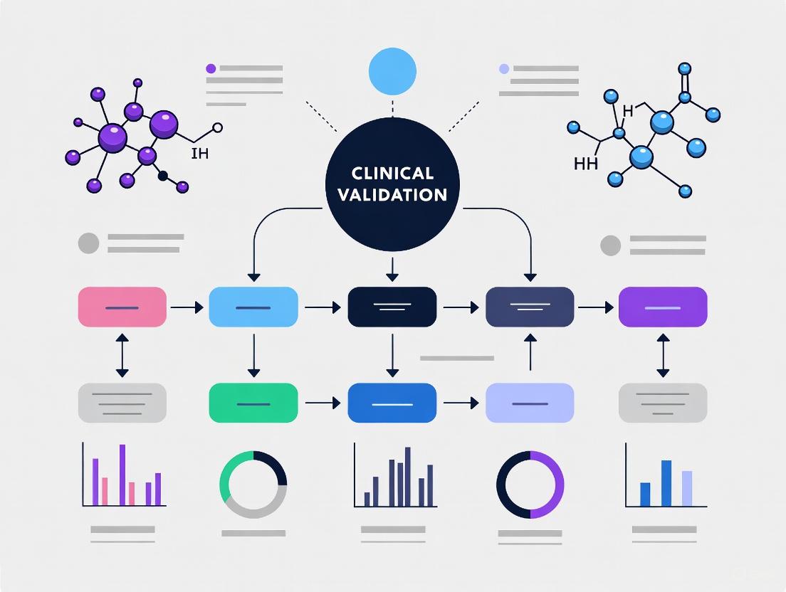
One primary technological approach involves analyzing methylation patterns in ctDNA. Cancer cells exhibit distinct DNA methylation landscapes, and profiling these patterns can simultaneously indicate the presence of a malignancy and predict its tissue of origin (TOO). Another approach, used by the Carcimun test, detects conformational changes in plasma proteins through optical extinction measurements, serving as a universal marker for general malignancy [4]. Other tests, such as OncoSeek, integrate a panel of protein tumor markers (PTMs) with clinical data, enhanced by artificial intelligence (AI) [5]. The following diagram illustrates the core logical workflow shared by many of these MCED technologies.
Comparative Performance Data: Single-Cancer Screening vs. MCED Tests
Clinical Performance of Leading MCED Tests
The potential of MCED tests is demonstrated in their clinical performance metrics. The following table summarizes key data from recent studies on several MCED tests, highlighting their ability to detect a broad range of cancers.
Table 3: Performance Metrics of Selected Multi-Cancer Early Detection (MCED) Tests
| Test Name (Company) | Core Technology | Sensitivity (Overall) | Specificity | Tissue of Origin (TOO) Accuracy | Number of Cancers Detected |
|---|---|---|---|---|---|
| Galleri (GRAIL) [6] | Targeted Methylation of ctDNA | 40.4% (All cancers); 73.7% for 12 high-mortality cancers | 99.6% | 92% | >50 types |
| OncoSeek (SeekIn) [5] | 7 Protein Tumor Markers + AI | 58.4% | 92.0% | 70.6% (for true positives) | 14 types (72% of global deaths) |
| Carcimun [4] | Optical Extinction of Plasma Proteins | 90.6% | 98.2% | Not specified in study | Multiple (Pan-cancer) |
| Cancerguard (Exact Sciences) [7] | DNA Methylation + Protein Biomarkers | 68% for 6 deadliest cancers*; Found >1 in 3 early-stage | 97.4% | Information not provided | >50 types and subtypes |
*Pancreatic, lung, liver, esophageal, stomach, and ovarian.
Direct Comparative Data from Interventional Studies
Recent large-scale interventional studies provide the most compelling evidence for the additive value of MCED tests. The PATHFINDER 2 study, the largest U.S. MCED interventional study in a cancer screening population to date, evaluated the Galleri test alongside standard-of-care screenings. The results, presented in 2025, demonstrated that adding the Galleri test to USPSTF A and B recommended screenings (for breast, cervical, colorectal, and lung cancers) led to a more than seven-fold increase in the number of cancers detected within a year [6] [8]. Critically, approximately 73% of the cancers detected by Galleri do not have standard-of-care screening options [6]. Furthermore, more than half (53.5%) of the new cancers detected by Galleri were early-stage (Stage I or II), indicating a shift toward earlier detection [6]. The following diagram visualizes the findings of this pivotal study.
Experimental Protocols and Methodologies in MCED Validation
Key Experimental Workflows
Robust clinical validation is paramount for MCED tests. The following section details the experimental protocols from cited studies.
The OncoSeek Multi-Centre Validation Study: This study integrated seven cohorts totaling 15,122 participants (3,029 cancer patients and 12,093 non-cancer individuals) from three countries [5]. The experimental workflow was as follows:
- Sample Collection and Processing: Blood samples were collected from participants. Plasma or serum was separated and distributed to different testing laboratories.
- Biomarker Quantification: The seven protein tumor markers (PTMs) were measured using different quantification platforms, including Roche Cobas e411/e601 and Bio-Rad Bio-Plex 200.
- AI-Based Risk Assessment: An algorithm incorporating the PTM concentrations and individual clinical data (e.g., age, gender) computed a probability score for the presence of cancer.
- Blinded Analysis: To assess consistency, a subset of samples was analyzed across different laboratories and platforms in a blinded manner, demonstrating a high Pearson correlation coefficient (0.99-1.00) for PTM results [5].
The Carcimun Test Validation Study: This prospective, single-blinded study included 172 participants (80 healthy, 64 cancer patients, 28 with inflammatory conditions) [4]. The methodology was based on a biochemical property shift:
- Sample Preparation: 26 µl of blood plasma was added to 70 µl of 0.9% NaCl solution, followed by 40 µl of distilled water.
- Thermal Equilibration: The mixture was incubated at 37°C for 5 minutes.
- Baseline Measurement: A blank measurement was recorded at 340 nm.
- Acidification and Final Measurement: 80 µl of 0.4% acetic acid solution was added, and the final absorbance was measured at 340 nm using a clinical chemistry analyzer.
- Cut-off Application: A pre-defined cut-off value of 120 was used to differentiate between healthy and cancer subjects. Mean extinction values were 23.9 (healthy), 315.1 (cancer), and 62.7 (inflammatory conditions), with the difference being statistically significant (p<0.001) [4].
The Scientist's Toolkit: Key Research Reagent Solutions
The development and validation of MCED tests rely on a suite of specialized reagents and tools. The following table details essential materials used in the featured experiments.
Table 4: Key Research Reagent Solutions for MCED Test Development
| Reagent / Material | Function in MCED Research | Example from Search Results |
|---|---|---|
| Blood Collection Tubes | Standardized collection and stabilization of blood samples for plasma/serum separation. | Used across all cited MCED studies for initial sample acquisition [5] [6] [4]. |
| Protein Assay Kits/Reagents | Quantification of specific protein tumor markers (PTMs) in plasma/serum. | Roche Cobas e411/e601 and Bio-Rad Bio-Plex 200 systems used to measure 7 PTMs in the OncoSeek study [5]. |
| ctDNA Extraction Kits | Isolation of high-quality, unfragmented circulating tumor DNA from plasma. | Core to the Galleri and Cancerguard tests, which analyze methylation patterns in ctDNA [6] [7]. |
| Bisulfite Conversion Reagents | Chemical treatment of DNA that converts unmethylated cytosines to uracils, allowing for methylation profiling. | Essential for the targeted methylation sequencing approach used by the Galleri test [6]. |
| PCR & NGS Library Prep Kits | Amplification and preparation of DNA libraries for next-generation sequencing. | Underpins the high-throughput sequencing required for ctDNA-based MCED tests [6]. |
| Optical Absorbance Reagents | Chemicals that induce conformational changes in proteins for optical detection. | Acetic acid solution used in the Carcimun test to induce aggregation measured at 340 nm [4]. |
Discussion and Future Directions
The data presented herein clearly illustrates the critical limitations of the current single-cancer screening paradigm and the transformative potential of MCED technologies. While traditional screenings for breast, cervical, colorectal, and lung cancers have provided significant population-level benefits, their restricted scope means they address only a fraction of the cancer burden [1] [2]. MCED tests, by contrast, offer a paradigm shift by simultaneously screening for a multitude of cancers from a single blood draw, many of which lack any current screening option.
The most compelling evidence for this additive benefit comes from real-world interventional studies like PATHFINDER 2, which demonstrated a more than seven-fold increase in cancer detection when an MCED test was combined with standard screenings [6]. The high accuracy of Cancer Signal Origin prediction (e.g., 92% for Galleri) is a critical feature that helps guide efficient diagnostic workups, mitigating the risk of prolonged and invasive procedures [6]. However, challenges remain. The variable sensitivity across cancer types and stages indicates that these tests are not yet a replacement for existing, highly effective single-cancer screenings [5] [8]. Furthermore, the risk of overdiagnosis, a known issue in cancer screening, must be thoroughly evaluated in the context of MCED tests [3] [8]. Finally, demonstrating a mortality reduction—the ultimate goal of any screening program—requires long-term follow-up, and this evidence is still being gathered for MCEDs [8].
In conclusion, the limitations of single-cancer modalities are well-documented and significant. The emergence of MCED tests represents a promising advancement to expand the reach of cancer screening. For researchers, scientists, and drug development professionals, the future landscape will involve refining the sensitivity and specificity of these tests, validating them in diverse populations, establishing cost-effective diagnostic pathways for positive results, and ultimately, generating definitive evidence that their integration into screening programs reduces cancer-specific mortality.
The landscape of cancer detection is being transformed by liquid biopsy technologies, which analyze circulating biomarkers in bodily fluids to identify the presence of cancer. Three principal biomarker classes—circulating tumor DNA (ctDNA), DNA methylation patterns, and protein biomarkers—form the technological foundation of modern multi-cancer early detection (MCED) tests. These approaches offer complementary strengths in sensitivity, specificity, and clinical applicability for detecting cancers at earlier, more treatable stages. The clinical validation of these technologies represents a critical research frontier in oncology, with numerous large-scale studies demonstrating their potential to significantly increase cancer detection rates when combined with standard screening methods.
Recent evidence from interventional trials highlights this transformative potential. For instance, the GRAIL PATHFINDER 2 study, the largest U.S. MCED interventional study to date, found that adding the Galleri test to recommended screenings for breast, cervical, colorectal, and lung cancers led to a more than seven-fold increase in the number of cancers detected within a year [6]. Importantly, approximately three-quarters of the cancers detected by this methylation-based test currently lack standard screening options, addressing a critical gap in our cancer screening capabilities [6]. Similarly, protein-based assays like OncoSeek have demonstrated robust performance across diverse populations and platforms, showing particular promise for making MCED more accessible and affordable, especially in low- and middle-income countries [5].
Circulating Tumor DNA (ctDNA) Detection
Fundamental Principles and Detection Methodologies
Circulating tumor DNA (ctDNA) comprises fragmented DNA molecules shed by tumor cells into the bloodstream and other bodily fluids. These fragments carry tumor-specific genetic and epigenetic alterations, offering a minimally invasive window into the cancer's molecular landscape. The detection of ctDNA presents significant technical challenges due to its extremely low concentration relative to total cell-free DNA (cfDNA) – often constituting less than 0.1% of total cfDNA in early-stage cancers [9]. This limitation has driven the development of increasingly sensitive detection methodologies.
The fundamental workflow for ctDNA analysis begins with sample collection, typically from blood (where plasma is preferred over serum due to higher ctDNA enrichment and better stability), but also from local sources like urine, saliva, or bile depending on the cancer type [10]. Following DNA extraction, two primary approaches are employed: tumor-informed assays, which require prior knowledge of mutations from tumor tissue sequencing to guide ctDNA detection, and tumor-agnostic approaches that detect cancer-associated alterations without prior tumor sequencing [9]. Key detection methods include PCR-based techniques (including digital PCR) for targeted analysis and next-generation sequencing (NGS) for broader mutation profiling. The analytical challenge lies in distinguishing true tumor-derived signals from sequencing errors and biological noise, necessitating sophisticated bioinformatic approaches.
Clinical Applications and Validation Data
ctDNA assays are developing a strong evidence base across the cancer care continuum, from early detection to monitoring treatment response in advanced disease [9]. In early-stage disease, the detection of ctDNA after curative-intent therapy, termed molecular residual disease (MRD), is strongly prognostic for clinical relapse [9]. Studies presented at ASCO 2025 confirmed that ctDNA dynamics post-operatively are strongly prognostic for patient outcomes. However, a critical unanswered question is whether ctDNA assays can predict response to treatment and better select patients for escalated or de-escalated adjuvant therapy.
The DYNAMIC-III clinical trial, the first prospective randomized study of ctDNA-informed management in resected stage III colon cancer, yielded important insights. Primary analysis demonstrated that treatment escalation strategies for ctDNA-positive patients did not improve Recurrence Free Survival (RFS), suggesting limitations in current treatment modalities rather than the assay's predictive capability [9]. In advanced disease, ctDNA analysis is increasingly adopted for molecular profiling across multiple tumor types. The SERENA-6 trial, a prospective randomized double-blind study in advanced HR-positive HER2-negative breast cancer, demonstrated that switching therapies upon detection of ESR1 mutations in ctDNA improved Progression Free Survival (PFS) and Quality of Life (QoL) compared to continuing standard therapy [9]. This represents the first registrational study demonstrating clinical utility for switching therapies based on ctDNA findings.
Table 1: Clinical Performance of ctDNA Assays in Recent Trials
| Trial Name | Cancer Type | Clinical Setting | Key Findings | Limitations |
|---|---|---|---|---|
| DYNAMIC-III [9] | Stage III Colon Cancer | Adjuvant (post-resection) | Treatment escalation for ctDNA+ patients did not improve RFS | Likely limitations of available treatments, not assay |
| SERENA-6 [9] | Advanced HR+/HER2- Breast Cancer | Advanced (1st-line CDK4/6i + AI) | Switching to camizestrant upon ESR1 mutation improved PFS & QoL | Potential lead-time bias; lack of crossover in design |
| VERITAC-2 [9] | Advanced HR+/HER2- Breast Cancer | Advanced | Benefit of vepdegestrant restricted to ESR1 mutation-positive patients | - |
| Bladder Cancer Study [11] | Muscle-invasive Bladder Cancer | Post-surgery | ctDNA-negative patients had low recurrence risk without adjuvant therapy | - |
Experimental Protocols for ctDNA Analysis
Sample Collection and Processing Protocol:
- Blood Collection: Draw blood into EDTA or specialized cfDNA collection tubes (e.g., Streck Cell-Free DNA BCT) to prevent cell lysis and preserve cfDNA integrity.
- Plasma Separation: Centrifuge at 800-1600 × g for 10 minutes within 2 hours of collection to separate plasma from cellular components.
- Secondary Centrifugation: Perform high-speed centrifugation at 16,000 × g for 10 minutes to remove residual cells and debris.
- cfDNA Extraction: Use commercial cfDNA extraction kits (e.g., QIAamp Circulating Nucleic Acid Kit) following manufacturer protocols.
- Quality Control: Quantify cfDNA using fluorometric methods (e.g., Qubit) and assess fragment size distribution (typically 160-180 bp) via bioanalyzer.
Tumor-Informed ctDNA Assay Protocol (e.g., Signatera):
- Tumor Sequencing: Perform whole-exome or comprehensive genomic sequencing of tumor tissue to identify patient-specific mutations.
- Assay Design: Design a personalized multiplex PCR panel targeting 16 selected mutations.
- Library Preparation: Prepare sequencing libraries from plasma cfDNA using the customized panel.
- Sequencing: Perform ultra-deep sequencing (typically >100,000X coverage) to detect low-frequency variants.
- Variant Calling: Use specialized algorithms to distinguish true variants from sequencing noise, with a typical analytical sensitivity of 0.01% variant allele frequency.
DNA Methylation Biomarkers
Fundamental Principles and Biological Significance
DNA methylation involves the addition of a methyl group to the 5' position of cytosine bases, primarily at CpG dinucleotides, resulting in 5-methylcytosine without altering the underlying DNA sequence [12]. This epigenetic mechanism plays crucial roles in gene regulation, genomic imprinting, X-chromosome inactivation, and cellular differentiation [10]. In cancer, DNA methylation patterns undergo profound alterations characterized by genome-wide hypomethylation, which can induce chromosomal instability, and localized hypermethylation at CpG-rich gene promoters, particularly those of tumor suppressor genes, leading to their silencing [10].
The stability of DNA methylation patterns and their emergence early in tumorigenesis make them ideal biomarkers for cancer detection [10]. Methylation patterns offer several advantages over genetic mutations for liquid biopsy applications: they are highly cancer-specific, occur more frequently than mutations, and provide information about tissue of origin through well-defined methylation signatures [10]. Furthermore, methylated DNA demonstrates enhanced resistance to degradation during sample processing due to nucleosome interactions that protect methylated DNA fragments from nuclease degradation [10].
Methylation Detection Technologies
Multiple technologies have been developed for methylation analysis, each with distinct strengths and limitations. A recent comparative evaluation of four major approaches highlights their performance characteristics [13]:
Table 2: Comparison of DNA Methylation Detection Methods
| Method | Resolution | Coverage | DNA Integrity | Cost & Throughput | Key Applications |
|---|---|---|---|---|---|
| Whole-Genome Bisulfite Sequencing (WGBS) | Single-base | ~80% of CpGs | Substantial degradation | High cost, moderate throughput | Comprehensive methylome mapping |
| Enzymatic Methyl-Seq (EM-seq) | Single-base | Comparable to WGBS | Preserves DNA integrity | Moderate cost, moderate throughput | Alternative to WGBS with better DNA preservation |
| Illumina EPIC Array | Single-CpG site | ~935,000 sites | Minimal requirements | Low cost, high throughput | Population studies, biomarker validation |
| Nanopore Sequencing | Single-base | Full genome with long reads | No conversion needed | Variable cost, emerging technology | Long-range methylation profiling, complex regions |
Bisulfite conversion-based methods like WGBS have been the gold standard but cause substantial DNA fragmentation (up to 90% degradation) [13]. EM-seq has emerged as a robust alternative that uses enzymatic conversion rather than harsh chemical treatment, thereby better preserving DNA integrity – a critical advantage for liquid biopsy applications where DNA is already limited [13]. Microarrays offer cost-effective profiling of predefined CpG sites, while nanopore sequencing enables real-time methylation detection without conversion and provides long-range methylation context [13].
Clinical Applications and Performance
Methylation-based MCED tests have demonstrated significant promise in large clinical studies. The Galleri test (GRAIL), which targets methylation patterns at approximately 100,000 genomic regions, exemplifies this approach [6]. In the PATHFINDER 2 registrational study involving 23,161 participants, the test demonstrated 73.7% episode sensitivity for the 12 cancers responsible for two-thirds of cancer deaths in the U.S., with a 99.6% specificity (0.4% false positive rate) [6]. The test accurately predicted the tissue of origin (Cancer Signal Origin) in 92% of true-positive cases, facilitating efficient diagnostic workups [6].
Different bodily fluids offer varying advantages for methylation-based detection. While blood provides systemic coverage, local fluids like urine for urological cancers, bile for biliary tract cancers, and cerebrospinal fluid for central nervous system tumors often yield higher biomarker concentrations with reduced background noise [10]. For example, urine-based tests for bladder cancer detection demonstrate significantly higher sensitivity than plasma-based approaches due to direct contact between tumors and urine [10].
Experimental Protocols for Methylation Analysis
EM-seq Protocol for Liquid Biopsies:
- DNA Input: Use 1-100 ng of plasma-derived cfDNA.
- Enzymatic Conversion: Treat DNA with TET2 enzyme and T4-BGT to convert and protect methylated cytosines while glucosylating 5hmC.
- Deamination: Use APOBEC to deaminate unmodified cytosines to uracils while modified cytosines remain protected.
- Library Preparation: Prepare sequencing libraries using commercial kits compatible with enzymatically converted DNA.
- Sequencing & Analysis: Sequence on Illumina platforms and analyze methylation patterns using alignment to bisulfite-converted reference genomes.
Targeted Methylation PCR Protocol:
- Bisulfite Conversion: Treat DNA with sodium bisulfite using commercial kits (e.g., Zymo EZ DNA Methylation Kit).
- PCR Amplification: Design primers specific to converted DNA sequences, with one primer pair specific for methylated sequences and another for unmethylated sequences.
- Detection: Use quantitative real-time PCR or digital PCR for absolute quantification of methylated alleles.
- Data Analysis: Calculate methylation ratios based on threshold cycles or absolute counts from methylated versus unmethylated assays.
Protein Biomarkers
Fundamental Principles and Detection Methodologies
Protein biomarkers represent another cornerstone of cancer detection, reflecting functional alterations in cellular processes driven by malignant transformation. Unlike genomic and epigenomic alterations, protein expression patterns provide direct insight into the functional state of tumors and their microenvironment. The proteomic biomarker analytics market is experiencing significant growth, driven by technological advancements and increasing demand for personalized medicine [14] [15].
Protein biomarkers for cancer detection include tumor-associated antigens, autoantibodies, cytokines, growth factors, and aberrantly expressed structural proteins. These biomarkers can be detected in various bodily fluids, with blood (plasma or serum) being the most common source due to its systemic circulation through tumor sites [5]. Protein biomarkers offer several advantages: they are often more abundant than nucleic acid biomarkers, can be measured using established clinical laboratory platforms, and may reflect broader tumor heterogeneity than single genetic alterations.
Clinical Applications and Performance
Protein-based MCED tests have demonstrated robust performance in large validation studies. The OncoSeek test exemplifies this approach, combining a panel of seven protein tumor markers (PTMs) with artificial intelligence to provide a cost-effective MCED solution [5]. In a comprehensive multi-centre validation study encompassing 15,122 participants (3,029 cancer patients and 12,093 non-cancer individuals) from seven centers in three countries, OncoSeek demonstrated an area under the curve (AUC) of 0.829, with 58.4% sensitivity and 92.0% specificity at predicting tissue of origin in true positives [5].
The test detected 14 common cancer types representing over 60% of worldwide cancer cases and more than 72% of cancer-related mortalities, with varying sensitivities across cancer types: pancreatic cancer (79.1%), lung cancer (66.1%), liver cancer (65.9%), colorectal cancer (51.8%), and breast cancer (38.9%) [5]. Importantly, the test demonstrated consistent performance across different laboratories, sample types (plasma and serum), and analytical platforms, highlighting its robustness and potential for widespread implementation, particularly in resource-limited settings [5].
Table 3: Performance of Protein-Based MCED Test (OncoSeek) Across Cancer Types
| Cancer Type | Sensitivity (%) | Cancer Type | Sensitivity (%) |
|---|---|---|---|
| Bile Duct | 83.3 | Stomach | 57.9 |
| Gallbladder | 81.8 | Colorectum | 51.8 |
| Endometrium | 80.0 | Esophagus | 46.0 |
| Pancreas | 79.1 | Lymphoma | 42.9 |
| Cervix | 75.0 | Breast | 38.9 |
| Ovary | 74.5 | All Cancers | 58.4 |
| Lung | 66.1 | ||
| Liver | 65.9 |
Experimental Protocols for Protein Biomarker Analysis
Multiplex Immunoassay Protocol:
- Sample Collection: Collect blood in EDTA tubes and separate plasma within 2 hours by centrifugation at 800-1600 × g for 15 minutes.
- Antibody Coating: Coat microplate wells with capture antibodies specific to each protein biomarker in the panel.
- Sample Incubation: Add plasma samples to wells and incubate to allow antigen-antibody binding.
- Detection Antibody: Add biotinylated detection antibodies that bind to different epitopes on the captured proteins.
- Signal Amplification: Add enzyme-conjugated streptavidin (typically horseradish peroxidase or alkaline phosphatase).
- Signal Development: Add chemiluminescent or colorimetric substrate and measure signal intensity.
- Data Analysis: Calculate protein concentrations from standard curves using specialized software.
Mass Spectrometry-Based Proteomics Protocol:
- Protein Extraction: Denature and reduce plasma proteins using urea and dithiothreitol.
- Digestion: Digest proteins with trypsin overnight at 37°C.
- Desalting: Purify peptides using C18 solid-phase extraction cartridges.
- Liquid Chromatography: Separate peptides using nano-flow liquid chromatography.
- Mass Spectrometry Analysis: Analyze eluting peptides using high-resolution mass spectrometry (e.g., Orbitrap platforms).
- Data Processing: Identify and quantify proteins using database search algorithms (e.g., MaxQuant) and statistical analysis.
Comparative Analysis of Detection Platforms
Performance Metrics Across Biomarker Classes
Direct comparison of the three biomarker classes reveals complementary strengths and limitations for MCED applications. ctDNA methylation assays generally offer the highest sensitivity for cancer detection across multiple cancer types, particularly for lethal cancers, while protein-based assays provide a more cost-effective alternative with adequate performance for resource-limited settings. ctDNA mutation-based assays excel in tumor-informed settings for monitoring minimal residual disease and treatment response.
Table 4: Comparative Performance of MCED Biomarker Platforms
| Parameter | ctDNA Methylation | ctDNA Mutations | Protein Biomarkers |
|---|---|---|---|
| Analytical Sensitivity | High (detects 0.1% ctDNA) | Very High (detects 0.01% ctDNA) | Moderate |
| Tissue of Origin Accuracy | High (>90%) | Limited without prior tumor knowledge | Moderate (70.6% in OncoSeek) |
| Stage I Sensitivity | Moderate (improving) | Low to Moderate | Low to Moderate |
| Cost Profile | High | High (especially tumor-informed) | Low to Moderate |
| Platform Requirements | Advanced sequencing | Advanced sequencing | Standard clinical platforms |
| Turnaround Time | Days to weeks | Days to weeks | Hours to days |
| Recommended Applications | Population screening, cancer detection | MRD monitoring, therapy selection | Resource-limited settings, symptomatic triage |
Integration of Multi-Modal Approaches
The integration of multiple biomarker classes represents the frontier of MCED development, leveraging the complementary strengths of each approach. Multi-modal strategies combining methylation patterns with protein biomarkers or mutational signatures offer the potential for enhanced sensitivity and specificity across diverse cancer types and stages. Companies like PrognomiQ are exploring multi-omic markers for various cancer diagnostic applications, while Exact Sciences has bolstered its plasma proteomics capabilities through strategic acquisitions [14] [15].
The emerging evidence suggests that different biomarker classes may excel in specific clinical contexts. Methylation-based tests appear particularly powerful for population screening, while protein-based tests offer practical advantages for symptomatic patient triage in resource-limited settings [5]. Tumor-informed ctDNA assays currently provide the highest sensitivity for monitoring minimal residual disease [9]. The optimal biomarker strategy depends on the specific clinical context, including the target population, healthcare infrastructure, and economic considerations.
The Scientist's Toolkit: Essential Research Reagents and Platforms
Successful development and validation of MCED tests require specialized reagents, instruments, and computational tools. The following table summarizes key components of the research toolkit for investigators in this field.
Table 5: Essential Research Reagents and Platforms for MCED Development
| Category | Specific Products/Platforms | Key Applications | Manufacturers |
|---|---|---|---|
| Sequencing Platforms | Illumina NovaSeq, Oxford Nanopore | Whole-genome methylation, mutation detection | Illumina, Oxford Nanopore |
| Protein Analyzers | Roche Cobas e411/e601, Bio-Rad Bio-Plex 200 | Multiplex protein biomarker quantification | Roche, Bio-Rad |
| Liquid Biopsy Kits | QIAamp Circulating Nucleic Acid Kit, cfDNA collection tubes | Sample collection, cfDNA extraction | Qiagen, Streck |
| Methylation Analysis | Infinium MethylationEPIC BeadChip, EM-seq kits | Genome-wide methylation profiling | Illumina, New England Biolabs |
| Data Analysis Tools | MethylomeMiner, CpGPT, MethylGPT | Methylation data processing, pattern recognition | [12] [16] |
| Reference Materials | Horizon cfDNA Reference Standards | Assay validation, quality control | Horizon Discovery |
Signaling Pathways and Experimental Workflows
The following diagrams illustrate key signaling pathways and experimental workflows in MCED test development and validation.
Diagram 1: MCED Development and Biomarker Integration Pathways
Diagram 2: Liquid Biopsy Workflow and Analysis Methods
The clinical validation of multi-cancer early detection tests represents a paradigm shift in oncology, with ctDNA methylation, ctDNA mutation, and protein biomarker approaches each offering distinct advantages for different clinical scenarios. Methylation-based assays currently lead in population screening applications with their high sensitivity and accurate tissue of origin prediction, while protein-based tests offer a cost-effective alternative for resource-limited settings. Tumor-informed ctDNA assays provide the highest sensitivity for monitoring minimal residual disease and treatment response.
The future of MCED lies in the intelligent integration of multiple biomarker classes, leveraging their complementary strengths to maximize sensitivity and specificity across diverse cancer types and stages. As these technologies continue to mature and demonstrate clinical utility in large prospective studies, they hold the potential to fundamentally transform cancer screening paradigms and significantly reduce cancer mortality through earlier detection.
Cancer represents a critical and growing global public health challenge, with the burden expected to rise dramatically in the coming decades. Current estimates indicate that in 2022, there were approximately 20 million new cancer cases and 9.7 million cancer deaths worldwide [17]. Projections suggest a concerning increase, with the global cancer burden expected to exceed 27 million new cases annually by 2040, representing a 50% increase from 2018 estimates [18]. Even more alarming are forecasts predicting over 30 million cases and more than 18 million deaths by 2050 [19]. This escalating burden is not distributed equally across nations, with low- and middle-income countries (LMICs) expected to experience the most substantial proportional increases [18] [17]. These disparities highlight the urgent need for innovative detection strategies that can address the growing global cancer burden efficiently and equitably, providing the fundamental rationale for multi-cancer early detection (MCED) technologies.
The Human Development Index (HDI) serves as a crucial framework for understanding disparities in cancer burden. Countries with low HDI levels face a disproportionately high burden of infection-related cancers, such as cervical and liver cancers, while nations with very high HDI contend with higher rates of cancers associated with industrialized lifestyles, including lung, colorectal, and breast cancers [18]. Strikingly, while women in low-HDI countries are 50% less likely to be diagnosed with breast cancer than women in high-HDI countries, they face a much higher risk of dying from the disease due to late diagnosis and inadequate access to quality treatment [17]. These disparities underscore the limitations of current single-cancer screening approaches and emphasize the need for accessible, comprehensive detection technologies that can transcend healthcare infrastructure limitations.
The Current Landscape of Cancer Screening and Detection
Limitations of Established Screening Modalities
The current paradigm of cancer screening relies predominantly on single-cancer tests endorsed by organizations such as the U.S. Preventive Services Task Force (USPSTF). These include biennial screening mammography for breast cancer, cervical cytology and/or primary high-risk human papillomavirus (hrHPV) testing for cervical cancer, various stool-based tests and direct visualization methods for colorectal cancer, and low-dose computed tomography (LDCT) for lung cancer screening [5]. While these screening methods have demonstrated success in reducing mortality for specific cancer types, their population-level impact is constrained by several fundamental limitations:
Limited Scope: Currently recommended screening tests cover only four cancer types (breast, cervical, colorectal, and lung), which account for approximately 40% of cancer incidence in the United States [6]. This leaves the majority of cancer types without recommended screening options.
Infrastructure and Resource Requirements: Many established screening modalities require sophisticated equipment, specialized healthcare facilities, and trained specialists, making implementation challenging in resource-limited settings [5]. For instance, LDCT for lung cancer screening requires specialized radiology equipment and expertise that may be unavailable in LMICs.
Access Barriers and Inequities: Significant global inequities exist in cancer service availability. Lung cancer-related services are 4-7 times more likely to be included in health benefit packages in high-income countries compared to lower-income nations [17]. The disparity is even more pronounced for advanced services such as stem-cell transplantation, which is 12 times more likely to be covered in high-income countries [17].
Variable Adherence: Even in high-resource settings, adherence to recommended cancer screening varies considerably across populations and cancer types, limiting overall effectiveness.
The Potential of Multi-Cancer Early Detection Technologies
MCED tests represent a paradigm shift in cancer screening by simultaneously detecting signals for multiple cancer types from a single blood sample. This approach addresses several limitations of current screening methods:
Broad Cancer Coverage: MCED tests can potentially detect more than 50 cancer types from a single blood draw, including many cancers that currently lack recommended screening methods [6].
Accessibility and Scalability: Blood-based tests require less specialized infrastructure than many current screening modalities, potentially increasing accessibility in diverse healthcare settings [5].
Integration with Existing Screening: MCED tests are designed to complement rather than replace existing evidence-based screening, potentially creating a more comprehensive early detection framework.
The development of MCED technologies is particularly timely given the projected increases in global cancer burden, especially in regions with limited healthcare resources. By 2040, the predicted increases in cancer incidence using demographic changes will be proportionately greatest in countries with low and medium HDI [18], precisely the settings where traditional screening infrastructure is most limited.
Methodologies in MCED Test Development
Analytical Platforms and Biomarker Integration
MCED tests leverage advanced molecular technologies to analyze circulating cell-free DNA (cfDNA) and other biomarkers in blood samples. The leading approaches in development utilize distinct but complementary methodological frameworks:
Table 1: Core Methodological Approaches in MCED Development
| Test Name | Primary Technology | Biomarkers Analyzed | Sample Type |
|---|---|---|---|
| Galleri (GRAIL) | Targeted Methylation Sequencing | DNA Methylation Patterns | Blood Plasma |
| OncoSeek (SeekIn) | Multi-Modal Analysis | Protein Tumor Markers (7) + Clinical Data | Blood Plasma/Serum |
| SeekInCare (SeekIn) | Shallow Whole-Genome Sequencing + Protein Markers | Copy Number Aberration, Fragment Size, End Motif, Oncogenic Virus + 7 Protein Tumor Markers | Blood |
The Galleri test employs a targeted methylation sequencing approach, analyzing patterns of DNA methylation in cfDNA. This method exploits the fact that cancer cells exhibit distinct methylation patterns compared to normal cells, allowing for both cancer detection and prediction of the tissue of origin [6]. The test uses bisulfite conversion followed by next-generation sequencing of targeted genomic regions, with machine learning algorithms trained to distinguish cancer from non-cancer signals and predict the cancer signal origin.
In contrast, the OncoSeek test utilizes a multi-modal approach that integrates measurements of seven protein tumor markers (CA 125, CA 15-3, CA 19-9, CA 72-4, CEA, CYFRA 21-1, and NSE) with individual clinical data, enhanced by artificial intelligence algorithms [5]. This approach demonstrated consistent performance across different quantification platforms, including Roche Cobas e411/e601 and Bio-Rad Bio-Plex 200 systems, enhancing its potential applicability across diverse laboratory settings [5].
The SeekInCare test represents a more comprehensive multi-omics approach, integrating multiple genomic and epigenetic features from shallow whole-genome sequencing of cfDNA, including copy number aberration, fragment size profiles, end motifs, and oncogenic virus detection, alongside the same seven protein tumor markers used in the OncoSeek test [20]. This multi-analyte approach aims to capture complementary aspects of cancer biology to improve overall detection performance.
Experimental Workflows and Validation Frameworks
Robust validation across diverse cohorts and settings is essential for establishing the clinical utility of MCED tests. The following diagram illustrates a generalized workflow for MCED test validation:
The validation of MCED tests requires carefully designed studies that address multiple methodological considerations:
Multi-Center Design: Recruitment of participants from multiple clinical sites across different geographic regions to ensure population diversity and generalizability. For example, the OncoSeek validation included participants from seven centers across three countries [5].
Platform Validation: Assessment of test performance across different laboratory instrumentation and sample types to establish robustness. The OncoSeek test demonstrated high consistency (Pearson correlation coefficient of 0.99-1.00) across different laboratories and platforms [5].
Blinded Analysis: Prospective blinded studies where the cancer status of participants is unknown at the time of testing, providing unbiased performance estimates. The PATHFINDER 2 study of the Galleri test employed this design in over 25,000 participants [6].
Diverse Cancer Representation: Inclusion of participants with various cancer types and stages to characterize performance across the spectrum of disease. The OncoSeek validation encompassed 15,122 participants (3,029 cancer patients) across 15 common cancer types [5].
Longitudinal Follow-up: Prospective monitoring of participants with negative test results to identify interval cancers and assess false negative rates. The SeekInCare prospective cohort study included a median follow-up of 753 days [20].
Comparative Performance of MCED Technologies
Detection Performance Across Cancer Types and Stages
The clinical utility of MCED tests depends on their performance characteristics across the spectrum of cancer types and stages. The following table summarizes key performance metrics for leading MCED tests based on recent validation studies:
Table 2: Comparative Performance Metrics of MCED Tests
| Performance Metric | Galleri (PATHFINDER 2) | OncoSeek (All Cohorts) | SeekInCare (Retrospective) |
|---|---|---|---|
| Overall Sensitivity | 40.4% (Episode Sensitivity) | 58.4% | 60.0% |
| Specificity | 99.6% | 92.0% | 98.3% |
| Positive Predictive Value | 61.6% | Not Reported | Not Reported |
| Stage I Sensitivity | Not Reported | Not Reported | 37.7% |
| Stage II Sensitivity | Not Reported | Not Reported | 50.4% |
| Stage III Sensitivity | Not Reported | Not Reported | 66.7% |
| Stage IV Sensitivity | Not Reported | Not Reported | 78.1% |
| Cancer Signal Origin Accuracy | 92% | 70.6% | Not Reported |
| Sample Size | 23,161 (Performance Cohort) | 15,122 Total Participants | 1,197 Total Participants |
Cancer type-specific sensitivity varies considerably across MCED tests. The OncoSeek test demonstrated particularly high sensitivity for bile duct cancer (83.3%), gallbladder cancer (81.8%), endometrial cancer (80.0%), and pancreatic cancer (79.1%), with more moderate sensitivity for breast cancer (38.9%) and lymphoma (42.9%) [5]. The Galleri test showed strong performance for cancers responsible for two-thirds of cancer deaths in the U.S., with 73.7% episode sensitivity for these malignancies [6].
Notably, the Galleri test demonstrated a more than seven-fold increase in cancer detection when added to USPSTF A and B recommended screenings (breast, cervical, colorectal, and lung cancers), detecting approximately three times as many cancers when added to standard-of-care screening for USPSTF A, B, and C recommendations (including prostate cancer) [6]. This additive detection capability is particularly significant given that approximately three-quarters of the cancers detected by Galleri do not have standard-of-care screening options [6].
Analytical Validation and Real-World Applicability
The transition from research settings to clinical implementation requires rigorous analytical validation and demonstration of robustness across pre-analytical and analytical variables:
Sample Type Consistency: The OncoSeek test demonstrated high correlation (Pearson coefficient = 1.00) between plasma and serum samples analyzed on different Roche instruments (Cobas e411 and e601), supporting flexibility in sample processing [5].
Inter-laboratory Reproducibility: Evaluation of the OncoSeek test across different laboratories showed maintained performance with a Pearson correlation coefficient of 0.99 for non-cancer samples and 1.00 for cancer patient samples [5].
Platform Transferability: The OncoSeek test maintained performance when deployed on different analytical platforms (Roche Cobas e411/e601 and Bio-Rad Bio-Plex 200), enhancing its potential for widespread adoption [5].
The diagnostic resolution process following a positive MCED test result is crucial for clinical utility. In the PATHFINDER 2 study, the Galleri test demonstrated a median time of 46 days from blood draw to diagnostic resolution, with only 0.6% of all participants requiring an invasive procedure [6]. This efficient diagnostic pathway highlights the potential for MCED tests to integrate effectively into clinical workflows without excessive burden on healthcare systems.
The development and implementation of MCED tests rely on specialized reagents and technical resources. The following table outlines key components essential for MCED research and development:
Table 3: Essential Research Reagents and Resources for MCED Development
| Resource Category | Specific Examples | Research Function |
|---|---|---|
| Analytical Platforms | Roche Cobas e411/e601, Bio-Rad Bio-Plex 200 | Quantification of protein tumor markers |
| Sequencing Technologies | Illumina Sequencing Platforms, Targeted Methylation Panels | Analysis of cfDNA methylation patterns and genomic features |
| Protein Biomarker Panels | CA 125, CA 15-3, CA 19-9, CA 72-4, CEA, CYFRA 21-1, NSE | Cancer signal detection and differentiation |
| Sample Collection Systems | Blood Collection Tubes for Plasma/Serum Separation | Standardized sample acquisition and preservation |
| Bioinformatics Tools | Machine Learning Algorithms, Methylation Analysis Pipelines | Cancer signal classification and tissue of origin prediction |
| Reference Materials | Commercial Quality Controls, Synthetic cfDNA Standards | Assay validation and quality control |
The selection of appropriate protein tumor markers in tests like OncoSeek is based on their established roles in cancer detection and monitoring. For instance, CEA (carcinoembryonic antigen) is associated with various carcinomas including colorectal cancer; CA 125 with ovarian cancer; CA 15-3 with breast cancer; and CA 19-9 with pancreatic and gastrointestinal cancers [5]. The combination of these markers with genomic features enhances the overall cancer detection capability beyond what either approach could achieve alone.
For methylation-based tests like Galleri, the targeted methylation panels are designed to cover genomic regions with differential methylation patterns between cancer and normal cells, as well as between different cancer types. The development of these panels requires extensive analysis of reference methylation databases from both normal tissues and cancer samples to identify optimally informative regions for cancer detection and tissue of origin prediction.
The growing global cancer burden, with projections exceeding 30 million annual cases by 2050, creates an urgent imperative for more effective and accessible early detection strategies [19]. MCED technologies represent a promising paradigm shift that could substantially address current limitations in cancer screening, particularly for cancers that lack recommended screening modalities and for populations with limited access to established screening infrastructure.
The validation data from tests such as Galleri, OncoSeek, and SeekInCare demonstrate the feasibility of detecting multiple cancer types from a single blood sample with clinically meaningful performance characteristics. The additive detection capability of MCED tests, with Galleri increasing cancer detection more than seven-fold when combined with standard screenings, highlights their potential to transform cancer screening paradigms [6]. Furthermore, the robustness of these tests across different platforms and populations supports their potential applicability across diverse healthcare settings [5].
Future directions for MCED development should focus on enhancing sensitivity for early-stage cancers, optimizing diagnostic pathways following positive tests, and demonstrating mortality reduction in large-scale screening studies. Additionally, implementation research is needed to establish the cost-effectiveness and equitable deployment of these technologies, particularly in resource-limited settings that face the greatest projected increases in cancer burden. As MCED technologies continue to evolve, they hold significant promise for reducing the global cancer burden through earlier detection, particularly for cancers that currently lack screening options and for populations with limited access to existing cancer screening infrastructure.
Despite significant advancements in oncology, routine population-based screening exists for only a few cancer types. Cancers of the breast, cervical, colorectal, prostate, and lung have established screening modalities, yet they represent less than 30% of cancers that people ultimately die from [21]. This leaves a substantial gap in early detection for many deadly malignancies. Multi-cancer early detection (MCED) tests represent an emerging technological solution to this critical public health challenge, utilizing circulating tumor DNA (ctDNA) and other biomarkers to detect cancer signals across multiple cancer types from a single blood sample [8]. This analysis systematically examines current screening gaps, evaluates the performance of emerging MCED technologies against standard approaches, and details the experimental methodologies driving this innovative field, providing researchers with a comprehensive overview of the current landscape and future directions.
Quantifying the Screening Gap
Cancers Lacking Established Screening Modalities
Current guideline-directed cancer screening options fail to cover the majority of potentially lethal cancers [21]. The PATHFINDER 2 study, the largest trial to date of an MCED test in patients undergoing routine screening, revealed that 73% of cancers detected had no existing screening tests [8]. These include:
- Pancreatic, liver, head-and-neck, and ovarian cancers: Not currently covered by established screening methods [8].
- Lung cancer: Despite available low-dose CT screening, current U.S. Preventive Services Task Force (USPSTF) guidelines miss approximately two-thirds of patients ultimately diagnosed with the disease. A Northwestern Medicine study of nearly 1,000 consecutive lung cancer patients found only 35% would have qualified for screening under current USPSTF criteria, which exclude many women and never-smokers [22].
- Prostate cancer: Many healthcare systems lack population-based screening programs. Italy, for example, had no national program until a pilot launched in Lombardy in November 2024 [23]. Furthermore, primary care providers often serve as gatekeepers, with one study showing only 6% believed PSA testing played a significant role in reducing mortality, creating a significant barrier for high-risk groups like Black men [24].
Disparities in Implementation and Access
Even for cancers with established screening methods, significant disparities persist in implementation and access across different populations and healthcare systems, as detailed in Table 1.
Table 1: Disparities in Global Implementation of Cancer Screening Programs
| Cancer Type | Country/Region | Screening Status | Specific Gaps and Disparities |
|---|---|---|---|
| Breast Cancer [25] | Germany | Organized program for ages 50-75, biennial mammography | 25.8% mortality reduction demonstrated, but access varies across Europe. |
| Chile | Ministry of Health recommends mammography for ages 50-69 | No established national program with certified radiologists; quality not guaranteed. | |
| India, Senegal, Philippines | No organized population-based screening program | Screening is opportunistic; uptake is low; lack of cancer registries and infrastructure. | |
| United States | Multiple society guidelines (USPSTF: biennial, 40-74) | Discrepancies in guidelines create confusion and variations in clinical practice. | |
| Lung Cancer [22] | United States | USPSTF guidelines (50-80, 20 pack-year history) | Only 35% of lung cancer patients qualify; disproportionately excludes women and never-smokers. |
| Prostate Cancer [23] | Italy (Lombardy) | Pilot digital, risk-stratified program launched Nov 2024 | No prior national program; initial uptake without active invitation was low (8.7%). |
| United States | Varied society guidelines; no universal "call-in" approach | Primary care providers often dismiss PSA testing, especially for high-risk Black men [24]. | |
| Colorectal Cancer [26] | United States (Kaiser Permanente) | System-wide outreach with FIT and colonoscopy | Prior to program, racial disparities in mortality were significant (30-40% higher for Black patients). |
Performance of Emerging MCED Technologies
Recent large-scale studies provide the first robust quantitative data on how MCED tests perform when added to standard screening, offering a potential solution to the identified screening gaps. Key performance metrics from recent studies are summarized in Table 2.
Table 2: Performance Metrics of Emerging Early Detection Technologies
| Technology / Test | Study / Context | Key Performance Metrics | Comparison to Standard Screening |
|---|---|---|---|
| Grail's Galleri (MCED) [8] | PATHFINDER 2 (n>35,000; added to standard screening) | - 7x more cancers found than screening alone- 73% of detected cancers had no screening test- 53.5% of detected cancers were Stage I or II- 90%+ accuracy in predicting tumor origin- 0.4% false-positive rate | Superior detection for cancers without screening options; does not replace traditional screening, which detected 40% of cancers missed by the MCED test. |
| Proteomics Blood Test (Astrin) [27] | Early data for breast cancer detection | - Reported sensitivity comparable to MRI for early-stage breast cancer- Designed to detect cancer as early as stage 0 | Aims to fill gap for dense breast tissue, where mammography sensitivity is low; results not yet peer-reviewed. |
| Next-gen mt-sDNA (Cologuard Plus) [28] | Modeling study (n=1 million hypothetical cohort) | - 3.7x more CRC cases detected vs. FIT (2,090 vs 440)- 5.5x more advanced precancerous lesions detected vs. FIT | Superior sensitivity for colorectal cancer compared to standard FIT; resulted in 2% lower total costs. |
| MRI-based Prostate Screening [29] | PROGRESS Trial (High-risk men) | - 83% sensitivity for clinically significant cancer (PI-RADS ≥4)- Positive Predictive Value: 56%- Detected ~1 additional significant cancer per 12 patients vs. PSA | Superior to age-adjusted PSA (58% sensitivity) and conventional PSA (>4.0 ng/mL), which missed >75% of significant cancers. |
The PATHFINDER 2 study demonstrated that adding an MCED test to standard screening can dramatically increase overall cancer detection, particularly for those malignancies without recommended screening modalities [8]. Furthermore, the high accuracy of tumor origin prediction (over 90%) is critical for guiding subsequent diagnostic workups [8].
Specialized screening approaches are also showing promise for high-risk populations. The PROGRESS trial, which uses MRI-based screening for men at high genetic risk, demonstrates a shift towards risk-adapted screening paradigms that maximize benefit while minimizing the harms of overdiagnosis [29].
Experimental Protocols and Methodologies
MCED Test Validation: The PATHFINDER 2 Protocol
The PATHFINDER 2 study provides a benchmark protocol for validating MCED tests in a screening population.
- Study Design: A prospective, multicenter, interventional study.
- Participants: More than 35,000 asymptomatic adults aged 50 and older from across the United States, who were already undergoing or planning to undergo routine cancer screening [8].
- Intervention: A single blood draw for the MCED test in addition to standard USPSTF-recommended screenings.
- Laboratory Methodology:
- Blood Sample Processing: Peripheral blood samples were collected in Streck Cell-Free DNA Blood Collection Tubes.
- Plasma Separation: Centrifugation to separate plasma from cellular components.
- Cell-free DNA (cfDNA) Extraction: Isolation of cfDNA from plasma.
- Bisulfite Sequencing: Treatment of cfDNA with bisulfite, which converts unmethylated cytosines to uracils, while methylated cytosines remain unchanged.
- Next-Generation Sequencing (NGS): High-throughput sequencing of the bisulfite-converted DNA to analyze methylation patterns across the genome.
- Bioinformatic Analysis: Proprietary machine learning algorithms were used to analyze the methylation patterns to detect the presence of a cancer signal and predict the tissue of origin [8].
- Primary Outcomes: Cancer detection rate, stage distribution of detected cancers, false-positive and false-negative rates, and the accuracy of tumor origin prediction.
Risk-Adapted Prostate Cancer Screening: The PROGRESS Protocol
The PROGRESS trial exemplifies a risk-adapted screening methodology for a high-risk population.
- Study Design: A prospective, early-detection study.
- Participants: Males aged 35-74 from three high-risk populations: (1) carriers of rare germline pathogenic variants (e.g., BRCA2), (2) individuals with a strong family history, and (3) individuals of self-reported Black American or Black Caribbean background [29].
- Screening Protocol:
- Annual Clinical Assessment: PSA test with age-adjusted cut-offs and digital rectal exam (DRE).
- Biennial Imaging: Multiparametric MRI (mpMRI) of the prostate every three years.
- Biopsy Indication: A biopsy was recommended for any of the following: abnormal DRE, PSA above age-adjusted cutoff, or an mpMRI with a PI-RADS score ≥3 [29].
- Primary Endpoint: Detection rate of clinically significant prostate cancer (Grade Group ≥2).
Deep Proteomics for Breast Cancer Detection
An emerging alternative to DNA-based tests uses deep proteomics and AI to detect breast cancer, particularly in individuals with dense breast tissue.
- Principle: Analyze hundreds of thousands of proteins from a blood draw, as proteins are the functional units of cells and can show cancer-associated changes long before symptoms appear [27].
- Workflow:
- Blood Draw and Plasma Isolation: Standard phlebotomy followed by centrifugation.
- Deep Proteomic Analysis: Use of nanotechnology and advanced mass spectrometry to achieve a proteomic depth 1000x greater than conventional methods, identifying low-abundance proteins.
- AI-Powered Classification: Machine learning models are trained to identify the subtle proteomic patterns indicative of early-stage breast cancer, potentially at stage 0 [27].
The following diagram illustrates the core logical relationship and workflow contrast between the dominant MCED approach (cfDNA methylation) and the emerging proteomics approach.
The Scientist's Toolkit: Key Research Reagents & Materials
The development and implementation of advanced cancer detection tests rely on a suite of specialized reagents and materials. The following table details key components used in the featured experimental protocols.
Table 3: Essential Research Reagents and Materials for MCED Development
| Reagent/Material | Function in Experimental Protocol | Specific Application Example |
|---|---|---|
| Cell-Free DNA Blood Collection Tubes (e.g., Streck) | Preserves blood sample integrity by stabilizing nucleated blood cells and preventing genomic DNA contamination during transport and storage. | Used in PATHFINDER 2 for stable blood draw shipping and processing [8]. |
| Bisulfite Conversion Reagents | Chemically modifies DNA, deaminating unmethylated cytosine to uracil while leaving methylated cytosine unchanged, enabling methylation analysis. | Critical step in Galleri test preparation for subsequent sequencing [8]. |
| Next-Generation Sequencing (NGS) Kits | Provides enzymes, buffers, and nucleotides for library preparation, target enrichment, and high-throughput sequencing of genetic material. | Used for whole-genome bisulfite sequencing of cfDNA in MCED tests [8]. |
| Methylation Panels & Probes | Target-specific oligonucleotides designed to capture and sequence genomic regions with known cancer-specific methylation patterns. | Component of the Galleri test to analyze informative methylation loci. |
| Multiparametric MRI (mpMRI) Contrast Agents | Intravenous contrast agents (e.g., gadolinium-based) that enhance visualization of vascularity and tissue structure in imaging. | Used in the PROGRESS trial and other MRI-based screening to improve prostate lesion detection (PI-RADS scoring) [29]. |
| Proteomic Assay Kits (e.g., Mass Spec Ready) | Contain reagents for digesting plasma proteins into peptides, labeling, and cleaning up samples for mass spectrometry analysis. | Essential for the deep proteomics workflow described by Astrin Biosciences [27]. |
| AI/Machine Learning Software Platforms | Computational environments for developing and training algorithms to identify complex patterns in large genomic, proteomic, or imaging datasets. | Used in both MCED (Galleri) and proteomic (Astrin) tests to classify cancer signals [8] [27]. |
The gap in early cancer detection for a majority of lethal malignancies represents a significant challenge and opportunity in public health. Evidence from large-scale studies like PATHFINDER 2 indicates that MCED tests can substantially increase the detection of early-stage cancers, particularly for those with no current screening options. However, these tests are not a panacea; they currently miss a proportion of cancers and are intended to complement, not replace, established screenings. The future landscape is likely to be characterized by risk-adapted, multi-modal strategies that combine traditional methods with blood-based MCED tests, advanced imaging, and deep proteomics. This integrated approach, validated through rigorous ongoing clinical trials, holds the promise of fundamentally shifting cancer diagnosis to earlier, more treatable stages across a broader spectrum of cancer types.
MCED Technological Frameworks: Biomarker Strategies and Analytical Methodologies
The emergence of liquid biopsy-based multi-cancer early detection (MCED) tests represents a paradigm shift in oncology, offering a minimally invasive means to detect cancers at stages when they are most treatable. DNA methylation, a stable epigenetic modification that occurs early in tumorigenesis, has surfaced as one of the most promising analytical targets for these tests [10]. Unlike genetic mutations, which can be heterogeneous and rare in early-stage disease, methylation patterns provide a consistent and abundant signal that can be leveraged for cancer detection and tissue-of-origin determination [30] [31]. This guide provides a comparative analysis of targeted sequencing approaches for DNA methylation profiling, focusing on their application in cancer signal detection for clinical validation studies. We objectively evaluate the performance characteristics of different technological platforms and methodologies, presenting experimental data to inform researchers and drug development professionals working in the MCED field.
Technological Platforms for Methylation Analysis
The selection of an appropriate methylation profiling platform involves careful consideration of multiple factors, including coverage, resolution, DNA input requirements, and compatibility with liquid biopsy samples where circulating tumor DNA (ctDNA) is often scarce.
Comparison of Major Profiling Methods
Table 1: Comparison of DNA Methylation Profiling Technologies
| Technology | Resolution | Genomic Coverage | DNA Input | Advantages | Limitations |
|---|---|---|---|---|---|
| Whole-Genome Bisulfite Sequencing (WGBS) | Single-base | ~80% of CpGs in genome [13] | High (≥1μg) [13] | Comprehensive, unbiased genome-wide coverage | DNA degradation from harsh bisulfite treatment; high cost [13] |
| Methylation Microarrays (e.g., Illumina EPIC) | Single-CpG site | 930,000 predefined CpG sites [32] | 250-500 ng [32] | Cost-effective for large cohorts; standardized analysis | Limited to predefined sites; unable to discover novel loci [33] [34] |
| Enzymatic Methyl-Seq (EM-seq) | Single-base | Comparable to WGBS [13] | Lower than WGBS [13] | Preserves DNA integrity; reduces sequencing bias [13] | Higher cost than targeted approaches |
| Targeted Bisulfite Sequencing | Single-base | Custom panels (e.g., 648 CpG sites) [33] | Low (can work with limited cfDNA) | Cost-effective; focused on biologically relevant regions; suitable for validation | Limited discovery potential; panel design critical |
Emerging Methods and Clinical Applications
Enzymatic conversion methods represent a significant advancement over traditional bisulfite treatment. A 2025 pan-cancer detection study utilized enzymatic conversion with a Twist pan-cancer methylation panel, demonstrating higher overall CpG methylation in cancer samples (1.82%) compared to controls (1.34%, p < 0.001) [30] [31]. Their approach achieved an AUC of 0.88 with 83.8% sensitivity and specificity, supporting targeted methylation sequencing as a robust method for cancer detection in clinically challenging populations [30].
Third-generation sequencing technologies, such as Oxford Nanopore Technologies (ONT), enable direct detection of DNA methylation without chemical conversion or amplification. ONT excels in long-range methylation profiling and accessing challenging genomic regions, though it currently shows lower agreement with WGBS and EM-seq compared to the concordance between WGBS and EM-seq [13].
Performance Comparison in Cancer Detection
Analytical Performance of Targeted Approaches
Table 2: Performance Metrics of Targeted Methylation Profiling in Cancer Detection
| Study & Platform | Cancer Types | Sensitivity | Specificity | AUC | Key Findings |
|---|---|---|---|---|---|
| Enzymatic Conversion + Targeted Sequencing [30] [31] | Pan-cancer (37 cancer patients) | 83.8% | 83.8% | 0.88 | 95.7% of 162 DMRs hypermethylated in cancer; 20 key DMRs identified for classification |
| Targeted Bisulfite Sequencing vs. Methylation Array [33] | Ovarian cancer (55 tissues, 25 cervical swabs) | Strong correlation between platforms | Strong correlation between platforms | N/A | BS reliably replicated array results; cost-effective for larger sample sets |
| OncoSeek (AI + Protein Biomarkers) [5] | 14 cancer types (3,029 cancer patients) | 58.4% | 92.0% | 0.829 | Integrated approach; symptomatic cohort sensitivity: 73.1% at 90.6% specificity |
| Galleri MCED Test [6] | >50 cancer types (23,161 participants) | 40.4% (all cancers); 73.7% (for 12 high-mortality cancers) | 99.6% | N/A | Seven-fold increase in cancer detection when added to standard screening; 92% CSO accuracy |
Tissue-of-Origin Prediction Accuracy
Accurate tissue-of-origin (TOO) or cancer signal origin (CSO) prediction is critical for clinical utility of MCED tests. The Galleri test demonstrated 92% accuracy in predicting CSO in the PATHFINDER 2 study, facilitating efficient diagnostic workups with a median time to diagnostic resolution of 46 days [6]. Similarly, the OncoSeek test achieved an overall accuracy of 70.6% in TOO prediction for true positive cases [5]. These performance metrics highlight the dual capability of modern methylation-based tests: not only detecting the presence of cancer but also guiding clinicians toward the likely primary site.
Experimental Protocols for Targeted Methylation Sequencing
Enzymatic Conversion-Based Workflow
A detailed 2025 study provides a robust protocol for enzymatic conversion-based targeted methylation sequencing [30] [31]:
- Sample Collection and cfDNA Extraction: Collect peripheral blood in EDTA or Streck tubes. Process within 4-6 hours with double centrifugation to obtain platelet-poor plasma. Extract cfDNA using silica membrane-based kits (e.g., QIAamp Mini Kit). Quantify using fluorometry.
- Enzymatic Conversion: Use the NEBNext Enzymatic Methyl-seq Conversion Module. The process involves:
- TET2 Catalysis: Oxidizes 5-methylcytosine (5mC) and 5-hydroxymethylcytosine (5hmC) to 5-carboxylcytosine (5caC).
- T4-BGT Glucosylation: Protects 5hmC from deamination.
- APOBEC Deamination: Deaminates unmodified cytosines to uracils, while modified cytosines (5mC, 5hmC, 5caC) are protected.
- Library Preparation and Target Enrichment: Perform library construction using the NEBNext Ultra II DNA Library Prep Kit. Enrich targets using the Twist Pan-Cancer Methylation Panel, which covers genomic regions with differential methylation across multiple cancer types.
- Sequencing and Bioinformatic Analysis: Sequence on Illumina platforms (minimum recommended coverage 30x). Process data using nf-core/methylseq pipeline for read alignment and methylation calling. Identify differentially methylated regions (DMRs) with DMRichR and implement machine learning classifiers (e.g., random forests) for cancer vs. control classification.
Targeted Bisulfite Sequencing Protocol
For targeted bisulfite sequencing, an alternative approach was validated against methylation arrays [33]:
- Sample Processing: Use fresh frozen tissue or cervical swabs. Extract DNA using Maxwell RSC Tissue DNA Kit or QIAamp DNA Mini Kit.
- Bisulfite Conversion: Treat DNA using the EpiTect Bisulfite Kit (QIAGEN), converting unmethylated cytosines to uracils while leaving methylated cytosines unchanged.
- Custom Panel Design: Design a custom QIAseq Targeted Methyl Panel covering 648 CpG sites, including both internal diagnostic signatures (23 CpGs) and external literature-based cancer-related regions.
- Library Preparation and Sequencing: Prepare libraries using the QIAseq Targeted Methyl Custom Panel kit. Perform quality control using Bioanalyzer High Sensitivity DNA Kit. Sequence on Illumina MiSeq with 300-cycle kits.
- Data Analysis and Validation: Analyze sequencing data using QIAGEN CLC Genomics Workbench with a custom workflow. Compare methylation beta values with those obtained from Infinium MethylationEPIC arrays using Spearman correlation and Bland-Altman analysis.
The Scientist's Toolkit: Essential Research Reagents
Table 3: Essential Research Reagents for Targeted Methylation Sequencing
| Category | Product Name | Specific Function |
|---|---|---|
| Sample Collection | Streck Cell-Free DNA Blood Collection Tubes | Preserves blood samples for cfDNA analysis by stabilizing nucleated blood cells |
| DNA Extraction | QIAamp DNA Mini Kit (QIAGEN) | Silica-membrane based extraction of high-quality DNA from various sample types |
| Bisulfite Conversion | EZ DNA Methylation Kit (Zymo Research) | Chemical conversion of unmethylated cytosines to uracils for bisulfite sequencing |
| Enzymatic Conversion | NEBNext Enzymatic Methyl-seq Conversion Module | Enzyme-based conversion protecting DNA integrity while identifying methylation status |
| Library Preparation | NEBNext Ultra II DNA Library Prep Kit (Illumina) | Preparation of sequencing libraries with optimized adapter ligation and amplification |
| Target Enrichment | Twist Pan-Cancer Methylation Panel | Hybridization capture baits for enriching cancer-relevant methylated genomic regions |
| Targeted Panels | QIAseq Targeted Methyl Custom Panel (QIAGEN) | Customizable amplicon-based panel for focused methylation analysis across many samples |
| Quantification | Qubit dsDNA HS Assay Kit (Thermo Fisher) | Fluorometric quantification of DNA concentration, crucial for low-input cfDNA workflows |
| Quality Control | Bioanalyzer High Sensitivity DNA Kit (Agilent) | Microfluidic electrophoresis for assessing DNA and library quality and size distribution |
Clinical Validation Considerations
The transition of methylation-based cancer detection tests from research to clinical practice requires rigorous validation in diverse populations. The OncoSeek test was validated across 15,122 participants from seven centers in three countries, demonstrating consistent performance (AUC 0.829) across different populations, platforms, and sample types [5]. Similarly, the Galleri test's PATHFINDER 2 study, involving 35,878 participants, showed a 0.93% cancer signal detection rate with 99.6% specificity and a 61.6% positive predictive value [6].
Key considerations for clinical validation include:
- Control Group Selection: Including individuals with confounding conditions (e.g., autoimmune, inflammatory diseases) to ensure biomarker specificity [30] [31].
- Sample Type Optimization: Choosing appropriate liquid biopsy sources (plasma, urine, CSF) based on cancer type to maximize signal-to-noise ratio [10].
- Analytical Validation: Establishing sensitivity, specificity, reproducibility, and limits of detection across multiple laboratories and platforms [5].
- Clinical Utility: Demonstrating that the test leads to earlier diagnosis, guides appropriate diagnostic workups, and ultimately improves patient outcomes [6].
Targeted sequencing approaches for DNA methylation profiling represent a powerful technological paradigm for multi-cancer early detection. Enzymatic conversion methods offer advantages in DNA preservation and analytical performance, while targeted bisulfite sequencing provides a cost-effective alternative for focused validation studies. The consistent demonstration of high sensitivity, specificity, and accurate tissue-of-origin prediction across multiple large-scale studies underscores the translational potential of these technologies. As the field advances, the integration of methylation profiling with other analyte types and the continued refinement of targeted panels promise to further enhance the clinical utility of liquid biopsy-based cancer screening, potentially transforming cancer detection paradigms and reducing global cancer mortality.
Cancer remains a critical global health challenge, causing approximately 10 million deaths annually worldwide [35]. Early detection is crucial for improving patient survival rates, yet conventional screening methods are limited to a few cancer types (e.g., breast, cervical, colorectal, and lung) and often suffer from variable sensitivity, specificity, and accessibility issues [36]. For approximately 45.5% of cancer cases, no routine screening methods exist, leading to frequent late-stage diagnoses and poor outcomes [36]. Multi-cancer early detection (MCED) tests represent a transformative approach that enables simultaneous screening for multiple cancers from a single, minimally invasive liquid biopsy [36]. By integrating measurements of circulating protein biomarkers with artificial intelligence (AI) algorithms, these tests can identify molecular signatures of cancer before symptoms appear, potentially revolutionizing cancer screening and risk assessment paradigms, particularly for cancers that currently lack recommended screening protocols [37] [6].
Comparative Analysis of Leading Protein Biomarker Panel Technologies
The landscape of MCED tests features diverse technological approaches, each with distinct biomarker strategies and AI integration methods. The following analysis compares three prominent protein-based MCED technologies, highlighting their experimental designs, performance metrics, and clinical applicability.
Table 1: Performance Comparison of Major Protein-Based MCED Tests
| Test Name | Biomarker Composition | Sensitivity (Overall) | Specificity | Cancer Types Detected | Tissue of Origin (TOO) Accuracy |
|---|---|---|---|---|---|
| OncoSeek [5] [38] | 7 protein tumor markers (PTMs) + clinical data (sex, age) | 58.4% (95% CI: 56.6-60.1%) | 92.0% (95% CI: 91.5-92.5%) | 14 cancer types representing ~72% of global cancer deaths | 70.6% |
| xPKA-Based Test [39] | 16 parameters (xPKA activity, additional kinase activities, cancer-associated IgG/IgM antibodies) | 100% (across all five cancer types) | 97% | Breast, lung, colorectal, ovarian, pancreatic | 98% |
| Galleri [6] | Targeted methylation patterns of cell-free DNA | 40.4% (all cancers); 73.7% (for 12 high-mortality cancers) | 99.6% | >50 cancer types | 92% |
Table 2: Stage-Specific Sensitivity of Protein-Based MCED Tests
| Test Name | Stage I Sensitivity | Stage II Sensitivity | Stage III Sensitivity | Stage IV Sensitivity |
|---|---|---|---|---|
| OncoSeek [5] | Not specified | Not specified | Not specified | Not specified |
| xPKA-Based Test [39] | 100% (all five cancer types) | 100% | 100% | 100% |
| Galleri [6] | 53.5% of detected cancers were Stage I/II | 53.5% of detected cancers were Stage I/II | 69.3% of detected cancers were Stage I-III | 30.7% of detected cancers were Stage IV |
Technology-Specific Performance Insights
The OncoSeek test demonstrates how AI can significantly enhance the performance of conventional protein biomarkers. In a large-scale validation across 15,122 participants from seven centers in three countries, the AI-powered algorithm increased specificity to 92.9% compared to just 56.9% using conventional single-threshold methods for each biomarker [38]. The test showed particularly high sensitivity for pancreatic cancer (79.1%), bile duct cancer (83.3%), and ovarian cancer (74.5%) - cancers that typically lack routine screening options [5].
The xPKA-Based Test achieved remarkable 100% sensitivity across all five cancer types tested (breast, lung, colorectal, ovarian, and pancreatic), including 100% detection of Stage I cancers [39]. This exceptional performance, demonstrated in a cohort of 141 cancer patients and 119 healthy controls, highlights the potential of kinase activity signatures combined with antibody profiles for detecting early-stage disease.
Galleri, while primarily focusing on methylation patterns, represents the clinical translation of MCED technology at scale. In the PATHFINDER 2 interventional study with 23,161 participants, adding Galleri to standard screenings increased cancer detection more than seven-fold, with 73.7% sensitivity for the 12 cancers responsible for two-thirds of cancer deaths in the U.S. [6].
Experimental Protocols and Methodological Frameworks
OncoSeek Testing Methodology
The OnCoSeek protocol employs a robust multi-center design with rigorous validation across diverse populations and platforms [5] [38]:
Sample Collection and Processing:
- Collection of one tube of peripheral blood from each participant
- Serum/plasma separation using standard centrifugation protocols
- Analysis on common clinical electrochemiluminescence immunoassay analyzers (Roche Cobas e411/e601, Bio-Rad Bio-Plex 200)
- Measurement of seven protein tumor markers (PTMs) - specific analytes not detailed in search results
AI Algorithm Development:
- Integration of PTM quantification results with clinical data (sex, age)
- Calculation of Probability of Cancer (POC) index using machine learning
- Implementation of ensemble methods to distinguish cancer patients from non-cancer individuals
- Tissue of origin (TOO) prediction through pattern recognition in biomarker profiles
Validation Framework:
- Multi-cohort design with training and independent validation sets
- Cross-platform consistency testing across different laboratory settings
- Demographic stratification to ensure representative performance
- Large-scale validation across 15,122 participants from diverse geographical locations
xPKA-Based Test Methodology
This innovative approach utilizes functional kinase activities and antibody profiles for cancer detection [39]:
Biomarker Quantification Protocol:
- Serum collection and processing under standardized conditions
- Extracellular PKA (xPKA) activity measurement using MESACUP Protein Kinase Assay Kit
- Serum activation with activating buffer (25 mM KH₂PO₄, 5mM EDTA, 150 mM NaCl, 50% glycerol w/v, 1 mg/mL BSA, 100 mM DTT, pH 6.5)
- Incubation with immobilized peptide substrate for 30 minutes at 25°C with agitation
- Detection using biotinylated phosphoserine antibodies and peroxidase-conjugated streptavidin
- Colorimetric detection with TMB substrate and absorbance reading at 450nm
- Additional kinase activities measured using analogous assays with appropriate peptide targets
- Cancer-associated antibodies (IgG, IgM) quantified using standard ELISA protocols
Analytical Framework:
- Supervised, rule-based classification strategy using if-then logic structures
- Initial pattern discovery through quantitative biomarker distribution analysis
- Optimal threshold establishment where separation between groups was maximized
- Cancer-type-specific conditional rules developed to resolve cross-reactivity
- Independent validation by qualified biostatisticians using SAS software
xPKA Test Workflow: Diagram illustrating the key steps in the extracellular protein kinase A activity-based biomarker test.
Machine Learning-Enhanced Biomarker Panel Development for Pancreatic Cancer
A separate research initiative focused specifically on pancreatic ductal adenocarcinoma (PDA) demonstrates the power of ML-driven biomarker selection [40]:
Experimental Design:
- Analysis of 47 serum protein biomarkers using Luminex bead-based multiplex immunoassays
- Cohort A (development set): 355 individuals (181 PDA patients, 174 healthy controls)
- Cohort B (validation set): 130 individuals (100 PDA patients, 30 healthy controls)
- Measurement using Luminex 200 system with xPONENT software
Machine Learning Pipeline:
- Application of multiple ML algorithms: Random Forest, XGBoost, LightGBM, CatBoost, SVM, KNN
- Five-fold cross-validation approach with stratification by gender and age
- Feature importance analysis using SHapley Additive exPlanations (SHAP)
- Model performance assessment via AUROC, F1 score, sensitivity, specificity, accuracy
Key Findings:
- CatBoost demonstrated the highest diagnostic accuracy
- SHAP analysis identified CA19-9, GDF15, and suPAR as key biomarkers
- Combined panel significantly outperformed CA19-9 alone (AUROC 0.992 vs. 0.952)
- Robust performance maintained in early-stage PDA (AUROC 0.976 vs. 0.868 for CA19-9 alone)
AI Integration and Computational Frameworks in Biomarker Analysis
Artificial intelligence serves as the critical enabling technology that transforms conventional protein biomarker measurements into powerful diagnostic tools. The integration of AI follows several sophisticated computational paradigms [37] [41]:
Machine Learning Approaches in Biomarker Discovery
Data Preprocessing and Feature Engineering:
- Quality control, normalization, and batch effect correction across multiple sites
- Handling of missing data through advanced imputation techniques
- Feature engineering to create derived variables capturing biologically relevant patterns
- Dimensionality reduction while preserving biological signal
Model Training and Validation:
- Employment of diverse algorithms including random forests, support vector machines, and gradient boosting methods
- Deep neural networks for capturing complex non-linear relationships in high-dimensional data
- Convolutional neural networks for analyzing medical images and pathology slides correlating with protein biomarkers
- Graph neural networks to model biological pathways and protein interactions
- Rigorous cross-validation and holdout testing to ensure model generalizability
Federated Learning Frameworks:
- Enable secure analysis across distributed datasets without moving sensitive patient data
- Maintain data privacy while leveraging diverse population datasets
- Facilitate multi-institutional collaboration while complying with regulatory requirements
AI Integration Framework: Computational pipeline showing how AI transforms biomarker data into clinical insights.
The Scientist's Toolkit: Essential Research Reagents and Platforms
Successful implementation of protein biomarker panels requires specialized reagents, instrumentation, and computational resources. The following table details essential research solutions derived from the analyzed experimental protocols.
Table 3: Essential Research Reagent Solutions for Protein Biomarker Panel Development
| Category | Specific Product/Platform | Experimental Function | Key Features/Benefits |
|---|---|---|---|
| Multiplex Immunoassay Platforms | Luminex 200 System [40] | Simultaneous quantification of multiple protein biomarkers in single sample | High-throughput multiplexing, reduced sample volume requirements, wide dynamic range |
| Electrochemiluminescence Analyzers | Roche Cobas e411/e601 [5] [38] | Clinical-grade quantification of protein tumor markers | Standardized clinical platforms, high reproducibility, established quality control |
| Protein Kinase Activity Assays | MESACUP Protein Kinase Assay Kit [39] | Measurement of extracellular PKA activity in serum | Specific kinase inhibition profiling, high sensitivity (LoD: 0.3 mU/mL) |
| Bead-Based Multiplex Panels | Millipore Human Angiogenesis/Growth Factor Panels [40] | Comprehensive profiling of angiogenesis-related proteins | Pre-configured biomarker panels, optimized antibody combinations, validated performance |
| Circulating Cancer Biomarker Panels | Millipore Human Circulating Cancer Biomarker Panels 1,3,4 [40] | Simultaneous measurement of established and novel cancer biomarkers | Broad coverage of cancer-associated proteins, standardized across research sites |
| Machine Learning Environments | Python/R with scikit-learn, XGBoost, CatBoost [40] | Development and validation of AI algorithms for biomarker integration | Open-source frameworks, comprehensive algorithm libraries, SHAP interpretability |
Clinical Validation and Implementation Challenges
The translation of protein biomarker panels from research settings to clinical practice requires rigorous validation and addressing of implementation barriers. The analyzed tests demonstrate varying stages of clinical maturity and evidence generation.
Validation Across Diverse Populations
The OncoSeek test has undergone extensive multi-center validation across 15,122 participants from three countries, demonstrating consistent performance despite variations in sample types, reagents, instruments, and operators [5]. The high Pearson correlation coefficients (0.99-1.00) between different laboratory sites indicate robust reproducibility essential for clinical deployment [5].
The Galleri test represents the most advanced clinical implementation, with data from the prospective, interventional PATHFINDER 2 study involving 35,878 enrolled participants across the United States and Canada [6]. The study design reflects real-world screening scenarios, with the test demonstrating a positive predictive value of 61.6% and a specificity of 99.6%, translating to a false positive rate of only 0.4% - a critical metric for population screening [6].
Addressing Implementation Barriers
Regulatory Considerations: As AI-powered biomarker tests evolve, regulatory frameworks must adapt to address unique challenges including algorithm transparency, reproducibility across diverse populations, and continuous learning systems [41]. The Galleri test is currently undergoing premarket approval (PMA) submission to the U.S. FDA, with completion expected in the first half of 2026 [6].
Integration with Existing Healthcare Workflows: Successful implementation requires seamless integration with standard clinical practice. The high accuracy of tissue of origin prediction (66.8-98% across different tests) enables efficient diagnostic workups, with Galleri demonstrating a median diagnostic resolution time of 46 days [5] [6].
Accessibility and Equity: Several MCED tests specifically aim to address healthcare disparities. The OncoSeek test was designed with practicality for low- and middle-income countries (LMICs) in mind, using affordable protein biomarkers and common clinical analyzers to ensure accessibility [38]. This addresses the critical need for early detection options in regions where 70% of cancer-related mortality occurs [37].
The integration of protein biomarker panels with artificial intelligence represents a paradigm shift in cancer risk assessment and early detection. Current technologies demonstrate compelling performance characteristics, with sensitivity ranging from 40.4% to 100% and specificity from 92% to 99.6% across different approaches [5] [39] [6]. The exceptional performance of certain tests in detecting early-stage cancers (100% for the xPKA-based test, 53.5% of Galleri-detected cancers being Stage I/II) highlights the potential for meaningful impact on cancer mortality through earlier intervention [39] [6].
Future development will likely focus on several key areas: expansion of detectable cancer types, improved performance in early-stage disease, enhanced accessibility for diverse global populations, and integration with other biomarker modalities (e.g., ctDNA, methylation patterns) for increased diagnostic power [36]. As these technologies mature and undergo additional validation, they hold the potential to fundamentally transform cancer screening from a limited, organ-specific approach to a comprehensive, population-wide strategy that could significantly reduce the global burden of cancer.
Multi-cancer early detection (MCED) represents a paradigm shift in oncology, moving beyond single-cancer screening to approaches that can identify multiple cancer types from a single liquid biopsy. Within this field, cell-free DNA (cfDNA) fragmentomics has emerged as a powerful analytical framework that interrogates the intricate fragmentation patterns of tumor-derived DNA circulating in blood. Unlike approaches that focus solely on genetic mutations, fragmentomics leverages the fact that cfDNA from cancer cells exhibits distinct fragmentation properties compared to healthy cfDNA due to differences in genomic organization, epigenetic regulation, and cell death mechanisms [42]. These patterns include characteristic fragment lengths, end motifs, and genomic distributions that serve as non-invasive biomarkers for cancer detection, tissue-of-origin prediction, and treatment monitoring [43] [44].
The integration of machine learning (ML) and artificial intelligence (AI) has been instrumental in advancing fragmentomic analyses, enabling the decoding of high-dimensional fragmentation features that would be imperceptible through conventional statistical methods [45] [42]. These computational approaches can identify subtle patterns across millions of DNA fragments, transforming fragmentomics into a powerful tool for clinical cancer detection. This review comprehensively compares the leading fragmentomics-based MCED methodologies, their underlying experimental protocols, performance metrics, and implementation challenges within the context of clinical validation studies.
Comparative Performance of Fragmentomics-Based MCED Approaches
The clinical utility of fragmentomics-based MCED tests is demonstrated through their performance across diverse validation studies. The table below summarizes key performance metrics for major approaches, highlighting their sensitivities, specificities, and cancer detection capabilities.
Table 1: Performance Comparison of Major Fragmentomics-Based MCED Approaches
| Test/Method | Core Technology | Sensitivity (Overall) | Stage I Sensitivity | Specificity | Cancer Types Detected | Tissue of Origin (TOO) Accuracy |
|---|---|---|---|---|---|---|
| Galleri (GRAIL) [6] [46] | Targeted Methylation Sequencing | 40.4% (All cancers) | Not specified | 99.6% | >50 types | 92% |
| PS Assay [43] | qPCR (ALU retrotransposons) | Not specified (AUC 0.93 for progression) | Not applicable (Therapy monitoring) | Not specified | Pan-cancer (therapy monitoring) | Not applicable |
| OncoSeek [5] | Protein Tumor Markers + AI | 58.4% | 37.7% (Stage I/II combined) | 92.0% | 14 types | 70.6% |
| SeekInCare [47] | Multi-omics (sWGS + proteins) | 60.0% (retrospective) | 37.7% (Stage I) | 98.3% | 27 types | Not specified |
| Repetitive Fragmentomics [44] | Low-pass WGS of repetitive elements | AUC 0.9824 (cancer vs. healthy) | Effective at early-stage (specifics not provided) | Implied by AUC | 5 types in study | 82.86% |
| Targeted Panel Fragmentomics [48] | Normalized depth on exon panels | AUROC 0.943-0.964 (cancer vs. healthy) | Varies by cancer type and panel | Varies by cancer type and panel | Multiple solid tumors | Not primary focus |
Table 2: Cancer Type Detection Sensitivity by Test (%)
| Cancer Type | Galleri [6] | OncoSeek [5] | Repetitive Fragmentomics [44] |
|---|---|---|---|
| Breast | Detected (specifics in combined metrics) | 38.9% | Detected (study included breast cancer) |
| Colorectal | Detected (specifics in combined metrics) | 51.8% | Detected (study included colorectal cancer) |
| Lung | Detected (specifics in combined metrics) | 66.1% | Detected (study included lung cancer) |
| Liver | Detected (specifics in combined metrics) | 65.9% | Not specified |
| Pancreatic | Detected (specifics in combined metrics) | 79.1% | Detected (study included pancreatic cancer) |
| Ovarian | Detected (specifics in combined metrics) | 74.5% | Not specified |
Performance data reveals distinct advantages across platforms. The Galleri test demonstrates exceptional specificity (99.6%) in a prospective interventional setting (PATHFINDER 2), critical for population screening to minimize false positives [6]. Its high positive predictive value (61.6%) means most positive results truly indicate cancer, reducing unnecessary diagnostic procedures. The OncoSeek platform shows robust performance across 15,122 participants from diverse geographical locations, demonstrating consistent sensitivity (58.4%) and specificity (92.0%) across different cohorts and quantification platforms [5]. Emerging approaches like repetitive element fragmentomics achieve remarkable sensitivity at ultra-low sequencing depths (AUC 0.9824 at 0.1× coverage), highlighting potential for cost-effective screening applications [44].
Experimental Protocols and Methodologies
Sample Collection and Processing
Standardized pre-analytical protocols are critical for reliable fragmentomics analysis. The following workflow illustrates the typical journey from blood draw to DNA sequencing:
Most protocols recommend collecting 8-10 mL of peripheral blood into specialized collection tubes (e.g., Streck Cell-Free DNA BCT) that stabilize nucleated blood cells and prevent genomic DNA contamination [43] [44]. Samples should be processed within 72-120 hours of collection, with a two-step centrifugation protocol (1600×g for 10 minutes followed by 16,000×g for 10 minutes) to obtain platelet-poor plasma [43]. Extracted cfDNA is typically stored at -80°C until library preparation. The QIAamp Circulating Nucleic Acid Kit is commonly used for extraction, with modifications such as omission of carrier RNA to improve recovery of small fragments [43].
Fragmentomics Analysis Platforms
qPCR-Based Fragmentomics (PS Assay)
The Progression Score (PS) assay utilizes quantitative PCR to target multi-copy retrotransposon elements (ALU repeats) in cfDNA [43]. This method specifically quantifies:
- Short cfDNA fragments (>80 bp but <105 bp) targeting ALU elements
- Medium fragments (>105 bp)
- Long fragments (>265 bp)
The DNA Integrity Index is calculated by integrating these quantities into a Progression Score (0-100), with higher values indicating probable disease progression. This approach requires only standard qPCR instrumentation, making it accessible for clinical laboratories without advanced sequencing capabilities [43].
Sequencing-Based Fragmentomics
Whole genome sequencing (WGS) and targeted sequencing approaches enable comprehensive fragmentomic profiling:
Table 3: Fragmentomics Metrics in Sequencing-Based Approaches
| Metric Category | Specific Metrics | Biological Significance | Application in Cancer Detection |
|---|---|---|---|
| Fragment Length Proportions [48] | Proportion of fragments <150 bp, Shannon entropy of fragment sizes | Tumor cfDNA tends to be shorter; diversity reflects nucleosome patterning | Distinguishes cancer from non-cancer with AUROC 0.943-0.964 |
| Normalized Fragment Depth [48] | Read depth normalized to region size and sequencing depth | Reflects nucleosome occupancy and chromatin accessibility | Best performing metric in targeted panels (AUROC 0.943-0.964) |
| End Motif Diversity [48] [44] | End motif diversity score (MDS) | Nuclease cleavage preferences in cancer vs. normal cells | Particularly effective for SCLC detection (AUROC 0.888) |
| Repetitive Element Patterns [44] | Fragment ratio, length, distribution, complexity, expansion | RE alterations occur early in tumorigenesis; Alu and STR elements show cancer-specific patterns | High prediction performance even at 0.1× sequencing depth (AUC 0.9824) |
| Transcription Factor Binding Sites [48] | Fragment size diversity at TFBS | TF binding alters local chromatin structure and fragmentation | Moderate performance for cancer typing |
Machine Learning Integration
Machine learning algorithms are essential for interpreting complex fragmentomics data. The typical analytical workflow involves:
Common ML approaches include:
- GLMnet elastic net models with 10-fold cross-validation for feature selection and regularization [48]
- Multimodal models that integrate multiple fragmentomic features (fragment ratio, length, distribution, complexity, and expansion) [44]
- AI-enhanced algorithms that combine fragmentomics with protein biomarkers and clinical data [5]
These models are trained on large retrospective cohorts and validated in prospective studies to ensure generalizability across diverse populations [5] [47].
The Scientist's Toolkit: Essential Research Reagents and Materials
Table 4: Key Research Reagents and Platforms for Fragmentomics Studies
| Reagent/Platform | Manufacturer/Provider | Function in Fragmentomics | Application Notes |
|---|---|---|---|
| Cell-Free DNA BCT Tubes | Streck | Stabilizes blood cells during transport & storage | Critical for preserving fragmentomics profiles; enables ambient temperature shipping |
| QIAamp Circulating Nucleic Acid Kit | Qiagen | Extracts high-quality cfDNA from plasma | Omission of carrier RNA improves recovery of small fragments [43] |
| KAPA Hyper Library Prep Kit | Roche | Prepares sequencing libraries from low-input cfDNA | Maintains fragment diversity with minimal bias |
| Cobas e411/e601 Analyzer | Roche | Quantifies protein tumor markers | Used in OncoSeek platform for multi-analyte detection [5] |
| Targeted Panels (Guardant360, FoundationOne Liquid CDx) | Multiple | Captures targeted genomic regions for deep sequencing | Enables fragmentomics analysis without whole-genome sequencing [48] |
| MGISEQ-2000/Illumina Platforms | MGI/Illumina | High-throughput sequencing | PE100 mode recommended for fragment end motif analysis [44] |
Fragmentomics represents a transformative approach in MCED, with multiple technologies demonstrating compelling performance in clinical validation studies. The Galleri test leads in prospective validation and specificity, while qPCR-based approaches offer accessibility for therapy monitoring. Repetitive element fragmentomics shows exceptional promise for cost-effective screening at ultra-low sequencing depths, and targeted panel fragmentomics enables leveraging existing clinical sequencing infrastructure [48].
Critical implementation challenges remain, including standardization of pre-analytical variables, interpretation of intermediate scores, integration with existing screening paradigms, and validation in diverse populations. The promising results from large prospective studies like PATHFINDER 2 (n=25,578) [6] and multi-centre validations (n=15,122) [5] provide strong evidence for the clinical validity of fragmentomics-based MCED tests. As these technologies continue to mature, they hold the potential to fundamentally reshape cancer screening by detecting malignancies at earlier, more treatable stages, particularly for cancers that currently lack effective screening methods.
Multi-cancer early detection (MCED) represents a transformative approach in oncology, moving beyond single-cancer, anatomy-specific screening methods. By integrating diverse molecular data—genomic, proteomic, and metabolomic biomarkers—with clinical variables, MCED tests aim to detect multiple cancer types simultaneously from a single liquid biopsy [36]. This paradigm is powered by advanced computational analytics, including artificial intelligence (AI) and machine learning algorithms, which reconcile these complex, multi-modal datasets into clinically actionable information [49] [5]. The clinical imperative is clear: many deadly cancers (e.g., pancreatic, ovarian) lack recommended screening tests and are often diagnosed at late stages. MCED technologies seek to close this gap, potentially detecting cancers earlier when treatments are more likely to be curative [36].
Comparative Performance of MCED Assays
The evolving landscape of MCED tests showcases a variety of technological approaches, each with distinct performance characteristics. The following table summarizes key assays based on recent clinical studies and validation data.
Table 1: Comparative Performance of Select MCED Tests
| Test Name (Developer) | Core Technology / Analytes | Reported Sensitivity | Reported Specificity | Key Detectable Cancers |
|---|---|---|---|---|
| OncoSeek (SeekIn) | AI-integrated panel of 7 protein tumor markers (PTMs) & clinical data [5] | 58.4% (All Cohorts) [5] | 92.0% (All Cohorts) [5] | Breast, lung, colorectal, stomach, liver, etc. (14 types) [5] |
| Galleri (GRAIL) | Targeted methylation sequencing of cell-free DNA [6] [36] | 51.5% (Overall) [36] | 99.5% (Overall) [36] | >50 cancer types [6] |
| CancerSEEK (Exact Sciences) | 16 gene mutations & 8 protein biomarkers [36] | 62% (Overall) [36] | >99% (Overall) [36] | Lung, breast, colorectal, pancreatic, etc. [36] |
| Shield (Guardant Health) | Genomic mutations, methylation, & DNA fragmentation patterns [36] | 83% (for Colorectal Cancer) [36] | -- | Currently focused on colorectal cancer [36] |
Performance metrics reveal critical trade-offs. For instance, the OncoSeek test demonstrates a balanced profile with 58.4% sensitivity and 92.0% specificity across a large, multi-centre cohort of 15,122 participants, showcasing its robustness across diverse populations and laboratory platforms [5]. In contrast, methylation-based approaches like Galleri achieve very high specificity (>99%), which is crucial for population screening to minimize false positives, albeit with a slightly lower overall sensitivity [36]. The PATHFINDER 2 study for Galleri, a large U.S. interventional trial, reported a 0.93% cancer signal detection rate and a 61.6% positive predictive value (PPV), meaning that over 60% of participants with a positive test result were subsequently diagnosed with cancer [6]. Integrating multiple biomarker types, as seen with CancerSEEK, can enhance sensitivity compared to using genomic mutations alone [36].
Experimental Protocols and Methodologies
Robust clinical validation of MCED tests relies on standardized, large-scale experimental protocols. The methodologies from two major tests, OncoSeek and the LucentAD Alzheimer's blood test, illustrate key principles in multi-analyte integration.
The OncoSeek Multi-Centre Validation Protocol
The OncoSeek test was validated through an extensive protocol across seven international cohorts [5].
- Sample Collection and Cohorts: The study integrated 15,122 participants (3,029 cancer patients, 12,093 non-cancer individuals) from three countries. Cohorts included retrospective case-control studies, a prospective blinded study, and a cohort of symptomatic individuals to simulate a real-world diagnostic setting [5].
- Biomarker Quantification: The core analytes are seven protein tumor markers (PTMs), quantified in blood using immunoassays. To ensure consistency, the protocol included cross-platform validation on four different analytical platforms (e.g., Roche Cobas e411/e601, Bio-Rad Bio-Plex 200). Repetitive experiments on sample subsets demonstrated a high correlation (Pearson coefficient up to 0.99) across different laboratories and technicians [5].
- Data Integration and AI Analysis: A critical step involves integrating the quantitative PTM data with individual clinical variables (e.g., age, gender). An AI-driven algorithm then processes this combined dataset to generate a probability score for the presence of cancer. The model was trained and validated using a holdout method, where it was trained on a subset of data and its performance was evaluated on a separate, unseen test set [5].
- Performance Assessment: The primary outcomes were sensitivity, specificity, and the area under the receiver operating characteristic curve (AUC). The test's ability to identify the tissue of origin (TOO) for true positives was also evaluated, achieving 70.6% accuracy in the combined cohort [5].
Algorithmic Integration of Multiple Blood Biomarkers
The development of a multi-analyte algorithmic test for Alzheimer's disease (LucentAD) provides a template for using biomarker combinations to reduce diagnostic uncertainty [50].
- Biomarker Selection: The test measures four plasma biomarkers via multiplexed digital immunoassays: phosphorylated tau 217 (p-tau217), amyloid beta 42/40 ratio (Aβ42/Aβ40), glial fibrillary acidic protein (GFAP), and neurofilament light chain (NfL). Each biomarker reflects a different aspect of the disease process [50].
- Algorithm Training and Cut-off Establishment: The algorithm was developed using a training set of 730 symptomatic individuals. It establishes two key cut-offs to define a tripartite result: "Positive," "Negative," and an "Intermediate" zone. The algorithm is designed to leverage the complementary information from all four biomarkers to classify samples in the p-tau217 borderline zone more definitively [50].
- Clinical Validation: The test was validated in an independent set of 1,082 symptomatic individuals from three cohorts. The multi-analyte approach significantly reduced the intermediate result zone by approximately 3-fold (from 34.4% to 11.9%) compared to using p-tau217 alone, while maintaining a high overall agreement (90%) with amyloid PET and CSF standards [50].
Diagram 1: Multi-Analyte MCED Workflow. TOO: Tissue of Origin.
The Central Role of Proteomic Biomarkers
While genomic alterations provide a blueprint for cancer, proteins often deliver a more direct reflection of dynamic disease activity and physiological state. A large-scale systematic comparison of genomic, proteomic, and metabolomic biomarkers from the UK Biobank demonstrated the superior predictive power of proteins. Machine learning models showed that a limited number of proteins (as few as five per disease) could achieve median area under the curve (AUC) values of 0.79 for disease incidence and 0.84 for prevalence, outperforming models based on genetic variants or metabolites [51].
This study analyzed 90 million genetic variants, 1,453 proteins, and 325 metabolites from 500,000 individuals. For example, in predicting atherosclerotic vascular disease (ASVD) prevalence, only three proteins—MMP12, TNFRSF10B, and HAVCR1—were sufficient to achieve an AUC of 0.88, highlighting the inflammation and matrix degradation pathways involved in atherogenesis [51]. This high predictive accuracy with a minimal marker panel is a significant advantage for developing cost-effective and clinically implementable diagnostic tests.
Essential Research Tools and Reagents
The development and validation of multi-analyte tests depend on a suite of specialized research tools and reagents. The following table catalogues key solutions utilized in the featured studies.
Table 2: Key Research Reagent Solutions for Multi-Analyte Integration
| Research Tool / Solution | Function / Application | Example Use Case |
|---|---|---|
| Simoa HD-X Analyzer | Fully automated digital immunoassay analyzer for ultrasensitive protein quantification [50]. | Measurement of low-abundance plasma biomarkers (p-tau217, GFAP, NfL) in the LucentAD test [50]. |
| Roche Cobas e411/e601 | Clinical immunoassay platforms for high-throughput quantification of protein biomarkers [5]. | Measurement of the 7-protein PTM panel in the OncoSeek multi-centre validation [5]. |
| Next-Generation Sequencing (NGS) | Platform for analyzing genomic alterations, including mutations and methylation patterns [36]. | Targeted methylation sequencing in the Galleri and other MCED tests [36]. |
| Reverse Phase Protein Array (RPPA) | High-throughput, antibody-based method for quantifying proteins and phosphoproteins [52]. | Used in cancer cell line profiling to identify drug-relevant protein targets and signaling networks [52]. |
| Quartet Reference Materials | Multi-omics reference materials (DNA, RNA, protein, metabolites) from a family quartet for data QC and integration [53]. | Provides "ground truth" for standardizing multi-omics measurements across labs and platforms, enabling ratio-based profiling [53]. |
Diagram 2: Multi-Analyte Data Integration Logic.
The integration of genomic, proteomic, and clinical variables marks a new frontier in cancer diagnostics. Evidence from large-scale studies indicates that multi-analyte approaches consistently outperform single-analyte tests, providing a more comprehensive view of the complex biology of cancer [5] [51] [36]. The consistent findings across independent cohorts and technologies underscore the robustness of this integrative paradigm.
However, the path to widespread clinical adoption requires overcoming several challenges. Future work must focus on demonstrating clinical utility—proof that testing leads to reduced cancer mortality—through large-scale prospective trials [6] [36]. Furthermore, standardizing methodologies and data integration pipelines, potentially using shared resources like the Quartet multi-omics reference materials, will be crucial for ensuring reproducibility and comparability across laboratories [53]. As these technologies mature and evidence of their life-saving potential accumulates, multi-analyte integration is poised to fundamentally reshape cancer screening and early detection.
The field of multi-cancer early detection (MCED) is rapidly evolving, moving beyond traditional genomic analyses to innovative approaches that probe the proteomic landscape of cancer. Among the most promising strategies are those that detect cancer-induced conformational changes in proteins using sensitive optical methods. These approaches leverage the fact that the malignant transformation of cells alters the microenvironment and protein structure, creating detectable signatures in easily accessible biofluids like blood plasma. This guide provides a comparative analysis of emerging technologies in this space, focusing on their underlying principles, experimental protocols, and performance metrics to inform researchers and drug development professionals. The clinical validation of these tests within the broader MCED research context is paramount for translating promising biomarkers into effective screening tools that can reduce cancer mortality [54].
Technology Comparison & Performance Data
The following technologies represent distinct approaches to detecting cancer through optical analysis of proteins and other biomarkers.
Comparative Performance Table
| Technology / Test Name | Core Detection Principle | Biomarker Target | Reported Sensitivity | Reported Specificity | Key Cancer Types Validated |
|---|---|---|---|---|---|
| Carcimun Test [4] | Optical extinction measurement | Conformational changes in plasma proteins | 90.6% | 98.2% | Various (Pancreatic, bile duct, colorectal, lung, etc.) |
| OncoSeek [5] | AI-powered protein tumor marker (PTM) panel | Levels of 7 protein tumor markers | 58.4% (Overall) | 92.0% (Overall) | 14 types (e.g., Bile duct: 83.3%, Pancreas: 79.1%, Breast: 38.9%) |
| FTIR Spectroscopy [55] | Infrared spectral analysis | Protein secondary structure (α-helix/β-sheet ratio) | 90% | 90% | Breast Cancer |
| Porphyrin Ratio Fluorometry [56] | Ratio fluorometry | Autofluorescence of erythrocyte porphyrins (630nm/590nm ratio) | 80-92% | 78-100% | Multiple cancer types (stages I-IV) |
| Proteome-based PEA Test [57] | Proximity extension assay | Sex-specific plasma protein panels (10 proteins) | 93% (Male), 84% (Female) at Stage I | 99% | 18 early-stage solid tumours |
Technology Readiness and Practical Considerations
| Technology / Test Name | Stage of Development | Key Advantages | Key Limitations / Barriers |
|---|---|---|---|
| Carcimun Test [4] | Clinical validation (includes inflammatory controls) | Robust against inflammatory conditions; simple, universal malignancy marker | Follow-up diagnostic pathway not fully defined |
| OncoSeek [5] | Large-scale multi-centre validation (n=15,122) | Cost-effective; uses established PTM platforms; high specificity | Moderate overall sensitivity; variable performance by cancer type |
| FTIR Spectroscopy [55] | Proof-of-concept pilot study | Low-cost; provides structural protein information; minimal sample prep | Limited to breast cancer in current study; requires sophisticated data analysis |
| Porphyrin Ratio Fluorometry [56] | Device prototype developed | Very low-cost, portable, and rapid; potential for point-of-care use | Does not identify cancer type; requires acetone extraction of porphyrins |
| Proteome-based PEA Test [57] | Technology development | Very high accuracy for early-stage; sex-specific panels | High-plex protein measurement can be complex and costly |
Experimental Protocols and Methodologies
A critical understanding of the experimental workflows is essential for evaluating and comparing these technologies.
Carcimun Test Protocol
The Carcimun test detects cancer-specific conformational changes in plasma proteins through a standardized optical extinction measurement protocol [4].
- Sample Preparation: Plasma is separated from a blood draw. A 26 µl aliquot of plasma is added to a reaction vessel containing 70 µl of 0.9% NaCl solution.
- Initial Measurement: The total volume is adjusted to 136 µl with distilled water, and the mixture is incubated at 37°C for 5 minutes for thermal equilibration. A blank absorbance measurement is taken at 340 nm to establish a baseline.
- Acidification and Final Measurement: 80 µl of a 0.4% acetic acid solution is added to the mixture. The final absorbance is measured again at 340 nm using a clinical chemistry analyzer (e.g., Indiko, Thermo Fisher Scientific).
- Data Analysis: The extinction value is calculated. A pre-defined cut-off value (previously determined as 120 via ROC analysis) differentiates between cancer and non-cancer samples. Values significantly above the cut-off indicate the presence of cancer-associated protein aggregates or conformational changes.
Second-Harmonic Generation (SHG) for Protein Conformational Change
While not yet a commercial MCED test, SHG is a powerful research tool for detecting real-time protein conformational changes, forming the basis for potential future diagnostic applications [58] [59].
- Protein Tethering and Labeling: Proteins of interest (e.g., Calmodulin, Maltose-Binding Protein) are recombinantly expressed with a poly-histidine tag. The proteins are labeled with a second-harmonic-active dye, either via amine-reactive (e.g., SHG1-SE) or thiol-reactive (e.g., SHG2-maleimide) chemistry.
- Surface Preparation: A supported lipid bilayer containing Ni-NTA (Nickel-Nitrilotriacetic acid) groups is formed on a clean glass slide. The His-tagged, labeled proteins are introduced and bind specifically to the Ni-NTA groups via their tag, creating an oriented monolayer on the surface.
- SHG Measurement: The prepared surface is irradiated with a pulsed fundamental beam from a laser. The immobilized, second-harmonic-active proteins generate a coherent signal at exactly half the wavelength (twice the energy) of the incoming light.
- Ligand Induction and Detection: Upon addition of a ligand (e.g., calcium for Calmodulin), the protein undergoes a conformational change. This alters the average orientation of the attached dye molecules relative to the surface, resulting in a measurable change in the intensity of the second-harmonic light. This change reports directly on the structural motion of the protein.
FTIR Spectroscopy for Serum Protein Analysis
This protocol outlines the use of Fourier Transform Infrared (FTIR) spectroscopy to detect breast cancer-associated changes in the secondary structure of serum proteins [55].
- Sample Acquisition and Processing: Serum is obtained from blood samples of healthy individuals and breast cancer patients. Serum samples are typically deposited on an Attenuated Total Reflectance (ATR) crystal.
- FTIR Spectral Acquisition: FTIR spectra are collected in the mid-infrared range (e.g., 4000 - 400 cm⁻¹). For protein conformational studies, the Amide I (1600-1700 cm⁻¹) and Amide II (1480-1570 cm⁻¹) regions are of primary interest, as they are sensitive to protein secondary structure.
- Spectral Deconvolution (Curve Fitting): The complex, overlapping absorbance peaks in the Amide I band are deconvoluted using Gaussian functions. This process allows for the quantification of different secondary structure elements, such as α-helices, β-sheets, turns, and random coils.
- Statistical Discrimination: Ratios of specific spectral features, such as the α-helix to β-sheet ratio or the ratio of absorbance at the Amide II (1556 cm⁻¹) and Amide III (1295 cm⁻¹) bands, are calculated. These ratios serve as spectral signatures to distinguish between cancerous and healthy samples with high sensitivity and specificity.
Diagram 1: Generalized workflow for optical MCED tests.
The Scientist's Toolkit: Key Research Reagents & Materials
Successful development and implementation of these technologies rely on a suite of specialized reagents and instruments.
Research Reagent Solutions
| Item Name | Function / Application | Example Use Case |
|---|---|---|
| SHG-Active Dyes (e.g., SHG1-SE, SHG2-maleimide) [58] | Labels proteins for Second-Harmonic Generation; provides the optical signal for detecting orientation changes. | Conformational change studies in model proteins like Calmodulin and Dihydrofolate Reductase. |
| Ni-NTA Supported Lipid Bilayers [58] | Provides a biomimetic surface for oriented immobilization of His-tagged proteins. | Creating a uniform monolayer of proteins for SHG measurements. |
| Proximity Extension Assay (PEA) Kits [57] | Enables highly multiplexed, specific quantification of proteins in solution. | Discovery and validation of low-abundance protein biomarker panels for early cancer detection. |
| Clinical Chemistry Analyzer (e.g., Indiko) [4] | Automates photometric measurements with high precision and throughput. | Performing standardized optical extinction measurements for the Carcimun test. |
| FTIR Spectrometer with ATR Accessory [55] | Allows for direct analysis of liquid biological samples (e.g., serum) without extensive preparation. | Acquiring infrared spectra from serum to analyze protein secondary structure changes in cancer. |
The landscape of MCED is being enriched by novel approaches that move beyond nucleic acids to target the proteomic hallmarks of cancer. Technologies based on protein conformational changes and optical detection, such as the Carcimun test and FTIR spectroscopy, offer the potential for robust, universal cancer signals. Meanwhile, methods like porphyrin ratio fluorometry demonstrate a path toward ultra-low-cost, point-of-care screening. While these approaches show high accuracy in validation studies, the broader clinical validation pathway requires large-scale prospective trials in asymptomatic populations to confirm that their use ultimately reduces cancer-specific mortality [54] [60]. For researchers, the choice of technology involves a strategic trade-off between factors like cost, sensitivity, specificity, information depth (e.g., tissue of origin), and practicality for widespread deployment.
Diagram 2: Logic of cancer-induced physiological changes to optical detection signals.
Navigating MCED Challenges: Optimization Strategies and Clinical Implementation Hurdles
The accurate detection of circulating tumor DNA (ctDNA) in early-stage cancer represents one of the most significant technical hurdles in liquid biopsy development. ctDNA refers to small fragments of DNA released by tumor cells into the bloodstream, typically through biological processes such as apoptosis, necrosis, and active secretion [61]. In early-stage malignancies, ctDNA often exists at extremely low concentrations, frequently constituting less than 0.1% of the total cell-free DNA (cfDNA) pool, which is dominated by DNA from hematopoietic and other normal cells [62]. This low abundance creates a formidable signal-to-noise challenge for detection technologies. The clinical implications are substantial; failure to detect these low-frequency variants can result in false negatives, undermining the effectiveness of multi-cancer early detection (MCED) tests and limiting their impact on mortality reduction. This article examines the technical limitations imposed by low ctDNA abundance and compares emerging solutions designed to overcome these barriers, providing researchers and drug development professionals with a critical analysis of current technological landscapes.
Technical Hurdles in Low ctDNA Detection
Biological and Analytical Limitations
The fundamental challenge in early cancer detection stems from the biological reality of minimal tumor burden. Early-stage tumors are physically small, contain fewer cells, and may shed DNA less efficiently than advanced malignancies. Furthermore, the half-life of cfDNA in circulation is brief, estimated between 16 minutes and several hours, creating a narrow window for detection [62]. Analytically, this low abundance pushes detection technologies to their limits, requiring sensitivity to identify a single mutant molecule among thousands of wild-type fragments. Traditional next-generation sequencing (NGS) approaches are constrained by PCR amplification errors and sequencing artifacts that can be misidentified as low-frequency variants, complicating accurate mutation calling [62]. The problem is compounded by biological factors such as tumor heterogeneity, where subclonal mutations may be present at even lower frequencies than clonal driver mutations, and variable shedding rates across different cancer types [62].
Impact on Clinical Test Performance
The technical limitations directly impact key performance metrics for MCED tests. Sensitivity inevitably decreases in early-stage cancers due to low ctDNA abundance. For instance, the Galleri test demonstrates how performance varies with disease progression, showing higher sensitivity for later-stage cancers [6]. Specificity can also be affected, as extremely sensitive detection methods may increase false positives from clonal hematopoiesis or other non-malignant sources of mutation. The limit of detection (LOD) becomes a critical parameter, with tests requiring increasingly sensitive thresholds to reliably identify early-stage malignancies. For comprehensive genomic profiling, low ctDNA abundance can result in "null reports" with no pathogenic or actionable results, potentially limiting treatment options for patients [63] [64].
Comparative Analysis of Technological Solutions
Multimodal Approach: SPOT-MAS
The SPOT-MAS (screening for the presence of tumor by methylation and size) assay represents an innovative multimodal approach that simultaneously profiles multiple features of ctDNA, including methylomics, fragmentomics, copy number alterations, and end motifs in a single workflow [65]. This method uses targeted and shallow genome-wide sequencing (~0.55×) to extract multiple cancer and tissue-specific signatures for detection and localization.
Experimental Protocol: Researchers applied SPOT-MAS to 738 non-metastatic patients with breast, colorectal, gastric, lung, and liver cancer, and 1,550 healthy controls. Cell-free DNA was extracted from plasma samples and subjected to bisulfite conversion to preserve methylation patterns. Sequencing libraries were prepared using protocols optimized for low-input DNA, followed by genome-wide sequencing at low coverage. Machine learning algorithms were employed to integrate the multidimensional data (methylation patterns, fragment size profiles, copy number variations, and end motif signatures) to distinguish cancer-derived signals from background noise [65].
Performance Data: The assay demonstrated a sensitivity of 72.4% at 97.0% specificity across five cancer types. Crucially, it maintained 73.9% and 62.3% sensitivity for stage I and II cancers, respectively, increasing to 88.3% for non-metastatic stage IIIA. The test achieved 0.7 accuracy for tumor-of-origin localization [65].
Ultra-Sensitive Targeted Sequencing: Northstar Select
Northstar Select addresses the low ctDNA challenge through an ultra-sensitive, tumor-naive comprehensive genomic profiling approach covering 84 genes. The assay detects multiple variant types including single nucleotide variants (SNVs), insertions/deletions (indels), copy number variations (CNVs), fusions, and microsatellite instability (MSI-H) [63] [64].
Experimental Protocol: The analytical validation involved 674 patient samples collected during routine care across various solid tumor types. The protocol utilizes unique molecular identifiers (UMIs) to tag individual DNA molecules before amplification, enabling bioinformatic correction of PCR errors and sequencing artifacts. For clinical validation, a prospective head-to-head comparison study of 182 patients compared Northstar Select performance against on-market CGP liquid biopsy assays using the same patient samples [63] [64].
Performance Data: Analytical validation demonstrated a 95% limit of detection of 0.15% variant allele frequency (VAF) for SNV/Indels, confirmed by digital droplet PCR. The assay sensitively detected CNVs down to 2.11 copies for amplifications and 1.80 copies for losses, and 0.30% for gene fusions. In direct comparison with existing CGP assays, Northstar Select identified 51% more pathogenic SNV/indels and 109% more CNVs, resulting in 45% fewer null reports. Notably, 91% of additional clinically actionable SNV/indels were detected below 0.5% VAF [63] [64].
Methylation-Based Profiling: Galleri Test
The Galleri test employs a targeted methylation-based approach to detect over 50 cancer types from a single blood draw. This method analyzes patterns of DNA methylation rather than individual mutations, leveraging the fact that methylation patterns are highly cancer-type specific [6].
Experimental Protocol: The PATHFINDER 2 study, a prospective, multi-center interventional study, evaluated Galleri's performance in 35,878 participants aged 50 and older with no clinical suspicion of cancer. The assay uses bisulfite sequencing to convert unmethylated cytosines to uracils while preserving methylated cytosines, allowing for precise mapping of methylation patterns across the genome. Proprietary machine learning algorithms then analyze these patterns to detect cancer signals and predict the tissue of origin [6].
Performance Data: In the analyzable cohort of 23,161 participants with 12 months of follow-up, Galleri demonstrated a cancer signal detection rate of 0.93% and a cancer detection rate of 0.57%. The test achieved a positive predictive value of 61.6% and a specificity of 99.6% (false positive rate of 0.4%). For the 12 cancers responsible for two-thirds of cancer deaths in the U.S., it showed 73.7% episode sensitivity, while overall episode sensitivity was 40.4% across all cancers. The test accurately predicted the cancer signal origin in 92% of cases [6].
Alternative Biofluid Analysis: Urine and Seminal Fluid ctDNA
Beyond plasma, research has explored alternative biofluids that may offer advantages for detecting ctDNA from malignancies in specific anatomical locations. A 2025 study investigated NGS-based mutation detection across multiple sample types in prostate cancer patients [66].
Experimental Protocol: The study enrolled 37 prostate cancer patients with intermediate to advanced stages (II-IV). Researchers collected matched samples including tissues (n=34), plasma (n=37), urine (n=32), and seminal fluids (n=9). All samples underwent targeted NGS of 437 cancer-related genes. DNA was extracted using specialized kits (QIAamp Circulating Nucleic Acid Kit for body fluids), and libraries were prepared using the KAPA Hyper DNA Library Prep Kit followed by hybridization capture [66].
Performance Data: While tissue samples achieved 100% mutation detection, plasma and urine demonstrated high detection sensitivities of 67.6% and 65.6%, respectively. Semen samples showed a lower detection rate of 33.3%. Mutations in FOXA1, SPOP, and TP53 were commonly detected across most sample types. Advanced disease stages correlated with increased ctDNA detection in both plasma and urine samples [66].
Table 1: Comparative Performance of ctDNA Detection Approaches for Early-Stage Cancers
| Technology/Assay | Detection Approach | Sequencing Depth | LOD (VAF) | Stage I/II Sensitivity | Specificity |
|---|---|---|---|---|---|
| SPOT-MAS [65] | Multimodal (methylation + fragmentomics) | ~0.55× genome-wide | Not specified | 73.9% (Stage I), 62.3% (Stage II) | 97.0% |
| Northstar Select [63] [64] | Ultra-sensitive targeted sequencing | Not specified | 0.15% (SNV/Indels) | Not specifically reported | Not specifically reported |
| Galleri Test [6] | Targeted methylation sequencing | Not specified | Not specified | 53.5% of detected cancers were Stage I/II (across all stages) | 99.6% |
| Urine Liquid Biopsy [66] | Targeted NGS (437 genes) | Not specified | Not specified | 65.6% detection sensitivity (all stages II-IV) | Not specified |
Table 2: Technical Specifications of Advanced ctDNA Detection Approaches
| Parameter | SPOT-MAS [65] | Northstar Select [63] [64] | Galleri Test [6] | Multi-Biofluid NGS [66] |
|---|---|---|---|---|
| Genes Covered | Genome-wide methylation + fragmentomics | 84 genes | Targeted methylation panels | 437 cancer-related genes |
| Variant Types | Methylation patterns, fragment size, CNV, end motifs | SNV/Indels, CNV, fusions, MSI | Methylation patterns | SNV/Indels, CNV, fusions |
| Tumor Informed | No | No | No | No |
| Sample Input | Plasma | Plasma | Plasma | Plasma, urine, seminal fluid |
| Key Innovation | Multi-analyte profiling with low-depth sequencing | Ultra-low VAF detection with UMIs | Cancer signal origin prediction | Multi-biofluid comparison |
Experimental Workflows and Methodologies
Workflow Diagram: ctDNA Analysis from Sample to Result
Diagram 1: Generic ctDNA Analysis Workflow. The process begins with blood collection and proceeds through plasma separation, cfDNA extraction, library preparation, sequencing, bioinformatic analysis, variant calling, and final clinical reporting. Key technical steps (library preparation and sequencing) and analytical steps (bioinformatic analysis and variant calling) are highlighted.
Methodological Details for Enhanced Sensitivity
Unique Molecular Identifiers (UMIs): Northstar Select and other sensitive assays implement UMIs - short random nucleotide sequences ligated to individual DNA molecules before PCR amplification. This allows bioinformatic distinction between true variants and PCR/sequencing errors by tracking amplification of original molecules [62].
Error Correction Sequencing: Advanced methods like Duplex Sequencing tag and sequence both strands of DNA duplexes, requiring mutations to appear on both strands for validation. Newer approaches like CODEC (Concatenating Original Duplex for Error Correction) achieve 1000-fold higher accuracy than conventional NGS while using fewer reads [62].
Multimodal Integration: SPOT-MAS employs machine learning to integrate multiple data types. Fragmentomics analyzes ctDNA size distributions (cancer-derived fragments tend to be shorter), while end-motif profiling examines sequence preferences at DNA fragment ends. Methylation patterns provide epigenetic signatures of malignancy [65].
Biofluid-Specific Protocols: The prostate cancer study implemented different filtering thresholds for various sample types: tissue samples required variant allele frequency (VAF) ≥1% with ≥5 supporting reads, while plasma samples used VAF ≥0.3% with ≥3 supporting reads to account for lower tumor fraction in liquid biopsies [66].
Research Reagent Solutions for Low ctDNA Studies
Table 3: Essential Research Reagents for Sensitive ctDNA Detection
| Reagent/Kit | Manufacturer | Primary Function | Application in Low ctDNA Context |
|---|---|---|---|
| QIAamp Circulating Nucleic Acid Kit | Qiagen | Extraction of cell-free DNA from plasma and other body fluids | Optimized for low-concentration samples, critical for low-abundance ctDNA [66] |
| KAPA Hyper DNA Library Prep Kit | Roche | Library preparation for NGS | Designed for low-input DNA, maintains complexity in limited samples [66] |
| DNeasy Blood & Tissue Kit | Qiagen | Genomic DNA extraction from cells and tissues | Used for reference samples and comparison with ctDNA results [66] |
| Unique Molecular Identifiers (UMIs) | Various | Molecular barcoding of DNA fragments | Enables error correction and accurate quantification of low-frequency variants [62] |
| Hybridization Capture Probes | Various (e.g., IDT, Twist) | Target enrichment for specific genomic regions | Allows deeper sequencing of relevant regions despite low overall ctDNA fraction [66] |
The technical challenge of low ctDNA abundance in early-stage cancers continues to drive innovation in liquid biopsy technologies. Each approach discussed offers distinct advantages: multimodal methods like SPOT-MAS provide complementary signals to enhance sensitivity without requiring deep sequencing; ultra-sensitive targeted approaches like Northstar Select push the boundaries of VAF detection; methylation-based profiling like Galleri offers pan-cancer application with tissue of origin prediction; and alternative biofluids may provide organ-specific advantages. The future trajectory points toward further integration of multiple analytes (fragmentomics, methylation, mutations), improved error-correction methodologies, and the application of artificial intelligence to extract subtle signals from complex datasets. As these technologies mature, standardization of protocols and validation in diverse populations will be essential for translating technical advances into clinically impactful early detection tools that ultimately reduce cancer mortality.
In the evolving landscape of cancer diagnostics, multi-cancer early detection (MCED) technologies represent a paradigm shift from single-cancer screening to a comprehensive approach. These tests aim to detect multiple cancer types simultaneously from a single blood sample, potentially identifying malignancies at earlier, more treatable stages. However, like all diagnostic tools, MCED tests are susceptible to two fundamental types of classification errors: false positives and false negatives. A false positive occurs when a test incorrectly indicates the presence of cancer in a healthy individual, while a false negative occurs when the test fails to detect an actual cancer [67] [68]. Within clinical oncology, these errors carry significant consequences—false positives can lead to unnecessary invasive procedures, patient anxiety, and increased healthcare costs, while false negatives can provide false reassurance and delay critical treatment [69] [70].
The analytical frameworks for mitigating these risks balance sensitivity (the ability to correctly identify those with cancer) and specificity (the ability to correctly identify those without cancer) [67]. This balance is particularly crucial in MCED tests, which are designed for population-scale screening of asymptomatic individuals. This article provides a comparative analysis of current MCED technologies, their methodological approaches to error mitigation, and performance data relevant to researchers and drug development professionals working in cancer diagnostics.
Fundamental Concepts and Clinical Consequences
Defining Error Types in Clinical Context
In statistical hypothesis testing, false positives correspond to Type I errors (rejecting a true null hypothesis), while false negatives correspond to Type II errors (failing to reject a false null hypothesis) [67]. In MCED testing, the null hypothesis typically states that no cancer is present. The practical implications of these errors vary significantly based on clinical context:
- False Positive Implications: Unnecessary diagnostic workups (e.g., advanced imaging, invasive biopsies), psychological distress, increased healthcare expenditures, and potential iatrogenic harm from follow-up procedures [70].
- False Negative Implications: Delayed diagnosis and treatment, disease progression, and potentially reduced survival outcomes due to missed early intervention opportunities [69].
The relative acceptability of these error types depends on cancer prevalence, disease aggressiveness, and available treatments. For most screening scenarios, minimizing false positives is prioritized when confirmatory testing is invasive or costly, while minimizing false negatives is critical for highly treatable cancers detected early [68].
Performance Metrics for Test Evaluation
Researchers evaluate MCED tests using several interconnected metrics that quantify the relationship between false positives, false negatives, and correct results:
- Sensitivity (Recall): Proportion of actual cancer cases correctly identified = TP / (TP + FN) [69]
- Specificity: Proportion of cancer-free individuals correctly identified = TN / (TN + FP) [69]
- Positive Predictive Value (PPV): Probability that a positive test result truly indicates cancer = TP / (TP + FP) [71]
- Negative Predictive Value (NPV): Probability that a negative test result truly indicates no cancer = TN / (TN + FN) [6]
These metrics are inversely related—increasing sensitivity typically decreases specificity, and vice versa. The prevalence of cancer in the tested population directly impacts PPV and NPV, with rarer diseases producing lower PPV even with high specificity [67].
MCED Technology Platforms and Methodological Approaches
Analytical Frameworks Across MCED Platforms
Current MCED platforms utilize distinct molecular approaches and analytical frameworks to achieve cancer detection while managing false positive and false negative rates:
Table 1: Comparison of MCED Technology Platforms and Error Mitigation Approaches
| Test Name | Technology Platform | Molecular Target | Key False Positive Mitigation | Key False Negative Mitigation |
|---|---|---|---|---|
| Galleri (GRAIL) | Targeted Methylation Sequencing | Cell-free DNA methylation patterns | High specificity (99.5%) via machine learning classification [72] [6] | Multi-feature methylation analysis to detect low tumor fraction [71] [72] |
| OncoSeek | Protein Biomarkers + AI | 7 Protein Tumor Markers (PTMs) | Specificity 92.0% via AI algorithm combining PTMs with clinical data [5] | Sensitivity 58.4% across multiple cancer types via multi-analyte approach [5] |
| Shield (Guardant) | Methylation Sequencing | Cell-free DNA methylation | Specificity 98.5% through methylation pattern recognition [73] | 74% sensitivity for aggressive cancers with poor prognosis [73] |
| Carcimun | Protein Conformation | Plasma protein structural changes | Specificity 98.2% by detecting cancer-specific protein aggregates [4] | Sensitivity 90.6% utilizing universal malignancy marker [4] |
Detailed Experimental Protocols
Methylation-Based MCED Protocol (Galleri/Shield)
The methylation-based approach represents the most extensively validated MCED methodology. The core experimental workflow involves:
Sample Collection and Processing: Peripheral blood collection (typically 2-4 tubes) into cell-free DNA blood collection tubes, followed by plasma separation within 24-48 hours through double centrifugation (e.g., 800-1600 × g for 10-20 minutes) [71] [72].
Cell-free DNA Extraction: Isolation of circulating cell-free DNA from plasma using magnetic bead-based or column-based extraction kits, with quantification via fluorometric methods to ensure sufficient input material (typically 10-30 ng) [72].
Library Preparation and Methylation Sequencing: Conversion of cell-free DNA using bisulfite treatment (or enzymatic methylation preservation), followed by library preparation targeting 100,000+ methylation sites. For the Galleri test, targeted methylation sequencing captures ~100,000 informative regions [71] [72].
Bioinformatic Analysis and Machine Learning: Sequencing data processing through customized pipelines including alignment, methylation calling, and quality control. Machine learning classifiers (e.g., random forest, gradient boosting) analyze methylation patterns to detect cancer signals and predict tissue of origin [71] [5].
Statistical Calibration and Result Reporting: Application of predefined score thresholds to determine positive/negative results, with calibration to achieve target specificity (e.g., >99%) while maximizing sensitivity [72] [6].
Protein Biomarker-Based Protocol (OncoSeek)
The protein-based MCED approach utilizes a different methodological framework:
Multiplex Immunoassay Analysis: Simultaneous measurement of seven protein tumor markers (AFP, CA15-3, CA19-9, CA72-4, CA125, CEA, CYFRA21-1) using automated immunoassay platforms (e.g., Roche Cobas e411/e601, Bio-Rad Bio-Plex 200) [5].
Clinical Data Integration: Collection of basic demographic and clinical variables (age, sex) to contextualize biomarker levels [5].
AI-Powered Risk Assessment: Application of machine learning algorithms that integrate protein concentrations with clinical features to calculate a cancer risk score, optimized to maintain 92% specificity while maximizing detection across multiple cancer types [5].
Threshold Optimization: Determination of risk score thresholds through receiver operating characteristic (ROC) analysis on training cohorts, with validation in independent populations [5].
Performance Data Comparison Across MCED Tests
Comprehensive Performance Metrics
Recent clinical validation studies and real-world implementation provide comparative performance data for current MCED tests:
Table 2: Comparative Performance Metrics of Leading MCED Tests
| Test Name | Overall Sensitivity | Specificity | Positive Predictive Value (PPV) | Cancer Signal Origin Accuracy | Key Validating Study |
|---|---|---|---|---|---|
| Galleri | 51.5% (all cancers) [72] | 99.5% [72] | 61.6% (PATHFINDER 2) [6] | 88.7% [72] | CCGA Substudy (n=4,077) [72] |
| 40.4% (all cancers, episode sensitivity) [6] | 99.6% [6] | 43.1% (real-world) [71] | 92% (PATHFINDER 2) [6] | PATHFINDER 2 (n=25,578) [6] | |
| OncoSeek | 58.4% (all cancers) [5] | 92.0% [5] | Not reported | 70.6% (Tissue of Origin) [5] | Multi-center (n=15,122) [5] |
| Shield | 60% (10 cancer types) [73] | 98.5% [73] | Not reported | 89% [73] | Case-control (n=778) [73] |
| Carcimun | 90.6% [4] | 98.2% [4] | Not reported | Not applicable | Prospective blinded (n=172) [4] |
Stage-Stratified Sensitivity Analysis
Early detection requires adequate sensitivity at initial cancer stages when treatment is most effective. Stage-specific performance varies significantly across MCED approaches:
Table 3: Stage-Specific Sensitivity of MCED Tests
| Test Name | Stage I Sensitivity | Stage II Sensitivity | Stage III Sensitivity | Stage IV Sensitivity | Study Reference |
|---|---|---|---|---|---|
| Galleri | 16.8% [72] | 40.4% [72] | 77.0% [72] | 90.1% [72] | CCGA Validation [72] |
| 53.5% of detected cancers were Stage I-II [6] | - | - | - | PATHFINDER 2 [6] | |
| OncoSeek | Not stage-stratified | Not stage-stratified | Not stage-stratified | Not stage-stratified | Multi-center [5] |
| Shield | Not reported | Not reported | Not reported | Not reported | AACR 2025 [73] |
| Carcimun | Included Stages I-III only, not stratified [4] | - | - | - | Prospective study [4] |
Visualizing MCED Test Workflows and Decision Pathways
Core MCED Testing Workflow
The following diagram illustrates the generalized workflow for MCED testing, from sample collection to clinical decision-making:
Diagram Title: MCED Testing Workflow
Error Mitigation Decision Pathway
This diagram outlines the decision logic pathways that influence false positive and false negative rates in MCED tests:
Diagram Title: Error Mitigation Decision Pathway
Essential Research Reagents and Materials
Successful MCED test development and validation requires specialized reagents and materials optimized for sensitive biomarker detection:
Table 4: Essential Research Reagents for MCED Test Development
| Reagent Category | Specific Examples | Function in MCED Testing | Implementation Example |
|---|---|---|---|
| Blood Collection Systems | Cell-free DNA BCT tubes (Streck), PAXgene Blood cDNA tubes | Stabilize nucleated blood cells to prevent genomic DNA contamination | Used in Galleri test for sample stability during transport [71] |
| Nucleic Acid Extraction Kits | QIAamp Circulating Nucleic Acid Kit (Qiagen), MagMAX Cell-Free DNA Isolation Kit (Thermo Fisher) | Isolation of high-quality cell-free DNA from plasma | Essential for all methylation-based MCED approaches [72] |
| Bisulfite Conversion Reagents | EZ DNA Methylation kits (Zymo Research), EpiTect Fast DNA Bisulfite kits (Qiagen) | Convert unmethylated cytosines to uracils while preserving methylated cytosines | Critical for methylation-based detection in Galleri and Shield tests [72] [73] |
| Targeted Sequencing Panels | Custom hybridization capture panels, amplicon-based methylation panels | Enrich cancer-relevant genomic regions for sequencing | Galleri test targets ~100,000 methylation regions [71] [72] |
| Protein Immunoassay Kits | Multiplex immunoassay panels (Luminex, MSD), automated immunoassay systems (Roche Cobas) | Quantify protein cancer biomarkers in serum/plasma | OncoSeek utilizes 7-protein panel on multiple platforms [5] |
| Bioinformatic Tools | BWA-meth, Bismark for alignment; SeSAMe, MethylKit for methylation analysis | Process sequencing data, call methylation status, perform classification | Custom machine learning pipelines for cancer signal detection [71] [5] |
The evolving landscape of MCED technologies demonstrates a consistent focus on mitigating false positives and false negatives through advanced molecular methodologies and machine learning approaches. Current data indicates that methylation-based assays achieve the highest specificities (>99%), critical for population-scale screening, while protein-based approaches offer cost-effective alternatives with reasonable performance characteristics. The Galleri test currently shows the most extensive clinical validation across large, prospective studies, with PATHFINDER 2 data demonstrating a 0.4% false positive rate and 61.6% PPV when used in a screening population [6].
Future directions in MCED development will likely focus on improving stage I sensitivity while maintaining high specificity, reducing costs through methodological refinements, and validating clinical utility through randomized controlled trials demonstrating mortality reduction. Additionally, integration of multi-analyte approaches (combining methylation, protein, and fragmentomic markers) may further enhance performance while providing frameworks for optimal risk mitigation between false positive and false negative errors. For researchers and drug development professionals, these advances offer promising pathways for early cancer detection that balances the critical tradeoffs between diagnostic sensitivity and specificity.
The promise of Multi-Cancer Early Detection (MCED) tests to revolutionize cancer screening is tempered by a significant diagnostic challenge: distinguishing true cancer signals from the biological noise generated by benign inflammatory conditions. Inflammation, a fundamental immune response, produces molecular and cellular changes that can closely mimic cancer signatures in blood-based assays, potentially leading to false positive results and unnecessary patient anxiety. This confounder represents a critical hurdle in the clinical validation of MCED tests, as the tests must demonstrate sufficient specificity to be viable for population-wide screening [4] [36].
The clinical implications of this challenge are substantial. Chronic inflammation drives cancer progression through cytokine-mediated immune dysregulation, angiogenesis, and tissue remodeling [74]. This biological interplay creates a complex diagnostic landscape where inflammatory biomarkers such as white blood cell (WBC) count and C-reactive protein (CRP) become elevated in both malignant and benign conditions [74] [75]. For MCED tests to achieve clinical utility, they must successfully navigate this overlap with high precision. Research involving 6,568 cancer patients confirms that inflammatory activity, as measured by combined WBC and CRP z-scores, significantly impacts patient outcomes, with severe inflammation conferring a 60.4% increased mortality risk compared to mild inflammation [74]. This underscores the biological plausibility of inflammation as a key confounder in cancer detection and the urgent need for diagnostic strategies that can differentiate these signals.
Inflammatory Biomarkers and Their Cancer-Mimicking Pathways
Established Inflammatory Indices in Oncology
Systemic inflammatory indices derived from routine complete blood count (CBC) tests have emerged as valuable prognostic tools in oncology due to their accessibility, cost-effectiveness, and reflection of the host immune response. The Systemic Inflammation Response Index (SIRI), calculated as (neutrophil count × monocyte count)/lymphocyte count, has demonstrated significant association with cancer survival outcomes [75]. A comprehensive study of 3,733 cancer survivors from the National Health and Nutrition Examination Survey (NHANES) revealed a nonlinear positive correlation between SIRI values and all-cause mortality, with the highest SIRI group experiencing a 52% increased mortality risk compared to the lowest SIRI group after full adjustment for covariates [75].
Other inflammatory indices show similar diagnostic and prognostic capabilities. In a prospective multicenter Italian cohort study focusing on ovarian cancer diagnosis, SIRI demonstrated superior diagnostic accuracy (AUC = 0.71) compared to the traditional biomarker CA-125 (AUC = 0.59) for differentiating benign ovarian masses from borderline ovarian tumors and malignant neoplasms [76]. This highlights the potential of inflammatory indices to enhance diagnostic precision beyond conventional single-marker approaches.
Table 1: Inflammatory Indices and Their Prognostic Value in Cancer
| Index Name | Calculation Formula | Clinical Utility | Performance Evidence |
|---|---|---|---|
| Systemic Inflammation Response Index (SIRI) | (Neutrophils × Monocytes)/Lymphocytes | All-cause mortality predictor in cancer survivors | HR: 1.52 (95% CI: 1.28-1.81) for highest vs. lowest quartile [75] |
| Inflammatory Score | Sum of z-scores for WBC and CRP | Predicts survival and nutritional deterioration | Severe inflammation: 60.4% increased mortality risk (HR=1.604) vs. mild inflammation [74] |
| Neutrophil-to-Lymphocyte Ratio (NLR) | Neutrophils/Lymphocytes | Prognostic across multiple malignancies | Associated with poor survival in lung, GI, and breast cancers [74] |
| Platelet-to-Lymphocyte Ratio (PLR) | Platelets/Lymphocytes | Prognostic across multiple malignancies | Associated with poor survival in lung, GI, and breast cancers [74] |
Molecular Mechanisms of Inflammation-Associated Interference
The molecular pathways through which inflammatory processes confound cancer detection are multifaceted. Pro-inflammatory cytokines, including IL-6 and TNF-α, drive muscle catabolism and anorexia, contributing to the cachexia affecting 30-50% of cancer patients [74]. These cytokines also stimulate the production of acute-phase proteins such as C-reactive protein (CRP) by hepatocytes and influence leukocyte proliferation and differentiation, altering the cellular composition of peripheral blood [74] [75].
At the epigenetic level, inflammation can induce global hypomethylation and site-specific hypermethylation, potentially creating overlapping methylation patterns between inflammatory conditions and cancer [36]. This is particularly problematic for MCED tests that rely on methylation-based detection methods. Protein biomarkers commonly used in cancer detection, including those in the OncoSeek test (AFP, CA-125, CA-15-3, CA-19-9, CEA, CYFRA 21-1, and PSA), can also be elevated in non-malignant inflammatory conditions, further complicating the diagnostic picture [5].
Figure 1: Inflammation-Induced Molecular Pathways That Can Confound MCED Tests. Inflammatory processes trigger cytokine release, cellular changes, protein alterations, and epigenetic modifications that mimic cancer signatures detected by MCED tests.
Comparative Performance of MCED Platforms Against Inflammatory Confounders
Analytical Methodologies for Inflammation Discrimination
MCED tests employ diverse technological approaches with varying capabilities to distinguish cancer signals from inflammatory noise. The leading methodologies include:
Methylation-Based Profiling (Galleri Test): This approach analyzes 100,000+ methylation regions in cell-free DNA (cfDNA) using bisulfite sequencing and machine learning algorithms. The test leverages methylation patterns specific to organs, tissues, or cell lineages to detect cancer signals and predict the cancer signal origin (CSO). Validation in the PATHFINDER 2 study (n=23,161) demonstrated a specificity of 99.6%, indicating robust performance against inflammatory confounders [6] [71].
Protein Marker Panels (OncoSeek Test): This methodology combines seven protein tumor markers (AFP, CA-125, CA-15-3, CA-19-9, CEA, CYFRA 21-1, and PSA) with artificial intelligence algorithms. The multi-biomarker approach helps mitigate false positives from individual markers that might be elevated in inflammatory conditions. In a validation across 15,122 participants, OncoSeek achieved 92.0% specificity [5].
Protein Conformational Changes (Carcimun Test): This innovative approach detects conformational changes in plasma proteins through optical extinction measurements at 340nm following acetic acid exposure. The method claims to identify a universal marker for general malignancy that can be distinguished from acute inflammation. In a prospective blinded study including participants with inflammatory conditions, the test demonstrated 98.2% specificity [4].
T-Cell Receptor Sequencing (MIL Methods): This emerging approach applies multiple instance learning (MIL) to T-cell receptor (TCR) sequences from peripheral blood. The method leverages the immune system's natural ability to detect cancer, potentially making it less susceptible to non-specific inflammation confounders. Early proof-of-concept studies have achieved AUC above 80% for five out of ten cancers [77].
Comparative Performance Metrics in Real-World Settings
Table 2: MCED Test Performance in Symptomatic and Asymptomatic Populations
| Test Name | Study Population | Sensitivity | Specificity | PPV | Key Findings Regarding Inflammation |
|---|---|---|---|---|---|
| Galleri | PATHFINDER 2 (N=23,161 asymptomatic) [6] | 40.4% (all cancers) 73.7% (high-mortality cancers) | 99.6% | 61.6% | Low false positive rate (0.4%) suggests good discrimination from inflammatory conditions |
| Galleri | SYMPLIFY (N=6,238 symptomatic) [78] | - | - | 84.2% (updated) | 35.4% of initial "false positives" were later diagnosed with cancer (within 24 months) |
| OncoSeek | Multi-centre (N=15,122) [5] | 58.4% | 92.0% | - | Validated across multiple platforms and populations |
| Carcimun | Including inflammatory conditions (N=172) [4] | 90.6% | 98.2% | - | Effectively distinguished cancer from fibrosis, sarcoidosis, and pneumonia |
| CancerSEEK | - [36] | 62% | >99% | - | Combines protein biomarkers with gene mutations |
Real-world evidence from over 100,000 Galleri tests provides insights into clinical performance. The overall cancer signal detection rate was 0.91%, consistent with modeled expectations. In patients with positive MCED results and follow-up data, 49.4% of asymptomatic individuals and 74.6% of symptomatic individuals were diagnosed with cancer, demonstrating the test's ability to maintain predictive value even in symptomatic patients who often have comorbid inflammatory conditions [71].
The Carcimun test specifically addressed inflammatory confounders by including participants with verified inflammatory conditions (fibrosis, sarcoidosis, pneumonia) or benign tumors. The test achieved clear separation with mean extinction values of 315.1 in cancer patients compared to 62.7 in those with inflammatory conditions and 23.9 in healthy individuals (p<0.001) [4].
Experimental Approaches to Validate Specificity Against Inflammation
Methodological Framework for Assessing Inflammatory Interference
Robust validation of MCED tests requires deliberate inclusion of participants with non-malignant inflammatory conditions in study cohorts. The experimental protocol should encompass:
Participant Cohort Design: Prospective inclusion of individuals with documented inflammatory conditions (e.g., fibrosis, sarcoidosis, pneumonia, autoimmune disorders) and benign tumors alongside cancer patients and healthy controls [4]. Sample size calculation should ensure sufficient statistical power for subgroup analyses.
Blinded Analysis: All samples should be processed and analyzed with personnel blinded to the clinical or diagnostic status of participants to prevent interpretation bias [4].
Reference Standard Verification: Cancer diagnoses must be confirmed using standard clinical methods (imaging, histopathology) according to established guidelines [76] [71]. Inflammatory conditions should be similarly verified through appropriate diagnostic criteria.
Follow-up Duration: Extended follow-up (at least 24 months) for participants with positive MCED results but initial negative standard diagnostic workup to identify cancers that may have been initially missed [78].
Statistical Analysis: Performance metrics (sensitivity, specificity, PPV, NPV) should be calculated with confidence intervals. ROC curve analysis determines optimal cut-off values. Multivariable regression analyses assess the independent predictive value of the test after adjusting for inflammatory markers [4] [76].
Figure 2: Experimental Workflow for Validating MCED Test Specificity Against Inflammatory Conditions. Robust validation requires deliberate cohort design, blinded analysis, reference standard verification, extended follow-up, and comprehensive statistical analysis.
The Scientist's Toolkit: Essential Research Reagents and Platforms
Table 3: Key Research Reagents and Platforms for MCED Development
| Category | Specific Examples | Research Function | Considerations for Inflammation Studies |
|---|---|---|---|
| Protein Biomarkers | AFP, CA-125, CA-15-3, CA-19-9, CEA, CYFRA 21-1, PSA [5] | Multi-marker panels improve specificity over single markers | Establish inflammation-specific reference ranges for each marker |
| Methylation Panels | 100,000+ methylation regions (Galleri) [6] [71] | Detect cancer-specific epigenetic signatures | Identify methylation patterns unique to inflammation vs. cancer |
| Sequencing Platforms | Roche Cobas e411/e601, Bio-Rad Bio-Plex 200 [5] | Quantify protein or nucleic acid biomarkers | Ensure platform consistency across multicenter studies |
| Cell Fraction Analysis | Neutrophil, monocyte, lymphocyte counts [75] | Calculate inflammatory indices (SIRI, NLR, PLR) | Correlate with MCED results to identify confounding patterns |
| Inflammatory Markers | C-reactive protein (CRP), IL-6, TNF-α [74] | Quantify systemic inflammatory burden | Establish thresholds for significant inflammatory interference |
The discrimination between true cancer signals and benign inflammatory conditions remains a central challenge in the clinical validation of MCED tests. Current evidence demonstrates that leading MCED platforms employing diverse technological approaches—including methylation profiling, protein marker panels, and protein conformational changes—can achieve specificities exceeding 90-99% in real-world settings [5] [4] [6]. The incorporation of machine learning algorithms to analyze complex multi-dimensional data has been instrumental in achieving this performance.
Future research directions should focus on several key areas: First, prospective studies specifically designed to include participants with active inflammatory conditions will provide more robust evidence of real-world performance. Second, the development of integrated algorithms that incorporate both traditional inflammatory markers and novel cancer signals may further enhance discrimination capability. Third, standardized methodologies for reporting and validating MCED performance against inflammatory confounders would enable more direct comparisons between platforms. As these technologies continue to evolve, their successful integration into clinical practice will depend on transparent demonstration of their ability to navigate the complex biological interplay between inflammation and cancer, ensuring that the promise of early cancer detection is not compromised by false alarms from benign conditions.
The pursuit of early cancer detection represents a paradigm shift in oncology, aimed at reducing late-stage diagnoses and improving survival outcomes. However, this endeavor is intrinsically linked to the risk of overdiagnosis, defined as the identification of conditions that would not otherwise cause symptoms or death during a patient's lifetime [79]. This phenomenon is particularly relevant in the emerging field of multi-cancer early detection (MCED) tests, which screen for multiple cancers simultaneously from a single blood sample [71]. For researchers and drug development professionals, understanding this balance is crucial for developing clinically meaningful diagnostic technologies that provide net patient benefit while minimizing harm from unnecessary diagnoses and subsequent overtreatment.
The clinical validation of MCED tests requires careful consideration of their potential to detect indolent cancers alongside aggressive malignancies. This review examines the current landscape of MCED technologies, their performance characteristics, and the methodological frameworks necessary to evaluate their contribution to overdiagnosis in cancer screening programs.
Understanding Overdiagnosis in Cancer Screening
Definitions and Mechanisms
Overdiagnosis occurs when a test correctly identifies a biologically real condition that would not have produced symptoms or led to premature mortality if left undetected [79]. This is distinct from false positives (where an initial positive test result is not confirmed by subsequent diagnostic evaluation) and misdiagnosis (an incorrect diagnosis) [79]. The central challenge lies in the inability of current technologies to reliably distinguish between progressively dangerous cancers and those that are indolent or non-progressive [80].
The prerequisites for overdiagnosis include conditions with heterogeneous progression (including very slow or no progression to symptoms or death) and tests capable of detecting these conditions during the asymptomatic phase [79]. This is inherent to any effective screening program, as not all individuals with asymptomatic conditions will eventually become symptomatic.
Consequences and Magnitude of the Problem
Overdiagnosis leads to multiple harms including psychological and behavioral effects of labeling, consequences of subsequent invasive testing, overtreatment with its associated toxicities, and financial burdens on both individuals and healthcare systems [79]. Notably, overdiagnosis creates a self-perpetuating cycle where improved survival statistics (due to including non-progressive cases) encourage more testing, leading to further overdiagnosis [79].
The magnitude of overdiagnosis varies significantly across cancer types, as illustrated in Table 1.
Table 1: Documented Rates of Overdiagnosis in Various Cancer Types
| Cancer Type | Screening Method | Reported Overdiagnosis Rate | Key Harms |
|---|---|---|---|
| Thyroid Cancer | Thyroid imaging | 75% of cases in Canada [79] | Surgery and its complications |
| Prostate Cancer | PSA test | 33.2% (ERSPC trial) to 50.4% [79] | Disease labeling, unnecessary surgery |
| Abdominal Aortic Aneurysm | Abdominal ultrasound | 49 per 10,000 screened men [79] | Labeling, surveillance, unnecessary surgery |
| Breast Cancer | Mammography | Persistent increased incidence in screening group [79] | Unnecessary treatment, anxiety |
Multi-Cancer Early Detection (MCED) Technologies: Comparative Performance
Technology Platforms and Methodological Approaches
Current MCED platforms utilize distinct technological approaches to detect cancer signals in blood samples:
Methylation-Based Profiling (Galleri Test) This approach analyzes methylation patterns of cell-free DNA (cfDNA) to detect cancer signals and predict the tissue of origin (cancer signal origin) [71]. The test uses targeted methylation sequencing and machine learning algorithms trained on large clinical datasets including the Circulating Cell-Free Genome Atlas (CCGA) study [71]. The laboratory process involves cfDNA extraction from plasma, library preparation, targeted methylation sequencing, and algorithmic analysis to determine cancer signal status and origin.
Protein Biomarker Panel with AI Integration (OncoSeek Test) This methodology integrates a panel of seven protein tumor markers (PTMs) with artificial intelligence algorithms [5]. Unlike methylation-based approaches, OncoSeek measures conventional protein biomarkers but enhances performance through machine learning interpretation that incorporates individual clinical data. The test can be performed on various clinical chemistry analyzers including Roche Cobas e411/e601 and Bio-Rad Bio-Plex 200 platforms, demonstrating consistency across different laboratory settings [5].
Protein Conformation Detection (Carcimun Test) This innovative approach detects conformational changes in plasma proteins through optical extinction measurements at 340nm, offering a different mechanism for cancer detection [4]. The test protocol involves sample preparation with NaCl solution, thermal equilibration at 37°C, baseline measurement, acetic acid addition, and final absorbance reading using clinical chemistry analyzers such as the Thermo Fisher Scientific Indiko system [4].
Table 2: Comparison of MCED Technological Approaches
| Test Platform | Biomarker Type | Analytical Platform | Sample Type | Key Advantages |
|---|---|---|---|---|
| Galleri | cfDNA methylation | Targeted sequencing | Plasma | Broad cancer coverage (50+ types), high CSO accuracy |
| OncoSeek | Protein biomarkers + AI | Clinical chemistry analyzers | Plasma/Serum | Cost-effective, accessible across laboratory settings |
| Carcimun | Protein conformation | Optical extinction | Plasma | Novel mechanism, rapid results |
Performance Metrics Across MCED Platforms
Recent large-scale studies provide comparative performance data for MCED tests:
Galleri Test Performance Real-world data from 111,080 individuals demonstrated a cancer signal detection rate of 0.91% (0.82% in females, 0.98% in males) [71]. In clinical validation, the test achieved 87% accuracy in predicting cancer signal origin, facilitating efficient diagnostic workups with a median of 39.5 days from result to diagnosis [71]. The PATHFINDER 2 study (n=25,578) showed a positive predictive value (PPV) of 61.6%, specificity of 99.6%, and 73.7% episode sensitivity for the 12 cancers responsible for two-thirds of cancer deaths [6]. When added to USPSTF-recommended screenings, Galleri increased cancer detection more than seven-fold, with 53.5% of detected cancers at stage I-II [6].
OncoSeek Test Performance A large validation across 15,122 participants from seven centers in three countries demonstrated an area under the curve (AUC) of 0.829, with 58.4% sensitivity and 92.0% specificity [5]. The test showed varied sensitivity across cancer types: 83.3% for bile duct, 79.1% for pancreas, 66.1% for lung, 51.8% for colorectal, and 38.9% for breast cancer [5]. In a symptomatic cohort, it achieved higher sensitivity of 73.1% at 90.6% specificity, suggesting utility in diagnostic settings [5].
Carcimun Test Performance A prospective study including 172 participants (80 healthy, 64 cancer patients, 28 with inflammatory conditions) showed significant differentiation between groups with 90.6% sensitivity and 98.2% specificity [4]. Mean extinction values were 315.1 in cancer patients versus 23.9 in healthy individuals and 62.7 in those with inflammatory conditions, demonstrating effective discrimination even in challenging clinical scenarios [4].
Table 3: Comparative Performance Metrics of MCED Tests
| Performance Metric | Galleri | OncoSeek | Carcimun |
|---|---|---|---|
| Sensitivity (Overall) | 40.4% (all cancers) [6] | 58.4% [5] | 90.6% [4] |
| Specificity | 99.6% [6] | 92.0% [5] | 98.2% [4] |
| Positive Predictive Value | 61.6% [6] | Not reported | Not reported |
| Cancer Signal Origin Accuracy | 92% [6] | 70.6% [5] | Not applicable |
| Stage I-II Detection | 53.5% [6] | Not reported | Not reported |
Methodological Frameworks for Evaluating Overdiagnosis
The "Fair Umpire" Conceptual Framework
A novel methodological framework for detecting and quantifying overdiagnosis addresses both prognosis-based and utility-based aspects [81]. This approach considers two central questions: (1) whether people diagnosed by a new diagnostic strategy but not by a conventional strategy have an increased risk of adverse clinical outcomes if untreated (prognosis evidence), and (2) whether diagnosis and treatment of these individuals results in beneficial reduction of adverse outcomes that outweighs harms (utility evidence) [81].
The framework utilizes Causal Directed Acyclic Graphs to illuminate relationships between diagnostic strategies and overdiagnosis frequency, providing a structure for quantifying overdiagnosis in both cancer and non-cancer conditions [81].
Analytical Approaches to Quantification
Methods to quantify overdiagnosis include both prospective and retrospective approaches, with significant variation in methodologies producing widely diverging results [82]. The most established approaches include:
- Excess incidence methods: Comparing cumulative incidence between screened and non-screened populations in randomized controlled trials, accounting for lead-time bias [81]
- Population-level temporal trend analyses: Monitoring incidence rates before and after screening implementation
- Autopsy studies: Identifying the prevalence of undiagnosed cancers in individuals dying from other causes
- Microsimulation models: Projecting expected cancer incidence and mortality with and without screening
A scoping review identified 46 studies quantifying overdiagnosis, primarily focused on breast and prostate cancer, utilizing methods ranging from randomized clinical trials to simulation models [82]. The lack of standardized methodology remains a significant challenge in comparing overdiagnosis rates across studies and technologies.
Visualizing MCED Test Workflows and Overdiagnosis Relationships
Core MCED Experimental Workflow
The following diagram illustrates the generalized workflow for MCED test development and validation, synthesizing common elements across the different technological platforms:
Overdiagnosis Risk Assessment Framework
The relationship between screening, detection, and potential overdiagnosis can be visualized through the following conceptual framework:
Essential Research Reagent Solutions for MCED Development
The development and implementation of MCED tests require specialized research reagents and platforms. The following table details key solutions used in the featured technologies:
Table 4: Essential Research Reagent Solutions for MCED Development
| Reagent/Platform | Manufacturer | Function in MCED Testing |
|---|---|---|
| Roche Cobas e411/e601 | Roche Diagnostics | Automated clinical chemistry analyzer for protein biomarker quantification in OncoSeek platform [5] |
| Bio-Rad Bio-Plex 200 | Bio-Rad Laboratories | Multiplexing platform for simultaneous analysis of multiple protein biomarkers [5] |
| Indiko Clinical Chemistry Analyzer | Thermo Fisher Scientific | Automated system for optical extinction measurements in Carcimun test [4] |
| Targeted Methylation Sequencing Panel | GRAIL, Inc. | Custom sequencing panel for cfDNA methylation analysis in Galleri test [71] |
| Cell-free DNA Extraction Kits | Multiple | Specialized kits for isolation of high-quality cfDNA from plasma samples [71] [5] |
| Protein Stabilization Reagents | Multiple | Chemical solutions to maintain protein conformation and stability during testing [4] |
Discussion and Future Directions
The integration of MCED tests into cancer screening programs offers the potential to detect cancers that lack recommended screening modalities, potentially addressing approximately 70% of cancer deaths that result from cancers without standard screening options [6]. However, the balance between early detection benefits and overdiagnosis risks remains a central consideration for researchers and clinicians.
Future development in this field should focus on several key areas:
- Refined risk stratification to identify individuals most likely to benefit from MCED testing
- Improved differentiation of indolent versus aggressive cancers through integration of multiple biomarker classes
- Standardized methodologies for quantifying overdiagnosis in MCED contexts [82]
- Long-term outcome studies to establish the relationship between MCED detection and mortality reduction
The remarkable technological progress in MCED tests must be matched by rigorous assessment of their clinical utility and potential for overdiagnosis. As these technologies evolve, maintaining focus on patient-centered outcomes will be essential to realizing the promise of early cancer detection while minimizing the harms of overdiagnosis.
The advent of multi-cancer early detection (MCED) tests represents a transformative shift in oncology, moving from single-organ screening to a comprehensive approach that can detect multiple cancers from a single blood draw [36]. These tests analyze circulating tumor DNA (ctDNA) and other biomarkers in liquid biopsies to identify cancer signals and predict the tissue of origin (TOO) or cancer signal origin (CSO) [83] [36]. While these technologies show significant promise for improving early cancer detection, their clinical utility ultimately depends on establishing efficient, standardized diagnostic pathways for confirming positive results. The development of such pathways is essential for translating MCED findings into actionable clinical outcomes while minimizing patient anxiety, unnecessary procedures, and healthcare costs [83] [84].
Current population-based screening programs target only a limited number of cancers—primarily breast, cervical, colorectal, and lung—leaving approximately 71% of cancer deaths caused by cancers without recommended screening tests [83] [71]. MCED tests have the potential to address this critical gap, with some tests capable of detecting more than 50 cancer types [6]. However, as these tests are screening tools rather than diagnostic instruments, positive results require confirmation through established diagnostic imaging and tissue biopsy [83] [84]. The emerging challenge lies in optimizing the post-test diagnostic workup to achieve diagnostic resolution efficiently and reliably across diverse healthcare settings and patient populations.
Performance Comparison of Leading MCED Technologies
Multiple MCED platforms have demonstrated promising results in clinical studies, utilizing different technological approaches and biomarkers. The table below summarizes the performance characteristics of several prominent MCED tests based on recent clinical validations.
Table 1: Comparative Performance of MCED Tests in Clinical Studies
| Test Name | Technology/ Biomarkers | Sensitivity (%) | Specificity (%) | PPV (%) | NPV (%) | TOO/CSO Accuracy (%) | Key Cancer Types Detected |
|---|---|---|---|---|---|---|---|
| Galleri [85] [6] | Targeted methylation sequencing | 51.5 (All), 73.7 (for high-mortality cancers) | 99.5-99.6 | 61.6 (PATHFINDER 2) | 98.6-99.9 | 87.0-92.0 | >50 cancer types |
| SPOT-MAS [83] | ctDNA methylation, fragment size, copy number, end motif | 78.1 | 99.8 | 58.1 | 99.9 | 84.0 | Liver, lung, breast, colon, stomach |
| OncoSeek [5] | 7 protein tumor markers + AI | 58.4 (All), 73.1 (Symptomatic) | 92.0 | N/R | N/R | 70.6 | 14 cancer types (72% of global cancer deaths) |
| CancerSEEK [36] [86] | 16 gene mutations + 8 protein biomarkers | 62.0 | >99 | 28.3-43.1 | N/R | N/R | Lung, breast, colorectal, pancreatic, gastric, hepatic, esophageal, ovarian |
| Shield [36] | Genomic mutations, methylation, DNA fragmentation | 65 (Stage I), 88 (Stage I-III) | N/R | N/R | N/R | N/R | Colorectal cancer |
PPV: Positive Predictive Value; NPV: Negative Predictive Value; TOO: Tissue of Origin; CSO: Cancer Signal Origin; N/R: Not Reported
Recent real-world evidence from over 100,000 Galleri tests demonstrated a cancer signal detection rate of 0.91%, with an empirical PPV of 49.4% in asymptomatic patients and 74.6% in symptomatic patients [71]. The test correctly predicted the CSO in 87% of cases with a reported cancer type, enabling efficient diagnostic workups with a median of 39.5 days from result receipt to cancer diagnosis [71]. Performance variations across cancer types and stages are expected, with sensitivities generally higher for advanced-stage cancers and those with higher cfDNA shed rates [36] [86].
Table 2: Stage-Stratified Sensitivity of MCED Tests
| Test Name | Stage I Sensitivity | Stage II Sensitivity | Stage III Sensitivity | Stage IV Sensitivity |
|---|---|---|---|---|
| Galleri [85] | 16.8% | 40.4% | 77.0% | 90.1% |
| Shield (Colorectal) [36] | 65% | 100% | 100% | 100% |
Standardized Diagnostic Workflow Protocol
The integration of MCED tests into clinical practice requires standardized diagnostic pathways to ensure appropriate management of patients with positive results. Based on published protocols and clinical experiences, the following workflow represents a consensus approach for diagnostic resolution after a positive MCED test.
Figure 1: Standardized Diagnostic Workflow Following Positive MCED Test
The SPOT-MAS protocol emphasizes that MCED tests are screening tools, not diagnostic substitutes, and requires consultation with an oncologist or genetic specialist for all positive results [83]. The diagnostic pathway should leverage the CSO/TOO prediction to guide a targeted imaging approach, followed by tissue biopsy for histological confirmation when suspicious lesions are identified [83] [6]. This standardized approach has demonstrated efficiency in real-world settings, with median times to diagnosis of 39.5-46 days from result receipt [6] [71].
Experimental Methodologies and Technical Approaches
Targeted Methylation Sequencing (Galleri/SPOT-MAS)
The targeted methylation approach utilizes bisulfite conversion and hybridization capture to analyze methylation patterns across thousands of genomic regions. The experimental workflow involves multiple standardized steps:
Table 3: Research Reagent Solutions for Targeted Methylation MCED Tests
| Research Reagent | Function | Example Implementation |
|---|---|---|
| Cell-free DNA Blood Collection Tubes | Stabilizes nucleated blood cells during shipment | Streck, PAXgene |
| Automated Nucleic Acid Extraction Kit | Isolates cfDNA from plasma | MagMax Kit (ThermoFisher) |
| Bisulfite Conversion Reagents | Converts unmethylated cytosines to uracils | EZ DNA Methylation kits (Zymo Research) |
| Hybridization Capture Probes | Enriches cancer-specific methylated regions | Custom panels targeting >100,000 regions |
| Dual-indexed Sequencing Libraries | Enables multiplexed high-throughput sequencing | Illumina compatible adapters |
| NovaSeq 6000 System | High-throughput sequencing | Illumina platform |
The Galleri test workflow begins with blood collection in cell-free DNA blood collection tubes, followed by plasma isolation within 2 days of collection [87]. Cell-free DNA is extracted using automated magnetic bead-based systems, with typical yields of approximately 1.5 ng/mL blood from a 10 mL sample [87]. Bisulfite treatment converts unmethylated cytosines to uracils while preserving methylated cytosines, after which dual-indexed sequencing libraries are prepared. These libraries are enriched for specific genomic regions with cancer- and tissue-specific methylation patterns using hybridization probes, targeting over 100,000 regions covering more than 1 million methylation sites [87]. Sequencing is performed on Illumina NovaSeq 6000 systems, achieving median unique on-target coverage of 139 reads per CpG site [87].
Bioinformatic analysis employs machine learning classifiers trained on methylation patterns from cancer and non-cancer samples. The classifier generates a score representing the percentile relative to non-cancer samples in the training set, with scores above the 99.4th percentile considered cancer signals [87]. Samples detected as cancer are further analyzed using tissue-specific methylation patterns to predict the anatomical location of the primary tumor.
Protein Biomarker Panel with AI (OncoSeek)
The OncoSeek methodology integrates a panel of seven protein tumor markers (PTMs) with artificial intelligence algorithms. This approach demonstrated consistency across different laboratories, sample types (serum and plasma), and analytical platforms (Roche Cobas e411/e601 and Bio-Rad Bio-Plex 200) with Pearson correlation coefficients reaching 0.99-1.00 [5]. The test achieved an area under the curve (AUC) of 0.829 across 15,122 participants from seven centers in three countries, with sensitivity of 58.4% and specificity of 92.0% [5].
Multi-analyte Approach (CancerSEEK)
CancerSEEK combines analysis of 16 cancer gene mutations with measurement of 8 protein biomarkers, using multiplex PCR and single immunoassay methodologies [36] [86]. This multi-analyte approach increases sensitivity from 43% with mutation analysis alone to 69% when combined with protein biomarkers [36]. The DETECT-A study, which combined CancerSEEK with PET-CT imaging, achieved a PPV of 28% with a false-positive rate of less than 1% across more than 10,000 women [86].
Clinical Validation and Real-World Implementation
Large-Scale Clinical Studies
Recent prospective studies have demonstrated the clinical feasibility of MCED tests in real-world settings. The PATHFINDER 2 study, the largest U.S. MCED interventional study to date with 25,578 participants, evaluated Galleri alongside standard-of-care screenings [6]. The results showed that adding Galleri to recommended screenings for breast, cervical, colorectal, and lung cancers increased cancer detection more than seven-fold, with 53.5% of detected cancers at stage I or II and approximately three-quarters representing cancer types without standard screening options [6]. The test demonstrated a PPV of 61.6% and specificity of 99.6%, with a false positive rate of only 0.4% [6].
The SPOT-MAS protocol, implemented across 75 hospitals in Vietnam, established a standardized consultation and work-up process for definitive diagnosis post-MCED testing [83]. Their approach included a structured timeline for diagnostic resolution within 12 months, with appropriate diagnostic tests based on TOO probability values [83]. The validation demonstrated the protocol's effectiveness across five true positive cases (liver, lung, breast, colon, and stomach cancers) and one false positive case [83].
Diagnostic Efficiency and CSO Guidance
A critical advantage of modern MCED tests is their ability to predict the cancer signal origin, which significantly streamlines the diagnostic process. Real-world data from over 100,000 Galleri tests showed that CSO prediction was 87% accurate, enabling efficient diagnostic workups [71]. The median time from result receipt to diagnosis was 39.5 days, with asymptomatic patients receiving a diagnosis in a median of 43 days and symptomatic patients in 30 days [71]. The PATHFINDER 2 study reported a median time to diagnostic resolution of 46 days, with only 0.6% of all participants requiring invasive procedures [6].
The establishment of efficient diagnostic pathways following positive MCED results is essential for realizing the potential of these innovative technologies in cancer screening. Standardized protocols that incorporate specialist consultation, CSO/TOO-directed imaging, and appropriate tissue confirmation have demonstrated feasibility across diverse healthcare settings. Current evidence shows that these pathways can facilitate diagnostic resolution within approximately 40-46 days while minimizing unnecessary invasive procedures.
As MCED technologies continue to evolve, further research is needed to optimize diagnostic pathways for specific patient populations and healthcare systems. Ongoing clinical trials, including the NHS-Galleri study, will provide additional evidence regarding the impact of MCED testing on cancer mortality and overall clinical utility. The integration of these tests into comprehensive cancer screening programs, complemented by efficient diagnostic pathways, holds significant promise for detecting cancers earlier and improving patient outcomes.
The rising global cancer burden, projected to reach 35 million cases and 18.5 million deaths annually by 2050, presents a critical challenge for healthcare systems worldwide [88]. Multi-cancer early detection (MCED) tests represent a paradigm shift in oncology, moving from single-organ screening to a comprehensive approach that can detect multiple cancers from a single blood draw. These tests leverage liquid biopsy to analyze circulating tumor DNA (ctDNA) and other biomarkers, identifying molecular changes before symptom onset [36]. The clinical validation of these tests is paramount, as detecting cancer before it reaches stage IV could potentially decrease cancer-related deaths by at least 15% within 5 years and reduce late-stage incidence by over 40% when combined with standard screenings [5] [88]. For healthcare systems, this transition requires substantial infrastructure adaptation across diagnostic, data, and care coordination domains to support widespread implementation.
Experimental Methodologies and Technological Platforms
MCED tests employ distinct but complementary technological approaches for cancer signal detection and tissue of origin (TOO) prediction. Understanding these methodologies is crucial for evaluating their clinical applicability and infrastructure needs.
Multi-Omics Approach: SeekInCare
The SeekInCare test utilizes a multi-omics approach, integrating multiple genomic and epigenetic hallmarks via shallow whole-genome sequencing of cell-free DNA. This includes analysis of:
- Copy number aberration (CNA)
- Fragment size patterns
- Fragment end motifs
- Oncogenic virus sequences
This genomic data is combined with a panel of seven protein tumor markers (PTMs) from a single blood sample [20]. Artificial intelligence algorithms then synthesize these multi-omics data streams to distinguish cancer patients from non-cancer individuals and predict the likely affected organ. In validation studies, this methodology achieved 60.0% sensitivity at 98.3% specificity in a retrospective cohort of 617 cancer patients and 580 non-cancer individuals across 27 cancer types, with an area under the curve (AUC) of 0.899 [20].
Targeted Methylation Analysis: Galleri
The Galleri test (GRAIL) employs a targeted methylation sequencing approach, analyzing methylation patterns at specific genomic sites to detect cancer signals and predict the tissue of origin [36] [6]. This platform can detect over 50 cancer types and was validated in the large-scale PATHFINDER 2 interventional study with 35,878 participants [6]. The test demonstrated a 92% accuracy for Cancer Signal Origin (CSO) prediction, which proved critical for guiding efficient diagnostic workups with a median resolution time of 46 days [6].
Combined Biomarker Analysis: OncoSeek
The OncoSeek test combines a panel of seven protein tumor markers with artificial intelligence algorithms, demonstrating consistent performance across diverse populations, platforms, and sample types [5]. In a large-scale validation across 15,122 participants (3,029 cancer patients and 12,093 non-cancer individuals) from seven centers in three countries, OncoSeek achieved an AUC of 0.829 with 58.4% sensitivity and 92.0% specificity [5]. The test detected 14 common cancer types accounting for 72% of global cancer deaths, with sensitivity for the true positives ranging from 38.9% (breast) to 83.3% (bile duct cancer) [5].
Table 1: Key Research Reagent Solutions for MCED Platforms
| Reagent Category | Specific Components | Primary Function in MCED Testing |
|---|---|---|
| Blood Collection Tubes | Cell-free DNA blood collection tubes | Stabilizes blood cells and preserves circulating tumor DNA integrity during transport and storage |
| Protein Assay Kits | Seven protein tumor marker (PTM) panels | Quantifies cancer-associated protein biomarkers (e.g., CEA, CA-19-9, PSA) to complement genomic signals |
| Nucleic Acid Extraction Kits | Cell-free DNA isolation kits | Isulates and purifies circulating tumor DNA from plasma for downstream genomic analyses |
| Sequencing Libraries | Methylation-specific panels, Whole-genome sequencing kits | Enables targeted or genome-wide analysis of methylation patterns, copy number alterations, and fragmentation profiles |
| Quality Control Reagents | Internal controls, reference standards | Monitors assay performance, batch-to-batch variation, and ensures result reproducibility across laboratories |
| Bioinformatics Pipelines | AI/ML algorithms, reference databases | Analyzes complex multi-omics data to distinguish cancer signals and predict tissue of origin |
Comparative Performance Analysis of MCED Tests
Robust clinical validation across diverse populations is essential for establishing MCED test performance characteristics and guiding appropriate implementation.
Large-Scale Validation Studies
Recent prospective studies demonstrate the evolving maturity of MCED technologies. The PATHFINDER 2 study, the largest U.S. MCED interventional study to date with 35,878 participants, evaluated Galleri test performance in a screening population. When added to standard screenings, Galleri increased cancer detection more than seven-fold and demonstrated 73.7% episode sensitivity for the 12 cancers responsible for two-thirds of cancer deaths in the U.S. [6]. The test showed 99.6% specificity, translating to a low false positive rate of 0.4%, with more than half (53.5%) of detected cancers being early-stage (I or II) [6].
The OncoSeek platform was validated across 15,122 participants from three countries using four analytical platforms and two sample types, demonstrating consistent performance with 58.4% sensitivity and 92.0% specificity in the combined ALL cohort [5]. This large-scale analysis demonstrated the test's robustness across diverse laboratory settings and populations.
Performance Across Cancer Stages and Types
A critical measure of MCED test utility is performance across cancer stages. SeekInCare demonstrated increasing sensitivity with cancer progression: 37.7% at stage I, 50.4% at stage II, 66.7% at stage III, and 78.1% at stage IV [20]. This pattern of improved detection with advancing stage is consistent across most MCED platforms but highlights the ongoing challenge of optimal early-stage sensitivity.
Table 2: Comparative Performance of MCED Tests in Validation Studies
| Test Name | Sensitivity (Overall) | Stage I Sensitivity | Specificity | Cancer Signal Origin Accuracy | Sample Size | Key Cancer Types Detected |
|---|---|---|---|---|---|---|
| Galleri [6] | 40.4% (All cancers)\n73.7% (High mortality cancers) | Not specified | 99.6% | 92% | 23,161 (Performance cohort) | >50 cancer types |
| OncoSeek [5] | 58.4% | Not specified | 92.0% | 70.6% (TOO for true positives) | 15,122 | 14 types covering 72% of global cancer deaths |
| SeekInCare [20] | 60.0% (Retrospective)\n70.0% (Prospective) | 37.7% | 98.3% (Retrospective)\n95.2% (Prospective) | Not specified | 1,197 (Retrospective)\n1,203 (Prospective) | 27 cancer types |
| CancerSEEK [36] | 62% | Not specified | >99% | Not specified | Not specified | 8 cancer types |
For cancers without recommended screening, MCED tests show particular promise. In the PATHFINDER 2 study, approximately three-quarters of Galleri-detected cancers were types without standard screening options [6]. OncoSeek demonstrated varying sensitivity across cancer types: high sensitivity for pancreatic cancer (79.1%), ovarian cancer (74.5%), and lung cancer (66.1%), but lower sensitivity for breast cancer (38.9%) and lymphoma (42.9%) [5].
Health System Infrastructure Requirements
Implementing MCED testing at scale requires significant infrastructure adaptations across multiple domains of healthcare delivery systems.
Diagnostic Infrastructure and Laboratory Networks
The complex analytical requirements of MCED tests necessitate sophisticated laboratory infrastructure. Key requirements include:
- High-throughput sequencing capabilities for processing thousands of samples annually
- Cross-platform validation to ensure consistent performance across different analytical systems [5]
- Multi-site reproducibility demonstrated through rigorous quality control measures
Quest Diagnostics exemplifies the scale needed, with infrastructure including 650,000 clinicians, 6,000 in-office phlebotomists, 2,000 service centers, and an extensive courier network [88]. Such distribution networks are essential for ensuring sample integrity during transport from collection sites to specialized laboratories.
Data Integration and Interoperability Systems
MCED implementation generates massive data volumes requiring sophisticated health information technology infrastructure:
- AI and machine learning platforms for analyzing complex multi-omics data [5] [20]
- Electronic Health Record (EHR) integration for seamless incorporation of test results into clinical workflows
- Population-level data systems for tracking outcomes and optimizing screening protocols
Quest's partnership with Epic demonstrates the importance of EHR integration for streamlining the testing process and ensuring results are readily available to clinicians [88]. Additionally, Guardant Health has built real-world biobanks of over one million patients to support ongoing test refinement and validation [88].
Care Coordination and Diagnostic Pathways
A positive MCED test requires efficient diagnostic pathways for confirmation and treatment planning:
- Rapid referral networks for specialist consultation
- Standardized diagnostic protocols for cancer signal origin evaluation
- Multidisciplinary tumor boards to review complex cases
In PATHFINDER 2, the high accuracy of Cancer Signal Origin prediction (92%) enabled efficient diagnostic workups with a median time to diagnostic resolution of 46 days [6]. This efficiency minimized unnecessary procedures, with only 0.6% of all participants requiring invasive procedures [6].
Implementation Barriers and Readiness Assessment
Despite promising performance, significant barriers impede widespread MCED implementation, requiring coordinated solutions across healthcare systems.
Evidence Generation and Reimbursement Challenges
Generating sufficient evidence for coverage decisions remains a substantial hurdle:
- Large-scale randomized trials with mortality endpoints are expensive and lengthy [89]
- Cost-effectiveness data must demonstrate value to payers and health systems
- Medicare coverage pathways require extensive validation and advocacy
Currently, diagnostics are significantly undervalued in the U.S. healthcare system, with only 10% of value assigned to early detection while the remainder is spent on surgery and expensive therapies [88]. All major MCED companies are actively advocating for Medicare legislation to support coverage, recognizing that reimbursement remains the primary challenge to widespread adoption [88].
Equity and Access Considerations
Ensuring equitable access to MCED technologies presents distinct challenges:
- High costs limit accessibility across developing economies [90]
- Geographic disparities in diagnostic infrastructure affect implementation
- Diverse population validation is needed to ensure generalizability
The OncoSeek test has specifically positioned itself as an affordable solution for low- and middle-income countries (LMICs), where cancer-related deaths constitute 70% of global annual mortality [5]. However, cost reduction efforts through partnerships, public-private collaborations, and government funding are essential for broad implementation [90].
Clinical Adoption and Workflow Integration
Integrating MCED tests into existing clinical workflows requires addressing several barriers:
- Provider education on appropriate use and interpretation of results
- Clinical guideline development for test utilization and follow-up protocols
- Result management systems for handling uncertain or unexpected findings
Surveys indicate significant enthusiasm for MCED adoption but highlight numerous barriers that must be addressed before the value proposition can be fully realized [89]. These include product confusion among providers, adherence to multiple annual tests, and the risk of MCED adoption cannibalizing existing standard-of-care testing [88].
Table 3: Health System Readiness Assessment for MCED Implementation
| Infrastructure Domain | Current Status | Implementation Requirements | Key Challenges |
|---|---|---|---|
| Laboratory Capacity | Specialized reference laboratories with NGS capability | High-throughput automation, multi-platform validation, quality control systems | High capital investment, reagent standardization, sample stability during transport |
| Data Analytics | Proprietary bioinformatics pipelines per manufacturer | EHR integration, clinical decision support, population health analytics | Data interoperability, computational storage needs, algorithm transparency |
| Care Coordination | Variable by institution; some pilot programs | Standardized diagnostic pathways, specialist networks, multidisciplinary review | Diagnostic resolution timelines, managing false positives, specialist capacity |
| Reimbursement Models | Limited coverage; mostly out-of-pocket or research funding | Evidence generation for payers, Medicare advocacy, value-based payment models | High test development costs, demonstrating mortality benefit, fragmented coverage |
| Provider Education | Early awareness stage; limited structured training | Clinical decision tools, guideline development, continuing medical education | Keeping pace with rapid technological advances, interpreting complex results |
Health system readiness for widespread MCED implementation requires coordinated development across technological, clinical, and financial infrastructures. The compelling clinical validation evidence from large-scale studies demonstrates the potential of these tests to transform cancer screening, particularly for cancers without current screening options. When added to standard screenings, MCED tests can increase cancer detection more than seven-fold, with over half of detected cancers found in early stages when treatment is most effective [6].
Successful implementation will depend on addressing key infrastructure requirements: (1) establishing robust laboratory networks capable of high-throughput testing with consistent quality; (2) developing sophisticated data integration systems for managing complex multi-omics data; (3) creating efficient care pathways for diagnostic resolution following positive tests; and (4) implementing sustainable reimbursement models that recognize the value of early detection. The economic case is strong, with early-stage (stage 1/2) cancer detection estimated to be four-to-seven times less costly than treating advanced-stage cancers [88].
As health systems prepare for MCED integration, focused investments in infrastructure, stakeholder education, and evidence generation will be essential. Deep, multi-pronged partnerships across diagnostic companies, healthcare providers, payers, and policymakers will accelerate this transformation, ultimately enabling a new paradigm in cancer detection that addresses the growing global cancer burden.
Evidence Assessment: Comparative Performance Metrics Across MCED Platforms and Validation Studies
Multi-cancer early detection (MCED) technology represents a paradigm shift in oncology, moving from single-cancer screening to a unified approach that can detect multiple cancers from a single blood sample. For researchers and drug development professionals, the clinical validation of these tests is paramount. Large-scale studies, particularly those exceeding 15,000 participants, provide the necessary statistical power to evaluate performance across diverse cancer types, stages, and patient demographics. These studies are critical for assessing the real-world robustness, generalizability, and potential clinical utility of MCED tests, ultimately informing their integration into cancer screening programs and guiding future diagnostic development [91] [36].
This review objectively compares the performance of MCED tests that have undergone such extensive validation, focusing on the methodologies and results from pivotal large-cohort studies. The data presented herein are essential for evaluating the maturity of the evidence base and directing future research and development efforts in the field.
Comparative Performance of MCED Tests in Large Studies
The following table summarizes the key performance metrics from major large-scale validation studies of MCED tests. The data demonstrates the current state of evidence for tests that have been validated in cohorts of over 15,000 participants.
Table 1: Large-Scale Clinical Validation Studies of MCED Tests
| Test Name (Company) | Study Name/Type | Participants (n) | Sensitivity (Overall) | Specificity | Positive Predictive Value (PPV) | Key Cancer Types Detected |
|---|---|---|---|---|---|---|
| OncoSeek [92] | Multi-centre validation (7 centres) | 15,122 | 58.4% | 92.0% | Information missing | 14 common types (e.g., breast, colorectal, lung, liver) |
| Galleri [6] [71] | PATHFINDER 2 (Interventional) | 35,878 (25,578 in analysis) | 40.4% (Episode Sensitivity) | 99.6% | 61.6% | >50 types, including those without standard screening |
| Galleri [71] | Real-World Data | 111,080 | Information missing | Information missing | 49.4% (in asymptomatic) | 32 cancer types reported in outcome cohort |
| Galleri [93] | CCGA (Substudy 3, Case-Control) | 15,254 | 51.5% | 99.5% | 44.6% | >50 cancer types |
Analysis of Comparative Data
The data reveals a trade-off between sensitivity and specificity across different test methodologies. OncoSeek demonstrates a higher overall sensitivity but a lower specificity compared to the Galleri test. This difference is largely attributable to their distinct technological foundations; OncoSeek integrates a panel of seven protein tumor markers (PTMs) with clinical data using AI [92], whereas Galleri relies on targeted methylation sequencing of cell-free DNA (cfDNA) [71] [93].
A critical metric for screening tests is the Positive Predictive Value (PPV). The Galleri test shows a notably high PPV of 61.6% in the PATHFINDER 2 study, meaning that over six in ten positive test results were confirmed to be cancer [6]. This is substantially higher than the PPV of many current single-cancer screenings, such as mammography (PPV 4.4-28.6%) or low-dose CT for lung cancer (PPV 3.5-11%) [71]. Furthermore, large-scale real-world data for Galleri, involving over 111,000 individuals, confirms a cancer signal detection rate of 0.91%, which aligns with expected cancer incidence, demonstrating the test's performance in clinical practice [71].
Detailed Experimental Protocols and Methodologies
Understanding the experimental design of these large studies is crucial for interpreting their results and assessing the validity of the findings.
OncoSeek Multi-Centre Validation Protocol
The OncoSeek test was evaluated in a comprehensive validation study integrating seven cohorts from three countries, with samples processed on four different analytical platforms and using two sample types (serum and plasma) [92].
- Objective: To assess the robustness and consistency of the AI-empowered blood test across diverse populations, platforms, and sample types.
- Study Design: The analysis combined four new validation cohorts (a case-control cohort of symptomatic patients, a prospective blinded study, and two retrospective cohorts) with three previously published cohorts (one training and two validation sets) [92].
- Biomarker & Technology: The test quantifies seven protein tumor markers (PTMs) and integrates them with individual clinical data using an AI algorithm to calculate a probability index for cancer presence [92].
- Consistency Validation: To evaluate inter-laboratory consistency, a subset of samples was tested across different sites using different Roche Cobas platforms (e411/e601) and sample types. The results showed a high Pearson correlation coefficient of 0.99 to 1.00, demonstrating robust reproducibility [92].
Galleri PATHFINDER 2 Study Protocol
The PATHFINDER 2 study is a landmark prospective, multi-center, interventional study designed to evaluate the implementation of the Galleri test in a screening population.
- Objective: To evaluate the safety and performance of the Galleri MCED test when used alongside standard-of-care cancer screenings, and to assess the efficiency of the subsequent diagnostic workup [6] [94].
- Study Design: The study enrolled 35,878 adults aged 50 and older with no clinical suspicion of cancer across the U.S. and Canada. Participants provided a single blood draw and were followed for 12 months to determine cancer status. This prospective, return-of-results design is a key strength, as it reflects real-world clinical application [6] [94].
- Biomarker & Technology: The test uses targeted bisulfite sequencing of cfDNA to analyze methylation patterns at approximately one million genomic sites. A machine learning classifier detects the presence of a cancer signal and predicts the Cancer Signal Origin (CSO) [71] [93].
- Endpoint Analysis: The study reported "episode sensitivity," defined as the test's ability to detect cancer that could be confirmed within 12 months after the blood draw. This is a relevant performance measure for a prospective screening trial where cancer status is unknown at enrollment [6].
Signaling Pathways and Experimental Workflows
The following diagrams illustrate the core technological principles and validation workflows for the featured MCED tests.
Core MCED Bioassay Workflow
The Galleri test workflow, representative of cfDNA-based assays, involves the isolation of cell-free DNA from blood, preparation of sequencing libraries, and targeted analysis to generate data for machine learning interpretation.
Multi-Center Validation Logic
Large-scale validation requires a structured approach to combine data from multiple independent cohorts and assess consistency across different testing conditions.
The Scientist's Toolkit: Key Research Reagents and Platforms
The development and validation of MCED tests rely on a suite of specialized reagents, instruments, and software. The table below details essential materials used in the featured large-scale studies.
Table 2: Essential Research Reagents and Platforms for MCED Development
| Category | Item | Specific Example from Search Results | Function in MCED R&D |
|---|---|---|---|
| Analytical Platforms | Immunoassay Analyzer | Roche Cobas e411/e601/e401 [92] | Quantification of protein tumor markers (PTMs) in assays like OncoSeek. |
| ELISA Platform | GBI ELISA [92] | An alternative platform for protein biomarker quantification. | |
| Immunoassay System | Abbott I2000 [92] | An alternative platform for protein biomarker quantification. | |
| Next-Generation Sequencer | Not Specified (Implied for methylation sequencing) [71] [93] | Enables high-throughput targeted methylation sequencing of cfDNA. | |
| Biomarker Reagents | Protein Tumor Markers (PTMs) | Panel of 7 selected PTMs (OncoSeek) [92] | Serves as the protein biomarker input for AI-based cancer signal detection. |
| Methylation Probe Panels | Targeted methylation panels covering ~1M sites (Galleri) [71] [93] | Enriches for genomic regions informative for cancer detection and tissue origin. | |
| Bioinformatics | Machine Learning Algorithm | Logistic Regression / Random Forest [92] [36] | Classifies biomarker data to distinguish cancer from non-cancer and predict tissue of origin. |
Discussion and Future Directions
The large-scale validation studies reviewed here mark a significant advancement in the MCED field. Tests like Galleri and OncoSeek demonstrate that a single blood test can detect multiple cancers with a specificity exceeding 90% and a PPV that surpasses many existing single-cancer screenings [92] [6]. The ability of these tests, particularly Galleri, to detect a high proportion of cancers that lack recommended screening tests (approximately 75% in PATHFINDER 2) underscores their potential to address a major unmet need in oncology [6].
However, critical questions remain for the research community. A recent systematic review highlighted that no controlled studies have yet demonstrated a mortality benefit from MCED screening, and evidence on potential harms is still limited [95]. The ultimate validation of MCED tests will come from randomized controlled trials with endpoints such as stage-shift (reduction in late-stage incidence) and cancer-specific mortality. The ongoing NHS-Galleri trial in the UK is designed to address these endpoints, with results anticipated in 2026 [94]. Furthermore, improving sensitivity for early-stage (Stage I) cancers remains a key technical challenge, necessitating the discovery of novel biomarkers and the application of even more sensitive analytical techniques [36].
For researchers and drug developers, the evolving landscape presents opportunities in refining analytical sensitivity, reducing costs, integrating multi-omics data, and developing robust follow-up protocols for positive results. As the evidence base matures, the focus will shift from technical validation to the practical challenges of implementation, cost-effectiveness, and ensuring equitable access to this transformative technology.
The landscape of cancer screening is being transformed by the advent of multi-cancer early detection (MCED) tests, which aim to detect multiple cancer types through a simple blood draw. Unlike traditional single-cancer screening methods, MCED technologies can identify signals from numerous cancers simultaneously, potentially addressing the critical diagnostic gap for malignancies that currently lack recommended screening options [6]. The clinical validation of these tests requires rigorous performance benchmarking across four key metrics: sensitivity (the ability to correctly identify cancer), specificity (the ability to correctly identify non-cancer), positive predictive value (PPV, the probability that a positive test truly indicates cancer), and negative predictive value (NPV, the probability that a negative test truly indicates no cancer) [96]. This guide provides an objective comparison of current MCED technologies, their performance characteristics, and the experimental methodologies underlying their clinical validation.
Performance Metrics Comparison of MCED Tests
The following tables summarize key performance metrics from recent clinical studies and real-world implementations of prominent MCED tests. These metrics provide critical insights for researchers and clinicians evaluating the relative strengths and limitations of each approach.
Table 1: Overall Performance Metrics of MCED Tests
| Test Name | Technology Platform | Sensitivity (%) | Specificity (%) | PPV (%) | NPV (%) | Study Population |
|---|---|---|---|---|---|---|
| Galleri (PATHFINDER 2) | Targeted methylation sequencing | 40.4 (All cancers); 73.7 (12 high-mortality cancers) | 99.6 | 61.6 | Not reported | 23,161 participants aged ≥50 [6] |
| OncoSeek (All Cohort) | AI with 7 protein tumor markers | 58.4 | 92.0 | Not reported | Not reported | 15,122 participants (3,029 cancer, 12,093 non-cancer) [5] |
| Carcimun | Protein conformation (optical extinction) | 90.6 | 98.2 | Not reported | Not reported | 172 participants (64 cancer, 80 healthy, 28 inflammatory) [4] |
| Galleri (Real-World) | Targeted methylation sequencing | Not reported | Not reported | 49.4 (asymptomatic) | Not reported | 111,080 tests; 459 with outcome data [71] |
Table 2: Cancer Signal Origin (CSO) or Tissue of Origin (TOO) Prediction Accuracy
| Test Name | CSO/TOO Prediction Accuracy (%) | Number of Cancer Types Detected | Key Cancers Detected |
|---|---|---|---|
| Galleri (PATHFINDER 2) | 92.0 | >50 | Cancers without recommended screening [6] |
| OncoSeek | 70.6 | 14 | Pancreas (79.1%), lung (66.1%), breast (38.9%) [5] |
| Galleri (Real-World) | 87.0 | 32 | Lymphoid, colorectal, breast, lung, prostate [71] |
Technological Approaches and Methodologies
MCED tests employ distinct technological approaches to detect cancer signals in blood, each with unique methodologies and biomarker targets.
Methylation-Based Profiling (Galleri Test)
The Galleri test utilizes targeted methylation sequencing of cell-free DNA (cfDNA). Cancer-specific methylation patterns are identified through high-throughput DNA sequencing and machine learning algorithms [6] [71]. The test detects abnormal methylation signatures indicative of malignancy while simultaneously predicting the tissue of origin based on organ-specific methylation patterns.
Key Experimental Protocol:
- Blood Collection and Processing: Peripheral blood samples are collected in standard blood collection tubes.
- Plasma Separation: Centrifugation separates plasma from cellular components.
- cfDNA Extraction: Cell-free DNA is isolated from plasma.
- Library Preparation and Targeted Methylation Sequencing: Bisulfite conversion or enzymatic methylation detection followed by next-generation sequencing focusing on ~100,000 informative methylation regions.
- Bioinformatic Analysis: Machine learning algorithms classify cancer vs. non-cancer and predict Cancer Signal Origin (CSO) based on methylation patterns.
- Statistical Analysis: Performance metrics are calculated against confirmed clinical diagnoses [6] [71].
Protein Biomarker with AI Integration (OncoSeek Test)
The OncoSeek test employs a different approach, measuring seven protein tumor markers (PTMs) in blood and integrating these measurements with clinical data using artificial intelligence algorithms [5]. This method aims to provide a more affordable and accessible MCED solution, particularly for low- and middle-income countries.
Key Experimental Protocol:
- Sample Collection: Blood samples collected from participants across multiple centers.
- Protein Biomarker Quantification: Seven protein tumor markers are measured using immunoassay platforms (Roche Cobas, Bio-Rad Bio-Plex).
- Data Integration: Clinical data (age, sex) is combined with protein biomarker levels.
- AI Algorithm Analysis: Machine learning model calculates cancer probability score.
- Validation Across Platforms: Test performance is validated across different laboratory settings and sample types (plasma and serum) [5].
Protein Conformation Analysis (Carcimun Test)
The Carcimun test utilizes a distinct methodology based on detecting conformational changes in plasma proteins through optical extinction measurements. This approach identifies structural alterations in plasma proteins associated with malignancy [4].
Key Experimental Protocol:
- Sample Preparation: 26 μL of blood plasma is mixed with 70 μL of 0.9% NaCl solution.
- Incubation: Mixture incubated at 37°C for 5 minutes for thermal equilibration.
- Baseline Measurement: Blank measurement recorded at 340 nm.
- Acid Addition: 80 μL of 0.4% acetic acid solution added to induce protein conformational changes.
- Final Measurement: Absorbance measured at 340 nm using clinical chemistry analyzer.
- Interpretation: Extinction values >120 indicate high probability of cancer [4].
Figure 1: Comparative Workflows of MCED Test Technologies. The diagram illustrates the shared initial steps and distinct analytical pathways of three major MCED testing approaches.
Performance Metrics in Context
Sensitivity and Specificity Trade-offs
MCED tests demonstrate varying balances between sensitivity and specificity. The Galleri test achieves very high specificity (99.6%) with moderate overall sensitivity (40.4%), though sensitivity increases substantially for high-mortality cancers (73.7%) [6]. This high specificity is crucial for population screening to minimize false positives. In contrast, OncoSeek shows moderate sensitivity (58.4%) with good specificity (92.0%) across a wide range of cancer types [5]. The Carcimun test reports high sensitivity (90.6%) and specificity (98.2%), though in a smaller, more limited cohort [4].
Sensitivity varies significantly by cancer type and stage. For example, OncoSeek demonstrates higher sensitivity for pancreatic (79.1%), gallbladder (81.8%), and ovarian (74.5%) cancers compared to breast (38.9%) and lymphoma (42.9%) [5]. Early-stage cancers generally show lower detection rates across all platforms, highlighting an ongoing challenge in MCED development.
Positive Predictive Value (PPV) and Clinical Utility
PPV represents the probability that a positive test truly indicates cancer and is highly dependent on cancer prevalence in the tested population. In the PATHFINDER 2 study, Galleri demonstrated a PPV of 61.6%, meaning approximately 62% of positive tests resulted in a cancer diagnosis [6]. Real-world data from over 111,000 Galleri tests showed a slightly lower PPV of 49.4% in asymptomatic individuals [71].
These PPV values substantially exceed those of many current single-cancer screening tests. For comparison, mammography has a PPV of 4.4-28.6%, fecal immunochemical test (FIT) for colorectal cancer has a PPV of approximately 7.0%, and low-dose CT for lung cancer has a PPV of 3.5-11% [71]. The higher PPV of MCED tests reduces the false positive burden on healthcare systems, a significant advantage when implementing population-level screening.
Cancer Signal Origin/Tissue of Origin Prediction
Accurate prediction of the cancer's origin is critical for guiding diagnostic follow-up. Galleri demonstrates high CSO prediction accuracy (92% in PATHFINDER 2, 87% in real-world data) [6] [71]. OncoSeek shows moderate TOO prediction accuracy (70.6%) across the cancer types it detects [5]. This capability enables more efficient diagnostic pathways, with median times to diagnosis of 39.5-46 days following a positive MCED test [6] [71].
Research Reagent Solutions and Essential Materials
Table 3: Key Research Reagents and Materials for MCED Development
| Category | Specific Examples | Research Application |
|---|---|---|
| Sample Collection | Cell-free DNA blood collection tubes; Standard EDTA tubes | Sample integrity preservation for plasma separation [6] [5] |
| Analytical Platforms | Roche Cobas e411/e601; Bio-Rad Bio-Plex 200; Indiko Clinical Chemistry Analyzer | Biomarker quantification and analysis [5] [4] |
| Sequencing Reagents | Targeted methylation panels; Bisulfite conversion kits; Library preparation reagents | Methylation-based cancer signal detection [6] [71] |
| Protein Assays | Immunoassay kits for protein tumor markers (e.g., CEA, CA19-9, PSA) | Protein biomarker quantification for AI integration [5] |
| Reference Materials | Certified reference materials for biomarkers; Control plasma samples | Assay validation and quality control [5] [4] |
Performance benchmarking across current MCED technologies reveals distinct trade-offs between sensitivity, specificity, PPV, and clinical applicability. Methylation-based approaches like Galleri offer broad cancer detection coverage with high specificity and CSO prediction accuracy. Protein-based methods like OncoSeek provide more accessible alternatives with moderate performance across common cancers. The varying technological approaches demonstrate complementary strengths, suggesting potential future applications in different clinical contexts and resource settings.
As MCED tests continue to evolve, ongoing clinical validation in diverse populations remains essential. Future directions include improving early-stage cancer sensitivity, validating clinical utility through mortality reduction endpoints, and developing strategies for equitable implementation across healthcare systems. The integration of MCED tests with existing cancer screening modalities holds promise for transforming cancer detection paradigms and reducing global cancer mortality.
Multi-cancer early detection (MCED) testing represents a transformative approach in oncology, capable of detecting multiple cancer types from a single biological sample, typically blood. These tests leverage advanced technologies like liquid biopsy to identify circulating tumor DNA (ctDNA) and other cancer-derived biomarkers [36] [97]. The "stage-shift" analysis—measuring how diagnostic technologies shift cancer detection to earlier stages—is crucial for evaluating their potential clinical impact, as early-stage diagnosis dramatically improves survival outcomes and reduces treatment costs [36] [98]. This analysis objectively compares the stage-shift capabilities of leading MCED tests within the broader context of clinical validation research, providing researchers, scientists, and drug development professionals with comparative performance data and methodological insights.
Performance Comparison of MCED Tests
The stage-shift impact of an MCED test is primarily quantified through its sensitivity (ability to detect cancer when present), specificity (ability to correctly identify non-cancer cases), and the distribution of cancer stages it detects. The following analysis compares these metrics across several MCED tests for which substantial clinical data are available.
Table 1: Comparative Performance Metrics of MCED Tests
| Test Name (Developer) | Study Design | Overall Sensitivity | Stage I/II Sensitivity | Specificity | Key Stage-Shift Findings |
|---|---|---|---|---|---|
| Galleri (GRAIL) [6] | Prospective interventional (PATHFINDER 2, N=23,161) | 40.4% (Episode Sensitivity) | 53.5% of detected cancers were Stage I/II [6] | 99.6% [6] | More than 7-fold increase in cancer detection when added to standard screening; >50% of Galleri-detected cancers were early-stage [6]. |
| OncoSeek (SeekIn) [5] | Multi-centre validation (N=15,122) | 58.4% [5] | Data not specified | 92.0% [5] | Detects 14 common cancer types, accounting for 72% of global cancer deaths [5]. |
| SPOT-MAS [99] | Prospective cohort (N=9,057) | Data not specified | NNS to detect one Stage I/II cancer: 1,000 [99] | Data not specified | Evaluation in an average-risk, asymptomatic population [99]. |
| Shield (Guardant Health) [36] | Retrospective (ECLIPSE, N>20,000) | 83% for CRC [36] | 65% sensitivity for Stage I CRC [36] | Data not specified | Currently focused on colorectal cancer (CRC) detection [36]. |
Analysis: The data reveals different stages of clinical validation and performance profiles. Galleri demonstrates its impact in a large, prospective interventional study, showing a substantial capacity to find more cancers, with over half at an early stage [6]. OncoSeek shows robust overall sensitivity across a wide spectrum of cancers in a large validation set, though with a lower specificity that would lead to a higher false-positive rate compared to Galleri [5]. Tests like SPOT-MAS provide a practical metric like the Number Needed to Screen (NNS), which contextualizes the real-world effort required for early detection [99].
Detailed Experimental Protocols and Methodologies
Understanding the experimental designs that generate stage-shift data is critical for interpreting results. Below are the methodologies for key studies cited.
- Objective: To assess the robustness and performance of the AI-enabled OncoSeek test across diverse populations, platforms, and sample types.
- Study Design: A large-scale, multi-centre validation study integrating seven independent cohorts from three countries, totaling 15,122 participants (3,029 cancer patients, 12,093 non-cancer individuals).
- Methods:
- Biomarker Analysis: Measured seven protein tumor markers (PTMs) from blood samples.
- AI Integration: An algorithm integrated the PTM levels with individual clinical data to calculate a cancer risk score.
- Platform Consistency: Verified consistency across four different quantification platforms (e.g., Roche Cobas e411/e601, Bio-Rad Bio-Plex 200) and two sample types (plasma and serum).
- Performance Evaluation: Sensitivity and specificity were calculated against clinical cancer diagnosis. Tissue of origin (TOO) prediction accuracy was assessed for true-positive cases.
- Objective: To evaluate the safety and performance of the Galleri test when used alongside standard-of-care (SOC) cancer screenings in an intended-use population.
- Study Design: A prospective, multi-center, interventional study with 35,878 enrolled participants aged 50+ from the US and Canada with no clinical suspicion of cancer.
- Methods:
- Blinded Testing: Blood draws were taken and analyzed using the Galleri test, which uses targeted methylation sequencing of cell-free DNA.
- Outcome Measurement: The primary measure was "episode sensitivity"—the ability to detect cancer confirmed within 12 months of the blood draw.
- Diagnostic Workup: Participants with a "Cancer Signal Detected" result underwent diagnostic evaluations based on the test's Cancer Signal Origin (CSO) prediction.
- Stage Assignment: Cancer stage at diagnosis was determined through the diagnostic workup.
MCED Signaling Pathways and Experimental Workflow
The core technology of most advanced MCED tests involves analyzing epigenetic and genetic alterations in cell-free DNA (cfDNA). The following diagram illustrates the primary signaling pathway and analytical workflow.
Diagram: MCED Core Signaling and Workflow. The process begins with the release of ctDNA from tumors into the blood. After cfDNA extraction, the core analysis involves examining specific biomarkers (e.g., DNA methylation), followed by sequencing and AI-driven classification to determine cancer signal status and predict its tissue of origin [36] [6] [97].
Essential Research Reagent Solutions
The development and validation of MCED tests rely on a suite of specialized research reagents and platforms. The following table details key materials essential for conducting research in this field.
Table 2: Key Research Reagents and Platforms for MCED Development
| Reagent/Solution | Primary Function | Examples/Platforms |
|---|---|---|
| Cell-free DNA Extraction Kits | Isolate high-quality, un-fragmented cfDNA from blood plasma samples. | Kits from Qiagen, Roche Cobas, Bio-Rad [5]. |
| Targeted Methylation Panels | Enrich and sequence specific methylated regions of ctDNA critical for cancer signal detection and origin prediction. | Galleri's targeted methylation panel [6]. |
| Protein Tumor Marker Assays | Quantify cancer-associated proteins to augment DNA-based detection, improving sensitivity for some cancer types. | OncoSeek's 7-PTM panel [5]. |
| Next-Generation Sequencing (NGS) | High-throughput sequencing of genetic and epigenetic biomarkers. | Platforms used by Guardant Health, GRAIL, and others [36]. |
| AI/ML Analytical Software | Classify complex biomarker patterns to distinguish cancer from non-cancer and predict the tissue of origin. | Proprietary algorithms from GRAIL, SeekIn, etc. [5] [6]. |
Stage-shift analysis confirms that MCED technologies can significantly increase the detection of early-stage cancers, particularly for those malignancies that currently lack standard screening methods [6] [100]. While performance metrics vary, the collective evidence underscores the potential of these tests to shift the diagnostic paradigm. The consistent observation that adding MCED to standard care more than doubles cancer detection rates and identifies a majority of new cancers in early stages provides a compelling argument for their clinical value [6]. For the research community, the critical path forward involves continuing rigorous validation in large, prospective, representative trials to firmly establish the impact of this stage-shift on ultimate patient outcomes, including cancer-specific mortality.
Multi-cancer early detection (MCED) technologies represent a paradigm shift in oncology, moving from single-cancer screening to a comprehensive approach that can identify multiple malignancies from a single blood sample. The clinical validation of these tests is a critical focus for researchers, scientists, and drug development professionals working to transform cancer detection paradigms. Current guideline-recommended screening tests cover only five cancer types (breast, cervical, colorectal, lung, and in specific cases, prostate), leaving approximately 70% of cancer deaths without standard screening options [6] [97]. This review examines real-world evidence from the clinical application of MCED tests in over 100,000 individuals, providing critical insights into their performance, implementation challenges, and potential to reshape cancer screening protocols.
Technological Landscape of MCED Tests
MCED tests utilize distinct technological approaches to detect cancer signals in blood samples. The current landscape is dominated by platforms analyzing circulating tumor DNA (ctDNA) through different molecular features.
Table 1: Core Technologies in MCED Testing
| Test Name | Technology Platform | Primary Biomarker | Key Analytical Method |
|---|---|---|---|
| Galleri | Targeted Methylation Sequencing | DNA Methylation Patterns | Machine Learning Algorithms |
| OncoSeek | Protein Biomarker Panel | Seven Protein Tumor Markers | AI-Enhanced Algorithm |
| Carcimun | Conformational Protein Changes | Plasma Protein Structural Shifts | Optical Extinction Measurement |
The Galleri test (GRAIL, Inc.) employs targeted methylation sequencing of cell-free DNA (cfDNA) to identify cancer-specific DNA methylation patterns, using machine learning algorithms to detect cancer signals and predict the tissue of origin (Cancer Signal Origin) [71] [6]. This approach analyzes over 100,000 methylation regions in the genome, creating a distinctive "fingerprint" for malignancy [101].
The OncoSeek test (SeekIn Inc.) utilizes a different approach, integrating a panel of seven protein tumor markers (PTMs) with artificial intelligence to calculate a cancer risk score [5] [102]. This method provides a potentially more affordable and accessible alternative for MCED, particularly in resource-limited settings.
A more novel approach is embodied by the Carcimun test, which detects conformational changes in plasma proteins through optical extinction measurements at 340nm, offering a distinct mechanism for identifying malignancy-associated molecular alterations [103].
Real-World Performance Data from Large-Scale Studies
The Galleri Test Experience: 100,000+ Clinical Tests
A study published in Nature Communications evaluating 111,080 individuals who underwent Galleri testing provides substantial real-world evidence. The cohort had a median age of 58 years, with 55.5% males and 44.5% females [71].
Table 2: Real-World Performance of Galleri MCED Test (n=111,080)
| Performance Metric | Result | Details |
|---|---|---|
| Overall Cancer Signal Detection Rate | 0.91% (1011/111,080) | Consistent with clinical studies and independent modeled values |
| Sex-Based Difference | 0.82% in females vs. 0.98% in males | Statistically significant (p=0.005, Fisher test) |
| Cancer Signal Origin (CSO) Prediction Accuracy | 87% | Consistent with previous clinical studies |
| Median Time to Diagnosis | 39.5 days | From result receipt to cancer diagnosis (IQR: 17-74 days) |
| Empirical Positive Predictive Value (PPV) | 49.4% in asymptomatic patients | 95% CI: 43.2-55.7% |
| PPV in Symptomatic Patients | 74.6% | 95% CI: 62.9-84.2% |
Among patients with a positive MCED test and follow-up data, 63% (258/411) were diagnosed with invasive cancer, spanning 32 different cancer types [71]. Notably, 74% of diagnosed cancers have no United States Preventive Services Task Force (USPSTF) grade A/B recommended screening, highlighting the potential of MCED tests to address significant gaps in current cancer screening paradigms [71].
The PATHFINDER 2 study, a prospective interventional study with 23,161 participants, demonstrated that adding Galleri to recommended screenings increased cancer detection more than seven-fold [6]. The test achieved a positive predictive value of 61.6% with a specificity of 99.6% (false positive rate of 0.4%), substantially higher than many existing single-cancer screening tests [6].
Comparative Performance Across MCED Platforms
Table 3: Cross-Platform Performance Comparison of MCED Tests
| Test | Study Population | Sensitivity | Specificity | PPV | CSO/TOO Accuracy |
|---|---|---|---|---|---|
| Galleri | 111,080 real-world tests | Not reported for full cohort | Not reported for full cohort | 49.4% (asymptomatic) | 87% |
| Galleri (PATHFINDER 2) | 23,161 screening population | 40.4% (all cancers); 73.7% (12 high-mortality cancers) | 99.6% | 61.6% | 92% |
| OncoSeek | 15,122 participants (7 cohorts) | 58.4% (all cancers) | 92.0% | Not reported | 70.6% |
| Carcimun | 172 participants | 90.6% | 98.2% | Not reported | Not applicable |
The OncoSeek platform demonstrated consistent performance across diverse populations and platforms, with an area under the curve (AUC) of 0.829 in a combined analysis of 15,122 participants (3,029 cancer patients and 12,093 non-cancer individuals) from seven centers in three countries [5] [102]. Sensitivity varied across cancer types, with highest detection for bile duct (83.3%), gallbladder (81.8%), and pancreatic (79.1%) cancers, and lowest for breast cancer (38.9%) [5].
The Carcimun test showed notably high sensitivity (90.6%) and specificity (98.2%) in a smaller study of 172 participants that included individuals with inflammatory conditions [103]. This performance in distinguishing cancer from inflammatory conditions addresses a significant limitation of many cancer detection tests.
Methodological Frameworks for MCED Testing
Sample Processing and Analytical Workflows
Figure 1: MCED Test Methodological Workflow
For DNA-based MCED tests like Galleri, the process begins with blood collection in K3EDTA tubes, followed by plasma separation through centrifugation within 24 hours of draw [104]. Cell-free DNA is extracted from 3.5-4mL of plasma using specialized kits, with bisulfite conversion applied to detect methylation patterns [104]. For tests analyzing protein biomarkers, plasma samples undergo quantitative analysis using immunoassay platforms like Roche Cobas e411/e601 or Bio-Rad Bio-Plex 200 [5].
Analytical Validation Across Platforms
Consistency across different laboratories and platforms is crucial for MCED test implementation. Studies evaluating the OncoSeek test demonstrated high correlation (Pearson correlation coefficient = 0.99-1.00) across different sites and instrument platforms (Roche Cobas e411/e601), indicating robust transferability of the assay [5]. This reproducibility is essential for widespread clinical adoption and reliable performance across diverse healthcare settings.
Signaling Pathways and Molecular Mechanisms
DNA Methylation in Cancer Detection
Figure 2: Methylation-Based Cancer Detection Pathway
DNA methylation is an epigenetic mechanism involving the addition of methyl groups to cytosine bases in CpG islands, which plays a crucial role in gene regulation. In cancer cells, specific regions of the genome undergo hypermethylation (increased methylation) or hypomethylation (decreased methylation) in patterns distinct from normal cells [104] [101]. The Galleri test targets over 100,000 methylation regions in the genome, using machine learning to recognize cancer-specific methylation "fingerprints" that not only indicate the presence of cancer but also predict its tissue of origin through patterns specific to organs, tissues, or cell lineages [71].
Promoter methylation of specific genes like SEPT9 (v2 transcript) has been validated as highly specific to colorectal carcinogenesis and other cancer types [104]. GRAIL's ctDNA-based targeted methylation assay has demonstrated potential for detecting robust promoter methylation signals in plasma samples, enabling both cancer detection and subtyping across multiple cancer types from a single blood draw [101].
Research Reagent Solutions for MCED Development
Table 4: Essential Research Reagents for MCED Test Development
| Reagent/Category | Function | Example Specifications |
|---|---|---|
| Cell-free DNA Extraction Kits | Isolation of high-quality cfDNA from plasma | 3.5-4mL plasma input volume [104] |
| Bisulfite Conversion Kits | Chemical conversion of unmethylated cytosines | Converts unmethylated C to U while preserving methylated C [104] |
| Methylation-Specific PCR Assays | Targeted amplification of methylated regions | Multiplexed panels covering 100,000+ regions [71] |
| Protein Biomarker Panels | Quantitative measurement of tumor-associated proteins | 7-protein panel (OncoSeek) analyzed on immunoassay platforms [5] |
| Next-Generation Sequencing Library Prep Kits | Preparation of sequencing libraries | Targeted methylation sequencing panels [71] |
| Quality Control Materials | Monitoring assay performance | Reference samples for instrument calibration [5] |
The development and implementation of MCED tests require specialized reagents optimized for sensitive detection of molecular biomarkers. For methylation-based assays, bisulfite conversion efficiency is critical, as it differentially converts unmethylated cytosine to uracil while preserving methylated cytosine, creating sequence differences that can be detected through sequencing or PCR-based methods [104]. For protein-based assays, high-sensitivity immunoassay platforms with minimal cross-reactivity are essential for accurate quantification of low-abundance protein biomarkers in plasma [5].
Clinical Implications and Future Directions
Real-world evidence from over 100,000 clinical tests demonstrates that MCED technologies can successfully integrate into clinical practice, providing clinically actionable information that addresses significant gaps in current cancer screening. The high accuracy of Cancer Signal Origin prediction (87-92%) enables efficient diagnostic workups, with median times to diagnosis of 39.5-46 days from test result receipt [71] [6].
Modeling studies suggest that widespread implementation of MCED testing could substantially reduce late-stage cancer diagnoses. One study projected a 45% decrease in stage IV diagnoses over 10 years with annual MCED testing, with the largest absolute reductions in lung, colorectal, and pancreatic cancers [105]. Importantly, the cumulative number of cancer diagnoses increased by only 2.8% with supplemental MCED testing, suggesting overdiagnosis may not be a significant concern with this technology [105].
The National Cancer Institute's newly funded Cancer Screening Research Network (CSRN) has launched the Vanguard study to assess the feasibility of using MCD tests in future larger trials, testing two different MCED platforms (Avantect test by ClearNote Health and Shield test by Guardant Health) [97]. This coordinated research effort reflects the growing recognition of MCED technologies' potential to transform cancer screening paradigms.
Real-world evidence from over 100,000 clinical tests provides substantial validation for MCED technologies as feasible and effective tools for comprehensive cancer screening. The consistent performance across diverse populations and clinical settings, combined with the ability to detect cancers that lack recommended screening tests, positions MCED as a transformative approach in oncology. As research continues to refine these technologies and establish optimal implementation pathways, MCED tests hold significant promise for reducing late-stage cancer diagnoses and improving patient outcomes across a broad spectrum of malignancies.
Multi-cancer early detection (MCED) tests represent a transformative approach in oncology, aiming to identify multiple cancer types from a single blood sample before symptoms appear. Unlike traditional single-cancer screening methods, MCED technologies leverage advanced genomic sequencing and artificial intelligence to detect circulating tumor DNA (ctDNA) or other cancer-related biomarkers in the bloodstream. The clinical validation of these tests through large-scale prospective trials is crucial for establishing their reliability, performance characteristics, and potential integration into routine cancer screening programs. This guide objectively compares the performance data and methodological approaches from major MCED studies, including PATHFINDER, ASCEND, and other significant trials, providing researchers and drug development professionals with a comprehensive analysis of the current evidence base. The field has evolved from initial proof-of-concept studies to large, interventional trials that assess real-world clinical utility, with ongoing research addressing key questions about optimal implementation and impact on cancer mortality.
Comparative Performance Data from Major MCED Trials
Table 1: Key Performance Metrics from Major MCED Prospective Trials
| Trial / Test Name | Sponsor | Study Design | Participant Count | Sensitivity (Overall) | Specificity | Positive Predictive Value (PPV) | Cancer Signal Origin Accuracy |
|---|---|---|---|---|---|---|---|
| PATHFINDER 2 [6] | GRAIL (Galleri) | Prospective interventional | 23,161 (performance cohort) | 40.4% (all cancers); 73.7% for 12 high-mortality cancers [6] | 99.6% [6] | 61.6% [6] | 92.7% [6] |
| ASCEND-2 [106] | Exact Sciences (Cancerguard) | Prospective case-control | >11,000 | 50.9% (overall); 63.7% for aggressive cancers [106] | 98.5% [106] | Data not reported | Data not reported |
| OncoSeek Validation [5] | (OncoSeek) | Multi-cohort validation | 15,122 | 58.4% [5] | 92.0% [5] | Data not reported | 70.6% (Tissue of Origin) [5] |
| Real-World Galleri Experience [71] | GRAIL (Galleri) | Real-world clinical use | 111,080 | Not reported | Not reported | 49.4% (asymptomatic) [71] | 87% [71] |
Table 2: Cancer Detection Capabilities and Stage Distribution
| Trial / Test Name | Number of Cancer Types Detected | Cancers Without Screening Options Detected | Early-Stage Detection (Stage I/II) | Median Time to Diagnosis |
|---|---|---|---|---|
| PATHFINDER 2 [6] | >50 types [6] | 73% of detected cancers [6] [8] | 53.5% [6] [8] | 46 days [6] |
| ASCEND-2 [106] | Multiple types (incidence-targeted) | 54.8% sensitivity for cancers without standard screening [106] | Not specified | Not reported |
| OncoSeek Validation [5] | 14 common types | Covers 72% of global cancer deaths [5] | Not specified | Not reported |
| Real-World Galleri Experience [71] | 32 cancer types diagnosed | 74% of diagnosed cancers [71] | Not specified | 39.5 days [71] |
Detailed Methodologies of Key Trials
PATHFINDER 2 Trial (GRAIL's Galleri Test)
Experimental Protocol and Design: PATHFINDER 2 (NCT05155605) is a prospective, multi-center, interventional registrational study designed to evaluate the safety and performance of the Galleri MCED test in a screening population [6] [107]. The trial enrolled 35,878 participants across the United States and Canada, with a pre-specified analysis of the first 25,578 participants with at least 12 months of follow-up [6]. The study population consisted of adults aged 50 and older with no clinical suspicion of cancer, representing a broad intended-use population for cancer screening [6].
Technology and Biomarker Analysis: The Galleri test utilizes targeted methylation sequencing of cell-free DNA (cfDNA) [108] [71]. The test isolates cfDNA from peripheral blood samples and analyzes cancer-specific DNA methylation patterns using high-throughput sequencing and machine learning algorithms [71]. This approach allows the test to detect the presence of a cancer signal and predict the tissue of origin or Cancer Signal Origin (CSO) by recognizing methylation patterns specific to organs, tissues, or cell lineages [71]. The methylation-based platform was initially developed and validated in the Circulating Cell-Free Genome Atlas (CCGA) study before advancement to interventional trials [71].
Primary Endpoints and Outcomes: The co-primary objectives focused on evaluating the safety and performance of the Galleri MCED test based on: (1) the number and type of diagnostic evaluations performed in participants with a "cancer signal detected" result, and (2) performance across various measures including PPV, negative predictive value (NPV), sensitivity, specificity, and CSO prediction accuracy [6]. Secondary objectives included assessment of guideline-recommended cancer screening utilization after MCED testing and participant-reported outcomes across multiple timepoints [6].
Diagram: Galleri Test Workflow - This diagram illustrates the key technical steps in the Galleri MCED testing process, from blood draw to diagnostic guidance.
ASCEND-2 Trial (Exact Sciences' Cancerguard Test)
Experimental Protocol and Design: ASCEND-2 (Ascertaining Serial Cancer patients to Enable New Diagnostic 2) is a large, multi-center, prospective case-control study designed to develop the algorithm and identify biomarkers for Exact Sciences' investigational Cancerguard test [106]. The trial enrolled over 11,000 participants across 151 sites in the U.S. and Europe, with a study population including male and female subjects 50 years and over with known cancer, suspicion of cancer, and controls without cancer [106].
Technology and Biomarker Analysis: Unlike the methylation-based approach of the Galleri test, the Cancerguard test employs a multi-biomarker class approach that analyzes multiple analyte types [106]. While specific biomarker classes weren't detailed in the available results, previous iterations of Exact Sciences' MCED approach (CancerSEEK) combined mutations in 16 genes with circulating levels of 9 proteins [106]. This multi-analyte approach aims to leverage the additive sensitivity of different biomarker classes to detect more cancers in earlier stages.
Primary Endpoints and Outcomes: The primary outcomes reported from the first analysis included overall sensitivity and specificity of the refined multi-biomarker class MCED test [106]. The test demonstrated particular effectiveness in detecting aggressive cancers with poor 5-year survival rates (63.7% sensitivity for pancreas, esophagus, liver, lung and bronchus, stomach, and ovary cancers) and cancers without standard-of-care screening (54.8% sensitivity) [106].
OncoSeek Multi-Cohort Validation Study
Experimental Protocol and Design: The OncoSeek validation study integrated seven cohorts from three countries with a total of 15,122 participants (3,029 cancer patients and 12,093 non-cancer individuals) [5]. This comprehensive analysis included a case-control cohort of symptomatic cancer patients, a prospective blinded study, and two retrospective case-control cohorts conducted on two distinct platforms, combined with previously published training and validation cohorts [5].
Technology and Biomarker Analysis: The OncoSeek test employs a distinct approach that integrates a panel of seven selected protein tumor markers (PTMs) with individual clinical data, enhanced by artificial intelligence algorithms [5]. The test was performed on four different quantification platforms (Roche Cobas e411/e601, Bio-Rad Bio-Plex 200, and others) and demonstrated consistent performance across diverse laboratory settings, sample types, and populations [5]. This approach was specifically designed to offer an affordable and accessible solution for MCED, particularly relevant for low- and middle-income countries.
Primary Endpoints and Outcomes: The study demonstrated an area under the curve (AUC) of 0.829, with 58.4% sensitivity and 92.0% specificity across the combined cohort [5]. The test showed varying sensitivity across cancer types, with highest performance for bile duct (83.3%), gallbladder (81.8%), and pancreatic (79.1%) cancers, and lower sensitivity for breast (38.9%) and lymphoma (42.9%) cancers [5].
Research Reagent Solutions and Experimental Materials
Table 3: Key Research Reagents and Materials in MCED Test Development
| Reagent / Material | Function in MCED Development | Example Platforms/Assays |
|---|---|---|
| Cell-free DNA Collection Tubes | Stabilizes blood samples to prevent genomic DNA contamination and preserve cfDNA integrity [71] | Streck Cell-Free DNA BCT, PAXgene Blood cDNA Tubes |
| Methylation Capture Reagents | Enriches for methylated DNA regions for targeted sequencing [71] | Illumina Methylation EPIC BeadChip, Agilent SureSelect Methyl-Seq |
| Protein Tumor Marker Panels | Measures circulating protein biomarkers for cancer signal detection [5] | Roche Cobas e411/e601, Bio-Rad Bio-Plex 200 |
| Next-Generation Sequencing Library Prep Kits | Prepares cfDNA libraries for high-throughput sequencing [71] | Illumina DNA Prep, KAPA HyperPrep |
| Machine Learning Algorithms | Analyzes complex biomarker patterns to detect cancer signals and predict tissue of origin [5] [71] | Custom algorithms (GRAIL, Exact Sciences, OncoSeek) |
| Quality Control Metrics | Ensures sample adequacy and assay performance [71] | Sample library concentration, depth of sequencing |
Research Gaps and Future Directions
Despite promising results from these major trials, several important research gaps remain. A key limitation noted across studies is that current evidence primarily demonstrates detection capability rather than mortality reduction [8] [109]. As noted in critical analyses, "It is not yet clear whether finding more cancers through MCED testing will reduce cancer deaths, since the study measured detection, not long-term survival" [8]. The ongoing NHS-Galleri trial in the UK, with over 140,000 participants, aims to address this fundamental question with mortality as an endpoint [108].
Additional challenges include the risk of overdiagnosis, variable sensitivity across cancer types and stages, and the need to optimize target populations and screening intervals [8]. The Vanguard Study, led by Fred Hutch Cancer Center as part of the National Cancer Institute's Cancer Screening Research Network, represents the next phase of MCED validation [110]. This study will randomly assign adults ages 45 to 75 to receive either the Avantect MCD Test (ClearNote Health), the Shield MCD Test (Guardant Health), or to a control group, providing comparative effectiveness data for emerging MCED technologies [110].
Diagram: MCED Evidence Evolution - This diagram shows the progression of evidence needed for MCED tests, from technical validation to clinical utility and implementation guidance.
The evolution of MCED tests continues with emerging technologies investigating different analytical approaches, including cell-free RNA, DNA fragmentation patterns, circulating exosomes, repeat DNA elements, proteins, and metabolites [111]. Future research directions emphasize the importance of targeting high-risk populations rather than using age as the sole criterion, potentially incorporating family history, polygenic risk scores, whole genome sequencing, and other risk stratification methods to improve detection yield and cost-effectiveness [111].
Multi-cancer early detection (MCED) tests represent a paradigm shift in oncology, moving beyond single-cancer screening to simultaneously detect multiple malignancies from a single blood sample [36]. A defining feature that differentiates advanced MCED tests from simple cancer signal detectors is their ability to predict the Cancer Signal Origin (CSO) or Tissue of Origin (TOO). This capability transforms a generic "cancer signal detected" result into a clinically actionable finding by guiding physicians toward appropriate diagnostic pathways [112] [71]. Without accurate CSO prediction, the diagnostic workup for a positive MCED result would require extensive, costly, and potentially invasive testing across multiple organ systems, creating significant clinical and economic burdens [113]. This review quantitatively compares the CSO prediction accuracy of leading MCED technologies and evaluates their impact on diagnostic efficiency within the broader context of clinical validation for multi-cancer early detection research.
Comparative Performance of MCED Tests in CSO Prediction
Galleri Test (GRAIL)
The Galleri test utilizes targeted methylation sequencing of cell-free DNA to detect cancer signals and predict their origin [71]. Recent large-scale studies demonstrate its robust CSO prediction capabilities:
Table 1: Galleri Test CSO Performance Across Studies
| Study Name | Study Design | Participants | CSO Accuracy | Clinical Context |
|---|---|---|---|---|
| PATHFINDER 2 [6] | Prospective, interventional | 23,161 (performance cohort) | 92% | Asymptomatic screening |
| Real-World Evidence [71] | Retrospective, clinical use | 111,080 tests | 87% | Clinical practice |
| SYMPLIFY (Symptomatic) [114] | Prospective, observational | 5,461 (evaluable) | 84.8% - 93.4%* | Symptomatic patients |
*In the SYMPLIFY 24-month follow-up, the CSO was correct in 27 of 28 cases initially considered false positives [114].
The Galleri test's high CSO accuracy enables efficient diagnostic workups, with a median time of 39.5 days from test result to cancer diagnosis in real-world clinical practice [71]. The test's positive predictive value (PPV) of 61.6% in the PATHFINDER 2 study indicates that when a cancer signal is detected with a CSO prediction, there is a high probability of confirming cancer through targeted diagnostics [6] [112].
OncoSeek Test
The OncoSeek test employs a different technological approach, integrating seven protein tumor markers (PTMs) with artificial intelligence [5]. In a large pooled analysis of 15,122 participants (3,029 cancer patients) across seven cohorts, the test demonstrated:
Table 2: OncoSeek Test Performance Metrics
| Metric | Performance | Context |
|---|---|---|
| Overall TOO Accuracy | 70.6% | For true positive cases |
| AUC | 0.829 | ALL cohort |
| Sensitivity | 58.4% (95% CI: 56.6%-60.1%) | ALL cohort |
| Specificity | 92.0% (95% CI: 91.5%-92.5%) | ALL cohort |
The OncoSeek approach is notable for its cost-effectiveness and accessibility, utilizing platforms already available in clinical laboratories (Roche Cobas, Bio-Rad Bio-Plex) [5]. This may make it particularly suitable for low- and middle-income countries (LMICs), though its TOO accuracy is modest compared to methylation-based methods.
Other MCED Platforms
Other emerging MCED tests show varying CSO prediction capabilities:
Table 3: Alternative MCED Test Performance
| Test Name | Company | Technology | Reported CSO/TOO Accuracy |
|---|---|---|---|
| CancerSEEK [36] | Exact Sciences | Protein biomarkers + mutation analysis | Limited published data on TOO |
| Shield [36] | Guardant Health | Genomic mutations, methylation, fragmentation | Focused on colorectal cancer |
| PanTum Detect [36] | Zyagnum AG | EDIM technology | Limited published CSO data |
Experimental Protocols and Methodologies
Methylation-Based CSO Prediction (Galleri Test)
Figure 1: Methylation-Based CSO Prediction Workflow
The Galleri test protocol involves several meticulously optimized steps:
Blood Collection and Processing: Two 10mL blood samples are collected in Streck Cell-Free DNA BCT tubes, which preserve cell-free DNA (cfDNA) by stabilizing nucleated blood cells [6] [71]. Plasma is separated via centrifugation within a specific timeframe to prevent genomic DNA contamination.
cfDNA Extraction and Library Preparation: cfDNA is extracted from plasma, and sequencing libraries are prepared using a targeted approach that enriches for approximately 100,000 informative methylation regions [6].
Targeted Methylation Sequencing: Next-generation sequencing is performed on the Illumina platform to determine methylation patterns at single-base resolution across the targeted regions [71].
Bioinformatic Analysis and Machine Learning: The sequenced reads are processed through a proprietary bioinformatic pipeline. Machine learning classifiers, trained on reference methylation patterns from specific cancer types, analyze the methylation data to simultaneously detect a cancer signal and predict its origin [71].
This methodology leverages the tissue-specific nature of DNA methylation patterns, which are highly conserved and can identify the tissue origin of cfDNA fragments with high precision [6] [71].
Protein Biomarker and AI Approach (OncoSeek Test)
Figure 2: Protein Biomarker and AI-Based TOO Prediction
The OncoSeek methodology employs a multi-analyte approach:
Protein Biomarker Quantification: Seven cancer-associated protein tumor markers (PTMs) are measured in blood serum or plasma using standard clinical immunoassay platforms (Roche Cobas, Bio-Rad Bio-Plex) [5].
Clinical Data Integration: The protein biomarker levels are combined with basic clinical features such as age and sex.
Artificial Intelligence Analysis: An AI algorithm processes the multi-parametric data to generate both a cancer probability score and a Tissue of Origin (TOO) prediction [5].
This approach benefits from using existing clinical laboratory infrastructure, potentially enabling broader accessibility and lower costs compared to sequencing-based methods [5].
The Scientist's Toolkit: Essential Research Reagents and Materials
Table 4: Key Research Reagents and Materials for MCED Development
| Reagent/Material | Function | Example in Current Tests |
|---|---|---|
| Streck Cell-Free DNA BCT Tubes | Preserves cfDNA and prevents background genomic DNA release | Used in Galleri test for blood sample stabilization [71] |
| Methylation-Specific Sequencing Enzymes | Enzymatic conversion of unmethylated cytosines for methylation analysis | Bisulfite conversion reagents or enzymatic conversion kits |
| Targeted Methylation Panels | Custom oligonucleotide panels for capturing cancer-informative regions | Galleri's ~100,000 region targeted panel [6] |
| Protein Tumor Marker Assays | Quantification of cancer-associated proteins in serum/plasma | OncoSeek's 7-PTM panel on Roche Cobas/Bio-Rad platforms [5] |
| Bioinformatic Pipelines | Processing sequencing data and generating classification results | Custom machine learning algorithms for CSO prediction [6] [5] |
| Validation Reference Standards | Control materials with known methylation patterns or cancer markers | Synthetic cfDNA references with defined methylation patterns |
Clinical Impact and Diagnostic Efficiency
The accuracy of CSO prediction directly influences diagnostic efficiency and patient outcomes. In the PATHFINDER 2 study, the high CSO accuracy of 92% facilitated a median diagnostic resolution time of 46 days [6]. Importantly, only 0.6% of all participants underwent invasive diagnostic procedures, with procedures being twice as common in participants with confirmed cancer than those without, indicating appropriate targeting of interventions [6].
The SYMPLIFY study provides particularly compelling evidence for the clinical value of CSO prediction in symptomatic patients [114] [78]. In this population, 35.4% (28 of 79) of participants initially classified as "false positives" were subsequently diagnosed with cancer within 24 months. Crucially, in 27 of these 28 cases, the original Galleri CSO prediction correctly matched the eventually diagnosed cancer type [114]. In more than half of these cases, the cancer type diagnosed was not congruent with the original diagnostic pathway chosen based on clinical symptoms alone, highlighting how CSO prediction could redirect and accelerate diagnosis [78].
Cancer Signal Origin prediction represents a critical advancement in multi-cancer early detection technology, transforming generic cancer signals into clinically actionable information. Among currently available platforms, methylation-based approaches like the Galleri test demonstrate superior CSO accuracy (87-92%) across both asymptomatic and symptomatic populations [6] [71] [114]. Protein-based alternatives like OncoSeek offer a more accessible platform with moderate TOO accuracy (70.6%) [5].
The clinical impact of accurate CSO prediction is substantial, enabling more efficient diagnostic pathways, reducing time to diagnosis, and potentially directing investigations toward cancers that would otherwise be missed in standard diagnostic workflows [114]. As MCED tests continue to evolve, further improvements in CSO accuracy, particularly for cancers with low prevalence or those that shed limited cfDNA, will enhance their clinical utility and solidify their role in cancer screening and diagnostic paradigms.
Conclusion
The clinical validation of MCED tests demonstrates significant progress in detecting multiple cancers simultaneously, with robust performance metrics emerging from large-scale studies. Current evidence confirms the potential of these tests to substantially shift cancer diagnosis to earlier stages, particularly for cancers lacking standard screening methods. However, challenges remain in optimizing sensitivity for early-stage diseases, managing false positives, and establishing efficient diagnostic pathways. Future directions require continued validation through randomized controlled trials demonstrating mortality reduction, refinement of biomarker panels, development of standardized regulatory frameworks, and implementation strategies ensuring equitable access. The ongoing research, including the NCI's Cancer Screening Research Network trials, will be crucial in determining the ultimate clinical utility and population-level impact of MCED technologies in transforming cancer screening paradigms.
References
- 1. The aggregate value of cancer screenings in the United ... [https://pmc.ncbi.nlm.nih.gov/articles/PMC10405449/]
- 2. Limitations to SOC Screening [https://www.exactsciences.com/exactacademy/colorectal-cancer-screening/cancer-screening-limitations-barriers/limitations-to-crc-screening]
- 3. Revisiting Cancer Screening Guidelines: Too Much, ... [https://globalrph.com/2025/10/revisiting-cancer-screening-guidelines-too-much-too-little-or-just-right/]
- 4. Evaluation of an innovative multi-cancer early detection test [https://pmc.ncbi.nlm.nih.gov/articles/PMC11922694/]
- 5. A large-scale, multi-centre validation study of an AI- ... [https://www.nature.com/articles/s41698-025-01105-2]
- 6. GRAIL PATHFINDER 2 Results Show Galleri® Multi ... [https://grail.com/press-releases/grail-pathfinder-2-results-show-galleri-multi-cancer-early-detection-blood-test-increased-cancer-detection-more-than-seven-fold-when-added-to-uspstf-a-and-b-recommended-screenings/]
- 7. Cancerguard™ Multi-Cancer Early Detection Test [https://www.exactsciences.com/cancer-testing/cancerguard-mced-providers]
- 8. New Study Finds Multi-Cancer Blood Test May Detect More ... [https://privatehealth.com/new-study-finds-multi-cancer-blood-test-may-detect-more-cancers-earlier/]
- 9. Circulating tumour DNA assays in focus: highlights from ... [https://www.nature.com/articles/s44276-025-00181-y]
- 10. a roadmap for DNA methylation biomarkers in liquid biopsies [https://www.nature.com/articles/s41388-025-03624-5]
- 11. ESMO 2025: ctDNA blood test could change how cancer is ... [https://www.statnews.com/2025/10/21/ctdna-blood-test-cancer-esmo-study-natera-signatera/]
- 12. DNA methylation and machine learning - PubMed Central - NIH [https://pmc.ncbi.nlm.nih.gov/articles/PMC12512482/]
- 13. Comparison of current methods for genome-wide DNA ... [https://epigeneticsandchromatin.biomedcentral.com/articles/10.1186/s13072-025-00616-3]
- 14. Proteomic Biomarker Analytics Market Forecast Report 2025 [https://finance.yahoo.com/news/proteomic-biomarker-analytics-market-forecast-080100476.html]
- 15. Proteomic Biomarker Analytics Market Forecast Report I ... [https://bisresearch.com/industry-report/proteomic-biomarker-analytics-market.html]
- 16. MethylomeMiner: A novel tool for high-resolution analysis ... [https://www.sciencedirect.com/science/article/pii/S2001037025004507]
- 17. Global cancer burden growing, amidst mounting need for ... [https://www.who.int/news/item/01-02-2024-global-cancer-burden-growing--amidst-mounting-need-for-services]
- 18. Transitions in human development and the global cancer ... [https://www.ncbi.nlm.nih.gov/books/NBK606511/]
- 19. Q&A: Global burden of cancer and forecasts to 2025 [https://www.healthdata.org/news-events/newsroom/videos/qa-global-burden-cancer-and-forecasts-2025]
- 20. Clinical Validation of a Noninvasive Multi-Omics Method for ... [https://pubmed.ncbi.nlm.nih.gov/40311780/]
- 21. MCD Clinic helps fill a critical gap in novel cancer testing [https://www.mayoclinic.org/medical-professionals/cancer/news/mcd-clinic-helps-fill-a-critical-gap-in-novel-cancer-testing/mac-20587737]
- 22. New study shows lung cancer surveillance in the United ... [https://news.nm.org/new-study-shows-lung-cancer-surveillance-in-the-united-states-misses-most-patients-northwestern-medicine-researchers-urge-universal-age-based-screening/]
- 23. Early Activation of a Multilevel Prostate Cancer Screening Model [https://pmc.ncbi.nlm.nih.gov/articles/PMC12385802/]
- 24. New hurdle seen to prostate screening: primary-care docs [https://newsroom.uw.edu/news-releases/new-hurdle-seen-to-prostate-screening-primary-care-docs]
- 25. National policies for screening and early detection of ... [https://pmc.ncbi.nlm.nih.gov/articles/PMC12640182/]
- 26. System-Wide Outreach Strategy Halves CRC Mortality and ... [https://news.ddw.org/news/system-wide-outreach-strategy-halves-crc-mortality-and-closes-racial-gaps/]
- 27. New era in breast cancer detection will end swirling post ... [https://www.statnews.com/sponsor/2025/10/06/new-era-in-breast-cancer-detection-will-end-swirling-post-mammogram-confusion/]
- 28. Exact Sciences Presents New Findings at ACG 2025 ... [https://investor.exactsciences.com/investor-relations/press-releases/press-release-details/2025/Exact-Sciences-Presents-New-Findings-at-ACG-2025-Demonstrating-How-the-Cologuard-Test-Helps-Improve-Adherence-and-Close-Screening-Gaps/default.aspx]
- 29. Risk-Adapted Screening With MRI for Early Detection ... [https://auanews.net/issues/articles/2025/november/december-2025/risk-adapted-screening-with-mri-for-early-detection-of-clinically-significant-prostate-cancer]
- 30. Pan-Cancer Detection Through DNA Methylation Profiling ... [https://pubmed.ncbi.nlm.nih.gov/41155454/?utm_source=FeedFetcher&utm_medium=rss&utm_campaign=None&utm_content=10MSL6XygFtCE0XpaQpog-pEt8342fXkY9lzrR7q6H0wVRyEa2&fc=None&ff=20251029125225&v=2.18.0.post22+67771e2]
- 31. Pan-Cancer Detection Through DNA Methylation Profiling ... [https://www.mdpi.com/1422-0067/26/20/10165]
- 32. Analyze methylation sites across the genome [https://www.illumina.com/techniques/microarrays/methylation-arrays.html]
- 33. Comparative Performance of Methylation Array and ... [https://pmc.ncbi.nlm.nih.gov/articles/PMC12589796/]
- 34. How to Choose the Right Methylation Array for Your ... [https://www.cd-genomics.com/resource-choose-right-methylation-array-illumina-affymetrix.html]
- 35. Emerging biomarkers for early cancer detection and diagnosis [https://eurjmedres.biomedcentral.com/articles/10.1186/s40001-025-03003-6]
- 36. the potential of multi-cancer early detection (MCED) ... [https://pmc.ncbi.nlm.nih.gov/articles/PMC11785667/]
- 37. AI-Powered Biomarker Discovery: 7 ... [https://lifebit.ai/blog/ai-powered-biomarker-discovery/]
- 38. A panel of seven protein tumour markers for effective and ... [https://www.sciencedirect.com/science/article/pii/S2589537023002183]
- 39. A Protein-Based Blood Test for Multi-Cancer Diagnostics [https://pmc.ncbi.nlm.nih.gov/articles/PMC12561768/]
- 40. Development of a serum protein biomarker panel for the ... [https://www.nature.com/articles/s41598-025-19631-1]
- 41. AI and the future of biomarker analysis in early R&D [https://www.drugtargetreview.com/article/190428/ai-and-the-future-of-biomarker-analysis-in-early-rd/]
- 42. Review Cell-free DNA fragmentomics in cancer [https://www.sciencedirect.com/science/article/pii/S1535610825003988]
- 43. A Novel Cell-Free DNA Fragmentomic Assay and Its ... [https://pmc.ncbi.nlm.nih.gov/articles/PMC12606779/]
- 44. Comprehensive fragmentation of cell-free repetitive DNA ... [https://www.frontiersin.org/journals/cell-and-developmental-biology/articles/10.3389/fcell.2025.1630231/full]
- 45. Deep Learning Applications in Clinical Cancer Detection [https://www.sciencedirect.com/science/article/pii/S2949761225000604]
- 46. A multi-cancer early detection blood test using machine ... [https://pmc.ncbi.nlm.nih.gov/articles/PMC11024170/]
- 47. Clinical Validation of a Noninvasive Multi-Omics Method for ... [https://www.sciencedirect.com/science/article/pii/S1525157825001060]
- 48. Analysis of cfDNA fragmentomics metrics and commercial ... [https://www.nature.com/articles/s41467-025-64153-z]
- 49. integrative analysis of multi-omic data in clinical research [https://pubmed.ncbi.nlm.nih.gov/40207843/]
- 50. Clinical performance of scalable automated p‐tau 217 multi ... [https://pmc.ncbi.nlm.nih.gov/articles/PMC12635865/]
- 51. An interactive atlas of genomic, proteomic, and ... [https://www.nature.com/articles/s41598-024-63399-9]
- 52. Integrated multi-dimensional comparison of proteomic ... [https://pmc.ncbi.nlm.nih.gov/articles/PMC12483590/]
- 53. Multi-omics data integration using ratio-based quantitative ... [https://www.nature.com/articles/s41587-023-01934-1]
- 54. Multicancer Early Detection: A Promise Yet to Be Proven [https://www.aafp.org/pubs/afp/issues/2023/0300/editorial-multicancer-early-detection.html]
- 55. Protein Conformational Changes in Breast Cancer Sera ... [https://www.mdpi.com/2072-6694/12/7/1708]
- 56. A Low-Cost, Portable, Multi-Cancer Screening Device ... [https://www.mdpi.com/2079-6374/14/10/482]
- 57. Novel proteomics-based plasma test for early detection of ... [https://pmc.ncbi.nlm.nih.gov/articles/PMC11235013/]
- 58. Protein Conformational Changes Are Detected and Resolved ... [https://pmc.ncbi.nlm.nih.gov/articles/PMC4547196/]
- 59. Detection of protein conformational change by optical ... [https://pubmed.ncbi.nlm.nih.gov/16942358/]
- 60. Exploring Barriers and Facilitators to the Buy-In of Multi- ... [https://pubmed.ncbi.nlm.nih.gov/40875952/]
- 61. Fundamentals of ctDNA Biomarkers in Tumor Precision Medicine [https://www.sciencepublishinggroup.com/article/10.11648/j.sjcm.20251404.12]
- 62. Circulating tumor DNA to monitor treatment response in solid tumors... [https://www.nature.com/articles/s41698-025-00876-y?error=cookies_not_supported&code=f4628838-4f57-42ef-8c79-cc00c592e46c]
- 63. Validation of a liquid biopsy assay with increased ... [https://pubmed.ncbi.nlm.nih.gov/40980342/]
- 64. Validation of a liquid biopsy assay with increased ... [https://www.sciencedirect.com/science/article/pii/S2950195425000384]
- 65. Multimodal analysis of methylomics and fragmentomics in plasma... [https://pubmed.ncbi.nlm.nih.gov/37819044/]
- 66. Evaluating sensitivity of NGS-based mutation detection ... [https://pmc.ncbi.nlm.nih.gov/articles/PMC12486888/]
- 67. False positives and false negatives [https://en.wikipedia.org/wiki/False_positives_and_false_negatives]
- 68. Practices of Science: False Positives and False Negatives [https://manoa.hawaii.edu/exploringourfluidearth/chemical/matter/properties-matter/practices-science-false-positives-and-false-negatives]
- 69. False Positives and False Negatives [https://www.geeksforgeeks.org/machine-learning/false-positives-and-false-negatives/]
- 70. False negatives and false positives [https://learn.snyk.io/lesson/false-negatives-and-false-positives/]
- 71. Real-world data and clinical experience from over ... [https://www.nature.com/articles/s41467-025-64094-7]
- 72. Clinical validation of a targeted methylation-based multi ... [https://pubmed.ncbi.nlm.nih.gov/34176681/]
- 73. Press Release [https://investors.guardanthealth.com/press-releases/press-releases/2025/Guardant-Health-Presents-Data-Demonstrating-Strong-Performance-of-Shield-Multi-Cancer-Detection-Test-Across-10-Tumor-Types/default.aspx]
- 74. Inflammatory score as a predictor of survival and nutritional ... [https://pmc.ncbi.nlm.nih.gov/articles/PMC12358745/]
- 75. Association between the systemic inflammation response ... [https://www.nature.com/articles/s41598-025-99790-3]
- 76. Inflammatory Indices vs. CA 125 for the Diagnosis of Early ... [https://www.mdpi.com/2075-4426/15/9/426]
- 77. A comparative study of multiple instance learning methods ... [https://www.sciencedirect.com/science/article/pii/S2001037021002191]
- 78. GRAIL and University of Oxford to Present Long-Term Data ... [https://www.prnewswire.com/news-releases/grail-and-university-of-oxford-to-present-long-term-data-from-the-symplify-study-evaluating-the-galleri-multi-cancer-early-detection-test-in-symptomatic-individuals-at-the-early-detection-of-cancer-conference-edcc-302588885.html]
- 79. Overdiagnosis: causes and consequences in primary ... [https://pmc.ncbi.nlm.nih.gov/articles/PMC6135119/]
- 80. Overdiagnosis in primary care: framing the problem and ... [https://pmc.ncbi.nlm.nih.gov/articles/PMC6889862/]
- 81. A novel methodological framework was described for ... [https://www.sciencedirect.com/science/article/abs/pii/S0895435622001111]
- 82. Data-driven overdiagnosis definitions: A scoping review [https://pubmed.ncbi.nlm.nih.gov/37769829/]
- 83. A consultation and work-up diagnosis protocol for a ... [https://pmc.ncbi.nlm.nih.gov/articles/PMC11389743/]
- 84. What are multi-cancer early detection (MCED) tests, and ... [https://www.mdanderson.org/cancerwise/what-are-multi-cancer-early-detection--mced--tests--and-should-you-get-one.h00-159779601.html]
- 85. Clinical validation of a targeted methylation-based multi- ... [https://www.sciencedirect.com/science/article/pii/S0923753421020469]
- 86. Putting early cancer detection to the test [https://www.nature.com/articles/d41586-025-00530-4]
- 87. Analytical validation of a multi-cancer early detection test with ... [https://pmc.ncbi.nlm.nih.gov/articles/PMC10104288/]
- 88. Columbia Business School Healthcare Symposium [https://gabelli.com/research/7th-annual-gabelli-funds-columbia-business-school-heal/]
- 89. Multi-Cancer Early Detection - 2025 Archive [https://www.triconference.com/25/precision-oncology]
- 90. Multi-Cancer Early Detection Market Size & Trends 2025 ... [https://www.metatechinsights.com/industry-insights/multi-cancer-early-detection-market-1532]
- 91. Review Liquid biopsy-based multi-cancer early detection [https://www.sciencedirect.com/science/article/pii/S2095927325006565]
- 92. A large-scale, multi-centre validation study of an AI ... [https://pmc.ncbi.nlm.nih.gov/articles/PMC12508103/]
- 93. A multi-cancer early detection blood test using machine ... [https://www.nature.com/articles/s41698-024-00568-z]
- 94. PATHFINDER 2: What to Expect from the Largest U.S. Multi ... [https://grail.com/stories/pathfinder-2-what-to-expect-from-the-largest-u-s-multi-cancer-early-detection-mced-interventional-trial/]
- 95. Multicancer Detection Tests for Screening : A Systematic ... [https://pubmed.ncbi.nlm.nih.gov/40953446/]
- 96. Performance with Metrics in Early Detection [https://www.bloodpac.org/bloodpac-blog/performancemetrics-in-earlydetection]
- 97. Are we ready for multi-cancer detection tests? [https://www.fredhutch.org/en/news/center-news/2025/09/are-we-ready-for-multi-cancer-detection-tests.html]
- 98. Cost-Effectiveness of a Multicancer Early Detection Test in ... [https://www.ajmc.com/view/cost-effectiveness-of-a-multicancer-early-detection-test-in-the-us]
- 99. Multicancer Early Detection: Numbers Needed to Screen = ... [https://www.aafp.org/pubs/afp/issues/2025/0900/poems-multicancer-early-detection.html]
- 100. Multi-cancer early detection: Advancing cancer screening [https://www.aafp.org/about/sponsored-resources/grail-multi-cancer-early-detection-advancing-cancer-screening.html]
- 101. GRAIL to Present New Data on Galleri® and its Methylation ... [https://grail.com/press-releases/grail-to-present-new-data-on-galleri-and-its-methylation-platform-at-american-association-for-cancer-research-aacr-annual-meeting/]
- 102. A large-scale, multi-centre validation study of an AI ... [https://pubmed.ncbi.nlm.nih.gov/41062647/]
- 103. Evaluation of an innovative multi-cancer early detection test [https://www.frontiersin.org/journals/oncology/articles/10.3389/fonc.2025.1520869/full]
- 104. Performance Comparison Between Plasma and Stool ... [https://pmc.ncbi.nlm.nih.gov/articles/PMC7176978/]
- 105. Multi-Cancer Testing Could Transform Cancer Diagnosis, ... [https://www.medpagetoday.com/hematologyoncology/othercancers/118477]
- 106. Exact Sciences to Present Late-Breaking Data from ... [https://www.exactsciences.com/newsroom/press-releases/exact-sciences-to-present-late-breaking-data-from-ascend-2-study]
- 107. GRAIL Announces Positive Top-Line Results From The Galleri ... [https://grail.gcs-web.com/news-releases/news-release-details/grail-announces-positive-top-line-results-gallerir-pathfinder-2]
- 108. Experimental Blood Test Shows Promise For Detecting ... [https://www.forbes.com/sites/greglicholai/2025/10/19/experimental-blood-test-shows-promise-for-detecting-cancers-earlier/]
- 109. New Grail cancer test data show progress but raise questions [https://www.statnews.com/2025/10/17/grail-galleri-blood-test-cancer-screening-study-results-2025/]
- 110. Fred Hutch leads new Vanguard Study for Cancer ... [https://www.fredhutch.org/en/news/releases/2025/07/vanguard-study-launch.html]
- 111. The Largest Study of a Multi-Cancer Early Detection Blood Test [https://erictopol.substack.com/p/the-largest-study-of-a-multi-cancer]
- 112. Galleri Test Sensitivity & Specificity | Galleri® for HCPs [https://www.galleri.com/hcp/galleri-test-performance]
- 113. Multi-cancer Detection (MCD) Tests [https://www.cancer.org/cancer/screening/multi-cancer-early-detection-tests.html]
- 114. Release Details - Investor Relations - GRAIL, Inc. [https://grail.gcs-web.com/news-releases/news-release-details/grail-and-university-oxford-present-long-term-data-symplify]