Decoding Cancer Complexity: How Single-Cell Genomics Reveals Intratumoral Heterogeneity and Drives Therapeutic Innovation
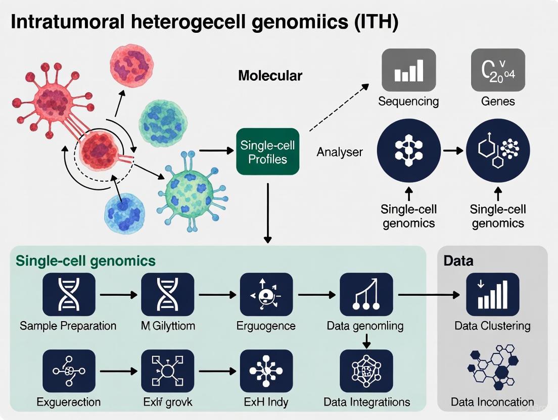
This article explores the transformative impact of single-cell genomics on our understanding of intratumoral heterogeneity (ITH) in cancer.
Decoding Cancer Complexity: How Single-Cell Genomics Reveals Intratumoral Heterogeneity and Drives Therapeutic Innovation
Abstract
This article explores the transformative impact of single-cell genomics on our understanding of intratumoral heterogeneity (ITH) in cancer. Aimed at researchers and drug development professionals, it synthesizes current evidence to illustrate how single-cell technologies decipher the cellular diversity, spatial architecture, and molecular mechanisms within tumors. The content covers foundational concepts of ITH, advanced methodological applications for dissecting it, key challenges in data analysis and integration, and the crucial validation of biological and clinical significance. By providing a comprehensive overview of how ITH influences tumor evolution, immune evasion, and therapy resistance, this resource aims to bridge cutting-edge research with the development of novel, targeted therapeutic strategies.
The Multifaceted Nature of Intratumoral Heterogeneity: From Basic Concepts to Clinical Impact
Defining Spatial and Temporal Heterogeneity in Cancer Ecosystems
Intratumoral heterogeneity (ITH) represents a fundamental challenge in clinical oncology, underlying tumor progression, metastatic potential, and therapeutic resistance. This heterogeneity manifests across multiple dimensions—spatial (variation across different tumor regions) and temporal (evolution over time and treatment courses)—creating complex cancer ecosystems that constantly adapt to selective pressures [1] [2]. The emergence of single-cell genomics has revolutionized our ability to dissect this complexity, revealing that ITH extends beyond genetic diversity to encompass transcriptional, epigenetic, and functional states across both malignant and non-malignant cell populations within the tumor microenvironment (TME) [3] [2].
Spatial heterogeneity arises from varied microenvironmental niches within tumors, where gradients of nutrients, oxygen, and signaling molecules create distinct ecological subregions. Temporal heterogeneity reflects clonal evolution, where cancer cells accumulate mutations and adapt under therapeutic selection pressures [1]. Understanding the dynamic interplay between these spatial and temporal dimensions is critical for developing effective therapeutic strategies that anticipate and counter adaptive resistance mechanisms. This technical guide synthesizes current methodologies, analytical frameworks, and insights from single-cell genomics research to provide a comprehensive resource for investigating cancer ecosystem heterogeneity.
Technological Foundations for Dissecting Heterogeneity
Single-Cell and Spatial Multi-Omics Technologies
Advanced genomic technologies now enable comprehensive profiling of ITH at multiple molecular layers while preserving crucial spatial and temporal context. The integration of these complementary approaches provides a powerful framework for reconstructing cancer ecosystems.
Table 1: Core Technologies for Analyzing Cancer Heterogeneity
| Technology | Key Applications in ITH Research | Resolution | Limitations |
|---|---|---|---|
| scRNA-seq | Identifying cell subtypes, transcriptional states, and rare populations [3] [4] | Single-cell | Loss of spatial context, technical noise |
| Spatial Transcriptomics | Mapping gene expression patterns in tissue architecture, identifying spatial niches [1] [5] [4] | 55μm (Visium) to subcellular | Lower resolution than scRNA-seq, limited sensitivity |
| scDNA-seq | Profiling copy number variations and single nucleotide variants [3] [5] | Single-cell | Incomplete genomic coverage, amplification artifacts |
| Spatial Multi-omics | Simultaneous measurement of multiple molecular layers in situ [1] | Varies by platform | Computational complexity, data integration challenges |
| scATAC-seq | Mapping chromatin accessibility and regulatory landscapes [3] | Single-cell | Sparse data, indirect epigenetic measurement |
The synergistic integration of single-cell RNA sequencing (scRNA-seq) with spatial transcriptomics (ST) has emerged as a particularly powerful approach. While scRNA-seq provides high-resolution characterization of cellular diversity, it loses native spatial context due to tissue dissociation. ST preserves spatial localization but traditionally at lower resolution. Computational integration strategies bridge this gap, enabling the mapping of cell types and states onto tissue architecture [4]. These integration methods include deconvolution approaches that infer cell type proportions within spatial spots, and mapping strategies that project single-cell data onto spatial coordinates using shared molecular features [1] [4].
Computational Integration Strategies
The effective integration of multi-modal single-cell and spatial data requires sophisticated computational approaches that address multiple challenges:
- Horizontal integration combines identical omics types across multiple tissue slices, using shared molecular features as reference points to align datasets and reconstruct three-dimensional tissue architecture [1].
- Vertical integration merges different omics data (e.g., gene expression and protein abundance) from the same tissue section, with individual cells serving as the natural reference for alignment [1].
- Diagonal integration addresses the most challenging scenario where different omics types originate from different tissue sections, requiring advanced algorithms to establish correspondence without shared features or common cells [1].
Tools such as PASTE apply optimal transport methods to align neighboring tissue slices, while GraphST and SPACEL use graph-based models to reconstruct 3D tissue structures and identify spatial domains across multiple samples [1]. Methods like SEDR, PRECAST, and STAligner employ autoencoders, projection-based alignment, and graph models to integrate data across diverse technologies and experimental conditions while preserving biological signals and removing technical batch effects [1].
Experimental Framework for Spatial and Temporal Analysis
Protocol 1: Comprehensive Pan-Cancer Ecosystem Mapping
The TabulaTIME framework represents a scalable approach for constructing pan-cancer single-cell atlases that capture ecosystem heterogeneity across cancer types, anatomical sites, and disease stages [6].
Experimental Workflow:
- Data Collection and Curation: Collect single-cell RNA sequencing datasets from public repositories and new samples, ensuring representation across multiple cancer types, tissue contexts (normal, precancerous, primary tumor, metastatic), and treatment histories. The TabulaTIME resource integrated 4,483,367 cells across 36 cancer types from 746 donors [6].
- Quality Control and Preprocessing: Implement rigorous quality control using the MAESTRO workflow or equivalent pipelines to remove doublets, filter low-quality cells, and mitigate technical artifacts [6].
- MetaCell Construction: Group transcriptionally similar cells into MetaCells (approximately 30 cells per group) to reduce technical noise and computational burden while preserving biological signals [6].
- Batch Effect Correction: Apply canonical correlation analysis (CCA) or other advanced integration methods to correct for batch effects across different studies, platforms, and experimental conditions [6].
- Lineage-Specific Integration: Perform separate integration analyses for major cellular lineages (e.g., cytotoxic lymphocytes, myeloid cells, fibroblasts) to achieve higher resolution of cell states within lineages [6].
- Multi-Omics Integration: Integrate single-cell maps with spatial transcriptomics, bulk tumor profiles from resources like TCGA, and clinical metadata to enable spatial localization and clinical correlation analyses [6].
Protocol 2: Resolving Intratumoral Heterogeneity with Multi-Modal Integration
This protocol focuses on deep characterization of ITH within individual tumors by combining single-cell genomics, spatial transcriptomics, and copy number variation analysis, as applied in studies of natural killer/T cell lymphoma (NKTCL) and high-grade serous ovarian carcinoma (HGSOC) [7] [5].
Experimental Workflow:
- Sample Processing and Quality Control: Process freshly collected tumor tissues for single-cell RNA sequencing, ensuring preservation of cell viability and RNA integrity. Perform initial quality control to remove damaged cells and technical artifacts.
- Cell Type Identification and Annotation: Identify major cell types using canonical marker genes and reference-based annotation. Distinguish malignant from non-malignant cells through copy number variation (CNV) inference from transcriptome data [7] [5].
- Malignant Cell Subtyping: Apply consensus non-negative matrix factorization (cNMF) or similar algorithms to identify meta-programs (MPs) representing distinct functional states within malignant cells. Correlate these programs with clinical outcomes [7].
- Spatial Transcriptomics Processing: Process matched tissue sections for spatial transcriptomics using platforms such as Visium (10x Genomics) or similar technologies. Align spatial data with histological features [5].
- Temporal Reconstruction: Apply pseudotime analysis algorithms (e.g., Monocle, PAGA) to reconstruct differentiation trajectories and infer temporal relationships between cellular states [7].
- Cell-Cell Communication Analysis: Infer ligand-receptor interactions using tools like CellPhoneDB or NicheNet to map communication networks between spatially co-localized cell populations [5] [4].
Table 2: Essential Research Reagents and Computational Tools
| Category | Specific Reagents/Tools | Application in ITH Research |
|---|---|---|
| Wet Lab Reagents | 10x Genomics Chromium Chip | Single-cell partitioning and barcoding [3] |
| Visium Spatial Gene Expression Slide | Spatial transcriptomics capture [1] | |
| Enzymatic Tissue Dissociation Kits | Preparation of single-cell suspensions [3] | |
| Bioinformatics Tools | Seurat | Single-cell data integration and analysis [5] |
| PASTE | Spatial alignment of tissue sections [1] | |
| CellPhoneDB | Ligand-receptor interaction analysis [4] | |
| cNMF | Meta-program identification [7] | |
| Reference Data | TabulaTIME | Pan-cancer single-cell reference [6] |
| TCGA | Bulk tumor molecular and clinical data [6] |
Key Findings and Analytical Insights
Spatially Organized Cancer-Associated Fibroblast and Macrophage Ecotypes
Pan-cancer single-cell analyses have revealed conserved, spatially organized cellular ecotypes that shape tumor ecosystems and influence clinical outcomes. The TabulaTIME resource identified CTHRC1+ cancer-associated fibroblasts (CAFs) as a hallmark of extracellular matrix-remodeling fibroblasts enriched at the leading edge between malignant and normal regions, where they may create physical barriers that prevent immune cell infiltration [6]. These specialized CAFs colocalize with SLPI+ macrophages to form profibrotic ecotypes that exhibit diminished phagocytic capacity but enhanced extracellular matrix remodeling activity [6].
Spatial analysis of these ecotypes reveals their coordinated role in shaping the tumor microenvironment. The colocalization of specific fibroblast and macrophage subtypes creates specialized niches that support tumor progression and immune evasion. These findings suggest that therapeutic targeting of these profibrotic ecotypes, rather than individual cell types, may represent a more effective strategy for disrupting protumorigenic microenvironmental niches [6].
Metabolic Heterogeneity and Therapeutic Vulnerabilities
Single-cell analyses have uncovered profound metabolic heterogeneity within tumor ecosystems, revealing novel therapeutic opportunities. In natural killer/T cell lymphoma (NKTCL), a distinct meta-program (MP3) characterized by MYC hyperactivation and elevated fatty acid metabolism was associated with poor prognosis and a less differentiated cellular state [7]. Within this aggressive subpopulation, fatty acid-binding protein 5 (FABP5) demonstrated a strong correlation with MYC signaling and differentiation status, with expression decreasing along differentiation trajectories [7].
Functional validation confirmed FABP5 as a therapeutic vulnerability, where pharmacological inhibition with SBFI-26 downregulated c-Myc expression and significantly impaired tumor growth both in vitro and in vivo [7]. This example illustrates how single-cell analysis can identify metabolic dependencies within specific malignant subpopulations, revealing context-specific vulnerabilities that may be masked in bulk analyses.
Spatial Hierarchy of Cancer Hallmarks
Spatial transcriptomic analysis across 63 primary untreated tumors from 10 cancer types has revealed that hallmark cancer capabilities are spatially organized within tumor ecosystems [8]. This organization follows distinct patterns, with cancer cells primarily contributing to seven of the thirteen established hallmarks, while the tumor microenvironment governs the remainder [8]. Genomic distance between tumor subclones correlates with differences in hallmark activity, leading to functional specialization where distinct subclones preferentially execute different hallmark capabilities [8].
These spatial patterns create ecological dynamics within tumors, where interdependent relationships between hallmarks emerge particularly at the interfaces between tumor and microenvironment compartments [8]. This spatial organization has direct therapeutic implications, as demonstrated in bladder cancer patients from the DUTRENEO trial, where spatial hallmark patterns correlated with sensitivity to different neoadjuvant treatments [8].
Clone-Specific Communication Networks
Analysis of high-grade serous ovarian carcinoma (HGSOC) has revealed that communication networks between tumor cell clusters exhibit unique patterns associated with the meta-programs governing these clusters [5]. The ligand-receptor pair MDK-NCL emerged as a highly enriched interaction in tumor cell communication, and functional studies confirmed that NCL overexpression enhanced tumor cell proliferation [5]. This finding illustrates how specific communication pathways are activated in particular clonal populations, creating autocrine and paracrine signaling networks that support tumor growth and ecosystem organization.
Copy number variation analysis further revealed intratumor heterogeneity through distinct tumor clones with unique evolutionary trajectories and spatial relationships [5]. By examining both heterogeneity and spatial relationships between clones, researchers can reconstruct the ecological and evolutionary dynamics that shape tumor progression and therapeutic resistance.
The integration of single-cell genomics with spatial transcriptomics has fundamentally transformed our understanding of cancer ecosystems, revealing previously unappreciated dimensions of spatial and temporal heterogeneity. The frameworks and methodologies outlined in this technical guide provide a roadmap for systematically dissecting this complexity, enabling researchers to move beyond cataloging heterogeneity toward understanding its functional consequences and therapeutic implications.
Future advances in this field will require continued technological innovation, particularly in achieving true single-cell resolution in spatial transcriptomics, capturing dynamic processes through live imaging integration, and developing more sophisticated computational models that can predict ecosystem dynamics in response to therapeutic perturbation. Additionally, standardized frameworks for data integration and sharing will be essential for building comprehensive atlases of tumor ecosystems across cancer types, disease stages, and therapeutic contexts.
As these technologies mature and become more accessible, spatially-resolved single-cell analysis is poised to transform cancer diagnostics and therapeutic development, enabling identification of novel biomarkers, patient stratification strategies, and ecosystem-targeted therapies that address the fundamental challenges of tumor heterogeneity and adaptation.
Genetic, Transcriptomic, and Epigenetic Drivers of Cellular Diversity
The emergence of single-cell genomics has revolutionized the study of cellular diversity, providing an unprecedented lens through which to view the genetic, transcriptomic, and epigenetic variation that underpins intratumoral heterogeneity (ITH). ITH is a fundamental property of most human cancers and a major cause of treatment resistance and disease progression [9] [10]. Whereas traditional bulk sequencing methods average signals across thousands of cells, obscuring rare but critical cell populations, single-cell technologies enable the dissection of tumors at the resolution of individual cells. This reveals a complex ecosystem of cellular states and lineages that coexist within a single tumor mass [9] [11]. Understanding the drivers of this diversity requires a multi-layered approach that integrates genomic alterations, transcriptomic programs, and epigenetic regulation. This technical guide examines the core mechanisms driving cellular diversity within the context of ITH, providing researchers with a comprehensive framework for studying this complex phenomenon using state-of-the-art single-cell methodologies.
Genetic Drivers of Cellular Diversity
Genetic evolution within tumors generates cellular diversity through the accumulation of somatic mutations, copy number alterations, and structural variations that are differentially distributed across cell populations. This genetic heterogeneity creates distinct subclones with varying functional capabilities within the same tumor mass.
Single-cell RNA sequencing (scRNA-seq) enables the investigation of ITH by capturing the transcriptional profiles of individual tumor cells from multiple regions within a single tumor. In a seminal study on pleural mesothelioma (PM), researchers analyzed tumor cells from three distant biopsies—costal, diaphragmatic, and mediastinal—using scRNA-seq. They identified three predominant cell states present across all regions: a stem-like state (C1), an epithelial-like state (C2), and a mesenchymal-like state (C3). Notably, the abundance of these states varied spatially, with the C1 state being less prominent in the mediastinal biopsy compared to the other regions [9]. This regional variation underscores how genetic diversity manifests in distinct microenvironments.
The merger of quantitative genetics with single-cell genomics has significantly enhanced the detection resolution of variants that control molecular traits. Single-cell population genomics not only identifies these genetic variants but also reveals the specific cell types in which they exert their effects. When combined with organism-level phenotype measurements, this approach elucidates which cellular contexts impact higher-order traits [12]. The implementation of single-cell genetics is advancing the investigation of the genetic architecture of complex molecular traits and providing new experimental paradigms for studying eukaryotic genetics.
Table 1: Genetic and Cell State Drivers of Intratumoral Heterogeneity - A Pleural Mesothelioma Case Study
| Driver Category | Specific Feature | Impact on ITH | Clinical/Functional Association |
|---|---|---|---|
| Cell State Identity | C1 (Stem-like) | Progenitor population with high plasticity | Potential sensitivity to anti-angiogenic therapies |
| Cell State Identity | C2 (Epithelial-like) | Differentiated state, epithelial characteristics | Not specified |
| Cell State Identity | C3 (Mesenchymal-like) | Differentiated state, mesenchymal characteristics | Associated with worse survival; reduced sensitivity to standard therapies |
| Spatial Distribution | Regional Biopsy Variation (Costal, Diaphragmatic, Mediastinal) | Distinct abundance of cell states (e.g., C1 less abundant in mediastinal region) | Suggests microenvironmental influence on cell state prevalence |
| Dynamic Process | Epithelial-Mesenchymal Plasticity (EMP) | Trajectory analysis suggested a dynamic continuum between states via a stem-like intermediate | Underpins cellular adaptability and potential for metastasis |
Transcriptomic Drivers of Cellular Diversity
Transcriptomic diversity represents the functional output of genetic and epigenetic variation, revealing distinct cellular states and identities within seemingly homogeneous tissues. Single-cell transcriptomics has uncovered remarkable complexity in both normal and diseased tissues, providing insights into disease mechanisms and potential therapeutic targets.
The power of single-cell transcriptomics lies in its ability to identify novel cell populations and disease-associated states. A landmark study on human heart failure demonstrated this capacity by integrating scRNA-seq and snRNA-seq data from 45 individuals. The analysis revealed that in dilated cardiomyopathy, different cell types undergo distinct transcriptional reprogramming. While cardiomyocytes were found to converge toward common disease-associated states, fibroblasts and myeloid cells underwent dramatic diversification, indicating cell-type-specific responses to disease stimuli [13]. This principle is equally applicable to cancer, where scRNA-seq of pancreatic ductal adenocarcinoma (PDAC) has revealed extensive heterogeneity encompassing various malignant and stromal cell types, with malignant subtypes consisting of multiple subpopulations with distinct proliferation and migration capabilities [10].
A key innovation in the transcriptomic analysis of cellular hierarchies is the CytoTRACE algorithm. This computational framework leverages a simple yet robust determinant of developmental potential—the number of expressed genes per cell (gene counts). Systematic analysis revealed that gene counts generally decrease with successive stages of differentiation across diverse tissues and organisms. CytoTRACE uses this feature to predict differentiation states from scRNA-seq data in an unsupervised manner, outperforming previous methods and nearly 19,000 annotated gene sets for resolving experimentally determined developmental trajectories [14]. This tool is particularly valuable for identifying stem-like cells and reconstructing differentiation trajectories within heterogeneous tumors.
Table 2: Key Single-Cell Transcriptomic Technologies and Applications
| Technique | Key Technical Features | Primary Applications in ITH Research |
|---|---|---|
| CEL-Seq2 | Introduces Unique Molecular Identifiers (UMIs) to eliminate PCR amplification bias; lower throughput. | Exploring cellular heterogeneity and molecular mechanisms with reduced technical artifacts. |
| MARS-Seq | High throughput; uses unique molecular tags for hybrid sequencing of multiple samples; lower cost. | Studying heterogeneity in tumors and capturing spatial transcriptomic information. |
| 10X Genomics | Droplet-based microfluidics for high-throughput single-cell partitioning and barcoding. | Large-scale atlas building (e.g., Human Cell Atlas); comprehensive profiling of complex tumor ecosystems. |
| Single-nucleus RNA-seq (snRNA-seq) | Sequences nuclear RNA, allowing profiling of cells difficult to isolate intact (e.g., cardiomyocytes, neurons). | Analyzing frozen or archived tissue; studying tissues resistant to dissociation. |
Epigenetic Drivers of Cellular Diversity
Epigenetic regulation constitutes a crucial layer of control over cellular identity and diversity, enabling heritable changes in gene expression without alterations to the DNA sequence itself. Single-cell epigenomic methods have revealed that epigenetic heterogeneity is a fundamental driver of ITH, contributing to tumor evolution, therapy resistance, and metastatic potential.
Various epigenetic layers can now be studied at single-cell resolution, including chromatin accessibility, DNA methylation, histone modifications, and nucleosome localization. The assay for transposase-accessible chromatin using sequencing (scATAC-seq) has been particularly transformative for mapping open chromatin regions genome-wide in individual cells. scATAC-seq utilizes a hyperactive Tn5 transposase to insert sequencing adapters directly into accessible chromatin regions, which are typically nucleosome-depleted and house regulatory DNA elements such as enhancers and promoters [12] [15]. When integrated with scRNA-seq data, which only loosely correlates with chromatin accessibility (Spearman's correlation coefficient 0.54-0.58), it provides a more comprehensive depiction of cellular states [12].
DNA methylation represents another critical epigenetic mark that can be mapped at single-cell resolution. Single-cell bisulfite sequencing (scBS-seq) uses a post-bisulfite adapter-tagging (PBAT) approach to overcome DNA degradation issues, enabling measurement of methylation at up to 50% of CpG sites in a single cell [15]. This has revealed high variability between single cells in distal enhancer methylation, even in seemingly homogeneous cell populations. Emerging multiomic technologies now allow parallel measurements of multiple epigenetic layers or the combination of epigenetic and transcriptomic profiling from the same single cell. For instance, scM&T-seq enables simultaneous BS-seq and RNA-seq from the same cell by physically separating poly-A mRNA from DNA [15]. These integrated approaches are essential for understanding how epigenetic states influence transcriptional output and cellular phenotype in cancer.
Integrated Multiomic Approaches and Experimental Design
The full complexity of ITH can only be captured through integrated approaches that simultaneously measure multiple molecular layers from the same cell. Multiomic single-cell technologies have emerged as powerful tools for unraveling the interconnected regulatory networks that govern cellular diversity in cancer.
Multiomic measurements typically rely on converting biological signals into DNA-level information that can be deconvoluted via sequencing. Techniques have been developed that measure two or more molecular traits from the exact same cell, such as simultaneous profiling of RNA expression and chromatin accessibility (scRNA-seq + scATAC-seq), genetic changes and genomic traits, or DNA methylation and transcriptomes [12]. The ability to correlate epigenetic states with transcriptional outputs from the same single cell is particularly valuable for distinguishing cause from effect in regulatory relationships and for identifying master regulators of cell fate decisions in cancer.
When designing single-cell studies of ITH, several methodological considerations are paramount. The choice between single-cell and single-nucleus sequencing depends on the tissue type and research questions. snRNA-seq offers advantages for profiling tissues that are difficult to dissociate (such as heart tissue [13]) or when working with frozen specimens, as it avoids biases introduced by enzymatic digestion and captures non-cytoplasmic transcripts. However, it ablates all cytoplasmic information, including protein signals [12]. For plant research, where cell walls present a barrier, single-nucleus techniques have enabled the migration of single-cell genomics from animal systems [12]. Experimental design must also account for technical variability through appropriate replication, sample multiplexing, and the implementation of rigorous quality control metrics, including thresholds for gene and feature counts per cell [13].
Cutting-edge research into cellular diversity relies on a suite of specialized reagents, technologies, and computational tools. The following table details key resources essential for conducting single-cell multiomic studies of intratumoral heterogeneity.
Table 3: Research Reagent Solutions for Single-Cell Multiomics
| Resource Category | Specific Item/Technology | Function/Application |
|---|---|---|
| Core Sequencing Technology | 10X Genomics Single Cell 5' Platform (e.g., used in [13]) | Enables high-throughput partitioning, barcoding, and preparation of single-cell or single-nucleus libraries for RNA-seq and ATAC-seq. |
| Epigenomic Profiling Reagent | Hyperactive Tn5 Transposase (for scATAC-seq [12] [15]) | Simultaneously fragments and tags accessible genomic DNA within nuclei, defining open chromatin landscapes. |
| Methylation Profiling Reagent | Sodium Bisulfite (for scBS-seq [15]) | Chemically converts unmethylated cytosines to uracils, allowing for single-base resolution mapping of DNA methylation (5mC). |
| Cell Isolation/Partitioning | Microfluidic Devices (e.g., Fluidigm C1) or Droplet-Based Systems | Physically isolates thousands of individual cells or nuclei into nanoliter-scale reactions for parallel processing. |
| Bioinformatic Tool | Seurat & Harmony [13] | Computational packages for the integration, quality control, unsupervised clustering, and differential expression analysis of single-cell data. |
| Developmental Trajectory Tool | CytoTRACE [14] | Computational framework that predicts cellular differentiation states and hierarchies based on the number of expressed genes per cell. |
| Color Palette Tool | Viz Palette [16] | Online tool to test and ensure that color palettes chosen for data visualization are accessible to audiences with color vision deficiencies (CVD). |
Data Visualization and Color Accessibility
Effective communication of single-cell data requires thoughtful visualization strategies that accurately represent complex relationships while remaining accessible to all readers, including those with color vision deficiencies (CVD). Scientific color palettes should be chosen not only for aesthetic appeal but as powerful tools for data storytelling [16].
For single-cell genomics, the type of color palette should match the nature of the data being visualized. Qualitative palettes, using distinct hues, are appropriate for categorical data such as cell types or clusters. Sequential palettes, which vary in lightness and optionally hue, are used for representing continuous numeric values with inherent ordering. Diverging palettes, which combine two sequential palettes with a shared central value, are ideal for highlighting deviations from a baseline (e.g., upregulated and downregulated genes) [17]. It is critical to avoid unnecessary usage of color and to maintain consistency across charts when colors refer to the same groups [17].
Given that approximately 1 in 12 men and 1 in 200 women experience some form of CVD, ensuring accessibility is essential for ethical scientific communication. Tools like Viz Palette allow researchers to test color combinations against simulations of different types of color blindness, such as deuteranopia (red-green confusion) [16]. A common misconception is that red and green should never be used together; however, if these colors are important for data storytelling (e.g., stop/go, positive/negative), they can be used together effectively by adjusting saturation and lightness to create sufficient contrast [16]. Grayscale remains a highly effective and accessible option, provided there is approximately a 15-30% difference in saturation between shades [16].
ITH as a Key Driver of Metastasis, Relapse, and Poor Prognosis
Intratumoral heterogeneity (ITH) represents a fundamental paradigm in cancer biology, driving metastatic progression, therapeutic relapse, and ultimately, poor patient prognosis. Through the lens of single-cell genomics, researchers can now decode the complex cellular ecosystems within tumors, revealing how genetic, transcriptomic, and epigenetic diversity fuels therapeutic resistance and disease advancement. This technical guide synthesizes cutting-edge research methodologies and analytical frameworks that empower researchers and drug development professionals to quantify, model, and target ITH. By integrating spatial metrics, computational modeling, and single-cell technologies, the field is progressing toward more effective therapeutic strategies that address the complex realities of tumor evolution.
Intratumoral heterogeneity (ITH) refers to the presence of distinct cancer cell populations within a single tumor that exhibit divergent genotypic and phenotypic properties [18]. This diversity arises through complex interactions between intrinsic factors (genetic mutations, transcriptomic variations, epigenetic modifications) and extrinsic factors (components of the tumor microenvironment) that collectively shape tumor evolution [18]. The clinical significance of ITH is profound—it represents a key biological mechanism underlying metastatic progression and therapeutic failure, with metastatic cancer accounting for approximately 80.9% of cancer-related deaths according to SEER database analyses [18].
The emergence of single-cell genomics has revolutionized our ability to dissect ITH at unprecedented resolution, moving beyond bulk tumor analysis to characterize the cellular and molecular diversity that drives cancer progression. This technical guide provides researchers and drug development professionals with the analytical frameworks and experimental methodologies required to investigate ITH within the context of modern cancer research, with particular emphasis on its role in metastasis, relapse, and poor clinical outcomes.
Molecular Mechanisms Driving ITH
Genetic and Transcriptomic Diversity
ITH manifests through multiple molecular layers that collectively drive tumor evolution and metastatic capability. Genetic heterogeneity arises from the accumulation of driver mutations (e.g., TP53, PTEN, PIK3CA) that provide selective advantages, and passenger mutations that contribute to clonal diversity without direct functional consequences [18]. Beyond genetic alterations, transcriptomic variations create phenotypic diversity within tumors, as demonstrated in hepatocellular carcinoma where 30% of stage II patients exhibited mixed transcriptomic subtypes with more aggressive phenotypes characterized by upregulated cell cycle pathways [18].
The following table summarizes the key dimensions of ITH and their functional consequences:
Table 1: Dimensions of Intratumoral Heterogeneity and Functional Consequences
| Dimension of Heterogeneity | Molecular Basis | Functional Consequences | Example Cancer Types |
|---|---|---|---|
| Genetic ITH | Copy number variations (CNV), single-nucleotide variants (SNV), indels, chromosomal aberrations | Differential drug sensitivity, metastatic potential, immune evasion | Colorectal cancer (BRAF/KRAS heterogeneity) [18] |
| Transcriptomic ITH | Gene expression profile variations, alternative splicing | Phenotypic plasticity, epithelial-mesenchymal transition (EMT) spectrum, metabolic adaptations | Hepatocellular carcinoma, breast cancer [18] |
| Epigenetic ITH | DNA methylation patterns, histone modifications (H3K27me3), chromatin remodeling | Therapy resistance, phenotype switching, stable non-genetic adaptations | Castration-resistant neuroendocrine prostate cancer [18] |
| Protein-level ITH | Differential expression of receptor proteins (ERα, HER2) and signaling molecules | Altered cell proliferation, invasion capacity, hormone dependence | Endometrial cancer, breast cancer [18] |
The Role of the Tumor Microenvironment
The tumor microenvironment (TME) serves as a critical extrinsic factor shaping ITH through dynamic interactions between cancer cells and stromal components. These include cancer-associated fibroblasts (CAFs), tumor-associated macrophages (TAMs), and various immune cell populations that create distinct microniches within the tumor [18]. The presence of both 'hot' (immune-enriched) and 'cold' (immunosuppressive) microenvironments within the same tumor further promotes selection of subclones with varying capacities for immune evasion [18]. Spatial organization of these components establishes pre-metastatic niches that support disseminated cells, with recent studies identifying specific stromal and immune cell subtypes (CCL2+ macrophages, exhausted cytotoxic T cells, FOXP3+ regulatory T cells) as critical to forming pro-tumor microenvironments in metastatic lesions [19].
Quantitative Assessment of ITH
Spatial Metrics from Computational Digital Pathology
Quantitative assessment of ITH requires specialized metrics adapted from computational digital pathology. These spatial metrics enable researchers to classify tumor immunoarchitecture and correlate spatial patterns with treatment outcomes [20].
Table 2: Spatial Metrics for Quantifying Intratumoral Heterogeneity
| Metric | Mathematical Basis | Interpretation | Application in Treatment Response |
|---|---|---|---|
| Mixing Score | Quantification of cell type intermixing | High values indicate well-mixed cell populations; low values indicate segregation | "Cold" tumors show poor mixing [20] |
| Average Neighbor Frequency | Probability analysis of adjacent cell types | Measures likelihood of specific cell-cell interactions | Compartmentalized tumors show structured neighbor relationships [20] |
| Shannon's Entropy | Information theory applied to cell distribution | Measures disorder in spatial organization | Higher entropy indicates greater randomness in cell distribution [20] |
| G-cross Function | Spatial statistics measuring clustering patterns | Quantifies accumulation of specific cell types at various distances | Area under curve (AUC) indicates degree of spatial clustering [20] |
| Cancer:Immune Cell Ratio | Simple count ratio of cell populations | Estimates overall immune infiltration | Lower ratios often correlate with better treatment response [20] |
These metrics have been successfully applied to classify TME immunoarchitecture into three distinct patterns: (1) "cold" tumors characterized by limited immune infiltration, (2) "compartmentalized" tumors showing structured but segregated immune regions, and (3) "mixed" tumors demonstrating high levels of immune-cancer cell intermixing [20]. Importantly, compartmentalized immunoarchitecture has been associated with more efficacious outcomes following immune checkpoint inhibitor therapy, providing a quantitative link between spatial heterogeneity and treatment response [20].
Single-Cell RNA Sequencing Analytical Frameworks
Single-cell RNA sequencing (scRNA-seq) provides the technological foundation for dissecting ITH at transcriptomic resolution. The standard analytical workflow encompasses multiple stages, each with specific methodological considerations:
Figure 1: scRNA-seq Data Analysis Workflow for ITH Research
Critical to this workflow is the experimental design phase, which must account for species-specific considerations, sample origin (tissue biopsies, PBMCs, or patient-derived organoids), and appropriate case-control structures [21]. Following data generation, quality control employs three key metrics: total UMI count (count depth), number of detected genes, and fraction of mitochondrial reads, with thresholds dependent on tissue type, dissociation protocol, and library preparation method [21].
Dimensionality reduction techniques present particular challenges for visualizing heterogeneous single-cell data. Different algorithms exhibit varying performance in preserving global versus local data structure, with input cell distribution (discrete versus continuous) largely determining method performance [22]. For instance, UMAP tends to compress local distances while maintaining global structure, whereas t-SNE may better preserve local neighborhoods—a critical consideration when analyzing continuous phenotypic transitions within tumors [22].
ITH in Metastasis and Therapy Resistance: Key Experimental Findings
Functional Evidence from Model Systems
Experimental models have provided compelling evidence linking ITH to metastatic progression. In a landmark study using the SUM149PT human breast cancer cell line, single-cell cloning revealed an epithelial-mesenchymal transition (EMT) spectrum encompassing epithelial (E), intermediate EMT (EM1, EM2, EM3), and mesenchymal (M1, M2) phenotypes [18]. Importantly, intermediate EMT cells—characterized by elevated CBFβ protein expression—exhibited significantly higher migratory and invasive capacity (2-10 fold) compared to fully mesenchymal clones [18]. In vivo metastasis assays demonstrated that these intermediate EMT populations predominantly contributed to metastatic lesions, with different EMT subtypes generating distinct metastatic patterns (micrometastases versus macrometastases) [18].
The relationship between ITH and therapy resistance has been systematically investigated in colorectal cancer models, where single-cell RNA sequencing of patient-derived organoids revealed heterogeneous populations of POU5F1-positive and POU5F1-negative cells with differential drug sensitivities [18]. Following anticancer drug treatment, chemo-resistant POU5F1-positive cells expanded significantly and demonstrated higher metastatic potential (4/4 liver metastases versus 0/4 in POU5F1-negative cells) through upregulation of the Wnt/β-catenin signaling pathway [18]. Therapeutic targeting of this pathway with the inhibitor XAV939 reduced β-catenin expression and led to tumor shrinkage, illustrating how understanding the molecular mechanisms underlying ITH can reveal novel therapeutic vulnerabilities [18].
Archetype Analysis in Small Cell Lung Cancer
In small cell lung cancer (SCLC), archetypal analysis has provided a novel framework for understanding phenotypic plasticity and its relationship to ITH. This approach models SCLC phenotypic heterogeneity through multi-task evolutionary theory, positioning cellular states within a five-dimensional convex polytope whose vertices optimize specific tasks reminiscent of pulmonary neuroendocrine cells [23]. These archetype tasks—including proliferation, slithering, metabolism, secretion, and injury repair—reflect fundamental cancer hallmarks and provide a quantitative basis for understanding cellular positioning along phenotypic continua [23]. SCLC subtypes can be characterized as task specialists or multi-task generalists based on their distance from archetype vertex signatures, with single-cell plasticity modeled as a Markovian process along an underlying state manifold [23].
Computational Modeling Approaches
Hybrid Spatio-Temporal Modeling
The integration of quantitative systems pharmacology (QSP) with agent-based models (ABM) has emerged as a powerful approach for simulating ITH dynamics and therapy response. Spatial QSP (spQSP) platforms combine whole-patient compartmental modeling with three-dimensional spatial resolution to capture the complex interactions between tumor cells and immune components [20]. These hybrid models typically comprise:
- QSP Module: A whole-body compartmental model (tumor, peripheral, tumor-draining lymph node, and central blood compartments) described by ordinary differential equations (ODEs) [20]
- ABM Module: A three-dimensional spatial model with discrete (cell-cell interactions) and continuum (cytokine distribution) layers [20]
- Coupling Framework: Bidirectional information exchange between QSP and ABM modules at each simulation time step [20]
This architecture enables simulation of virtual patient populations over clinical timescales while maintaining spatial resolution sufficient to quantify emergent heterogeneity patterns using the spatial metrics described in Section 3.1 [20].
Phylogenetic Reconstruction from Multi-region Sequencing
Phylogenetic analysis based on multi-region sequencing data enables reconstruction of tumor evolutionary histories and quantification of ITH spatial patterns. In endometrial carcinoma, whole-exome sequencing of multiple tumor regions has revealed extensive spatial heterogeneity, with phylogenetic trees illustrating divergent evolution across geographical locations within the same tumor [24]. Notably, while primary tumors exhibit substantial spatial ITH, metastatic lesions from the same patient often display genomic homogeneity, suggesting that metastatic seeding may originate from specific subclones or require particular genetic constellations [24]. These phylogenetic approaches have also decoded the molecular evolution of ambiguous endometrial cancers, guiding personalized therapy selection validated through patient-derived xenograft models [24].
The Scientist's Toolkit: Essential Research Reagents and Platforms
Table 3: Essential Research Reagents and Platforms for ITH Investigation
| Tool Category | Specific Examples | Primary Function | Technical Considerations |
|---|---|---|---|
| scRNA-seq Platforms | 10x Genomics Chromium, Singleron systems | High-throughput single-cell transcriptomic profiling | Cell Ranger/CeleScope for data processing; optimized for high-performance computing [21] |
| Data Processing Pipelines | Cell Ranger, CeleScope, scPipe, zUMIs | Raw data processing, demultiplexing, UMI count matrix generation | Choice less critical than downstream analysis; require massive computational resources [21] |
| Dimensionality Reduction Algorithms | t-SNE, UMAP, PCA, SIMLR | Visualization of high-dimensional data in 2D/3D space | Performance depends on input cell distribution; trade-offs between local/global structure preservation [22] |
| Spatial Metrics Software | Mixing score, G-cross function, Shannon's entropy algorithms | Quantification of spatial patterns in multiplexed imaging data | Implementation in Python/R; validation against known spatial patterns required [20] |
| Cell Type Annotation Tools | SCANVI, CellHint | Biology-aware integration and cell type identification | Leverage known cell type labels; incorporate sample-specific covariates [19] |
| Copy Number Inference | InferCNV, CaSpER, SCEVAN | CNV profiling from scRNA-seq data | T cells as reference; higher CNV scores indicate genomic instability [19] |
Intratumoral heterogeneity represents a fundamental challenge in clinical oncology, serving as both a biomarker of aggressive disease and a therapeutic target in its own right. The integration of single-cell genomics, spatial metrics, and computational modeling provides researchers with an unprecedented toolkit to dissect the molecular and cellular complexity of heterogeneous tumors. As these technologies mature, their translation into clinical applications promises to transform cancer diagnosis and treatment, moving beyond bulk tumor characterization toward precision approaches that address the diverse cellular ecosystems within each patient's cancer. Future research directions will likely focus on integrating multi-omic single-cell data (genome, epigenome, transcriptome, proteome) within spatial contexts, developing therapeutic strategies that explicitly target phenotypic plasticity, and validating ITH metrics as clinically actionable biomarkers for treatment selection and monitoring.
Intratumoral heterogeneity (ITH) represents a fundamental challenge in oncology, contributing to therapeutic resistance, disease progression, and metastatic potential. Single-cell genomics has revolutionized our understanding of ITH by enabling the dissection of tumor ecosystems at unprecedented resolution. This technical review examines ITH through case studies of three distinct malignancies: Natural Killer/T-cell Lymphoma (NKTCL), Uveal Melanoma, and Head and Neck Cancers. Each case study demonstrates how single-cell technologies reveal complex cellular hierarchies, transcriptional programs, and microenvironmental interactions that drive clinical outcomes. The integration of these multidimensional datasets provides a framework for identifying critical therapeutic vulnerabilities and developing personalized intervention strategies.
Natural Killer/T-Cell Lymphoma (NKTCL)
Single-Cell Characterization of the NKTCL Microenvironment
NKTCL is an aggressive Epstein-Barr virus-associated non-Hodgkin lymphoma with considerable heterogeneity and poor outcomes for resistant cases. A recent integrative multi-omics study analyzed tissues from 13 NKTCL patients using single-cell RNA sequencing (scRNA-seq) and spatial transcriptomics, comparing them with 7 non-malignant nasopharyngeal controls [7] [25]. The analysis of 66,873 cells from NKTCL tissues identified major cellular compartments, including epithelial cells, fibroblasts, endothelial cells, myeloid cells, T and B lymphocytes, NK/malignant cells, and plasma cells [7]. Computational analysis revealed significant proportional differences in the tumor microenvironment (TME), with NKTCL tissues exhibiting increased myeloid cells and decreased B and T cells compared to controls [7].
Table 1: Cellular Composition in NKTCL vs. Control Tissues
| Cell Type | NKTCL Tissues | Control Tissues | Significance |
|---|---|---|---|
| Myeloid Cells | Higher Proportion | Lower Proportion | Facilitates immune evasion |
| B Cells | Lower Proportion | Higher Proportion | Diminished humoral response |
| T Cells | Lower Proportion | Higher Proportion | Impaired cellular immunity |
| NK/Malignant Cells | Present with CNVs | Normal NK cells | Malignant population identified |
Malignant cells were distinguished from non-malignant cells through copy number variation (CNV) inference from transcriptome data, identifying 14,658 malignant cells from seven patients [7]. These cells exhibited characteristic chromosomal abnormalities, including deletions at chromosome 6q21, a region previously implicated in NKTCL pathogenesis [7].
Meta-Programs Reveal Functional Heterogeneity
Consensus non-negative matrix factorization (cNMF) analysis of malignant cells identified 37 intra-tumoral programs, which were consolidated into five meta-programs (MPs) with distinct functional attributes [7]:
- MP1: Cell cycle progression (G2M checkpoints: CCNB1, CDC20, TPX2)
- MP2: DNA replication and chromatin organization (E2F targets: HIST1H4C, HIST1H1D, NUSAP1)
- MP3: MYC signaling hyperactivation (NPM1, FABP5, PTMA)
- MP4: Undefined function (MACF1, NKTR, GOLGA4)
- MP5: Immune activity and inflammation (NKG7, CCL5, GZMK; enriched in TNF-α/NF-κB signaling)
Table 2: Characteristics of Malignant Meta-Programs in NKTCL
| Meta-Program | Key Marker Genes | Functional Pathways | Clinical Correlation |
|---|---|---|---|
| MP1 | CCNB1, CDC20, TPX2 | G2M Checkpoint | Proliferative phenotype |
| MP2 | HIST1H4C, HIST1H1D | E2F Targets | DNA replication |
| MP3 | NPM1, FABP5, PTMA | MYC Signaling | Poor prognosis (HR 3.71, p=0.022) |
| MP4 | MACF1, NKTR, GOLGA4 | Not significantly enriched | Unknown clinical significance |
| MP5 | NKG7, CCL5, GZMK | TNF-α via NF-κB, Cytotoxicity | Differentiated state |
Pseudotime trajectory analysis revealed a continuous differentiation continuum, with MP3 emerging at early differentiation stages characterized by low differentiation (CytoTRACE scores) and poor prognosis, while MP5 represented terminally differentiated cytotoxic phenotypes [7]. The MP3 subpopulation demonstrated particularly significant clinical relevance, showing association with worse prognosis (hazard ratio 3.71, p = 0.022) when evaluating its signature genes in an independent cohort of 97 bulk RNA-seq datasets [7].
FABP5 as a Therapeutic Target in MYC-Hyperactivated NKTCL
The MP3 program's strong association with MYC signaling prompted investigation of potential therapeutic targets. Fatty acid-binding protein 5 (FABP5), a lipid metabolism-related gene, demonstrated strong correlation with undifferentiated states (R = 0.67, p < 0.001 with MYC-targets) and decreased expression along differentiation trajectories [7]. Functional validation confirmed FABP5's role in NKTCL pathogenesis:
- Overexpression: FABP5 overexpression in YT cell lines significantly enhanced proliferation compared to vector controls [7].
- Pharmacological Inhibition: Treatment with the selective FABP5 inhibitor SBFI-26 suppressed cell growth in dose-dependent manner in vitro and significantly attenuated tumor progression in YT xenograft models [7].
- Mechanistic Insights: FABP5 inhibition downregulated c-Myc protein levels, providing a mechanistic link between lipid metabolism and oncogenic signaling [7].
- Histological Correlation: Immunohistochemical staining confirmed FABP5 overexpression in NKTCL patient tissues compared to healthy controls [7].
Immune Evasion Mechanisms
Ligand-receptor interaction analysis revealed sophisticated immune evasion mechanisms, with tumor-associated macrophages (TAMs), particularly APOE+ macrophages, facilitating immune suppression and T cell activity inhibition within the TME [7] [25]. This suppressive microenvironment likely contributes to the limited efficacy of current immunotherapeutic approaches in NKTCL.
Diagram 1: FABP5-MYC Signaling Axis in NKTCL. The diagram illustrates the central role of FABP5 in activating MYC signaling to promote tumor growth, while APOE+ macrophages mediate parallel immune evasion mechanisms. Dashed lines indicate inhibitory effects of FABP5 targeting.
Uveal Melanoma
Intratumoral Heterogeneity and Metastatic Subpopulations
Uveal melanoma (UM) is a highly metastatic ocular malignancy with pronounced liver tropism and limited therapeutic options once metastasized. Single-cell RNA sequencing of six primary UMs revealed significant intratumoral heterogeneity at genomic and transcriptomic levels, identifying distinct transcriptional cell states and tumor-associated populations [26]. Copy number variation analysis through array comparative genomic hybridization (a-CGH) showed characteristic chromosomal abnormalities, including monosomy 3 and chromosome 8q gain, associated with high metastatic risk [26].
A gene regulatory network underlying invasive, poor-prognosis states was identified, driven significantly by the transcription factor HES6 [26]. RNAscope assays validated heterogeneous HES6 expression within primary human UMs, revealing cellular subpopulations conveying dismal prognosis in tumors otherwise classified as favorable by bulk analyses [26].
Functional Validation of HES6 in UM Progression
Functional studies established HES6's critical role in UM pathogenesis:
- Depletion Experiments: HES6 depletion impaired proliferation, migration, and metastatic dissemination in vitro and in vivo using chick chorioallantoic membrane assays [26].
- Prognostic Significance: Heterogeneous HES6 expression identified metastatic subpopulations within primary tumors, explaining discordances between bulk molecular classification and clinical outcomes [26].
- Therapeutic Targeting: HES6 represents a valid therapeutic target to impede UM progression, potentially addressing the limited treatment options for metastatic disease [26].
Comprehensive scRNA-seq Analysis of UM Heterogeneity
A larger-scale integrated analysis of 37,660 malignant cells from 17 UM tumors further expanded understanding of UM heterogeneity [27]. Application of consensus non-negative matrix factorization to scRNA-seq data identified five prevalent expression programs across UM tumors:
- Program 1: Melanocytic differentiation (e.g., MLANA, MITF)
- Program 2: Cell cycle progression
- Program 3: Stress response
- Program 4: Invasive signature
- Program 5: Immune-regulatory phenotype
Malignant cells were classified into two distinct intra-tumoral subtypes (ITMHlo and ITMHhi) with different prognoses and immune microenvironments [27]. A machine learning-derived 9-gene signature was developed to translate single-cell heterogeneity information into bulk tissue transcriptomes for patient stratification, validated across multiple cohorts [27].
Tumor Microenvironment and Immunotherapeutic Insights
ScRNA-seq of 59,915 tumor and non-neoplastic cells from 8 primary and 3 metastatic UM samples revealed an immunosuppressive TME characterized by a previously unrecognized CD8+ T-cell subtype predominantly expressing the checkpoint marker LAG3 rather than PD-1 or CTLA-4 [28]. This finding suggests LAG-3 as a potential immunotherapeutic target in UM, possibly explaining the limited efficacy of anti-PD-1 and anti-CTLA-4 therapies in metastatic UM patients [28].
Table 3: UM Heterogeneity Programs and Clinical Correlations
| Program | Key Features | Metastatic Potential | Therapeutic Implications |
|---|---|---|---|
| HES6-Driven | Invasive phenotype, Poor prognosis | High | HES6 targeting may impede metastasis |
| LAG3+ T-cell | Immunosuppressive TME | Moderate-High | LAG-3 inhibition potentially beneficial |
| ITMHhi | High heterogeneity, Immune-rich | Variable | May require combination therapies |
| ITMHlo | Low heterogeneity | Variable | Possibly more amenable to targeted therapy |
Head and Neck Cancers
HPV-Associated Head and Neck Squamous Cell Carcinoma
Head and neck squamous cell carcinoma (HNSCC) encompasses heterogeneous malignancies with variable etiology, including HPV-associated and HPV-negative subtypes. Whole-genome sequencing of 51 HPV+ HNSCC tumors revealed extensive intratumor heterogeneity in HPV integration, with 44% of breakpoints being subclonal [29]. This heterogeneity significantly impacts oncogenic mechanisms and therapeutic responses.
HPV Integration Heterogeneity
Analysis identified 396 HPV16 integration breakpoints across 38 tumors, with distinctive patterns [29]:
- Clonal breakpoints: Significantly enriched in the E1 region of HPV16 (binomial two-tailed test P = 0.022)
- Subclonal breakpoints: More frequent in the L1 region (P = 0.013) and less frequent in E6 (P = 0.037)
- Human genome breakpoints: 63.4% occurred in intergenic regions, with no coding sequences disrupted
Four Physical States of HPV Genome
Tumors were classified into four distinct HPV physical states based on integration patterns and viral copy number [29]:
- Clonally-mixed: Episome + clonally integrated HPV genome (39.2%)
- Integrated-only: Solely integrated HPV genome (11.8%)
- Subclonally-mixed: Episome + subclonally integrated HPV genome (23.5%)
- Episomal-only: Solely episomal HPV genome (25.5%)
This classification has significant implications for disease behavior and therapeutic targeting, with at least 49% of tumors progressing without integration [29].
Genomic Instability and Mutational Patterns
HPV integration was associated with distinct genomic instability patterns:
- APOBEC-induced mutagenesis: Broad genomic instability linked to integration events
- Focal instability: Structural variants at integration sites
- Minimal smoking signatures: HPV+ HNSCCs exhibited almost no smoking-induced mutational signatures
- ATM haploinsufficiency: Heterozygous ATM loss in 67% of tumors, with downregulation confirmed by scRNA-seq and IHC [29]
Patient-Derived Organoids Model HNSCC Heterogeneity
Patient-derived tumor organoids (PDOs) from 31 HNSCC patients faithfully maintained genomic features and histopathologic traits of primary tumors, serving as robust representative models [30]. These PDOs demonstrated predictive capability for cisplatin treatment responses, with ex vivo drug sensitivity correlating with patient outcomes [30].
Bulk and single-cell RNA sequencing unveiled molecular subtypes and intratumor transcriptional heterogeneity in PDOs paralleling patient tumors [30]. Notably, a hybrid epithelial-mesenchymal transition (EMT)-like ITH program was associated with cisplatin resistance and poor survival [30]. Functional analyses identified amphiregulin as a potential regulator of this hybrid EMT state, contributing to cisplatin resistance via EGFR pathway activation [30].
Diagram 2: HPV Integration Heterogeneity in HNSCC. The diagram illustrates the clonal and subclonal patterns of HPV integration and their molecular consequences, including APOBEC-mediated mutagenesis and genomic instability, alongside the hybrid EMT program associated with cisplatin resistance.
Experimental Methodologies and Technical Approaches
Single-Cell RNA Sequencing Workflows
The case studies employed standardized scRNA-seq methodologies with variations tailored to specific research questions:
Sample Processing and Quality Control
- Tissue dissociation into single-cell suspensions
- Cell viability assessment (>80% typically required)
- Library preparation using 10X Genomics Chromium system
- Sequencing depth: 50,000-100,000 reads per cell
- Quality thresholds: >200 genes/cell, <10% mitochondrial reads [7] [26] [27]
Data Processing and Analysis
- Read alignment (STAR or CellRanger)
- Unique molecular identifier (UMI) counting
- Batch effect correction (Harmony, Seurat integration)
- Cell clustering (Louvain, Leiden algorithms)
- Differential expression analysis (Wilcoxon rank-sum test)
Specialized Computational Methods
Copy Number Variation Inference
- Algorithm: InferCNV
- Reference cells: T-cells or normal epithelial cells
- Output: Large-scale chromosomal alterations identifying malignant cells [7] [27]
Trajectory Inference
- Tool: Monocle2
- Method: Reverse graph embedding
- Application: Reconstruction of differentiation trajectories and cellular states [7] [27]
Consensus Non-negative Matrix Factorization (cNMF)
- Purpose: Decomposition of gene expression programs
- Parameter selection: k=3-10 factors tested per sample
- Program consolidation: Hierarchical clustering of Pearson correlations [7] [27]
Cell-Cell Communication Analysis
- Tool: CellChat or NicheNet
- Input: Ligand-receptor pairs from curated databases
- Output: Inferred intercellular signaling networks [7]
Spatial Transcriptomics Integration
- Technology: 10X Visium or similar platforms
- Resolution: 55-100 μm spot size
- Integration: Registration with H&E staining
- Application: Validation of cellular neighborhoods inferred from scRNA-seq [7] [25]
Functional Validation Approaches
- In vitro models: Cell lines (YT for NKTCL, UM cell lines), patient-derived organoids [7] [26] [30]
- Genetic manipulation: siRNA/shRNA knockdown, CRISPR-Cas9, overexpression vectors [26]
- Pharmacological inhibition: Dose-response assays (e.g., SBFI-26 for FABP5) [7]
- In vivo models: Xenografts (mouse), chick chorioallantoic membrane assays [7] [26]
The Scientist's Toolkit: Essential Research Reagents and Platforms
Table 4: Key Research Reagents and Platforms for ITH Studies
| Category | Specific Tool | Application | Key Features |
|---|---|---|---|
| Single-Cell Platform | 10X Genomics Chromium | scRNA-seq library prep | High-throughput, cell barcoding |
| Spatial Transcriptomics | 10X Visium | Spatial gene expression | Whole transcriptome, histology integration |
| Bioinformatics Tools | Seurat R package | scRNA-seq analysis | Dimensional reduction, clustering, visualization |
| CNV Inference | InferCNV | Malignant cell identification | Uses expression patterns to infer CNVs |
| Trajectory Analysis | Monocle2 | Pseudotime ordering | Reconstructs differentiation trajectories |
| Gene Program Analysis | cNMF algorithm | Expression program decomposition | Identifies co-regulated gene modules |
| Cell-Cell Communication | CellChat | Ligand-receptor interaction analysis | Models intercellular signaling networks |
| Functional Validation | Patient-derived organoids | Ex vivo therapeutic testing | Preserves tumor heterogeneity, drug screening |
The application of single-cell genomics to NKTCL, uveal melanoma, and head and neck cancers has revealed remarkable complexity in intratumoral heterogeneity across cancer types. Each malignancy demonstrates unique patterns of cellular diversity, transcriptional programs, and microenvironmental interactions that drive clinical outcomes. Common themes emerge, including the importance of metabolic adaptations (FABP5 in NKTCL), developmental transcription factors (HES6 in UM), and viral integration dynamics (HPV in HNSCC) as drivers of heterogeneity. The integration of single-cell and spatial transcriptomics provides unprecedented resolution of tumor ecosystems, enabling identification of novel therapeutic targets and biomarkers. Moving forward, standardized experimental and computational approaches will be essential for translating these insights into improved clinical strategies for cancer patients.
Advanced Single-Cell and Spatial Omics Technologies for Dissecting ITH
Intratumoral heterogeneity (ITH) is a fundamental characteristic of cancer that significantly contributes to carcinogenesis, tumor evolution, and therapeutic resistance [31]. Traditional bulk sequencing approaches, which provide averaged signals across cell populations, have limited capacity to resolve the cellular complexity within tumors [32]. Single-cell genomics has emerged as a transformative technology for dissecting ITH by enabling molecular profiling at the resolution of individual cells [31] [32]. Among these technologies, single-cell RNA sequencing (scRNA-seq) and single-cell assay for transposase-accessible chromatin using sequencing (scATAC-seq) have become core platforms for comprehensive cellular phenotyping. These methods allow researchers to characterize both the transcriptomic and epigenetic states of single cells, providing unprecedented insights into the molecular mechanisms driving tumor heterogeneity [31] [33]. The integration of these multi-omics approaches offers a powerful framework for unraveling the complex regulatory networks that govern cancer progression and treatment response.
Technology Fundamentals
Single-Cell RNA Sequencing (scRNA-seq)
ScRNA-seq enables the comprehensive profiling of gene expression at the single-cell resolution. A typical scRNA-seq workflow begins with tissue dissociation into single-cell suspensions, followed by single-cell isolation using microfluidic devices (e.g., 10X Genomics), nanowell systems, or fluorescence-activated cell sorting (FACS) [34] [32]. Individual cells are encapsulated in droplets or wells where cell lysis, reverse transcription, and cDNA amplification occur. During library preparation, mRNA transcripts are tagged with cell barcodes and unique molecular identifiers (UMIs) to distinguish individual cells and account for amplification biases [34]. The resulting libraries are sequenced using high-throughput platforms, and computational pipelines process the data to generate a cell-by-gene expression matrix [34].
Key scRNA-seq data analysis steps include quality control to remove low-quality cells and doublets, normalization to address technical variations, dimensionality reduction using principal component analysis (PCA), and clustering to identify cell subpopulations [34]. Visualization techniques such as t-distributed stochastic neighbor embedding (t-SNE) or uniform manifold approximation and projection (UMAP) enable the exploration of cellular heterogeneity [31] [34]. Differential expression analysis then identifies marker genes characterizing distinct cell states, providing insights into their functional identities and potential roles in tumor biology [35].
Single-Cell ATAC Sequencing (scATAC-seq)
ScATAC-seq maps genome-wide chromatin accessibility landscapes at single-cell resolution, providing epigenetic insights into gene regulation mechanisms [36]. This method leverages the Tn5 transposase enzyme, which preferentially inserts sequencing adapters into open chromatin regions where nucleosomes are displaced—a process known as tagmentation [36]. The workflow begins with nuclei isolation from fresh or frozen tissue, followed by tagmentation in bulk where Tn5 transposase cuts accessible chromatin and adds adapter sequences [36]. Tagmented nuclei are then partitioned using microfluidic systems where cell-specific barcodes are added to all fragments from each nucleus [36].
After sequencing, specialized computational tools process scATAC-seq data through several key steps: peak calling to identify significantly accessible chromatin regions, quality control filtering, cell clustering based on accessibility patterns, and motif analysis to predict transcription factor binding activities [37] [36]. The resulting data reveals active regulatory elements, including promoters, enhancers, and insulators, providing mechanistic insights into the epigenetic control of gene expression programs that define cellular states in tumor ecosystems [37] [36].
Figure 1: scATAC-seq Experimental Workflow. The diagram illustrates key steps from sample preparation through data analysis, highlighting the tagmentation process that specifically targets open chromatin regions.
Integrated Multi-Omics Approaches
The combination of scRNA-seq and scATAC-seq provides a more comprehensive view of cellular states by connecting epigenetic regulation with transcriptional outputs [33]. Integrated analysis can be performed computationally by harmonizing datasets collected from the same sample types or experimentally through multiome approaches that simultaneously profile both modalities in the same single cells [37] [36]. This integration enables the construction of regulatory networks by linking transcription factor binding motifs in accessible chromatin regions with the expression of their target genes [37]. For example, a recent pan-carcinoma study integrated scATAC-seq and scRNA-seq data from eight different carcinoma types to identify cell-type-specific transcription factors and their regulatory networks, revealing conserved epigenetic programs across cancer types [37]. Similarly, research on breast cancer endocrine resistance combined these modalities to define distinct cancer cell states and identify a heterogeneity-guided core signature associated with treatment resistance [33].
Experimental Design and Protocols
Sample Preparation Considerations
Successful single-cell experiments require careful sample preparation to maintain cell viability and integrity while preserving molecular information. The specific protocols vary between scRNA-seq and scATAC-seq, particularly in the initial processing steps:
scRNA-seq Sample Preparation:
- Tissue Dissociation: Fresh tissues are mechanically dissociated and treated with enzymatic cocktails (e.g., collagenase) to create single-cell suspensions while minimizing stress responses that could alter transcriptional profiles [34].
- Cell Viability: Critical for scRNA-seq, with viability >80% recommended to reduce background RNA from dead cells [34].
- Cryopreservation: Cells can be cryopreserved in DMSO-containing freezing media, though some protocols recommend using fresh samples for optimal RNA quality [34].
scATAC-seq Sample Preparation:
- Nuclei Isolation: Requires gentle extraction of intact nuclei using hypotonic lysis buffers and detergents (e.g., NP-40) [37] [36].
- Sample Compatibility: Compatible with cryopreserved cells and snap-frozen tissues, making it suitable for clinical archives [36].
- Quality Assessment: Nuclei integrity is verified by microscopy and counting using trypan blue exclusion [37] [33].
For complex tissues like breast cancer, protocols typically involve mincing tissue into 1-3 mm³ pieces followed by collagenase digestion (e.g., 60 minutes at 37°C), filtration through 40μm strainers, and centrifugation to collect cells or nuclei [33]. The use of viability dyes (e.g., 7-AAD) during fluorescence-activated cell sorting can help exclude dead cells and improve data quality [33].
Quality Control and Benchmarking Metrics
Rigorous quality control is essential for generating reliable single-cell data. The following metrics should be assessed during experimental optimization and data processing:
Table 1: Quality Control Metrics for scRNA-seq and scATAC-seq
| Parameter | scRNA-seq | scATAC-seq | Purpose |
|---|---|---|---|
| Cells/Nuclei Quality | >80% viability | Intact nuclei | Ensure input material integrity |
| Sequencing Depth | 20,000-50,000 reads/cell | 25,000-100,000 reads/cell | Sufficient molecular coverage |
| Unique Molecular Identifiers | 1,000-5,000 genes/cell | N/A | Assess library complexity (scRNA-seq) |
| Fragment Distribution | N/A | Periodicity ~200bp | Verify nucleosome patterning |
| Mitochondrial Reads | <10-20% | <5% | Monitor cell stress/quality |
| TSS Enrichment | N/A | >2-10 | Assess chromatin data quality |
| Doublet Rate | <5% with detection tools | <5% with detection tools | Identify multiple cells per barcode |
For scRNA-seq, additional quality checks include assessing the number of genes detected per cell (nFeature), total counts per cell (nCount), and percentage of mitochondrial genes [34] [37]. In scATAC-seq, key metrics include nucleosome signal (fragment size periodicity), transcription start site (TSS) enrichment, and fraction of fragments in peaks [37] [36]. Computational tools like DoubletFinder and others are routinely used to identify and remove multiplets [37].
Research Reagent Solutions
The successful implementation of single-cell technologies relies on a range of specialized reagents and platforms. The table below outlines essential solutions for scRNA-seq and scATAC-seq workflows:
Table 2: Essential Research Reagents for Single-Cell Genomics
| Reagent Category | Specific Examples | Function | Application |
|---|---|---|---|
| Nucleic Acid Isolation | Collagenase II, NP-40 detergent, sucrose buffers | Tissue dissociation and nuclei extraction | Both scRNA-seq and scATAC-seq |
| Cell Viability Assays | Trypan blue, 7-AAD viability staining | Assess sample quality and exclude dead cells | Both scRNA-seq and scATAC-seq |
| Library Preparation Kits | 10X Genomics Chromium Single Cell 3' Kit, 10X Single Cell Multiome ATAC + Gene Expression | Barcoding, reverse transcription, library construction | Platform-specific applications |
| Transposase Enzymes | Tn5 transposase (commercially engineered) | Fragments open chromatin and adds adapters | scATAC-seq |
| Amplification Reagents | PCR master mixes, custom primers | Amplify cDNA or tagmented DNA fragments | Both scRNA-seq and scATAC-seq |
| Sequencing Additives | Custom sequencing primers, PhiX control | Enhance sequencing quality and balance | Both scRNA-seq and scATAC-seq |
| Bioinformatic Tools | Cell Ranger, Seurat, Signac, MACS2 | Data processing, normalization, and analysis | Both scRNA-seq and scATAC-seq |
Commercial platforms have significantly standardized single-cell protocols, with 10X Genomics Chromium systems being widely adopted for both scRNA-seq and scATAC-seq [32] [36]. The Chromium Single Cell 3' Kit enables 3' transcript counting, while the Multiome ATAC + Gene Expression kit allows simultaneous profiling of chromatin accessibility and gene expression from the same nuclei [36]. For scATAC-seq specifically, the Tn5 transposase is a critical reagent that has been engineered for high activity and loaded with known adapter sequences to enable efficient tagmentation of open chromatin regions [36].
Data Analysis Frameworks
Computational Pipelines and Integration Strategies
The analysis of single-cell data requires specialized computational approaches to extract biological insights from high-dimensional datasets. The standard workflow encompasses multiple stages:
Data Preprocessing:
- scRNA-seq: Raw sequencing data is processed using tools like Cell Ranger, STARsolo, or Alevin to generate cell-by-gene count matrices [34]. Quality control filters remove low-quality cells based on UMI counts, gene detection, and mitochondrial percentage [37].
- scATAC-seq: Signac and ArchR are commonly used to process fragment files, perform peak calling (e.g., with MACS2), and create cell-by-peak matrices [37]. Quality thresholds include total fragments per cell, TSS enrichment, and nucleosome signal [37].
Dimensionality Reduction and Clustering: Both modalities use similar approaches after initial processing. Principal component analysis (PCA) reduces dimensionality, followed by graph-based clustering to identify cell populations [31] [34]. Nonlinear methods like UMAP and t-SNE enable visualization of cellular relationships in two dimensions [31].
Multi-Omic Data Integration: Several strategies enable the joint analysis of scRNA-seq and scATAC-seq data:
- Reference Mapping: Projecting scATAC-seq cells onto scRNA-seq-derived embeddings using methods like Seurat's label transfer [37].
- Multiomic Sequencing: Using 10X Multiome to simultaneously profile both modalities in the same nucleus [36].
- Regulatory Network Inference: Linking peaks to potential target genes based on correlation between accessibility and expression [37].
Figure 2: Single-Cell Data Analysis Pipeline. The workflow illustrates the standard computational processing steps for both scRNA-seq and scATAC-seq data, from raw sequencing reads to biological interpretation.
Analytical Approaches for Intratumoral Heterogeneity
Single-cell data enables multiple approaches to characterize ITH, each providing complementary insights:
Diversity Scoring: Quantifying heterogeneity using metrics like the "diversity score," which calculates the average distance of cells to their cluster centroid in PCA space [31]. This approach has revealed that 57% of cancer cell lines show discrete subpopulations while 43% exhibit continuous variation patterns [31].
Lineage Tracing: Inferring developmental trajectories using pseudotime algorithms that order cells along differentiation paths based on transcriptional similarity [32]. When combined with scATAC-seq, this can reveal epigenetic reprogramming events during cancer progression.
Copy Number Variation Inference: Computational tools like CopyKat and InferCNV infer large-scale chromosomal alterations from scRNA-seq data, enabling discrimination of malignant from non-malignant cells without separate DNA sequencing [34] [32].
Regulatory Network Analysis: Identifying transcription factor activities from scATAC-seq data by scanning for enriched motifs in accessible chromatin regions, then linking these to expression patterns of potential target genes [37].
Applications in Cancer Research and Drug Discovery
Characterizing Heterogeneity and Resistance Mechanisms
ScRNA-seq and scATAC-seq have dramatically advanced our understanding of ITH across cancer types. In breast cancer, integrated analysis of primary and recurrent tumors revealed distinct cancer cell states (CSs) with differential treatment sensitivity [33]. Researchers identified nine CSs—five primary tumor-specific, three recurrent tumor-specific, and one shared—with distinct epigenetic regulation and tumor microenvironment crosstalk [33]. Similarly, in osteosarcoma, scRNA-seq of primary, recurrent, and metastatic lesions uncovered cellular populations and gene signatures associated with metastatic potential, including TIGIT expression across immune populations suggesting T-cell exhaustion [35].
A pan-cancer study profiling 42 cell lines with scRNA-seq and 39 with scATAC-seq demonstrated that heterogeneity manifests as either discrete subpopulations or continuous phenotypic spectra [31]. This research established that copy number variation, epigenetic regulation, and extrachromosomal DNA distribution collectively drive ITH, with environmental stressors like hypoxia capable of reshaping transcriptional heterogeneity [31].
Advancing Drug Discovery and Development
Single-cell technologies are transforming multiple stages of the drug discovery pipeline:
Target Identification: Uncovering novel therapeutic targets by identifying cell-type-specific surface markers or critical transcription factors maintaining malignant states [34] [37]. For example, integrated analysis identified CEBPG, LEF1, SOX4, TCF7, and TEAD4 as tumor-specific transcription factors in colon cancer that represent potential therapeutic targets [37].
Mechanism of Action Studies: Characterizing drug responses at cellular resolution by profiling treated samples to identify responsive and resistant subpopulations [34] [38]. This approach can reveal compensatory pathways that mediate resistance.
Biomarker Discovery: Identifying expression signatures predictive of treatment response or patient outcomes [33] [35]. In breast cancer, a heterogeneity-guided core signature of 137 genes derived from single-cell data predicted tamoxifen resistance and provided insights into underlying MAPK signaling pathways [33].
Clinical Trial Optimization: Enabling patient stratification based on cellular heterogeneity patterns and enabling more precise monitoring of drug response through cellular composition changes [34].
Table 3: Single-Cell Multi-Omics Applications in Drug Discovery
| Application Area | scRNA-seq Contribution | scATAC-seq Contribution | Representative Findings |
|---|---|---|---|
| Target Identification | Identifies cell-type-specific markers and dysregulated pathways | Reveals transcription factors driving malignant programs | TEAD family TFs widely control cancer signaling pathways [37] |
| Resistance Mechanisms | Characterizes transcriptional programs in resistant subclones | Identifies epigenetic adaptations underlying resistance | Breast cancer recurrence involves BMP7-mediated MAPK modulation [33] |
| Biomarker Development | Defines expression signatures predictive of outcome | Uncovers chromatin accessibility patterns associated with progression | Epithelial signature from scRNA-seq predicts DSRCT survival [39] |
| Microenvironment Targeting | Maps cell-cell communication networks | Reveals epigenetic basis of stromal activation | Metabolic and profibrotic states localize to hypoxic niches [39] |
Future Perspectives
The integration of scRNA-seq and scATAC-seq with emerging spatial transcriptomics and proteomics technologies will provide increasingly comprehensive views of tumor ecosystems [38]. Computational method development remains crucial, particularly for integrating multi-omic datasets and leveraging artificial intelligence to predict drug responses [40] [38]. As these technologies become more accessible and analytical frameworks mature, single-cell multi-omics is poised to transform cancer research and clinical practice by enabling truly personalized therapeutic approaches based on a deep understanding of intratumoral heterogeneity.
Spatial transcriptomics (ST) has emerged as a pivotal technological advancement for elucidating molecular regulation and cellular interplay within the intricate tissue microenvironment, particularly in the context of intratumoral heterogeneity (ITH). While single-cell RNA sequencing (scRNA-seq) has become an indispensable tool across diverse fields including developmental biology, pathology, and immunology for its ability to delve into cellular heterogeneity, it inherently sacrifices spatial information, overlooking the pivotal role of extracellular and intracellular interplays in shaping cell fates and function within a tissue context [41]. ITH is defined as an uneven distribution, spatially or temporally, of genomic diversification in an individual tumor, fostered by accumulated genetic mutations [42]. This heterogeneity manifests through distinct subclones that can evolve at different stages during oncogenesis (temporal), and can also reside at different regions (spatial) [42]. The dynamic interactions between malignant cells and their microenvironment create distinct ecosystems within a tumor that shape evolutionary fitness and determine response to therapies, including immunotherapy [42].
Conventional "bulk" molecular profiling methods provide only an average scenery of the studied tumor, lacking information about inherent variation and spatial organization within the tumor mass [42]. Even single-cell technologies, while revealing transcriptional heterogeneity, require cell isolation that disrupts native spatial context [43]. Spatial transcriptomics technologies have significantly advanced our capacity to quantify gene expression within tissue sections while preserving crucial spatial context information, enabling researchers to dissect the complex cellular ecosystems that underlie cancer progression and treatment resistance [43] [44]. This technical guide explores the core technologies, analytical frameworks, and practical applications of spatial transcriptomics with a specific focus on addressing the challenges of intratumoral heterogeneity in cancer research and drug development.
Spatial Transcriptomics Technologies and Platforms
Spatial transcriptomics encompasses diverse technological approaches for measuring gene expression while preserving spatial information. These technologies generally fall into three main categories: in situ sequencing, in situ hybridization, and spatial barcoding [44]. In situ sequencing technologies, such as FISSEQ (Fluorescent In Situ Sequencing) and STARmap (Spatially-resolved Transcript Amplicon Readout Mapping), perform sequencing reactions directly within tissue sections to read out RNA sequences in their native spatial context [45]. These methods typically use rolling circle amplification (RCA) to generate sufficient signal for detection and imaging. In situ hybridization approaches, including multiplexed error-robust FISH (MERFISH), sequential FISH (seqFISH), and seqFISH+, use fluorescently labeled probes that bind to specific RNA targets through complementary base pairing, allowing for precise localization of individual RNA molecules [45]. These methods achieve high resolution at the single-molecule level but are generally limited to targeted panels of genes.
Spatial barcoding technologies, such as 10x Genomics Visium, Slide-seq, and HDST (High-Definition Spatial Transcriptomics), use arrays of oligonucleotides containing spatial barcodes to capture mRNA molecules from tissue sections [44] [45]. After capture, the barcoded cDNA is sequenced using standard next-generation sequencing (NGS) platforms, and the spatial origin of each transcript is decoded based on its associated barcode. The Visium platform from 10x Genomics, for example, allows expression measurement of up to 5000 spots per slice, with each spot in the 2D space capturing between 1 and 30 cells [43]. More recent advancements like the CosMx FISH Platform from Bruker Spatial Biology and 10x Genomics' Xenium platform offer subcellular resolution while maintaining whole-transcriptome or large panel capabilities [46].
Table 1: Comparison of Major Spatial Transcriptomics Technologies
| Technology | Resolution | Sensitivity | Throughput | Key Advantages |
|---|---|---|---|---|
| 10x Visium | 55-100 μm (multi-cell) | ~10,000 genes | Whole transcriptome | Standardized workflow, compatible with standard NGS |
| MERFISH | Single-molecule | Targeted panels | High multiplexing | High detection efficiency, single-cell resolution |
| seqFISH+ | Single-molecule | ~10,000 genes | High multiplexing | Whole transcriptome, single-cell resolution |
| Slide-seq | 10 μm (near single-cell) | ~10,000 genes | Whole transcriptome | High spatial resolution, whole transcriptome |
| STARmap | Single-cell | ~1,000 genes | Targeted panels | 3D intact tissues, combined genetic and transcriptomic |
Computational Analysis of Spatial Transcriptomics Data
The analysis of spatial transcriptomics data requires specialized computational approaches that integrate gene expression information with spatial coordinates. Preprocessing of spatial transcriptomic data is an essential step prior to any analysis or visualization and typically includes alignment, tissue detection, barcode/UMI counting, and feature-spot matrix generation using tools like Space Ranger [44]. Normalization methods such as Scran and SCNorm are then applied to account for technical variations [44].
Key Analytical Approaches
Spatial Clustering and Domain Identification: Unlike conventional clustering algorithms that consider only gene expression similarity, spatial clustering methods incorporate spatial coordinates to identify tissue regions with coherent expression patterns while maintaining spatial continuity. Popular methods include hidden Markov random field (HMRF), which models spatial dependency between neighboring spots [41] [45], and graph-based approaches like Louvain and Leiden algorithms that can be adapted to incorporate spatial constraints [44]. These methods enable identification of spatially coherent domains that may correspond to histological regions, tumor subclones, or specialized microenvironments.
Spatially Variable Gene (SVG) Detection: Identifying genes with non-random spatial expression patterns is crucial for understanding regional specialization within tissues. Methods like SpatialDE use Gaussian process regression to decompose variability into spatial and non-spatial components [44]. These spatially variable genes often mark distinct functional regions or reveal gradients of cellular states within the tumor microenvironment.
Cell-Type Deconvolution and Mapping: Since many spatial transcriptomics platforms capture multiple cells per spot, computational deconvolution methods are essential for inferring cell-type composition at each spatial location. Tools like CARD, cell2location, RCTD, and SPOTlight integrate scRNA-seq data with spatial data to estimate the abundance of different cell types within each spot [41] [44]. More advanced methods like CMAP (Cellular Mapping of Attributes with Position) go beyond spot-level resolution to map individual cells to precise spatial locations by integrating single-cell and spatial data through a divide-and-conquer strategy [41].
Cell-Cell Communication and Interaction Analysis: The preserved spatial information enables inference of ligand-receptor interactions and signaling pathways between neighboring cells or distinct spatial domains. Tools like Squidpy and Giotto provide frameworks for analyzing spatial neighborhoods, cellular interactions, and ligand-receptor co-expression patterns [44] [46].
Diagram 1: Spatial transcriptomics computational workflow showing key analysis stages.
Advanced Integration Methods for Resolving Intratumoral Heterogeneity
Integrated Clonal Tracking with Spatial Transcriptomics
Resolving the complex relationship between genetic subclones and their spatial organization requires innovative integration of multiple technologies. A powerful approach combines DNA barcode-based clonal tracking with single-cell transcriptome analyses in patient-derived xenograft (PDX) models [47]. This integrated experimental system directly connects gene expression with cellular behavior at the single-cell level, enabling researchers to track how individual clones expand, circulate, and respond to therapies in distinct spatial contexts.
In practice, primary cancer cells (e.g., B-cell acute lymphoblastic leukemia cells) are genetically barcoded using lentiviral vectors, with a fraction analyzed by droplet-based single-cell transcriptome analysis and the rest xenografted into mice [47]. The clonal tracking barcodes are transcribed and can be recovered from single-cell cDNA libraries together with cellular indexes, enabling efficient mapping between single-cell gene expression and clonal activity [47]. This approach has revealed spatially confined clonal expansion in the bone marrow, where specific clones substantially expand at single anatomical sites without circulating [47]. Through comparison of gene expression profiles between spatially restricted versus circulating clones, researchers have identified genes such as BTK, DNAJC, and LRIF1 that are associated with spatially confined expansion, potentially regulating homing and adherence to the bone marrow niche [47].
The CMAP Framework for High-Resolution Spatial Mapping
The Cellular Mapping of Attributes with Position (CMAP) algorithm represents a significant computational advancement for precisely predicting single-cell locations by integrating spatial and single-cell transcriptome datasets [41]. This approach enables the reconstruction of genome-wide spatial gene expression profiles at single-cell resolution, unlocking the potential to explore tissue microenvironments with enhanced resolution beyond conventional spot-level analysis.
The CMAP workflow implements a three-level mapping strategy [41]:
- CMAP-DomainDivision (Level 1): Partitions cells into spatial domains using expression profiles and spatial coordinates from ST data to identify spatially specific genes and cluster spatial domains, typically using hidden Markov random field (HMRF). A classification model (e.g., support vector machine) then assigns spatial domain labels to individual cells.
- CMAP-OptimalSpot (Level 2): Aligns cells to optimal spots/voxels by identifying spatially variable genes within each spatial domain, generating a random alignment matrix between cells and spots, and iteratively refining this matrix through deep learning-based optimization.
- CMAP-PreciseLocation (Level 3): Determines exact cellular coordinates by building a nearest neighbor graph to represent relationships among spots and employing a Spring Steady-State Model learned from physical field to assign each cell an exact location within the spatial context.
Benchmarking analyses demonstrate that CMAP performs effectively across diverse data types and sequencing platforms, handling scenarios well where discrepancies exist between single-cell and spatial transcriptomics data [41]. In simulated mouse olfactory bulb data, CMAP achieved a 99% cell usage ratio and 73% weighted accuracy in correctly mapping cells to corresponding spots, outperforming CellTrek and CytoSPACE which showed cell loss ratios of 55% and 48% respectively [41].
Diagram 2: CMAP workflow for high-resolution single-cell spatial mapping.
Multi-Slice Alignment and 3D Reconstruction
Understanding the complete spatial architecture of tumors often requires alignment and integration of multiple tissue slices to reconstruct three-dimensional tissue context. This is a nontrivial task due to tissue heterogeneity and plasticity [43]. Currently, at least 24 different computational methodologies have been developed to address the challenge of aligning and integrating multiple tissue slices in ST, which can be categorized into three main approaches [43]:
Statistical Mapping Approaches: Tools including Splotch, GPSA, Eggplant, PRECAST, PASTE, PASTE2, OTVI, DeST-OT, ST-GEARS, and GraphST use statistical models such as Bayesian inference, optimal transport, and cluster-aware alignment to integrate multiple slices [43]. These methods typically model the similarity of gene expression patterns across slices while preserving spatial relationships.
Image Processing and Registration Approaches: Methods like STIM, STaCker, STalign, and STUtility leverage image registration techniques, either landmark-free or landmark-based, to align tissue slices based on their histological features or fiducial markers [43]. These approaches are particularly useful when integrating spatial transcriptomics data with high-resolution histology images.
Graph-Based Approaches: Tools including SpatiAlign, STAligner, Graspot, ATAT, MaskGraphene, STAIR, SLAT, SPIRAL, BiGATAE, and SPACEL use graph neural networks, contrastive learning, graph matching, or adversarial learning to align spatial networks constructed from neighboring relationships between spots or cells [43].
Table 2: Performance Comparison of Spatial Mapping Methods
| Method | Approach | Accuracy | Cell Retention | Key Advantage |
|---|---|---|---|---|
| CMAP | Hierarchical spatial mapping | 73% (weighted) | 99% | Precise single-cell coordinates |
| CellTrek | Multivariate random forests | Lower than CMAP | 45% (55% loss) | Direct cell-to-spot mapping |
| CytoSPACE | Deconvolution-based | Lower than CMAP | 52% (48% loss) | Uses spot composition estimates |
| CARD | Deconvolution only | N/A (spot-level) | N/A | Cell-type proportion estimation |
| cell2location | Deconvolution only | N/A (spot-level) | N/A | Bayesian cell-type mapping |
Experimental Design and Protocols
Integrated Single-Cell Genotypic and Transcriptomic Analysis
For correlating genetic mutations with transcriptional profiles in the spatial context, TARGET-seq represents a robust protocol for high-sensitivity detection of multiple mutations within single cells from both genomic and coding DNA, in parallel with unbiased whole-transcriptome analysis [48]. This method overcomes the limitation of conventional scRNA-seq protocols that do not allow reliable mutational analysis due to insufficient coverage across key mutation hotspots [48].
The TARGET-seq protocol involves [48]:
- Single-Cell Sorting: Individual cells are sorted into multi-well plates containing lysis buffer using fluorescence-activated cell sorting (FACS) with appropriate controls.
- Reverse Transcription and Preamplification: Cells are lysed and mRNA is reverse transcribed using oligo-dT primers, followed by limited preamplification of cDNA.
- Mutation Detection Assays: Parallel genotyping assays are performed on both genomic DNA and cDNA using high-sensitivity methods such as digital PCR or targeted sequencing.
- Whole-Transcriptome Amplification: The remaining cDNA is amplified using Smart-seq2 or similar whole-transcriptome amplification methods.
- Library Preparation and Sequencing: Libraries are prepared for both mutation detection and whole transcriptome analysis, followed by sequencing on appropriate NGS platforms.
Applying TARGET-seq to 4,559 single cells from myeloproliferative neoplasms has demonstrated how this technique uniquely resolves transcriptional and genetic tumor heterogeneity in cancer stem and progenitor cells, providing insights into deregulated pathways of mutant and non-mutant cells [48].
Spatial Visualization and Color Optimization
Effective visualization of spatial transcriptomics data is essential for accurate interpretation of cellular patterns and relationships. The Spaco (Spatially Aware Color Optimization) protocol provides a systematic approach for assigning contrastive colors to neighboring categories in spatial visualizations [49]. This method addresses the challenge where traditional color palettes and lexicographical color-category mapping often result in neighboring categories displaying similar colors, making visual differentiation difficult [49].
The Spaco protocol involves [49]:
- Calculate Cluster Interlacement: Construct a spatial interlacement matrix by calculating the Degree of Interlacement (DOI) metric between different categories based on their spatial proximity and neighborhood relationships.
- Generate Adaptive Color Palette: Select an appropriate color palette with sufficient perceptual contrast between colors, considering color vision deficiencies and visualization context.
- Calculate Color Contrast Matrix: Compute a color difference matrix whose values are the perceptual contrast between different colors in the selected palette.
- Align Interlacement and Contrast: Optimize the cluster-color assignment by aligning the spatial interlacement matrix with the color contrast matrix to ensure that spatially adjacent categories receive highly distinguishable colors.
This protocol is implemented in both Python (spaco package) and R (SpacoR package) and can significantly enhance the interpretability of spatial plots by reducing perceptual ambiguity [49].
Essential Research Reagents and Computational Tools
Table 3: Essential Research Reagent Solutions for Spatial Transcriptomics
| Reagent/Tool | Function | Application Context |
|---|---|---|
| 10x Visium Gene Expression Slide | Spatial barcoding array | Whole transcriptome spatial mapping |
| CosMx Cell Segmentation Kit | Cell boundary identification | High-plex FISH-based spatial analysis |
| TARGET-seq Genotyping Primers | Mutation detection in single cells | Integrated genotypic-transcriptomic analysis |
| Lentiviral Barcode Library | Clonal tracking | Lineage tracing in PDX models |
| MERFISH Encoding Probes | Multiplexed error-robust FISH | Targeted high-resolution spatial mapping |
| DNA Nanoballs (DNB) | High-density spatial array | Stereo-seq and related technologies |
| Spatial Indexed Primers | In situ cDNA synthesis | Spatial barcode incorporation |
Spatial transcriptomics technologies have fundamentally transformed our ability to map cellular communities and interactions within intact tissues, providing unprecedented insights into the spatial architecture of intratumoral heterogeneity. The integration of these approaches with clonal tracking, single-cell genomics, and computational mapping methods like CMAP enables researchers to resolve the complex relationships between genetic subclones, transcriptional states, and spatial localization that drive cancer progression and therapeutic resistance [41] [47]. As these technologies continue to evolve toward higher resolution, increased multiplexing, and improved accessibility, they hold tremendous promise for identifying novel therapeutic targets, understanding mechanisms of treatment resistance, and developing more effective strategies for personalized cancer therapy. The ongoing development of computational methods for spatial data analysis, integration, and visualization will be equally crucial for extracting biologically meaningful insights from these complex multidimensional datasets.
Intratumoral heterogeneity (ITH) is a fundamental characteristic of malignant tumors, arising from dynamic variations across genetic, epigenetic, transcriptomic, proteomic, metabolic, and microenvironmental factors [50]. This complexity drives tumor evolution and treatment resistance, directly undermining the accuracy of clinical diagnosis, prognosis, and therapeutic planning [50]. While conventional bulk tissue analysis often overlooks subtle cellular heterogeneity, recent advances in single-cell technologies have enabled unprecedented resolution in dissecting ITH across molecular layers [50] [31].
Multi-omics integration provides a powerful framework for addressing ITH by simultaneously analyzing multiple molecular dimensions from the same biological sample. Genomics identifies clonal architecture and somatic mutations, epigenomics reveals regulatory programs through DNA methylation and chromatin accessibility, while transcriptomics reflects gene expression states [50] [51]. None of these layers alone provides a comprehensive picture of tumor biology [50]. However, their integration facilitates cross-validation of biological signals, identification of functional dependencies, and construction of holistic tumor "state maps" that link molecular variation to phenotypic behavior [50] [52]. This approach is particularly valuable for resolving conflicting biomarker data and enhancing predictive models of treatment response [50].
This technical guide examines current methodologies, computational approaches, and applications of integrated genomics, epigenomics, and transcriptomics in ITH research, with specific emphasis on single-cell resolution analyses that are transforming our understanding of cancer evolution and therapeutic resistance.
Technological Foundations for Single-Cell Multi-Omics
Core Strategies for Multi-Omics Profiling
Several experimental strategies have been developed to concurrently profile multiple omics layers from the same single cells, each with distinct advantages and limitations [52]:
Table 1: Core Strategies for Single-Cell Multi-Omics Profiling
| Strategy | Principle | Example Methods | Advantages | Limitations |
|---|---|---|---|---|
| Separate | Biochemical extraction and separation of different molecule types from the same cell lysate | G&T-seq [51] [52], scTrio-seq [51] | Minimal cross-contamination between omics layers | Material loss during separation steps |
| Split | Physical partitioning of cell lysate into fractions for independent analysis | DR-seq [51] [52] | Applicable to virtually any omics combination | Potential loss of low-abundance molecules |
| Convert | Biochemical conversion of one molecular feature into another measurable form | Bisulfite treatment for DNA methylation [52] | Enables combined analysis of otherwise incompatible layers | May introduce technical artifacts |
| Combine | Simultaneous measurement of different molecular features in a single protocol | Nanopore sequencing for sequence and methylation [52] | Streamlined workflow | Requires extensive protocol optimization |
The "separate" strategy, exemplified by scTrio-seq, involves physical separation of the cytoplasm (containing mRNAs) and nucleus (containing gDNA) from the same single cells by centrifugation [51]. The separated gDNA and mRNAs are then independently amplified and sequenced using single-cell whole-genome sequencing (scWGS) protocols and Smart-seq2, respectively [51]. Similarly, G&T-seq separates poly-A-tailed mRNAs from gDNA using oligo-dT-coated magnetic beads before independent sequencing [51] [52].
In contrast, the "split" strategy, as implemented in DR-seq, involves simultaneous MALBAC-like quasilinear preamplification of gDNA and cDNA without initial separation [51]. The preamplified gDNA and cDNA are then split into two fractions for separate scRNA-seq and scWGS analysis [51]. This approach avoids potentially inefficient separation steps but may result in uneven distribution of molecular material.
Integrated Experimental Workflows
Several integrated experimental workflows have been specifically developed for cancer research applications:
Barcode-Based Clonal Tracking with Transcriptomics: This integrated system connects single-cell gene expression to heterogeneous cancer cell growth, metastasis, and treatment response by combining synthetic DNA barcode tracking with single-cell mRNA sequencing in patient-derived xenograft (PDX) models [47]. Primary leukemia cells are genetically barcoded using a GFP-encoding lentiviral vector, with a fraction analyzed by droplet-based single-cell transcriptomics while the remainder is xenografted into mice to assay cellular activities [47]. During transcriptome assays, cDNAs from each cell are tagged with a unique cellular index, enabling recovery of both clonal tracking barcodes and cellular indexes from single-cell cDNA libraries [47].
scRNA-seq with scATAC-seq Integration: This approach characterizes both transcriptomic and epigenetic heterogeneity within cancer cell lines [31]. Single-cell RNA sequencing (scRNA-seq) reveals heterogeneity in transcriptional programs, while single-cell sequencing assay for transposase-accessible chromatin (scATAC-seq) provides mechanistic insights into gene regulation modulated by transcription factors [31]. The joined application helps discern precise cis-regulatory elements and target genes while identifying key regulatory networks governing tumor development [31].
Diagram 1: Single-Cell Multi-Omics Workflow for ITH Analysis. This diagram illustrates the integrated experimental workflow from tumor sample processing through multi-omics data integration for intratumoral heterogeneity analysis.
Computational Methods for Data Integration
Advanced Integration Algorithms
The complexity of multi-omics data requires sophisticated computational approaches that can handle high dimensionality, technical noise, and biological variability. Several state-of-the-art methods have been developed specifically for this purpose:
GAUDI (Group Aggregation via UMAP Data Integration): This novel, non-linear, unsupervised method leverages independent UMAP embeddings for concurrent analysis of multiple data types [53]. GAUDI applies UMAP independently to each omics dataset, concatenates the individual UMAP embeddings into a unified dataset, then applies a second UMAP to this concatenated dataset to combine distinct omics layers into a single, lower-dimensional representation [53]. It subsequently employs Hierarchical Density-Based Spatial Clustering of Applications with Noise (HDBSCAN) for clustering, which handles clusters of varying densities and irregular shapes without assuming a predefined number of clusters [53]. Finally, GAUDI computes metagenes using XGBoost models to synthesize molecular features and extracts feature importance scores using SHapley Additive exPlanations (SHAP) values [53].
Other Integration Frameworks: Traditional approaches to multi-omics integration have primarily focused on dimension reduction techniques including Canonical Correlation Analysis (used in RGCCA), Co-Inertia Analysis (used in MCIA), Bayesian Factor Analysis (underpinning MOFA+), Negative Matrix Factorization (central to intNMF), Principal Components Analysis (used in JIVE), and Independent Components Analysis (basis of tICA) [53]. However, these methods often rely on linear assumptions that can be inadequate for capturing complex, non-linear interplay among different omics layers [53].
Table 2: Performance Comparison of Multi-Omics Integration Methods
| Method | Underlying Algorithm | Clustering Capability | Non-Linear Handling | Clinical Interpretation |
|---|---|---|---|---|
| GAUDI | UMAP + HDBSCAN | Native | Excellent | High (via SHAP values) |
| intNMF | Non-negative Matrix Factorization | Native | Limited | Moderate |
| MOFA+ | Bayesian Factor Analysis | Requires additional clustering | Limited | High |
| RGCCA | Canonical Correlation Analysis | Requires additional clustering | Limited | Moderate |
| MCIA | Co-Inertia Analysis | Requires additional clustering | Limited | Moderate |
In benchmark evaluations using artificial datasets with predefined reference clusters, GAUDI achieved perfect clustering accuracy (Jaccard index of 1) across all scenarios, regardless of cluster count or sample distribution heterogeneity [53]. When applied to TCGA multi-omics data from eight cancer types, GAUDI demonstrated enhanced sensitivity in detecting critical survival differences, particularly in acute myeloid leukemia (AML), where it identified a small high-risk group with median survival of only 89 days—a threshold not reached by other methods [53].
Visualization and Interpretation Tools
Effective visualization is crucial for interpreting complex multi-omics datasets. The Cellular Overview tool enables simultaneous visualization of up to four types of omics data on organism-scale metabolic network diagrams [54]. This interactive, web-based metabolic charts depict metabolic reactions, pathways, and metabolites of a single organism as described in metabolic pathway databases, with each omics dataset painted onto different "visual channels" of the diagram [54]. For example, transcriptomics data might be displayed by coloring reaction arrows, while proteomics data is shown as reaction arrow thickness, and metabolomics data as metabolite node colors [54].
The tool supports semantic zooming that provides more detail as users zoom in, animation for multi-timepoint datasets, and interactive adjustment of data-to-visual mapping [54]. This approach enables researchers to directly observe changes in activation levels of different metabolic pathways in the context of the full metabolic network, facilitating hypothesis generation about metabolic adaptations in heterogeneous tumor subpopulations [54].
Applications in Intratumoral Heterogeneity Research
Dissecting Clonal Architecture and Evolution
Integrated multi-omics approaches have revealed fundamental insights into the clonal architecture and evolutionary dynamics of tumors:
Spatially Confined Clonal Expansion: Integrated clonal tracking and single-cell transcriptome analyses in patient-derived xenograft models of B-cell acute lymphoblastic leukemia (B-ALL) have uncovered a form of leukemia expansion that is spatially confined to the bone marrow of single anatomical sites and driven by cells with distinct gene expression [47]. This clonal disparity challenges the conventional use of single biopsy during diagnosis, which assumes that "liquid" cancers like leukemia uniformly spread throughout the body [47]. Researchers identified three genes—BTK, DNAJC, and LRIF1—that were significantly differentially expressed in clones exhibiting spatially confined expansion, with functional validation showing that DNAJC or LRIF1 knockout significantly reduced B-ALL cell adherence to stroma cells, while BTK or LRIF1 knockout increased cell migration [47].
Extramedullary Expansion Patterns: The same integrated system demonstrated that leukemia clones at extramedullary sites (such as enlarged kidney, stomach, or ovaries) were often different from those in hematopoietic tissues, indicating clonal selection during extramedullary expansion [47]. Analysis revealed that B-ALL clones that expanded in the ovary expressed elevated levels of CMC2 prior to transplantation, suggesting distinct gene expression predispositions for microenvironmental adaptation [47].
Mapping Transcriptomic and Epigenetic Heterogeneity
Large-scale single-cell multi-omics studies of cancer cell lines have provided systematic insights into the molecular mechanisms driving ITH:
Pan-Cancer Cell Line Analysis: Single-cell RNA-sequencing of 40 human cancer cell lines and 2 normal cell lines revealed that transcriptomic heterogeneity is frequently observed across different tissue origins, often driven by multiple common transcriptional programs [31]. Cell lines could be classified into discrete (57%) and continuous (43%) heterogeneity patterns based on their single-cell transcriptome profiles [31]. The discrete pattern showed distinct subclusters likely due to subclones, while the continuous pattern exhibited a "hairball" pattern without clear borders between subclusters [31].
Multi-Layer Heterogeneity Drivers: Integrated scRNA-seq and scATAC-seq analyses demonstrated that copy number variation only partially contributes to observed transcriptomic heterogeneity [31]. Both epigenetic diversity and extrachromosomal circular DNA (ecDNA) distribution significantly contribute to intra-cell-line heterogeneity [31]. Furthermore, lineage tracing and stress treatment experiments demonstrated that transcriptomic heterogeneity is plastic and can be reshaped under environmental stress such as hypoxia [31].
Diagram 2: Multi-Omics Contributions to ITH. This diagram illustrates how different molecular layers contribute to intratumoral heterogeneity manifestations and clinical consequences.
Prognostic and Therapeutic Applications
Multi-omics integration has demonstrated significant value in prognostic stratification and therapeutic targeting:
Pancreatic Cancer Prognostic Modeling: Integration of bulk and single-cell RNA sequencing in pancreatic cancer revealed that patients exhibiting lower intratumoral heterogeneity levels demonstrated poorer clinical outcomes [55]. Researchers applied the DEPTH2 algorithm with differential expression analysis to identify genes associated with ITH, then used univariate Cox regression and multiple machine learning techniques to establish a reliable prognostic model [55]. The resulting 11-gene signature successfully stratified patients into high- and low-risk categories with significant survival differences, with immune profiling revealing notable differences in immune cell composition between groups [55]. Single-cell RNA sequencing identified greater ITH scores in epithelial cells, highlighting key interactions involving Galectin signaling pathways [55].
Drug Response Prediction: In B-ALL PDX models, integrated clonal tracking and transcriptomics showed that leukemia cells exhibiting unique gene expression respond to different chemotherapies in distinct but consistent manners across multiple mice [47]. This approach can identify transcriptional programs associated with pre-existing resistant subpopulations that expand under therapeutic pressure [47].
Research Reagent Solutions Toolkit
Table 3: Essential Research Reagents and Platforms for Multi-Omics ITH Research
| Reagent/Platform | Function | Application in Multi-Omics |
|---|---|---|
| Oligo-dT Magnetic Beads | Poly-A RNA capture from cell lysate | Physical separation of mRNA from gDNA in G&T-seq [51] |
| Barcode Lentiviral Vectors | Heritable genomic labeling | Clonal tracking in PDX models [47] |
| Tn5 Transposase | Tagmentation of accessible chromatin | scATAC-seq library preparation [31] |
| MALBAC Primers | Quasilinear whole-genome amplification | Simultaneous gDNA/cDNA amplification in DR-seq [51] |
| Cell Hashing Antibodies | Sample multiplexing | Pooling multiple samples in single scRNA-seq runs [31] |
| Chromium Controller (10X Genomics) | Microfluidic partitioning | Single-cell barcoding for 3' RNA-seq and ATAC-seq [31] |
| Smart-seq2 Reagents | Full-length RNA sequencing | High-sensitivity transcriptome coverage [51] |
| Cellular Overview (Pathway Tools) | Multi-omics visualization | Painting multiple datatypes on metabolic maps [54] |
| GAUDI Algorithm | Non-linear data integration | UMAP-based multi-omics clustering [53] |
| CellChat R Package | Cell-cell communication analysis | Inference of signaling networks from scRNA-seq [55] |
The integration of genomics, epigenomics, and transcriptomics at single-cell resolution has fundamentally advanced our understanding of intratumoral heterogeneity in cancer. The experimental strategies and computational methods reviewed here provide powerful approaches for dissecting the complex molecular architecture of tumors and identifying the drivers of therapeutic resistance and disease progression. As these technologies continue to evolve, with improvements in throughput, sensitivity, and analytical sophistication, they promise to enable increasingly precise patient stratification and personalized therapeutic interventions targeting the specific cellular subpopulations that drive cancer mortality.
Intratumoral heterogeneity (ITH) represents a fundamental challenge in oncology, driving therapeutic resistance and disease progression. Cancer is not a disease of a single malignant cell population but rather a complex ecosystem comprising multiple cellular states with distinct molecular features and functional properties [56]. This heterogeneity occurs at genomic, transcriptomic, and proteomic levels, creating a constantly evolving landscape that often renders targeted therapies ineffective against all cellular subpopulations within a tumor [56]. Single-cell genomics has emerged as a transformative approach for deconvoluting this complexity, enabling researchers to characterize ITH at unprecedented resolution and identify master regulators of oncogenic programs that may serve as vulnerable points for therapeutic intervention [57].
The shift from bulk sequencing to single-cell analysis represents a paradigm change in cancer research. Traditional bulk sequencing methods only measure average profiles across cell populations, obscuring rare but critical cell types such as cancer stem cells or resistant subclones that drive disease progression and recurrence [56]. In contrast, single-cell technologies provide a high-resolution view of cell-to-cell variation, allowing researchers to detect functional cell populations in the tumor microenvironment, understand the effects of epigenetic heterogeneity in cancer progression, and construct the evolution of somatic variants from tumor samples [58]. This technical revolution now enables the identification of specific therapeutic vulnerabilities within complex tumor ecosystems.
Single-Cell Technologies for Dissecting ITH
Technological Foundations
Advanced single-cell technologies now enable comprehensive profiling of the multi-layered complexity within tumors. The table below summarizes the core methodological approaches for single-cell analysis in cancer research:
Table 1: Single-Cell Analysis Technologies in Cancer Research
| Method Type | Amplification Technique | Application | Coverage/Bias | Key References |
|---|---|---|---|---|
| Genomic Analysis | GenomePlex PCR | Copy number variation | Low coverage | [56] |
| MDA (Multiple Displacement Amplification) | Genome/exome sequencing | High coverage | [56] | |
| MALBAC (Multiple Annealing and Looping-Based Amplification Cycles) | Copy number/genome | High coverage, uniform amplification | [56] | |
| Transcriptomic Analysis | Single-cell qPCR | Transcriptome | Targeted regions | [56] |
| Smart-seq | Transcriptome | Full-length | [56] | |
| CEL-seq | Transcriptome | 3' bias | [56] | |
| Drop-seq/inDrop | High-throughput transcriptome | 3' bias | [56] | |
| Epigenomic Analysis | scATAC-seq | Chromatin accessibility | Open chromatin regions | [59] [58] |
| Proteomic Analysis | Mass Cytometry | Proteomic analysis | Targeted proteins | [56] |
Each of these technologies addresses specific challenges in single-cell analysis. For genomic studies, the primary hurdle is amplifying minute amounts of DNA while maintaining accuracy and uniformity. Methods like MALBAC provide more uniform coverage, making them suitable for detecting both single nucleotide variants and copy number alterations [56]. For transcriptomic applications, the choice between full-length transcript methods (e.g., Smart-seq) and high-throughput 3'-biased approaches (e.g., Drop-seq) depends on whether the research goal requires complete isoform information or maximal cell numbers [56].
Recent advances have enabled multi-omic approaches that combine multiple data types from the same cells. For example, single-cell multiome ATAC + Gene Expression sequencing allows simultaneous profiling of chromatin accessibility and transcriptome in individual cells, providing unprecedented insights into gene regulatory mechanisms [59]. These integrated approaches are particularly powerful for linking non-coding genetic variants with their potential target genes and understanding how epigenetic states influence cellular phenotypes in cancer.
Experimental Workflow Design
The following diagram illustrates a comprehensive single-cell multi-omics workflow for identifying oncogenic programs and therapeutic vulnerabilities:
This integrated workflow highlights the critical steps from sample processing through computational analysis to target identification. Proper sample preparation is crucial, as the quality of single-cell suspensions directly impacts data quality. For tumor tissues, optimization of dissociation protocols is needed to preserve cell viability while minimizing stress responses that could alter transcriptional states [19]. The incorporation of multi-omic measurements from the same cells provides complementary data layers that enable more robust identification of cellular states and their regulatory drivers [59] [60].
Analytical Frameworks for Oncogenic Program Identification
Data Processing and Integration
The analytical journey begins with processing raw sequencing data into meaningful biological insights. Primary analysis involves demultiplexing barcoded reads, aligning sequences to reference genomes, and generating gene-cell expression matrices [58]. For scATAC-seq data, this includes identifying accessible chromatin regions through peak calling [59]. Secondary analysis focuses on dimensionality reduction (e.g., PCA, UMAP), cell clustering, and cell type annotation using marker genes [19] [59]. A critical step in cancer single-cell analysis is distinguishing malignant from non-malignant cells, often achieved through inference of copy number variations (CNV) from gene expression data [19].
Batch effect correction is particularly important when integrating datasets from multiple patients or experimental batches. Methods such as Harmony effectively integrate scATAC-seq data across samples while preserving biological heterogeneity [59]. Similarly, for scRNA-seq data, tools like SCVI (Single-Cell Variational Inference) enable metadata-aware integration that accounts for technical variability while preserving biological signals [19]. These approaches are essential for comparing cellular states across patient cohorts to distinguish consistent oncogenic programs from patient-specific variation.
Trajectory Inference and Regulatory Network Analysis
Trajectory inference methods reconstruct cellular transition processes such as stem cell differentiation, epithelial-mesenchymal transition, or drug resistance evolution. In pleural mesothelioma, trajectory analysis revealed an epithelial-mesenchymal plasticity dynamic with a stem-like intermediate state, providing insights into cellular state transitions that may drive tumor progression [9]. Similarly, in breast cancer, trajectory analysis can reconstruct the evolution from primary to metastatic states, identifying transcriptional programs associated with metastatic competence [19].
Regulatory network analysis integrates scATAC-seq and scRNA-seq data to identify transcription factors (TFs) driving oncogenic states. By analyzing chromatin accessibility and gene expression in parallel, researchers can construct peak-gene link networks that reveal distinct cancer gene regulation patterns [59]. In colon cancer, this approach identified tumor-specific TFs including CEBPG, LEF1, SOX4, TCF7, and TEAD4 that are pivotal in driving malignant transcriptional programs and represent potential therapeutic targets [59]. The TEAD family of TFs, in particular, was found to widely control cancer-related signaling pathways in tumor cells across multiple carcinoma types [59].
Cell-Cell Communication and Tumor Microenvironment Analysis
The tumor microenvironment (TME) consists of complex interactions between malignant cells and non-malignant components including immune cells, fibroblasts, and vascular cells. Single-cell RNA sequencing enables systematic mapping of these cellular interactions through cell-cell communication analysis based on ligand-receptor expression patterns [19]. In ER+ breast cancer, comparisons between primary and metastatic lesions revealed a marked decrease in tumor-immune cell interactions in metastatic tissues, likely contributing to an immunosuppressive microenvironment [19].
The cellular composition of the TME differs significantly between primary and metastatic sites. Primary breast tumors show enrichment for FOLR2 and CXCR3-positive macrophages associated with pro-inflammatory phenotypes, while metastatic lesions contain more CCL2 and SPP1-positive macrophages linked to pro-tumorigenic functions [19]. Understanding these ecosystem-level differences is critical for developing effective therapeutic strategies that target both cancer cells and their supportive microenvironment.
Case Studies: From Single-Cell Data to Therapeutic Insights
Pleural Mesothelioma: Linking Cellular States to Clinical Outcomes
In pleural mesothelioma, scRNA-seq analysis of multi-site tumor specimens identified three main cellular states: stem-like (C1), epithelial-like (C2), and mesenchymal-like (C3) [9]. These states exhibited distinct spatial distribution patterns, with the stem-like C1 state most prominent globally but less abundant in mediastinal biopsies compared to costal and diaphragmatic regions [9]. Critically, the researchers developed gene expression signatures for each state (SigC1, SigC2, SigC3) and validated their clinical significance in a large cohort of mesothelioma patients.
The translational impact of this classification became evident when correlating these signatures with patient outcomes. Patients with tumors enriched in the mesenchymal-like SigC3 signature experienced significantly worse survival and reduced sensitivity to standard mesothelioma regimens [9]. Conversely, the stem-like SigC1 signature appeared to predict potential sensitivity to anti-angiogenic therapies [9]. This study demonstrates how single-cell-derived cellular states can inform both prognostic stratification and therapeutic selection.
Breast Cancer: Primary-Metastasis Evolution
A comprehensive scRNA-seq study of ER+ breast cancer analyzing 99,197 cells from 23 patients (12 primary, 11 metastatic) revealed significant remodeling of both cancer cells and their microenvironments during progression [19]. Malignant cells from metastatic sites showed increased genomic instability, with higher CNV scores compared to primary tumors [19]. Specific chromosomal regions including chr7q34-q36, chr2p11-q11, and chr16q13-q24 were more frequently altered in metastases, encompassing cancer-related genes such as BIRC3, MSH2, MSH6, and MYCN [19].
The tumor microenvironment also undergoes dramatic reprogramming during metastatic progression. Metastatic lesions exhibited specific immune cell alterations, including enrichment for exhausted cytotoxic T cells and FOXP3+ regulatory T cells, creating an immunosuppressive niche [19]. In contrast, primary breast cancers showed increased activation of the TNF-α signaling pathway via NF-κB, suggesting a potential therapeutic target for early-stage disease [19]. These findings highlight how single-cell analyses can reveal both cancer cell-intrinsic and microenvironmental factors driving disease progression.
Multi-Carcinoma Analysis Reveals Conserved Regulators
A pan-cancer analysis integrating scATAC-seq and scRNA-seq data from eight carcinoma types (breast, skin, colon, endometrium, lung, ovary, liver, and kidney) identified conserved epigenetic regulation across cell types within cancer [59]. This study established a comprehensive catalog of candidate cis-regulatory elements (cCREs) based on chromatin accessibility profiles from 380,465 cells [59]. By constructing gene regulatory networks, the researchers identified cell-type-associated transcription factors that regulate key cellular functions across multiple cancer types.
The TEAD family of transcription factors emerged as widespread regulators of cancer-related signaling pathways in tumor cells across diverse carcinoma types [59]. In colon cancer, further validation through in vitro experiments confirmed the functional importance of tumor-specific TFs including CEBPG, LEF1, SOX4, TCF7, and TEAD4 in driving malignant transcriptional programs [59]. This cross-cancer analysis demonstrates how single-cell multi-omics can reveal conserved regulatory principles and master regulators that may represent therapeutic vulnerabilities across multiple cancer types.
The Scientist's Toolkit: Essential Research Reagents and Platforms
Table 2: Essential Research Reagents and Platforms for Single-Cancer Cancer Analysis
| Category | Specific Product/Platform | Key Function | Application Examples |
|---|---|---|---|
| Library Preparation | 10x Genomics Chromium Next GEM Chip | Single-cell partitioning & barcoding | Single-cell multiome ATAC + Gene Expression [59] |
| Illumina Single Cell 3' RNA Prep | scRNA-seq library preparation | Gene expression profiling in tumor ecosystems [58] | |
| Sequencing Platforms | Illumina NovaSeq X Series | High-throughput sequencing | Large-scale single-cell transcriptomic studies [58] |
| Analysis Software/Packages | Seurat R package | scRNA-seq data analysis | Cell clustering, visualization, differential expression [59] |
| Signac R package | scATAC-seq data analysis | Chromatin accessibility peak calling, integration [59] | |
| InferCNV | Copy number variation inference | Distinguishing malignant from non-malignant cells [19] | |
| DoubletFinder | Doublet detection | Quality control for single-cell data [59] | |
| Integrated Platforms | NCI Cancer Research Data Commons (CRDC) | Data access and analysis | Multi-omic data exploration and visualization [61] |
| UCSC Xena | Online multi-omic data exploration | Integration of public and private datasets [61] |
The selection of appropriate reagents and platforms is critical for successful single-cell studies. For transcriptomic applications, the choice between full-length and 3'-biased sequencing methods depends on the research objectives. Full-length methods (e.g., Smart-seq) enable isoform-level analysis but with lower throughput, while 3'-biased methods (e.g., 10x Genomics) provide higher cell throughput at the cost of transcript coverage [56]. For multi-omic studies, platforms that enable simultaneous measurement of multiple data types from the same cells, such as the 10x Genomics Multiome (ATAC + Gene Expression), provide powerful insights into gene regulatory mechanisms [59].
Computational tools represent an equally critical component of the single-cell toolkit. The Seurat package provides comprehensive functionality for scRNA-seq data analysis, including dimensionality reduction, clustering, and differential expression [59]. For scATAC-seq data, the Signac package offers specialized methods for chromatin accessibility analysis and integration with transcriptomic data [59]. Tools like InferCNV leverage gene expression patterns to infer large-scale chromosomal alterations, enabling discrimination between malignant and non-malignant cells without direct DNA sequencing [19].
Visualization and Data Interpretation Strategies
Effective visualization is essential for interpreting complex single-cell datasets and communicating insights. The following diagram illustrates the core analytical pathway from raw single-cell data to therapeutic target identification:
Standardized visualization approaches are critical for exploring and presenting single-cell data. UMAP plots effectively visualize cellular heterogeneity, with coloring schemes to represent cell types, patients, or gene expression patterns [19]. Heatmaps display expression patterns across cell populations, effectively revealing gene programs that define cellular states [61]. Violin plots illustrate expression distribution of marker genes across clusters, while dot plots simultaneously show expression level and percentage of expressing cells [19]. For chromatin accessibility data, browser tracks visualize peak intensities across genomic regions of interest [59].
More specialized visualizations include trajectory plots that reconstruct cellular transition processes using tools like Monocle or PAGA [9]. Network diagrams illustrate regulatory relationships between transcription factors and target genes or cell-cell communication networks [61] [59]. When presenting results to diverse audiences, proportional bubble charts can effectively show how cell type frequencies change between conditions, such as primary versus metastatic tumors [19]. The NCI's 3DVizSNP tool extends visualization into three dimensions, enabling evaluation of missense mutations in structural context [61].
Single-cell genomics has fundamentally transformed our understanding of intratumoral heterogeneity, revealing previously unappreciated complexity in cancer ecosystems. The approaches outlined in this guide provide a roadmap for going from complex single-cell datasets to actionable therapeutic insights. By identifying cellular states, lineage relationships, and regulatory drivers within tumors, researchers can now pinpoint master regulators of oncogenic programs that represent vulnerable points for therapeutic intervention [57].
The future of this field lies in strengthening the connection between single-cell discoveries and clinical applications. This will require larger patient cohorts, standardized analytical pipelines, and functional validation of candidate targets. As single-cell technologies continue to evolve, they promise to further refine cancer classification, reveal novel therapeutic vulnerabilities, and ultimately enable more personalized approaches to cancer treatment based on the specific cellular composition and regulatory programs operating within each patient's tumor.
Navigating Computational and Technical Challenges in Single-Cell Analysis
Intratumoral heterogeneity (ITH) is a fundamental characteristic of cancer, driven by the continuous accumulation of somatic mutations that lead to distinct cellular populations, or clones, within a single tumor mass [62]. This heterogeneity is a primary cause of therapeutic relapse and treatment resistance, as different clones may exhibit varying sensitivities to drugs [62] [63]. The natural history of cancers, such as small cell lung cancer (SCLC), includes a rapid evolution from initial chemosensitivity to chemoresistance, a transition underpinned by the emergence and selection of diverse cellular subpopulations [63]. Understanding the architecture and composition of these clones is therefore not merely an academic exercise but a critical endeavor for improving cancer treatment outcomes.
Clone reconstruction refers to the computational process of identifying, characterizing, and mapping these distinct cellular populations from genomic data. The goal is to move beyond viewing a tumor as a uniform entity and instead to decipher its complex cellular ecosystem. This involves inferring the phylogenetic relationships between clones, estimating their prevalence, and understanding their spatial distribution within a tissue [62]. With the advent of high-throughput single-cell and spatial genomics technologies, rich datasets are becoming increasingly available, enabling the inference of high-resolution tumor clones and their prevalences across different spatial and temporal coordinates [62] [64]. Computational methods are essential to distill this complexity into actionable biological insights, revealing the dynamic trajectories and evolutionary principles that govern tumor progression.
Multi-Omics Data Foundations for Clone Inference
The accurate reconstruction of tumor clones is predicated on the generation of high-quality, multi-faceted genomic data. Single-cell RNA sequencing (scRNA-seq) has emerged as a powerful tool for dissecting cellular heterogeneity, state transitions, and their roles in complex biological processes [65] [31]. By capturing the gene expression profiles of individual cells, scRNA-seq can resolve subtle differences that define cell types and states, enabling the precise characterization of clones and their transcriptional programs [65] [63].
However, transcriptomic data alone often provides an incomplete picture. The integration of multiple data modalities, or multi-omics, offers a more comprehensive view of the molecular mechanisms driving clonal diversity. For instance, single-cell sequencing assay for transposase-accessible chromatin (scATAC-seq) profiles the epigenetic landscape of individual cells, providing mechanistic insights into gene regulation modulated by transcription factors [31]. As demonstrated in a pan-cancer study of 42 human cell lines, the combined application of scRNA-seq and scATAC-seq helps discern precise cis-regulatory elements and target genes, thereby identifying the key regulatory networks that govern tumor development and intra-cell-line heterogeneity [31]. This multi-omics approach revealed that copy number variation (CNV), epigenetic diversity, and extrachromosomal DNA distribution all contribute significantly to the observed heterogeneity within individual cell lines [31].
Table 1: Key Sequencing Technologies for Clone Reconstruction
| Technology | Data Type | Application in Clone Reconstruction | Key Considerations |
|---|---|---|---|
| scRNA-seq | Transcriptomics | Identifies transcriptionally distinct cell populations; infers cell states and functional heterogeneity. | High data sparsity; technical noise (e.g., from 10x Genomics, Smart-seq platforms) [65]. |
| scATAC-seq | Epigenomics | Reveals open chromatin regions; infers regulatory landscapes and transcription factor activity driving clonal phenotypes. | Data is even sparser than scRNA-seq; requires specialized analysis for peak calling. |
| Single-cell DNA-seq | Genomics | Identifies subclonal genetic alterations (e.g., SNVs, CNVs) that define clonal lineages. | Cannot directly link genotype to transcriptome phenotype in the same cell. |
Experimental Protocol: Multi-Omics Profiling of Cancer Cell Lines
The following protocol, adapted from a study investigating intra-cell-line heterogeneity, outlines the steps for generating a multi-omics dataset suitable for clone reconstruction [31]:
- Cell Line Selection and Culture: Select human cancer cell lines of interest, ensuring they represent different lineages or molecular subtypes. Culture cells under standard conditions, maintaining strict quality control to monitor for contamination or phenotypic drift.
- Single-Cell Suspension Preparation: Harvest cells and dissociate them into a single-cell suspension. For scATAC-seq, nuclei are isolated instead of whole cells. It is critical to optimize dissociation protocols to minimize stress and preserve cell viability.
- Library Preparation and Sequencing:
- For scRNA-seq, using a platform like 10x Genomics, mRNA from individual cells is barcoded, reverse-transcribed into cDNA, and amplified to construct sequencing libraries. The study by [31] pooled three cell lines from different lineages per sequencing run to increase throughput, with computational assignment to their cell line of origin based on expression features.
- For scATAC-seq, using a platform like 10x Genomics Chromium, permeabilized nuclei are tagmented by the Tn5 transposase, which preferentially fragments and tags open chromatin regions with sequencing adapters.
- Data Processing and Integration:
- scRNA-seq Data: Process raw sequencing data using pipelines (e.g., Cell Ranger) for demultiplexing, alignment, and gene expression counting. Perform quality control to remove low-quality cells and doublets.
- scATAC-seq Data: Process data using pipelines (e.g., Cell Ranger ATAC) for alignment, peak calling, and creation of a count matrix for accessibility in genomic regions.
- Data Integration: Use tools like Seurat [31] to integrate the scRNA-seq and scATAC-seq datasets, aligning cells based on a common latent space to enable joint analysis of transcriptional and epigenetic states.
Core Computational Frameworks for Reconstruction
Computational methods for clone reconstruction can be broadly categorized into several strategic frameworks, each designed to address specific aspects of the problem, from lineage tracing to spatial mapping.
Trajectory Inference and Dynamic Modeling with Growth
A significant challenge in analyzing time-series scRNA-seq data is linking the destructive snapshots sampled at different time points. Methods based on optimal transport (OT) have been developed to infer dynamic trajectories between these snapshots. TIGON (Trajectory Inference with Growth via Optimal transport and Neural network) is a dynamic, unbalanced OT model that simultaneously reconstructs dynamic trajectories and population growth, as well as the underlying gene regulatory network [64].
TIGON models a group of cells as a time-dependent density, ρ(x,t), in gene expression space. It solves a hyperbolic partial differential equation that incorporates both a velocity field, v(x,t), describing the instantaneous change in gene expression for each cell, and a growth term, g(x,t), describing the net change in cell population due to division and death [64]:
∂ₜρ(x,t) + ∇⋅(v(x,t)ρ(x,t)) = g(x,t)ρ(x,t)
This model is solved by minimizing the Wasserstein-Fisher-Rao (WFR) cost, which balances the kinetic energy of cell state transition and the energy of population growth. TIGON uses neural networks to approximate the velocity and growth functions, and neural ordinary differential equations (ODEs) to solve the system efficiently [64]. Beyond trajectory inference, TIGON can also reconstruct temporal, causal gene regulatory networks (GRNs) by calculating the Jacobian of the velocity field, which describes the regulatory strength between genes [64].
Spatial Deconvolution of Mixed Signals
While single-cell technologies provide granularity, they often lose the spatial context of cells within a tissue. Spatial transcriptomic technologies measure gene expression within minute regions of a tissue but typically profile a mixture of cells. Spatial deconvolution addresses this by quantifying the abundance of specific cell types within each spatially resolved region [66].
SpatialDecon is an algorithm developed for this purpose. It advances upon classical deconvolution methods by using log-normal regression instead of least-squares regression, which better accounts for the skewness and inconsistent variance of gene expression data, leading to improved performance [66]. The algorithm can be enhanced with custom cell profile matrices. For tumor-immune deconvolution, the SafeTME matrix includes only genes with minimal expression in cancer cells (as identified from TCGA data), preventing overestimation of immune populations [66]. Furthermore, by incorporating nuclei counts from platforms like GeoMx, SpatialDecon can estimate not just relative proportions but absolute counts of cell populations in each tissue segment [66].
Table 2: Comparison of Deconvolution Methods for Spatial Data
| Method | Core Algorithm | Key Feature | Applicability |
|---|---|---|---|
| SpatialDecon [66] | Log-normal regression | Models background noise; uses SafeTME matrix for tumors; estimates absolute cell counts. | Flexible for any tissue with a pre-defined cell profile matrix. |
| DeMixSC [67] | Weighted non-negative least squares (wNNLS) | Uses a benchmark dataset to identify and adjust for technological discrepancies between bulk and scRNA-seq. | Ideal for deconvolving large bulk cohorts when a small, matched benchmark dataset is available. |
| DWLS [66] | Dampened weighted least squares | Designed for data with unequal variance; performs well in cell line mixing experiments. | Suitable for data where gene variance is a primary concern. |
| NNLS [66] | Non-negative least squares | Classical approach; assumes unskewed, constant variance data. | Often inaccurate for real gene expression data due to statistical inefficiency. |
Visualizing Spatial Clonal Architecture
Once clones are identified and their abundances are estimated, visualizing their spatial distribution is crucial for interpretation. ClonArch is a web-based tool designed specifically to interactively visualize the phylogenetic tree and spatial distribution of clones in a single tumor mass [62]. It takes as input the phylogenetic trees and clone prevalences inferred from multiple spatial biopsies. Using the marching squares algorithm, ClonArch draws closed boundaries around clones that exceed a specified prevalence threshold at each spatial location [62]. This allows researchers to examine the spatial clonal architecture, study the relationship between clone prevalence and location, and assess the consistency of spatial patterns across multiple plausible phylogenetic trees, facilitating clinical and biological interpretations of intra-tumor heterogeneity.
Successful clone reconstruction relies on a suite of well-curated biological data resources and computational reagents. The table below details key components of this toolkit.
Table 3: Key Research Reagents and Resources for Clone Reconstruction
| Resource / Reagent | Type | Function in Clone Reconstruction | Example |
|---|---|---|---|
| Cell Profile Matrices | Pre-computed Data | Serve as a reference for cell type identity during deconvolution; contain average gene expression profiles for known cell types. | SafeTME matrix (for tumor immune/stromal cells) [66]; Library of 75 matrices for diverse tissues [66]. |
| Marker Gene Databases | Knowledge Base | Provide prior biological knowledge for manual or automated cell type annotation of scRNA-seq clusters. | CellMarker, PanglaoDB [65]. |
| Benchmark Datasets | Experimental Data | Used to calibrate methods and assess technological discrepancies between platforms (e.g., bulk vs. single-cell). | Matched bulk and snRNA-seq from 24 healthy retinal samples [67]. |
| scRNA-seq Platforms | Experimental Technology | Generate the primary single-cell transcriptomic data used for identifying transcriptional clones and building references. | 10x Genomics, Smart-seq2 [65]. |
| Spatial Transcriptomics Platforms | Experimental Technology | Provide gene expression data with retained spatial coordinates, enabling the mapping of clones within tissue architecture. | GeoMx Digital Spatial Profiler [66]. |
| Cancer Cell Lines | Biological Model | Provide controlled, reproducible systems for studying the principles of intra-cell-line heterogeneity and therapy response. | 42 cancer cell lines profiled by scRNA-seq and scATAC-seq [31]. |
The computational deconvolution of mixed signals for clone reconstruction represents a critical frontier in cancer genomics. By integrating multi-omics data, leveraging sophisticated mathematical models like optimal transport, and developing specialized tools for spatial deconvolution and visualization, the field is rapidly advancing our understanding of intratumoral heterogeneity. These methods are moving from descriptive to predictive, revealing not only the current architecture of a tumor but also its evolutionary history and potential future trajectories. As these computational frameworks continue to mature and integrate with ever more rich and complex multi-omics datasets, they hold the promise of uncovering novel therapeutic vulnerabilities rooted in the complex clonal architecture of cancer, ultimately paving the way for more effective and enduring treatments.
Addressing Technical Noise and Batch Effects in High-Throughput Data
In the field of intratumoral heterogeneity (ITH) research, single-cell genomics has revolutionized our ability to decipher the complex cellular states and evolutionary trajectories within tumors. However, this powerful approach faces a significant challenge: technical noise and batch effects that can obscure true biological signals and compromise data interpretation. Batch effects are technical variations introduced into high-throughput data due to differences in experimental conditions, reagents, handling personnel, sequencing platforms, or processing times [68]. These non-biological variations are notoriously common in omics data and present particularly formidable obstacles in single-cell RNA sequencing (scRNA-seq) studies investigating ITH [68] [7].
The profound negative impact of batch effects in single-cell genomics cannot be overstated. In the most benign cases, batch effects increase variability and decrease statistical power to detect real biological signals. More alarmingly, when batch effects correlate with biological outcomes of interest, they can lead to misleading conclusions and irreproducible findings [68]. For instance, in clinical trial settings, batch effects introduced by changes in RNA-extraction solutions have resulted in incorrect risk classification for patients, leading to inappropriate treatment decisions [68] [69]. The problem is particularly acute in single-cell technologies compared to bulk RNA-seq due to lower RNA input, higher dropout rates, increased cell-to-cell variations, and a higher proportion of zero counts [68]. As single-cell genomics continues to transition from laboratory research to clinical applications, addressing these technical challenges becomes increasingly critical for ensuring reliable and actionable insights into tumor heterogeneity [70] [71].
Technical variations in single-cell genomics can arise at virtually every step of the experimental workflow, from sample preparation to data analysis. Recognizing these sources is essential for implementing effective mitigation strategies. Below are the primary sources of batch effects in single-cell studies of intratumoral heterogeneity:
Sample Preparation and Storage: Variations in sample collection, processing time prior to centrifugation, centrifugal forces during plasma separation, storage temperature, duration, and freeze-thaw cycles can significantly impact mRNA, protein, and metabolite stability [68]. For tumor samples, which often exhibit varying levels of necrosis and degradation, these factors can introduce substantial technical noise.
Experimental Procedures: Differences in tissue dissociation protocols, cell viability, enzymatic digestion times, and single-cell isolation methods (e.g., FACS, LCM, micromanipulators, microfluidics) can create batch-specific technical artifacts [72] [70]. In tumor ecosystems where cellular states exist along a viability continuum, these procedural differences can systematically alter observed cell type proportions.
Reagent and Platform Variations: Lot-to-lot reagent variability, especially in critical components like fetal bovine serum (FBS), enzymes, and amplification kits, can introduce significant batch effects [68]. Different sequencing platforms (10x Genomics, SMART-seq, Drop-seq) and analysis pipelines also contribute to technical variations [73].
Human and Environmental Factors: Differences in handling personnel, laboratory conditions, and processing times represent often-overlooked sources of technical noise [72]. In longitudinal studies of tumor evolution, technical variables may become confounded with time-varying biological exposures of interest.
The fundamental cause of batch effects can be partially attributed to the basic assumptions of data representation in omics data. In quantitative omics profiling, the absolute instrument readout or intensity is often used as a surrogate for analyte concentration or abundance. This relies on the assumption that there is a linear and fixed relationship between intensity and concentration under any experimental conditions. However, in practice, due to differences in diverse experimental factors, this relationship fluctuates, making intensity measurements inherently inconsistent across different batches and leading to inevitable batch effects [68].
Experimental Design Strategies for Batch Effect Mitigation
Proactive experimental design represents the most effective approach for minimizing batch effects before they occur. Well-designed experiments can significantly reduce the technical variations that confound biological interpretation in ITH studies.
Fundamental Principles of Batch-Aware Experimental Design
Randomization and Balancing: Whenever possible, distribute samples from different biological conditions (e.g., treatment vs. control, different tumor regions) across multiple batches rather than processing all samples from one condition in a single batch. This prevents complete confounding of biological and technical factors [72] [69].
Reference Material Integration: Incorporate well-characterized reference materials into each batch to facilitate downstream batch effect correction. In multiomics studies, the ratio-based method—scaling absolute feature values of study samples relative to those of concurrently profiled reference materials—has proven highly effective, especially when batch effects are completely confounded with biological factors [69]. The Quartet Project has established publicly available multiomics reference materials derived from B-lymphoblastoid cell lines that can be leveraged for this purpose [69].
Replication Strategies: Include technical replicates across batches to estimate and account for technical variability. For tumor heterogeneity studies, splitting samples and processing them in different batches provides valuable information about batch-specific effects.
Standardization and Documentation: Implement standardized protocols across all aspects of the experiment and meticulously document all potential sources of variation, including reagent lot numbers, equipment calibration dates, and processing times [72].
Laboratory and Sequencing Strategies
Technical factors that potentially lead to batch effects may be avoided with mitigation strategies in the lab and during sequencing. Laboratory strategies include processing samples simultaneously when possible, using the same handling personnel, employing consistent reagent lots and protocols, and reducing PCR amplification bias [72]. Sequencing strategies can include multiplexing libraries across flow cells to distribute technical variations evenly across samples [72].
For comprehensive batch effect management in single-cell studies of tumor heterogeneity, the following experimental workflow incorporates both preventive and corrective measures:
Computational Approaches for Batch Effect Correction
When preventive measures are insufficient, computational batch effect correction methods become essential for integrating data across batches. These algorithms aim to remove technical variation while preserving biological signals, a particularly challenging task in ITH studies where biological and technical variations may exhibit similar patterns.
Multiple computational approaches have been developed to address batch effects in single-cell data, each with different underlying assumptions and methodologies:
Harmony: An efficient algorithm that iteratively clusters cells from different batches in a reduced dimensional space while maximizing batch diversity within each cluster [72] [73]. It has demonstrated strong performance in benchmark studies and is particularly noted for its computational efficiency.
Mutual Nearest Neighbors (MNN): Identifies pairs of cells that are mutual nearest neighbors across batches and uses these "anchors" to correct the data [72] [73]. This approach forms the basis for several methods, including fastMNN and Scanorama.
Seurat Integration: Employs canonical correlation analysis (CCA) to identify shared correlation structures across datasets, then identifies "anchors" (mutual nearest neighbors in the CCA space) to guide batch correction [72] [73].
LIGER: Uses integrative non-negative matrix factorization to decompose the data into shared and batch-specific factors, preserving biological heterogeneity while removing technical variations [72] [73].
Ratio-Based Methods: Transform absolute feature values to ratios relative to concurrently profiled reference materials, effectively eliminating batch-specific systematic variations [69]. This approach has shown exceptional performance in multiomics studies, particularly when batch and biological factors are confounded.
RECODE: A recently upgraded platform that addresses both technical noise and batch effects across diverse single-cell modalities, including scRNA-seq, single-cell Hi-C, and spatial transcriptomics [74].
ComBat: A traditional batch correction method that uses empirical Bayes framework to adjust for batch effects, originally developed for microarray data but sometimes applied to single-cell data [73].
Performance Comparison of Batch Correction Methods
Comprehensive benchmarking studies have evaluated various batch correction methods across multiple datasets and scenarios. The table below summarizes the performance characteristics of major algorithms based on these evaluations:
Table 1: Performance Comparison of Batch Effect Correction Methods for Single-Cell Data
| Method | Underlying Approach | Strengths | Limitations | Recommended Use Cases |
|---|---|---|---|---|
| Harmony | Iterative clustering in PCA space | Fast runtime, good scalability, handles multiple batches | May overcorrect in confounded designs | Large datasets, balanced batch-group designs |
| Seurat Integration | CCA + MNN anchoring | Preserves biological variance, returns corrected expression matrix | Computationally intensive for very large datasets | General purpose integration, multi-modal data |
| LIGER | Integrative NMF | Separates shared and batch-specific factors, preserves biological heterogeneity | Requires parameter tuning, complex implementation | When biological differences across batches are expected |
| Ratio-Based Methods | Reference-based scaling | Effective in confounded designs, simple implementation | Requires reference materials, may not capture all batch effects | When reference materials are available, confounded batch-group scenarios |
| MNN Correct | Mutual nearest neighbors | Directly models batch effects, returns corrected expression matrix | Computationally demanding, sensitive to parameter choices | Pairwise batch integration |
| RECODE | High-dimensional statistics | Comprehensive noise reduction, multiple data modalities | Newer method with less extensive validation | Diverse single-cell modalities, technical noise reduction |
Based on comprehensive benchmarking studies, Harmony, LIGER, and Seurat 3 are generally recommended for batch integration in single-cell data [73]. Due to its significantly shorter runtime, Harmony is recommended as the first method to try, with the other methods as viable alternatives [73]. However, method performance can vary depending on specific data characteristics, so trying multiple approaches may be necessary.
Special Considerations for Intratumoral Heterogeneity Studies
Correcting batch effects in ITH research presents unique challenges. The table below highlights key considerations and recommended approaches for addressing these challenges:
Table 2: Batch Effect Correction Considerations for Intratumoral Heterogeneity Studies
| Challenge | Impact on ITH Analysis | Recommended Approaches |
|---|---|---|
| Confounded Designs | Batch effects completely correlated with biological conditions of interest | Reference material-based ratio methods [69], careful experimental design |
| Rare Cell Populations | Technical variations may obscure rare subclones | Methods that preserve biological heterogeneity (LIGER, Seurat), quality-aware correction [75] |
| Multiple Data Modalities | Integrating scRNA-seq with spatial transcriptomics or epigenomics | RECODE platform [74], multiomics integration methods |
| Trajectory Analysis | Batch effects distort developmental trajectories | Methods that preserve continuous biological variations (Harmony, MNN) |
| Cross-Sample Comparisons | Technical variations mimic evolutionary relationships | Reference-based normalization, cohort-level batch correction |
Case Study: Batch Effect Management in Breast Cancer ITH Analysis
A recent study on estrogen receptor-positive (ER+) breast cancer provides an exemplary model of comprehensive batch effect management in single-cell ITH research [19]. The investigators analyzed scRNA-seq data from twenty-three female patients with either primary or metastatic disease to elucidate differences in the tumor ecosystem.
Experimental Workflow and Quality Control
The research team implemented a rigorous approach to minimize technical variability:
- All tumor biopsies were processed using a standardized experimental protocol for tissue dissociation, single-cell suspension generation, and scRNA-seq library construction [19].
- After quality control (mitochondrial content filtering, gene/UMI thresholds, doublet removal), each sample was processed and analyzed using consistent parameters to ensure comparability across individuals [19].
- To mitigate batch effects and account for inter-patient variability, they applied metadata-aware integration using SCVI, incorporating biopsy identity as a covariate to model sample-specific variation [19].
- They further implemented SCANVI and CellHint for biology-aware integration, leveraging known cell type labels to improve annotation accuracy and resolution [19].
This comprehensive approach allowed the researchers to successfully analyze a total of 99,197 cells from primary and metastatic breast cancer tissues, identifying distinct cellular states and microenvironmental changes associated with disease progression [19].
Key Findings Enabled by Effective Batch Management
Through their careful attention to technical variations, the researchers made several significant discoveries:
- They identified specific subtypes of stromal and immune cells critical to forming a pro-tumor microenvironment in metastatic lesions, including CCL2+ macrophages, exhausted cytotoxic T cells, and FOXP3+ regulatory T cells [19].
- Analysis of cell-cell communication revealed a marked decrease in tumor-immune cell interactions in metastatic tissues, likely contributing to an immunosuppressive microenvironment [19].
- Primary breast cancer samples displayed increased activation of the TNF-α signaling pathway via NF-κB, indicating a potential therapeutic target [19].
- Malignant cells exhibited the most remarkable diversity of differentially expressed genes, indicating pronounced transcriptional dynamics within these cellular populations [19].
This case study demonstrates how rigorous batch effect management enables robust biological insights into tumor heterogeneity and evolution.
Implementing effective batch effect control requires both experimental reagents and computational tools. The following table provides key resources for managing technical variations in single-cell ITH studies:
Table 3: Essential Research Reagents and Computational Tools for Batch Effect Management
| Resource Type | Specific Examples | Function/Purpose |
|---|---|---|
| Reference Materials | Quartet Project reference materials (DNA, RNA, protein, metabolite) [69] | Multiomics quality control and ratio-based batch correction |
| Single-Cell Isolation | FACS, LCM, micromanipulators, microfluidics [70] | Isolating individual cells with minimal technical variability |
| Amplification Kits | Whole Transcriptome Amplification (WTA), Whole Genome Amplification (WGA) [70] | Uniform amplification of genetic material from single cells |
| Batch Correction Algorithms | Harmony, Seurat, LIGER, MNN, RECODE [72] [74] [73] | Computational removal of technical variations |
| Quality Assessment Tools | seqQscorer [75] | Machine-learning-based quality evaluation and batch detection |
| Spatial Transcriptomics | 10x Genomics Xenium, MGI, Nanostring [70] [71] | Integrating spatial context with single-cell data |
| Multiomics Integration | SCVI, SCANVI, CellHint [19] | Integrating multiple data modalities while accounting for batch effects |
Signaling Pathways and Molecular Networks in Batch Effect Management
Understanding the molecular basis of batch effects can inform both experimental and computational mitigation strategies. The following diagram illustrates key signaling pathways and technical factors that influence data quality in single-cell studies:
Notably, recent research in Natural Killer/T-cell Lymphoma (NKTCL) has revealed intriguing connections between biological pathways and technical artifacts. Studies have shown that hyperactivation of MYC signaling in malignant cells is associated with poor prognosis [7]. Furthermore, fatty acid metabolism and specifically fatty acid-binding protein 5 (FABP5) demonstrate a strong correlation with a lower degree of differentiation in tumor cells [7]. These biological pathways can interact with technical factors in complex ways, potentially exacerbating batch effects or creating artifacts that mimic technical variations.
Addressing technical noise and batch effects remains a critical challenge in single-cell genomics studies of intratumoral heterogeneity. As the field continues to evolve, several promising directions are emerging:
Reference Material Development: Expanded suites of multiomics reference materials will enable more robust batch effect correction across diverse sample types and experimental conditions [69].
Machine Learning Approaches: Advanced machine learning methods, including quality-aware correction algorithms that automatically evaluate sample quality, show promise for detecting and correcting batch effects without prior knowledge of batch labels [75].
Multiomics Integration: As simultaneous measurement of multiple molecular layers becomes more common, integrated batch correction approaches that address technical variations across genomics, transcriptomics, epigenomics, and proteomics will be essential [68] [74].
Real-Time Quality Monitoring: Development of rapid quality assessment tools that provide immediate feedback during sample processing could help researchers identify potential batch effects early and adjust protocols accordingly [75].
The field is moving toward more standardized and systematic approaches to batch effect management. Consortium efforts such as the Quartet Project are establishing frameworks for quality control and data integration of multiomics profiling [69]. As single-cell technologies continue to transition from research tools to clinical applications, robust management of technical variations will be essential for generating reliable insights into tumor heterogeneity that can inform therapeutic strategies and improve patient outcomes [70] [71].
For researchers investigating intratumoral heterogeneity, a proactive approach that combines careful experimental design with appropriate computational correction methods provides the most effective strategy for addressing technical noise and batch effects. By implementing these practices, the scientific community can enhance the reliability and reproducibility of single-cell genomics studies, ultimately accelerating our understanding of tumor biology and evolution.
In single-cell genomics research, intratumoral heterogeneity (ITH) represents a fundamental challenge and opportunity for advancing cancer biology and therapeutic development. ITH is defined as the genomic diversification within an individual tumor, driven by accumulated genetic mutations and fostered by selective pressures such as therapeutic interventions [42]. This heterogeneity manifests both spatially, with distinct subclones residing in different tumor regions, and temporally, as subclones evolve throughout oncogenesis and treatment [42]. The complex ecosystem of a tumor comprises not only malignant cells but also diverse immune populations and stromal components whose dynamic interactions create distinct microenvironments that influence disease progression and treatment response [42].
Traditional bulk sequencing approaches have provided valuable but limited insights into ITH, as they average expression profiles across mixed cell populations, thereby obscuring the inherent variation within tumors [42]. Single-cell technologies have revolutionized this landscape by enabling researchers to dissect multicellular local ecosystems at unprecedented resolution. However, comprehensively characterizing ITH requires integrating data across multiple technological platforms—including single-cell RNA sequencing (scRNA-seq), single-cell ATAC-seq (scATAC-seq), proteomics, and spatial transcriptomics—to capture the full spectrum of cellular diversity and regulatory mechanisms [76] [77]. The integration of these multi-platform datasets presents both technical and analytical challenges that must be addressed through sophisticated computational methods and standardized workflows.
This technical guide examines the tools, methods, and best practices for effectively integrating multi-platform single-cell data, with particular emphasis on applications in ITH research. By providing a comprehensive framework for data integration, we aim to empower researchers to uncover the complex cellular architectures and molecular networks that underlie cancer progression, treatment resistance, and therapeutic opportunities.
Computational Methods for Multi-platform Data Integration
Categories of Integration Methods
The removal of batch effects and integration of datasets from different platforms are crucial preprocessing steps that enable joint analysis and focus on finding common biological structure across datasets [78]. Integration methods can be broadly categorized into four groups, each with distinct strengths and applications in ITH research:
Global models originate from bulk transcriptomics and model batch effects as consistent additive or multiplicative effects across all cells. A common example is ComBat [78]. These methods assume consistent batch effects across cell types, which may not hold true in complex tumor ecosystems with diverse cellular populations.
Linear embedding models were among the first single-cell-specific batch removal methods. These approaches use variants of singular value decomposition to embed data, then identify local neighborhoods of similar cells across batches to correct batch effects in a locally adaptive manner [78]. Prominent examples include Seurat integration, Scanorama, and Harmony [78]. These methods effectively handle moderate technical variations while preserving biological heterogeneity, making them suitable for integrating datasets from similar technologies.
Graph-based methods represent data from each batch using nearest-neighbor graphs and correct batch effects by forcing connections between cells from different batches, then pruning forced edges to account for differences in cell type compositions [78]. The most prominent example is BBKNN, which offers computational efficiency for large datasets [78].
Deep learning approaches represent the most recent and complex category of integration methods, typically based on autoencoder networks [78]. These include scVI, scANVI, and scGen, which either condition dimensionality reduction on batch covariates or fit locally linear corrections in embedded space [78]. These methods excel at handling complex integration tasks with nested batch effects and partially overlapping cell identities.
Benchmarking Integration Performance
Several independent benchmarks have evaluated integration methods using multiple metrics that assess both batch effect removal and conservation of biological variation [78] [77]. The package scIB provides a comprehensive implementation of these evaluation metrics [77]. Key findings from these benchmarks include:
For simple batch correction tasks with distinct batch structures and consistent cell identity compositions, linear embedding models such as Harmony and Seurat perform well [78] [77].
For complex data integration tasks involving datasets generated with different protocols and potentially non-overlapping cell identities, deep learning approaches (scANVI, scVI, scGen) and the linear embedding model Scanorama demonstrate superior performance [78] [77].
Methods that can incorporate cell identity labels (e.g., scANVI) generally perform better at conserving biological variation, though this requires prior knowledge that may not be available in discovery-stage ITH research [78].
Table 1: Performance of Selected Integration Methods for ITH Research
| Method | Method Type | Best Suited For | Output Format | Strengths for ITH Research |
|---|---|---|---|---|
| Harmony | Linear embedding | Simple batch correction | Corrected embedding | Fast, preserves fine population structure |
| Seurat | Linear embedding | Simple to moderate integration | Corrected counts or embedding | Handles multiple data modalities |
| Scanorama | Linear embedding | Complex integration | Corrected embedding | Effective for large, diverse datasets |
| BBKNN | Graph-based | Large dataset integration | Integrated graph | Computational efficiency |
| scVI | Deep learning | Complex integration | Corrected embedding | Models complex batch effects |
| scANVI | Deep learning | Complex integration (with labels) | Corrected embedding | Incorporates partial label information |
| scPairing | Deep learning | Multi-omics generation | Artificial multi-omics data | Generates paired multi-omics from unimodal data |
Specialized Methods for Multi-omics Integration
Single-cell multi-omics technologies enable joint profiling of multiple modalities within individual cells but present unique challenges due to higher costs, scarcer data, and potentially poorer quality for each individual modality [76]. The scPairing framework addresses these challenges by using contrastive learning to embed different modalities from the same single cells onto a common embedding space, then generating novel multi-omics data through bridge integration [76]. This approach can construct artificial multi-omics datasets from separate unimodal measurements, effectively expanding the analytical possibilities for ITH research where true multi-omics data may be limited.
Best Practices for Experimental Design and Data Generation
Study Design Considerations for ITH Research
Effective multi-platform integration begins with thoughtful experimental design that anticipates integration challenges. For ITH studies, key considerations include:
Batch structure planning: Deliberately distribute samples across processing batches to avoid confounding biological factors of interest (e.g., treatment conditions, spatial regions) with technical batches [78].
Reference panel inclusion: When designing large-scale ITH studies, include shared reference samples across batches to facilitate technical integration and normalization [31].
Multi-platform sampling strategy: For multi-omics studies, plan whether all assays will be performed on the same cells (true multi-omics) or on different aliquots from the same sample, as this determines the appropriate integration approach [76].
Replication design: Include technical replicates to assess reproducibility, particularly when investigating subtle heterogeneity patterns that might be confused with technical artifacts [31].
Quality Control and Preprocessing
Robust quality control is essential before integration, particularly for tumor samples that may contain stressed, dying, or low-quality cells that can obscure true biological signals [77] [79]. Key steps include:
Cell-level filtering: Remove low-quality cells based on thresholds for detected genes, count depth, and mitochondrial read percentage [77]. For tumor samples, these thresholds may need adjustment to account for inherent biological variation in metabolic activity.
Ambient RNA removal: Apply methods like SoupX or CellBender to address contamination from cell-free RNA, which can be particularly problematic in tumor samples with high cell death rates [77] [79].
Doublet detection: Use algorithms like scDblFinder to identify and remove multiplets, which can create artificial cell populations that misinterpret tumor heterogeneity [77].
Normalization and transformation: Select appropriate normalization methods based on downstream analysis goals. The shifted logarithm transformation works well for variance stabilization, Scran normalization performs effectively for batch correction tasks, and analytical Pearson residuals better support identification of rare cell populations [77].
Table 2: Essential Research Reagent Solutions for Multi-platform ITH Studies
| Reagent/Resource | Function | Application in ITH Research |
|---|---|---|
| Cell Ranger | Raw data processing | Processes 10x Genomics data to generate feature-barcode matrices [79] |
| SoupX | Ambient RNA correction | Estimates and removes background noise from lysed cells [77] |
| scDblFinder | Doublet detection | Identifies droplets containing multiple cells [77] |
| Scran | Normalization | Computes pool-based size factors for heterogeneous cell populations [77] |
| Tricycle | Cell cycle analysis | Maps cell cycle stages, crucial for proliferation heterogeneity in tumors [77] |
| Single-Cell Atlas | Reference database | Provides annotated reference for cell type annotation [80] |
| Polly | Curated data platform | Hosts harmonized, ML-ready single-cell datasets [81] |
Implementation Workflows for ITH Research
Multi-omics Integration Workflow for Cancer Cell Lines
Recent research applying scRNA-seq and scATAC-seq to 42 human cancer cell lines demonstrates a effective workflow for characterizing transcriptomic and epigenetic heterogeneity [31]. The implementation includes:
Experimental design: Pool multiple cell lines from different lineages in each sequencing run, then computationally assign cells to corresponding cell lines based on expression features [31].
Quality assessment: Evaluate cell line assignment effectiveness by matching scRNA-seq profiles with bulk RNA-seq references from resources like the Cancer Cell Line Encyclopedia [31].
Heterogeneity quantification: Systematically quantify intra-cell-line heterogeneity using diversity scores calculated as the average distance of cells to their cell line-specific centroids in principal component space [31].
Pattern classification: Categorize cell lines into discrete (distinct subclusters) or continuous (gradient patterns) heterogeneity groups to inform downstream analysis strategies [31].
Multi-omics correlation: Integrate scATAC-seq data to investigate epigenetic drivers of observed transcriptomic heterogeneity and identify regulatory mechanisms [31].
This workflow successfully revealed that copy number variation only partially explains transcriptomic heterogeneity, with epigenetic diversity and extrachromosomal DNA distribution contributing significantly to intra-cell-line heterogeneity [31].
Visualization and Interpretation of Integrated Data
Effective visualization is crucial for interpreting integrated data and communicating findings in ITH research. Standard methods include t-SNE and UMAP, though scalability to large datasets remains challenging [81] [82]. Net-SNE addresses these limitations by training a neural network to learn a mapping function from high-dimensional gene expression profiles to low-dimensional embeddings, enabling rapid visualization of new data in existing reference frameworks [82].
Advanced visualization platforms like CellxGene, BBrowserX, and Nygen Analytics provide interactive exploration capabilities that facilitate identification of rare cell populations and heterogeneity patterns in complex tumor ecosystems [81] [80]. These tools enable researchers to overlay additional data layers—such as gene expression, spatial context, or clinical annotations—onto integrated visualizations to generate comprehensive insights into ITH architecture.
Diagram 1: Multi-platform Data Integration Workflow for ITH Research
Applications in Intratumoral Heterogeneity Research
Resolving Tumor Subclones and Ecosystems
Integrated multi-platform approaches have dramatically advanced our ability to resolve the complex cellular architecture of tumors. By combining scRNA-seq with scATAC-seq, researchers can move beyond transcriptional profiling to identify regulatory mechanisms driving subclone formation and maintenance [31]. This integrated perspective reveals how genetic, epigenetic, and transcriptional heterogeneity collectively shape tumor evolution and therapeutic responses.
The tumor microenvironment represents another critical dimension of ITH that benefits from multi-platform integration. Simultaneous assessment of malignant cells and immune populations through CITE-seq (which combines transcriptome and surface protein profiling) enables comprehensive characterization of immune contexture and its spatial variation within tumors [42] [80]. These insights are particularly valuable for understanding mechanisms of response and resistance to immunotherapies.
Tracking Tumor Evolution and Plasticity
Longitudinal studies leveraging multi-platform single-cell analyses provide unprecedented windows into tumor evolution under therapeutic pressure. The TRACERx study exemplifies this approach, demonstrating how multi-region profiling combined with single-cell analyses can reconstruct evolutionary trajectories and identify drivers of treatment resistance [42].
Integrated analyses have also revealed the remarkable plasticity of tumor cells, demonstrating how environmental stresses like hypoxia can reshape transcriptomic heterogeneity [31]. These findings underscore the dynamic nature of ITH and highlight the importance of studying tumors as evolving ecosystems rather than static entities.
Integrating multi-platform data represents both a formidable challenge and tremendous opportunity in single-cell genomics research on intratumoral heterogeneity. The computational methods, best practices, and workflows outlined in this guide provide a framework for effectively combining diverse data types to generate comprehensive insights into tumor architecture and evolution.
As single-cell technologies continue to advance, we anticipate further innovation in integration methodologies, particularly in handling increasingly large and complex datasets, incorporating spatial information, and leveraging artificial intelligence approaches. By adopting robust integration strategies, researchers can fully leverage the potential of multi-platform single-cell data to unravel the complexities of intratumoral heterogeneity and accelerate the development of more effective cancer therapeutics.
Diagram 2: Multi-platform Approaches Reveal ITH Driving Mechanisms
Benchmarking Analysis Pipelines and Ensuring Reproducibility
The advancement of single-cell genomics has profoundly transformed our understanding of intratumoral heterogeneity (ITH), revealing the complex cellular diversity within tumors that drives cancer progression, metastasis, and therapeutic resistance. However, the analytical journey from raw sequencing data to biological insights involves numerous computational steps, each with methodological choices that significantly impact results. This technical guide examines the critical frameworks for benchmarking single-cell analysis pipelines and ensuring reproducibility, with specific focus on ITH research. We explore comprehensive benchmarking studies that evaluate computational methods for data integration and spatial deconvolution, quantify ITH using specialized algorithms, and provide actionable strategies to enhance the reliability and reproducibility of single-cell genomic analyses in cancer research.
Benchmarking Methodologies for Single-Cell Data Integration
The scIB Benchmarking Framework
Large-scale benchmarking studies provide objective guidance for selecting analytical methods in single-cell genomics. The single-cell Integration Benchmarking (scIB) study evaluated 68 method and preprocessing combinations on 85 batches of gene expression, chromatin accessibility, and simulation data, representing over 1.2 million cells across 13 atlas-level integration tasks [83]. This comprehensive assessment employed 14 performance metrics categorized into two primary classes: batch effect removal and biological conservation [83].
The benchmarking methodology addressed fundamental challenges in comparing diverse integration methods, including varying output formats and inconsistent preprocessing requirements. The evaluation pipeline treated different outputs from the same method as separate integration runs, developed consistent metric extensions for graph-based outputs, joint embeddings, and corrected data matrices, and systematically tested preprocessing decisions including scaling and highly variable gene (HVG) selection [83].
Table 1: Key Metrics for Evaluating Data Integration Methods
| Metric Category | Specific Metrics | Evaluation Purpose |
|---|---|---|
| Batch Effect Removal | kBET, kNN graph connectivity, ASW across batches, graph iLISI, PCA regression | Quantifies technical artifact removal while preserving biological variation |
| Label Conservation | graph cLISI, ARI, NMI, cell-type ASW, isolated label scores | Assesses preservation of known biological cell identities and structures |
| Label-Free Conservation | Cell-cycle variance, HVG overlap, trajectory conservation | Evaluates biological feature preservation beyond annotated cell labels |
Performance Findings and Method Selection
The benchmarking results demonstrated that method performance varies significantly based on task complexity. For scRNA-seq data integration, Scanorama and scVI consistently performed well, particularly on complex integration tasks, while scANVI and scGen outperformed other methods when cell annotations were available [83]. For scATAC-seq data integration, performance was strongly influenced by feature space selection, with Harmony and LIGER proving most effective on window and peak feature spaces [83].
Preprocessing decisions significantly impacted performance. Highly variable gene selection generally improved integration outcomes, while scaling pushed methods to prioritize batch removal over conservation of biological variation [83]. These findings underscore the importance of selecting methods and preprocessing strategies aligned with specific analytical goals, particularly in ITH research where preserving subtle biological variations is paramount.
Quantitative Assessment of Intratumoral Heterogeneity
Algorithmic Approaches to ITH Quantification
Several computational algorithms have been developed specifically to quantify ITH from genomic data. The DEPTH algorithm measures ITH based on mRNA alterations, evaluating the asynchrony of transcriptome alterations in tumor cells [84]. The method calculates heterogeneity by analyzing expression deviations across genes within individual tumors, with high DEPTH scores indicating greater heterogeneity where many genes exhibit divergent expression patterns [84].
DEPTH2 represents an advanced iteration that quantifies ITH without reference to normal controls, enhancing its applicability to datasets where matched normal samples are unavailable [85]. The algorithm computes the standard deviations of absolute z-scored expression values across genes in a tumor sample, effectively capturing transcriptome-wide heterogeneity patterns [85].
Table 2: Comparison of ITH Quantification Methods
| Method | Data Input | Underlying Principle | Key Applications |
|---|---|---|---|
| DEPTH | mRNA expression | Asynchrony of transcriptome alterations relative to normal tissue | Correlation with genomic instability, prognosis, immune evasion |
| DEPTH2 | mRNA expression | Standard deviation of absolute z-scored expression without normal reference | Pan-cancer ITH assessment, therapy response prediction |
| MATH | DNA sequencing | Mutant allele frequency heterogeneity | Genetic ITH assessment |
| EXPANDS | DNA sequencing | Clonal subpopulation prediction based on mutation profiles | Subclonal architecture reconstruction |
Validation and Biological Correlations
These mRNA-based ITH quantification methods demonstrate significant correlations with established ITH-associated features. DEPTH scores show strong associations with genomic instability markers including tumor mutation burden, TP53 mutations, microsatellite instability, and DNA damage response pathway alterations [84]. Additionally, high DEPTH scores correlate with unfavorable prognosis, immunosuppression, and altered drug responses across multiple cancer types [84].
The DEPTH2 algorithm maintains these biological correlations while expanding applicability, demonstrating significant associations with tumor progression, reduced antitumor immunity, immunotherapy response, and altered drug sensitivity in diverse cancers [85]. When compared to other ITH evaluation algorithms, DEPTH2 shows competitive performance in characterizing ITH properties, particularly in associating with unfavorable clinical outcomes [85].
Reproducibility Challenges in Single-Cell Genomics
Critical Gaps in Data Reporting
The reproducibility of single-cell genomic analyses faces substantial challenges, primarily stemming from incomplete metadata reporting. A systematic evaluation of 72 published scRNA-seq datasets revealed that only 49% could be fully reconstructed from publicly available data, despite 96% of raw sequencing reads and 94% of processed gene expression matrices being available [86]. The most significant gap was the absence of cell type annotations, which were missing in 45% of studies [86].
This metadata gap severely impedes analytical reproducibility, as cell type assignment in single-cell studies typically involves iterative, expert-guided clustering and sub-clustering processes that are difficult to reconstruct without original annotations [86]. The subjectivity inherent in this process means that without the original authors' annotations, reproducing published analyses becomes exceptionally challenging [86].
Technical Variability and Analytical Decisions
Technical variability represents another critical challenge to reproducibility in single-cell experiments. Sources including cell isolation methods, RNA capture efficiency, sequencing depth, and data preprocessing introduce variability that can mask true biological signals [87]. Specific impacts include:
- Dropout events where genes are undetected in certain cells due to technical limitations, creating false impressions of selective gene expression [87]
- Batch effects where systematic inconsistencies between samples processed separately falsely suggest biological differences [87]
- Misleading clustering complicating identification of true cell subtypes, states, or rare populations [87]
Analytical decisions further compound variability, as different clustering methodologies and parameters can yield substantially different results. Independent reanalysis of datasets often identifies 20% fewer or more clusters than originally reported, with only 50-70% equivalence in cell-type assignments [88]. This variability stems from decisions regarding quality control thresholds, normalization approaches, integration methods, highly variable gene selection, and clustering algorithms [88].
Experimental Protocols for Benchmarking Studies
Synthetic Data Generation for Method Evaluation
Rigorous benchmarking requires carefully designed synthetic data with known ground truth. The Spotless pipeline implements a simulation engine called synthspot that generates synthetic spatial transcriptomics data with defined tissue patterns for deconvolution method evaluation [89]. The protocol involves:
Reference Data Preparation: Using publicly available scRNA-seq datasets from relevant tissues (e.g., brain cortex, hippocampus, kidney, melanoma) with stratified splitting of cells into simulation and reference sets [89]
Pattern Definition: Creating artificial tissue regions with specific abundance patterns characterizing uniformity, distinctness, and rarity of cell types [89]
Spot Generation: Simulating spot composition based on frequency priors determined by selected abundance patterns, with each replicate containing approximately 750 spots [89]
This approach generates silver standard datasets that mimic realistic tissue architectures while maintaining known cellular compositions for method validation [89].
Gold Standard Establishment
For spatial transcriptomics deconvolution benchmarking, gold standards are generated from targeted ST data with single-cell resolution, such as seqFISH+ and STARMap datasets [89]. The protocol involves:
Cell Selection: Identifying single cells within defined spatial coordinates from imaging-based spatial transcriptomics data [89]
Spot Simulation: Summing counts from cells within circles of 55μm diameter to mimic spots from the 10x Visium platform [89]
Composition Ground Truth: Recording the exact cellular composition of each synthetic spot for method validation [89]
This approach provides high-quality validation data with biologically realistic spatial distributions for rigorous method assessment [89].
Performance Evaluation Metrics
Comprehensive benchmarking employs multiple evaluation metrics to assess different aspects of method performance:
Root-Mean-Square Error (RMSE): Quantifies overall accuracy in cell type proportion estimation [89]
Area Under the Precision-Recall Curve (AUPR): Evaluates performance in identifying specific cell types, particularly useful for rare cell types [89]
Jensen-Shannon Divergence (JSD): Measures similarity between predicted and true cell type distributions [89]
These metrics provide complementary insights into method performance across different biological scenarios and analytical challenges [89].
Standardization Frameworks for Enhanced Reproducibility
Community-Led Standardization Initiatives
Several initiatives address reproducibility challenges through standardized practices. The Human Cell Atlas (HCA) has established comprehensive frameworks including:
- Protocol Standardization: Detailed protocols for sample collection, cell dissociation, RNA extraction, library preparation, and sequencing to minimize technical variability [87]
- Reference Datasets: Gold standard datasets for various tissue types and cell populations enabling cross-study comparability [87]
- Quality Control Standards: Stringent QC measures assessing cell viability, RNA integrity, and sequencing depth to ensure data quality [87]
These efforts provide foundational standards that improve consistency across laboratories and studies [87].
Analytical Reproducibility Enhancements
Specific strategies to enhance analytical reproducibility include:
Complete Metadata Reporting: Requiring deposition of cell type annotations, experimental conditions, and biological replicate information alongside gene expression matrices [86]
Cross-Validation Approaches: Implementing sample-level cross-validation to ensure findings generalize beyond discovery datasets [88]
Cluster Reproducibility Assessment: Reporting robustness metrics such as Rand Index based on data partitioning to quantify clustering stability [88]
Transparent Reporting: Documenting all analytical decisions including quality control thresholds, normalization methods, and clustering parameters [88]
Adopting these practices significantly improves the reproducibility and reliability of single-cell genomic analyses in ITH research.
Visualization of Benchmarking Workflows
Benchmarking Pipeline Workflow
Table 3: Essential Resources for Single-Cell Benchmarking Studies
| Resource Category | Specific Tools/Methods | Primary Function |
|---|---|---|
| Data Integration Methods | Scanorama, scVI, scANVI, Harmony | Batch effect correction and data integration for multi-sample studies |
| Spatial Deconvolution | cell2location, RCTD, SpatialDWLS | Inferring cell type composition from spatial transcriptomics spots |
| ITH Quantification | DEPTH, DEPTH2, MATH, EXPANDS | Measuring intratumoral heterogeneity from genomic data |
| Benchmarking Pipelines | scIB, Spotless | Comprehensive method evaluation and comparison |
| Simulation Tools | synthspot, scDesign3 | Generating synthetic data with known ground truth |
| Reference Datasets | Human Cell Atlas, TCGA | Gold standard data for method validation and comparison |
Robust benchmarking of analytical pipelines and stringent reproducibility practices are fundamental to advancing ITH research in single-cell genomics. Comprehensive evaluations demonstrate that method performance varies significantly across contexts, emphasizing the need for task-specific selection guided by empirical evidence. The development of specialized algorithms for ITH quantification enables precise characterization of tumor heterogeneity and its clinical implications. However, persistent challenges in analytical reproducibility necessitate community-wide adoption of standardized practices, complete metadata reporting, and rigorous validation frameworks. By implementing these benchmarking methodologies and reproducibility enhancements, researchers can enhance the reliability and translational potential of single-cell genomic discoveries in cancer biology.
Translating Single-Cell Discoveries into Biological and Clinical Insights
Intratumoral heterogeneity (ITH), revealed through single-cell genomics, represents a fundamental challenge and opportunity in cancer research. The vast, descriptive catalogs of cell states, genetic variants, and gene expression patterns generated by single-cell RNA sequencing (scRNA-seq) and other omics technologies require rigorous functional validation to distinguish causal drivers from passive correlates [90]. This transition from computational finding to mechanistic insight is critical for bridging the "valley of death" between academic discovery and clinical application, ensuring that resources are focused on the most promising therapeutic targets [91]. In the context of ITH, functional validation provides the essential link between observed molecular heterogeneity and its functional consequences in tumor evolution, therapy resistance, and metastasis.
The challenge is particularly acute because single-cell studies typically generate long, ranked lists of putative marker genes and genetic variants with predicted biological functions, but without experimental validation, it remains unknown which markers truly exert the putative function [91]. Over 90% of disease-associated genetic variants are located in noncoding regions, making their functional impact especially challenging to assess without sophisticated methods that can link genotypes to phenotypes at single-cell resolution [92]. This technical guide provides a comprehensive framework for designing and executing functional validation studies that can effectively prioritize and test computational findings from ITH research, with particular emphasis on methods suitable for validating targets within rare but biologically critical cell subpopulations.
Computational Prioritization of Candidate Targets
Framework for Target Gene Prioritization
Before embarking on resource-intensive functional studies, computational prioritization is essential to identify the most promising candidates from typically extensive lists generated by single-cell analyses. The Guidelines On Target Assessment for Innovative Therapeutics (GOT-IT) provide a structured framework for this process, focusing on assessment blocks (ABs) evaluated through critical path questions (CPQs) [91].
Table 1: GOT-IT Framework for Target Prioritization in ITH Research
| Assessment Block | Key Considerations | Application to ITH |
|---|---|---|
| AB1: Target-Disease Linkage | Strength of association with disease, specificity to pathological cell states, conservation across models and species | Focus on targets enriched in therapy-resistant or metastatic subpopulations identified by scRNA-seq |
| AB2: Target-Related Safety | Genetic links to other diseases, expression in healthy tissues, essential biological functions | Exclude targets with genetic associations to non-cancer diseases or vital physiological processes |
| AB3: Target Tractability | Druggability, chemical feasibility, availability of perturbation tools | Prioritize targets with known druggable domains or available chemical probes |
| AB4: Strategic Issues | Novelty, intellectual property landscape, competitive environment | Focus on minimally characterized targets with strong therapeutic potential |
| AB5: Technical Feasibility | Protein localization, assay developability, biomarker potential | Prefer intracellular targets over secreted proteins; ensure targetability in relevant models |
Application of this framework to tip endothelial cells in tumor angiogenesis successfully prioritized six candidates (CD93, TCF4, ADGRL4, GJA1, CCDC85B, and MYH9) from over 50 top-ranking markers identified through scRNA-seq, demonstrating how systematic prioritization can narrow the candidate pool for functional validation [91].
Data Integration and Quality Assessment
Robust computational findings require integration of multiple datasets to distinguish consistent signals from batch effects or dataset-specific artifacts. Benchmarking studies have identified several high-performing integration methods for complex single-cell data: scANVI, Scanorama, scVI, and scGen perform particularly well on tasks with high biological complexity and nested batch effects [83]. The integration accuracy should be evaluated using metrics that balance batch effect removal with biological conservation, including:
- Batch effect removal: kBET (k-nearest-neighbor batch effect test), graph connectivity, average silhouette width (ASW) across batches [83]
- Biological conservation: graph cLISI (local inverse Simpson's Index), isolated label scores, trajectory conservation, cell-cycle variation conservation [83]
Proper data integration ensures that candidates selected for validation represent consistent biological signals rather than technical artifacts, significantly increasing the success rate of downstream functional studies.
Experimental Design for Functional Validation
SDR-seq: Integrating Genotype and Phenotype at Single-Cell Resolution
Single-cell DNA–RNA sequencing (SDR-seq) represents a significant advancement for functional validation of genomic variants in ITH research. This method simultaneously profiles up to 480 genomic DNA loci and the transcriptome in thousands of single cells, enabling direct linkage of coding and noncoding variants to their functional consequences on gene expression within the same cell [92].
Table 2: SDR-seq Experimental Protocol
| Step | Procedure | Key Considerations |
|---|---|---|
| Cell Preparation | Dissociate tissue into single-cell suspension, fix with glyoxal or PFA, permeabilize | Glyoxal fixation provides superior RNA sensitivity compared to PFA [92] |
| In Situ Reverse Transcription | Perform RT with custom poly(dT) primers containing UMI, sample barcode, and capture sequence | Enables cell-specific barcoding and reduces ambient RNA contamination |
| Droplet Generation | Load cells onto Tapestri platform (Mission Bio), generate first droplet, lyse cells, treat with proteinase K | Optimized cell lysis is critical for access to both gDNA and RNA |
| Multiplex PCR | Mix with target-specific primers, perform multiplexed PCR with barcoding beads in droplets | Separate overhangs on gDNA (R2N) and RNA (R2) primers enable optimized sequencing |
| Library Preparation | Break emulsions, prepare separate gDNA and RNA libraries with distinct overhangs | Enables full-length variant coverage and transcript + UMI information |
SDR-seq has demonstrated high sensitivity, detecting 82% of gDNA targets with high coverage in most cells, with minimal cross-contamination between cells (<0.16% for gDNA, 0.8-1.6% for RNA) [92]. The technology is scalable to 480 simultaneous targets, with only minor decreases in detection efficiency for larger panel sizes, making it particularly valuable for validating the functional impact of genetic heterogeneity within tumors.
Functional Phenotyping of Candidate Genes
For validated targets, functional characterization requires well-designed assays that recapitulate key cancer phenotypes. The following protocols provide a framework for assessing the functional contribution of prioritized candidates to ITH-relevant processes:
siRNA Knockdown and Phenotypic Assays
- Knockdown Validation: Transfect primary cells (e.g., HUVECs for angiogenesis studies) with three different non-overlapping siRNAs per target gene. Validate knockdown efficiency at both RNA and protein levels, then select the two most effective siRNAs for functional assays [91].
- Proliferation Assessment: Measure cellular proliferation using ³H-Thymidine incorporation assays or alternative methods like CFSE dilution. For tip endothelial cells, expect moderate effects as these cells are primarily migratory [91].
- Migration Capacity: Utilize wound healing assays or transwell migration chambers. For validated tip EC genes, expect significant impairment of migratory capacity following knockdown [91].
- Sprouting Angiogenesis: Employ fibrin bead assays or similar 3D models to assess endothelial sprouting capability. Quantify number and length of sprouts following target perturbation [91].
In Vivo Validation Models
- For targets with strong in vitro validation, proceed to orthotopic or transgenic mouse models to assess impact on tumor growth, metastasis, and vessel density.
- Utilize spatial transcriptomics to validate colocalization of target expression with functional niches, as demonstrated by the association of VEGFA+ mast cells with IL1B+ macrophages at the tumor-normal interface in ccRCC [93].
Visualization and Data Presentation Standards
Accessible Scientific Visualization
Effective communication of functional validation data requires adherence to established visualization principles. The following guidelines ensure clarity and accessibility for scientific audiences:
- Color Contrast: Maintain a minimum contrast ratio of 4.5:1 for text against background and 3:1 for adjacent data elements like bars or pie wedges [94]. Use solid borders between adjacent elements to enhance distinction.
- Color Independence: Never rely exclusively on color to convey meaning. Incorporate additional visual indicators such as patterns, shapes, or direct labels to ensure accessibility for color-blind readers [94].
- Direct Labeling: Position labels directly adjacent to corresponding data points rather than relying on legends whenever possible [94].
- Data Tables: Provide supplemental data tables alongside visualizations to accommodate diverse analytical preferences and ensure access to underlying values [94].
Signaling Pathways and Experimental Workflows
Complex signaling relationships and experimental workflows should be visualized using standardized approaches that maintain accessibility while conveying sophisticated biological information. The following diagrams illustrate key concepts using Graphviz with adherence to accessibility guidelines:
Functional Validation Workflow
Cell-Cell Signaling in TME
Essential Research Reagents and Tools
Table 3: Research Reagent Solutions for Functional Validation
| Reagent/Tool | Function | Application Example |
|---|---|---|
| Mission Bio Tapestri | Microfluidic platform for single-cell DNA-RNA sequencing | Simultaneous genotyping and transcriptome profiling [92] |
| Custom Poly(dT) Primers | In situ reverse transcription with cell barcoding | Introducing cell-specific barcodes during cDNA synthesis [92] |
| Multiple siRNAs | Target gene knockdown with validation | Using 3 non-overlapping siRNAs per gene to confirm phenotype specificity [91] |
| Fibrin Bead Assay | 3D model of sprouting angiogenesis | Assessing endothelial cell sprouting capacity [91] |
| Spatial Transcriptomics | Spatial mapping of gene expression | Validating colocalization of cell subtypes in tissue context [93] |
Functional validation represents the critical bridge between computational findings from ITH profiling and mechanistic understanding with therapeutic potential. By implementing a structured approach that integrates rigorous computational prioritization with sophisticated experimental models like SDR-seq and targeted functional phenotyping, researchers can effectively navigate the complexity of tumor ecosystems. The frameworks and methodologies outlined in this guide provide a pathway for transforming descriptive single-cell genomics observations into validated mechanistic insights, ultimately accelerating the development of novel therapeutic strategies that address the fundamental challenge of intratumoral heterogeneity in cancer.
Linking Cellular Subpopulations to Drug Response and Resistance Mechanisms
Intratumoral heterogeneity (ITH) represents a fundamental challenge in oncology, serving as the primary cause of tumor treatment failure and varying across disease sites (spatial heterogeneity) while evolving over time (temporal heterogeneity) [95]. This heterogeneity manifests at genetic, epigenetic, transcriptional, phenotypic, secretory, and metabolic levels, creating distinct cellular subpopulations within individual tumors [95]. The clinical consequence of this diversity is profound—therapeutic agents that effectively target one cellular subpopulation may leave other subpopulations unscathed, ultimately leading to treatment resistance and disease progression [95] [42].
The advent of single-cell genomics has revolutionized our ability to dissect this complexity, moving beyond bulk tumor analyses that merely averaged molecular signatures to approaches that reveal the intricate cellular ecosystem within malignancies [96] [42]. This technical guide explores how researchers can leverage these advanced methodologies to link specific cellular subpopulations to drug response and resistance mechanisms, ultimately informing more effective therapeutic strategies.
Core Mechanisms: How Cellular Subpopulations Drive Resistance
Genetic and Chromosomal Instability
Chromosomal instability (CIN) accelerates the evolution of resistance by generating diverse karyotypes within tumor populations. While aneuploidy generally diminishes cellular fitness under standard conditions, it provides phenotypic plasticity that enables adaptation to stressful environments, including anticancer therapies [97]. Transient periods of CIN reproducibly accelerate the acquisition of resistance to targeted therapies and chemotherapies, with single-cell sequencing revealing that resistant populations develop recurrent aneuploidies [97]. Extrachromosomal DNA (ecDNA) distribution further contributes to intratumoral heterogeneity by unequally distributing oncogenes to daughter cells during division, creating dramatic cellular diversity that fuels resistance [95] [31].
Non-Genetic Resistance Mechanisms
Drug-tolerant persister (DTP) cells represent a transient, adaptive state where subpopulations survive therapeutic exposure through non-genetic mechanisms. These cells exhibit distinct gene expression profiles characterized by cell cycle arrest, metabolic reprogramming, and epigenetic remodeling [96] [98]. Single-cell RNA sequencing has identified multiple DTP states that co-occur within treated cell populations, each expressing unique combinations of markers including epithelial-to-mesenchymal transition genes, vesicle-mediated transport components, and chromatin regulators [98]. Cellular plasticity enables transitions between different DTP states and more differentiated states, creating dynamic resistance patterns that complicate therapeutic targeting [98].
Table 1: Key Cellular Subpopulations in Drug Resistance
| Subpopulation | Defining Characteristics | Associated Resistance Mechanisms | Therapeutic Implications |
|---|---|---|---|
| Drug-tolerant persisters (DTPs) | Quiescent, epigenetic reprogramming, metabolic adaptation | Reduced drug uptake, enhanced DNA repair, altered metabolism | Epigenetic inhibitors may prevent resistance emergence |
| CIN-derived aneuploid cells | Chromosomal imbalances, gene copy number variations | Altered drug target expression, bypass signaling pathways | Targeting specific aneuploidy vulnerabilities |
| EMT-transitioned cells | Mesenchymal markers, stem-like properties | Enhanced survival signaling, immune evasion | EMT pathway inhibitors in combination |
| Tumor microenvironment niches | Protected anatomical locations, stromal interactions | Physical barrier to drug penetration, survival signals | Stromal-targeting agents to improve drug access |
Tumor Microenvironment-Driven Heterogeneity
The tumor microenvironment (TME) creates distinct ecological niches that shape subpopulation evolution through varied growth factors, cytokines, oxygen levels, nutrients, and extracellular matrix composition [95]. Immune cell infiltrates exhibit significant heterogeneity across tumor regions, with varying ratios of cytotoxic to suppressive immune populations creating immunologically distinct microenvironments [95] [42]. This spatial variation in immune contexture influences both targeted therapy and immunotherapy responses, as differential PD-L1 expression and T-cell exhaustion states across regions create pockets of immune evasion [42].
Experimental Approaches: Single-Cell Resolution of Resistance Subpopulations
Single-Cell RNA Sequencing Workflows
Cell Preparation and Sequencing: Begin with fresh or properly preserved tumor samples, including patient-derived xenografts, organoids, or clinical specimens. Dissociate tissues to single-cell suspensions while preserving viability and minimizing stress response artifacts. For scRNA-seq library preparation, select platforms based on required throughput (10X Genomics for high-cell numbers) or sensitivity (Parse Biosciences Evercode for complex designs) [99]. Include multiplexing options (cell hashing) when analyzing multiple samples to reduce batch effects [31].
Quality Control and Preprocessing: Process raw sequencing data through standard pipelines (Cell Ranger, STARsolo) to generate gene expression matrices. Apply rigorous quality control filters based on unique molecular identifiers (UMIs) per cell, genes detected, and mitochondrial percentage. Remove doublets using computational tools (DoubletFinder, Scrublet) and regress out cell cycle effects if they are not biologically relevant [98].
Table 2: Single-Cell Sequencing Technologies for Resistance Research
| Technology | Key Applications | Throughput | Considerations |
|---|---|---|---|
| scRNA-seq | Gene expression profiling of subpopulations, DTP identification | 10,000-1,000,000 cells | Captures transcriptome but not underlying mechanisms |
| scATAC-seq | Epigenetic regulation of resistant states | 10,000-100,000 cells | Reveals chromatin accessibility in resistant subpopulations |
| Multiome (RNA+ATAC) | Linked gene expression and regulatory elements | 10,000-100,000 cells | Directly connects regulatory changes to transcriptional outputs |
| CITE-seq | Protein surface marker validation | 10,000-100,000 cells | Adds protein-level validation to transcriptomic clusters |
| Spatial transcriptomics | Geographical mapping of resistant niches | Limited by capture areas | Preserves spatial context of resistant subpopulations |
Analytical Framework for Subpopulation Identification
Clustering and Cell Typing: Normalize and scale UMI counts, then perform dimensionality reduction using principal component analysis (PCA). Identify significant principal components through jackstraw analysis or elbow plots. Construct shared nearest neighbor graphs and cluster cells using algorithms (Louvain, Leiden) at multiple resolutions to capture hierarchical subpopulation structure [98]. Validate clusters through marker gene expression and comparison to reference datasets.
Trajectory and Pseudo-temporal Analysis: Reconstruct cellular evolution paths using trajectory inference tools (Monocle3, PAGA, Slingshot) to model transitions from drug-sensitive to resistant states [98]. Order cells along pseudo-temporal trajectories based on expression similarity, then identify genes dynamically regulated during resistance development.
Multi-sample Integration: When analyzing multiple patients or time points, employ integration methods (LIGER, Harmony, Seurat CCA) to align datasets while preserving biological variation [100]. LIGER (Linked Inference of Genomic Experimental Relationships) employs integrative non-negative matrix factorization to delineate both shared and dataset-specific features of cellular identity, enabling robust comparison across experimental conditions [100].
Data Integration and Interpretation: From Subpopulations to Mechanisms
Quantitative Heterogeneity Assessment
Heterogeneity Metrics: Quantify intratumoral heterogeneity using established metrics like the "diversity score," which calculates the average distance of cells to their cluster centroids in principal component space [31]. Alternatively, apply silhouette width analysis, where negative values indicate cells more similar to different clusters than their assigned cluster [101]. The fraction of cells with negative silhouette values (NSV) provides a quantitative measure of heterogeneity, with higher values indicating greater diversity [101].
Differential Expression Analysis: Identify genes differentially expressed between subpopulations using appropriate statistical frameworks (MAST, Wilcoxon rank-sum test) with multiple testing correction [98]. Focus not only on individual genes but also on coordinated pathway alterations and regulon activities that define functional differences between sensitive and resistant subpopulations.
Multi-omics Integration Strategies
Linked RNA and Epigenetic Analysis: Combine scRNA-seq with scATAC-seq to connect transcriptional states with underlying regulatory mechanisms in resistant subpopulations [31]. Identify transcription factors with differentially accessible binding sites in resistant versus sensitive cells, then link these to expression changes in their target genes.
Chromosomal Instability Signature Analysis: Derive CIN signatures from single-cell copy number variation profiles to classify tumors based on specific instability patterns [102]. Recent research has established that specific CIN signatures (CX2, CX3, CX5, CX8, CX9, CX13) can predict resistance to platinum-based chemotherapies, taxanes, and anthracyclines across multiple cancer types [102].
Diagram 1: Experimental workflow for identifying resistance mechanisms
Clinical Translation: Targeting Resistance Subpopulations
Biomarker Development and Patient Stratification
Single-cell analyses have revealed that traditional bulk biomarkers often miss critical subpopulation-level resistance predictors. CIN signature biomarkers now enable prediction of resistance to platinum-based chemotherapies, taxanes, and anthracyclines using a single genomic test [102]. In emulated clinical trials, these signatures identified patients with elevated treatment failure risk for taxane (hazard ratio of 7.44 in ovarian cancer) and anthracycline (HR of 3.69 in metastatic breast cancer) therapies [102].
Composite subpopulation scoring that integrates multiple resistance features (e.g., DTP signatures, CIN scores, microenvironment composition) provides more accurate prediction of therapeutic outcomes than single biomarkers. These approaches require validation in prospective trials but hold promise for selecting patients who may benefit from specific combination therapies.
Therapeutic Targeting of Resistant Subpopulations
Combination therapies represent the most promising approach to overcome subpopulation-mediated resistance. For EGFR-mutant NSCLC, scRNA-seq revealed that crizotinib could prevent the emergence of specific EGFR inhibitor-tolerant clones when used in combination [98]. Similarly, epigenetic inhibitors targeting chromatin modifiers can prevent or delay the acquisition of drug tolerance when administered with targeted therapies [98].
Sequential treatment strategies informed by single-cell trajectory analyses may effectively target evolving subpopulations. By understanding the evolutionary paths from sensitive to resistant states, clinicians can design adaptive therapy regimens that preemptively target emerging resistant clones before they dominate the tumor ecosystem.
Diagram 2: Resistance mechanisms in cellular subpopulations
Research Reagent Solutions
Table 3: Essential Research Tools for Subpopulation Analysis
| Reagent/Technology | Function | Application Notes |
|---|---|---|
| 10X Genomics Chromium | Single-cell partitioning and barcoding | Ideal for high-throughput profiling of heterogeneous samples |
| Parse Biosciences Evercode | Combinatorial barcoding for massive scaling | Enables 1,092 samples in single run (10M cells) [99] |
| Seurat Software Suite | Single-cell data analysis and integration | Comprehensive toolkit for clustering, integration, and visualization |
| LIGER Algorithm | Multi-dataset integration | Identifies shared and dataset-specific factors [100] |
| Cell Hashing Antibodies | Sample multiplexing | Redbles batch effects and costs in multi-sample studies |
| Feature Barcoding Oligos | Protein surface marker detection | Enables CITE-seq applications alongside transcriptome |
| Nuclei Isolation Kits | Preparation from frozen tissues | Essential for working with clinical biobank specimens |
The systematic dissection of cellular subpopulations driving drug resistance represents both a formidable challenge and unprecedented opportunity in cancer therapeutics. Single-cell technologies have revealed the stunning complexity of intratumoral heterogeneity, moving beyond simplified models of resistance to reveal intricate ecosystems where genetic, epigenetic, and microenvironmental factors interact to foster treatment failure. The research framework outlined here provides a roadmap for identifying, characterizing, and ultimately targeting the cellular subpopulations that undermine therapy efficacy. As these approaches mature and become more accessible, they promise to transform cancer treatment from a one-size-fits-all paradigm to a dynamic, adaptive process that anticipates and preempts resistance evolution.
Intratumoral heterogeneity (ITH) represents a fundamental challenge in oncology, driving tumor evolution, metastasis, and therapeutic resistance. While traditional bulk sequencing approaches have revealed broad differences between cancer types, recent advances in single-cell genomics now enable unprecedented resolution of both shared and distinct features across malignancies. This whitepaper synthesizes findings from cutting-edge single-cell studies to provide a systematic comparison of ITH patterns, cellular ecosystems, and molecular mechanisms across diverse cancer types. By integrating pan-cancer analyses with cancer-specific discoveries, we aim to identify unifying principles of tumor biology while highlighting unique characteristics that necessitate tailored therapeutic approaches. Our analysis focuses specifically on computational and experimental frameworks for delineating ITH, with implications for biomarker discovery, drug development, and clinical trial design.
Cellular Origins and Developmental Trajectories Across Cancers
Understanding the normal cell types that give rise to cancers provides critical insights into tumor behavior and therapeutic vulnerabilities. Recent studies leveraging single-cell chromatin accessibility and mutational patterns have revealed both expected and surprising cellular origins across cancer types.
Methodological Framework for Cell of Origin Identification
The SCOOP (Single-cell Cell Of Origin Predictor) framework represents a significant methodological advancement for identifying cellular origins at single-cell resolution [103]. This approach integrates three key data types:
- Whole Genome Sequencing (WGS) Data: 3,669 patient samples providing single-nucleotide variant (SNV) count profiles aggregated across cancer types
- Single-cell ATAC-seq: 559 cellular profiles spanning 32 adult and 15 fetal tissue types capturing chromatin accessibility landscapes
- Machine Learning Model: XGBoost algorithm trained to predict mutation density based on chromatin features, with iterative backward feature selection to identify the most informative cell subset
The fundamental principle underlying this methodology is that somatic mutations preferentially accumulate in closed chromatin regions of a cancer's cell of origin, as these areas are less accessible to DNA repair mechanisms [103]. By training the model on binned scATAC-seq profiles and corresponding mutational patterns, SCOOP can predict the cell of origin with high robustness and accuracy across 37 cancer subtypes.
Pan-Cancer Patterns in Cellular Origins
Table 1: Predicted Cellular Origins Across Cancer Types
| Cancer Type | Predicted Cell of Origin | Previously Established Origin | Biological Significance |
|---|---|---|---|
| Small Cell Lung Cancer (SCLC) | Basal cells | Pulmonary neuroendocrine cells (PNECs) | Challenges conventional theory; supported by lineage tracing in mouse models [103] |
| Lung Adenocarcinoma (LUAD) | Alveolar type II (AT2) cells | Breast epithelial cells (prior prediction) | Provides higher cellular resolution than previous tissue-level predictions [103] |
| Lung Squamous Cell Carcinoma (LUSC) | Lung basal cells | Breast epithelial cells (prior prediction) | Confirms known anatomical and cellular origins at cell subset resolution [103] |
| Multiple Myeloma (MM) | Bone marrow B cells | Hematopoietic cells (general) | Specific cell subset prediction supported by literature [103] |
| Hepatocellular Carcinoma (HCC) | Hepatoblasts (hepatic progenitor cells) | Hepatocytes vs. HPCs (competing theories) | Supports hepatic progenitor cells as primary origin [103] |
| Pleural Mesothelioma (PM) | Mesothelial cells | Mesothelial cells | Confirms current models of mesothelial oncogenesis [103] |
| Gastrointestinal Cancers | Metaplastic-like stomach goblet cell | Various gastrointestinal epithelial cells | Indicates convergent cellular trajectories during tumorigenesis [103] |
Notably, the SCOOP approach challenged the long-held theory that small cell lung cancer (SCLC) arises primarily from pulmonary neuroendocrine cells, instead demonstrating a predominantly basal cell origin [103]. This finding was subsequently validated by a landmark study employing cellular lineage tracing in SCLC genetically-engineered mouse models, highlighting the predictive power of this computational approach [103]. Interestingly, neuroendocrine cells were still implicated in the genesis of atypical SCLC and less aggressive carcinoid tumors, suggesting origin-dependent subtypes within this cancer.
The discovery of a metaplastic-like stomach goblet cell as the origin for five different gastrointestinal cancers indicates convergent cellular trajectories during tumorigenesis, with important implications for cancer prevention and early detection strategies targeting this shared precursor state [103].
Figure 1: Cellular Origins and Resulting Cancer Types. This diagram illustrates the relationship between normal cell types, their transformation events, and the resulting cancer types, highlighting both established and newly discovered origins.
Single-Cell Methodologies for Malignant Cell Identification
Accurately distinguishing malignant cells from their non-malignant counterparts represents a critical challenge in single-cancer genomics. Multiple computational approaches have been developed to address this challenge, each with distinct strengths and limitations.
Experimental and Computational Workflows
The fundamental workflow for malignant cell identification typically begins with cell type annotation using marker genes corresponding to the cell of origin (e.g., epithelial markers for carcinomas) [104]. However, since tumors often contain both malignant and normal cells of the same lineage, additional discriminatory features are required. The most common approach involves inferring copy number alterations (CNAs) from scRNA-seq data, as malignant cells typically exhibit characteristic large-scale chromosomal aberrations not present in normal cells [104].
Table 2: Computational Methods for Identifying Malignant Cells in scRNA-seq Data
| Method | Underlying Principle | Data Requirements | Strengths | Limitations |
|---|---|---|---|---|
| InferCNV | Compares smoothed gene expression along chromosomes to reference cells using Hidden Markov Models | scRNA-seq expression matrix + reference cells | First widely-used method; effective for detecting large CNAs | Requires appropriate reference cells; performance affected by technical noise [104] |
| CopyKAT | Gaussian mixture modeling to identify "confident normal" cells as baseline for CNA detection | scRNA-seq expression matrix | Can infer reference internally; better for aneuploid tumors | Limited performance in low-aneuploidy cancers [104] [105] |
| SCEVAN | Joint segmentation algorithm to identify breakpoints and deviations from diploid baseline | scRNA-seq expression matrix + reference cells | Robust to technical noise; identifies tumor subpopulations | Requires high-quality reference cells [104] |
| Numbat | Incorporates haplotype information and allelic imbalance with expression profiles | scRNA-seq reads (not just matrix) + haplotype phasing | Superior performance using allelic shift signals | Requires more complex data inputs [104] |
| scMalignantFinder | Logistic regression classifier trained on pan-cancer gene signatures from calibrated malignant cells | scRNA-seq expression matrix | No reference cells needed; captures transcriptional hallmarks | Limited to carcinomas; may miss genomically stable tumors [105] |
Recent benchmarks indicate that methods exploiting allelic shift signals (Numbat, CaSpER) generally outperform expression-only approaches, with CopyKAT representing the recommended method when only expression matrices are available [104]. However, the emerging approach of training supervised classifiers on pan-cancer gene signatures, as implemented in scMalignantFinder, shows particular promise for capturing shared transcriptional hallmarks of malignancy across cancer types [105].
Technical Considerations and Implementation
Regardless of the algorithm selected, several technical considerations are critical for accurate malignant cell identification:
Reference Cell Selection: Methods requiring reference cells perform best when true normal counterparts of the tumor cells are available (e.g., normal epithelial cells from adjacent tissue) [104].
Cluster-Level Analysis: Due to single-cell noise, CNA-based methods typically classify entire clusters of cells rather than individual cells, requiring integration with clustering results [104].
Integration with Prior Knowledge: Accuracy improves when incorporating known recurrent CNAs for specific cancer types (e.g., chromosome 3p loss in clear cell renal cell carcinoma) [104].
Multi-Modal Validation: When available, orthogonal validation using whole-exome sequencing or pathologist annotation of cell types strengthens confidence in classification results [104].
Figure 2: Computational Workflow for Malignant Cell Identification. This diagram outlines the decision process and methodological options for distinguishing malignant cells from normal cells in single-cell RNA sequencing data.
The Scientist's Toolkit: Essential Research Reagents and Computational Tools
Cutting-edge cancer heterogeneity research requires specialized reagents and computational resources. The following table summarizes key solutions for single-cell multi-omics studies.
Table 3: Essential Research Reagents and Computational Tools for ITH Studies
| Category | Specific Tool/Reagent | Function/Application | Example Use Case |
|---|---|---|---|
| Single-cell Technologies | 10x Genomics Chromium | Partitioning cells for barcoded scRNA-seq/library prep | Profiling cellular heterogeneity in tumor biopsies [106] [107] |
| Cell Type Markers | Epithelial (EPCAM, KRT), Immune (CD45, CD3), etc. | Distinguishing major cell lineages by flow cytometry/IF | Isulating epithelial cells from tumor digests [104] |
| Computational Tools | InferCNV, CopyKAT, scMalignantFinder | Identifying malignant cells from scRNA-seq data | Distinguishing cancer cells from normal epithelial counterparts [104] [105] |
| Pathway Analysis | Gene Set Enrichment Analysis (GSEA) | Identifying enriched biological pathways | Linking ITH scores to EMT pathway activation [108] |
| Spatial Transcriptomics | 10x Visium, MERFISH | Mapping gene expression in tissue context | Characterizing nerve-tumor interfaces in pancreatic cancer [106] |
| Cell-Cell Communication | NicheNet, CellChat | Inferring intercellular signaling networks | Identifying immune-suppressive interactions in bone metastases [109] |
Tumor Microenvironment Archetypes Across Cancer Types
The tumor microenvironment exhibits both conserved and tissue-specific organization patterns across cancer types. Recent single-cell studies have identified recurrent ecosystem archetypes that transcend traditional histopathological classifications.
Neural Invasion in Pancreatic Cancer
In pancreatic ductal adenocarcinoma (PDAC), integrated single-cell and spatial transcriptomics revealed distinct cellular neighborhoods associated with neural invasion [106]. Key findings include:
- Tertiary lymphoid structures (TLS) are abundant in low-neural invasion tissues and co-localize with non-invaded nerves
- NLRP3+ macrophages and cancer-associated myofibroblasts surround invaded nerves in high-neural invasion tissues
- TGFBI+ Schwann cells locate at the leading edge of neural invasion and promote tumor cell migration
- Basal-like and GABRP+ malignant cells exhibit heightened neural invasion potential
This study demonstrates how specialized microenvironmental niches enable specific invasion programs that may represent therapeutic targets.
Bone Metastasis Ecosystem Archetypes
A pan-cancer analysis of bone metastases across eight primary cancer types revealed three distinct immune ecosystem archetypes [109]:
- Macrophage and Osteoclast Dominated: Characterized by enrichment of TAMs and bone-resorbing osteoclasts
- Regulatory and Exhausted T Cell Enriched: Dominated by Tregs and exhausted CD8+ T cells
- Monocyte Abundant: Characterized by high monocyte infiltration with relatively few differentiated macrophages
Notably, archetype classification did not strictly follow tumor origin, with metastases from the same cancer type often falling into different archetypes, and metastases from different cancer types sometimes sharing the same archetype [109]. This suggests both convergent and divergent evolution pathways, where cancers originating from different organs can evolve similar immunosuppression mechanisms in the bone microenvironment.
Primary vs. Metastatic Ecosystem Shifts
Comparative analysis of primary and metastatic ER+ breast cancer revealed fundamental reorganization of tumor ecosystems during progression [107]:
- Macrophage polarization shifts: Primary tumors enrich for FOLR2+ and CXCR3+ pro-inflammatory macrophages, while metastases contain more CCL2+ and SPP1+ pro-tumorigenic macrophages
- Decreased tumor-immune interactions: Metastatic tissues exhibit marked reduction in tumor-immune cell communication networks
- Increased genomic instability: Metastatic malignant cells demonstrate higher CNV scores, reflecting accumulated genomic alterations
- TNF-α signaling activation: Primary tumors show increased activation of TNF-α signaling via NF-κB, suggesting a potential therapeutic target
These conserved ecosystem patterns across cancer types highlight opportunities for developing archetype-specific therapies that could benefit patients regardless of primary tumor origin.
Quantitative Imaging Approaches to Assess Intratumoral Heterogeneity
Radiomic approaches to quantify ITH provide non-invasive methods for characterizing heterogeneity that complement single-cell genomic analyses.
ITHscore Calculation and Validation
The ITHscore is computed from CT imaging data through a multi-step process [108] [110]:
- Tumor Segmentation: Semi-automatic delineation of tumor regions of interest (ROI) on CT scans
- Intratumoral Clustering: Application of simple linear iterative clustering (SLIC) to partition the ROI into subregions based on pixel-level features (intensity, texture, wavelet transforms)
- Feature Extraction: Calculation of 104 pixel-level features within each subregion using a 2×2 window
- Heterogeneity Quantification: Unsupervised clustering of subregions (typically 5 clusters) followed by ITHscore computation using the formula:
[ \text{ITHscore} = 1 - \frac{1}{S{\text{total}}} \sum{i=1}^{V} \frac{S{i,\text{max}}}{ni} ]
Where (V) is the total clusters, (S{\text{total}}) is the total lesion area, (ni) is the number of topologically distinct regions in cluster i, and (S_{i,\text{max}}) is the maximal contiguous area in cluster i [110].
Clinical Applications and Biological Correlations
The ITHscore has demonstrated significant clinical utility across multiple cancer types:
- Lung Adenocarcinoma: ITHscore independently predicts lymph node metastasis (AUCs of 0.813-0.718 across test sets) and correlates with epithelial-mesenchymal transition pathway activation [108]
- Ground-Glass Nodules: Integration of ITHscore with clinical-radiological features in a stacking ensemble classifier improves ternary classification of AIS, MIA, and IAC (macro-AUCs of 0.7850 internal, 0.7717 external validation) [110]
- Predictive Modeling: Fusion models incorporating ITHscore with deep learning features and traditional radiomics significantly outperform single-modality approaches for predicting aggressive behavior [108]
These imaging-based heterogeneity measures provide clinically actionable information that complements genomic ITH assessment, particularly for early-stage tumors where biopsy material is limited.
This comparative analysis across cancer types reveals both striking commonalities and important unique features in ITH patterns, cellular ecosystems, and molecular mechanisms. Conserved processes include recurrent microenvironmental archetypes in metastatic sites, shared transcriptional programs of malignancy, and consistent patterns of genomic evolution. Cancer-specific features encompass distinct cells of origin, tissue-specific microenvironmental interactions, and organ-specific metastatic adaptations.
Methodologically, the integration of single-cell genomics with computational approaches for malignant cell identification and spatial mapping provides unprecedented resolution of ITH. These advances enable both basic science discoveries and clinical applications, particularly when combined with radiomic assessment of heterogeneity. The emerging paradigm of targeting conserved ecosystem archetypes rather than solely tissue-specific markers offers promising avenues for therapeutic development.
Future research directions should focus on: (1) longitudinal tracking of ITH evolution through therapy, (2) developing integrated models that incorporate genetic, transcriptional, and microenvironmental heterogeneity, and (3) validating archetype-specific treatment approaches in clinical trials. As single-cell technologies continue to mature and become more accessible, their integration into standard oncological practice promises to transform cancer classification and therapeutic personalization.
The advent of single-cell genomics has fundamentally reshaped our understanding of cancer biology by revealing the profound complexity of intratumoral heterogeneity (ITH). ITH represents the presence of diverse cellular subpopulations with distinct molecular profiles within a single tumor, contributing to therapeutic resistance and disease progression [31] [111]. Traditional bulk sequencing approaches, which average signals across millions of cells, inevitably obscure this cellular diversity and mask critical minority cell populations that may drive clinical outcomes. Within this context, assessing the clinical utility of prognostic biomarkers and implementing effective patient stratification strategies presents both unprecedented challenges and opportunities. Single-cell RNA sequencing (scRNA-seq) and related technologies have emerged as powerful tools to dissect this heterogeneity at unprecedented resolution, enabling the identification of more precise biomarkers and the development of stratification strategies that account for the complex cellular ecosystem of tumors [111] [34]. This technical guide examines current methodologies, analytical frameworks, and implementation strategies for evaluating biomarker clinical utility within the paradigm of ITH, providing researchers and drug development professionals with a comprehensive roadmap for advancing precision oncology.
Defining Clinical Utility in the Context of ITH
Conceptual Framework for Clinical Utility Assessment
Clinical utility extends beyond mere analytical validity or prognostic association to encompass the actual usefulness of a biomarker in improving patient care and outcomes. The Fryback and Thornbury hierarchical model of efficacy provides a robust framework for assessing clinical utility across multiple domains, which can be specifically adapted to account for ITH [112]. This model progresses through technical efficacy, diagnostic accuracy, diagnostic thinking efficacy, therapeutic efficacy, patient outcome efficacy, and societal efficacy. When applied to biomarkers discovered through single-cell genomics, each level must be evaluated with consideration of cellular heterogeneity.
For prognostic biomarkers in oncology, clinical utility typically demonstrates value across several key domains. Diagnostic thinking efficacy refers to how biomarker testing impacts clinician understanding of disease prognosis or categorization. Therapeutic efficacy reflects how biomarker results guide more effective treatment selection, while patient outcome efficacy measures ultimate impact on survival, quality of life, or other clinically meaningful endpoints. Societal efficacy considers broader impacts on resource allocation and healthcare systems [112]. Within each domain, ITH introduces additional complexity, as a biomarker's utility may vary across cellular subpopulations within the same tumor.
Methodological Considerations for Utility Validation
Establishing clinical utility for biomarkers derived from single-cell studies requires specialized methodological approaches. Unlike traditional biomarkers identified through bulk analyses, heterogeneity-aware biomarkers must be validated using methods that account for cellular composition and minority populations. Key considerations include:
- Cell-type-specific effect sizes: Biomarker effects may be diluted when measured in bulk tissues if they are specific to rare subpopulations. Validation studies must employ single-cell or highly multiplexed methods to confirm cell-type-specific associations.
- Longitudinal stability: ITH is not static but evolves under therapeutic selection pressure. Biomarkers with clinical utility must demonstrate predictive value across disease stages and treatment courses.
- Spatial context: Single-cell dissociations lose spatial information critical to tumor biology. Emerging spatial transcriptomics methods should be incorporated into validation pipelines to preserve this dimension [31] [111].
Statistical frameworks for biomarker validation must also accommodate the high-dimensional nature of single-cell data and multiple testing considerations. Randomization-based tests, which hold the sequence of patient entries fixed and resample treatment assignments, can provide valid significance tests that are not dependent on model assumptions and are robust to complex data structures [113].
Single-Cell Technologies for Biomarker Discovery
Experimental Workflows for scRNA-seq
The standard scRNA-seq workflow encompasses three major phases: library generation, computational pre-processing, and post-processing analysis [34]. Library preparation begins with tissue dissociation and single-cell or nucleus isolation, followed by cell barcoding, reverse transcription, and cDNA amplification. The critical step of cellular dissociation requires optimization to minimize stress responses that can alter transcriptional profiles, with single-nucleus RNA sequencing (snRNA-seq) often preferable for frozen samples. Current dominant platforms include droplet-based systems (10X Genomics) and plate-based methods (SMART-seq2), each offering different tradeoffs in throughput, sensitivity, and cost [34].
Table 1: Comparison of Major scRNA-seq Platforms
| Platform | Throughput | Sensitivity | Cost per Cell | Ideal Applications |
|---|---|---|---|---|
| Droplet-based (10X) | High (10,000+ cells) | Moderate | Low | Large cohort studies, atlas building |
| Plate-based (SMART-seq) | Low (100-1,000 cells) | High | High | Deep characterization, isoform detection |
| Microfluidic | Medium | Variable | Medium | Integrated functional assays |
| Single-nucleus | High | Lower than whole-cell | Low | Frozen archives, difficult-to-dissociate tissues |
Following sequencing, computational pre-processing involves quality control, demultiplexing, alignment, and generation of a cell-by-gene count matrix. Critical quality metrics include genes per cell, unique molecular identifiers (UMIs) per cell, mitochondrial percentage, and doublet detection. Post-processing encompasses normalization, feature selection, dimensionality reduction, clustering, and cell-type annotation [34]. The entire workflow requires careful experimental design and appropriate computational resources to ensure robust biomarker discovery.
Figure 1: scRNA-seq Experimental Workflow
Multi-omics Integration for Comprehensive Biomarker Discovery
While scRNA-seq provides powerful transcriptional profiling, integrating multiple data modalities offers a more comprehensive view of ITH. Single-cell ATAC-seq (scATAC-seq) reveals chromatin accessibility landscapes and regulatory elements, while single-cell DNA sequencing identifies genetic heterogeneity. Multi-omics approaches that simultaneously measure multiple molecular layers from the same cells are particularly valuable for establishing mechanistic links between genetic alterations, epigenetic states, and transcriptional outputs [31].
In a pan-cancer study of 42 human cell lines, integrated scRNA-seq and scATAC-seq analysis demonstrated that both genetic and epigenetic heterogeneity contribute significantly to ITH. Copy number variations (CNVs), epigenetic diversity, and extrachromosomal DNA distribution all drive transcriptional heterogeneity, suggesting biomarkers capturing these complementary dimensions may have enhanced clinical utility [31]. Cellular indexing of transcriptomes and epitopes (CITE-seq) further enables simultaneous protein and RNA measurement at single-cell resolution, bridging transcriptomic signatures with surface marker expression.
Analytical Frameworks for Biomarker Development from Single-Cell Data
Quantifying and Classifying Intratumoral Heterogeneity
Single-cell studies have revealed that tumors exhibit distinct patterns of heterogeneity that may inform biomarker strategies. Analysis of cancer cell lines has identified two broad classifications: "discrete" heterogeneity, characterized by distinct subclonal populations, and "continuous" heterogeneity, showing a spectrum of cell states without clear boundaries [31]. The diversity score metric quantifies this heterogeneity by calculating the average distance of cells to their cluster centroids in principal component space, providing a quantitative framework for associating heterogeneity level with clinical outcomes.
Table 2: Heterogeneity Classification in Cancer Cell Lines (n=42)
| Heterogeneity Pattern | Number of Cell Lines | Percentage | Typical Diversity Score | Example Cell Lines |
|---|---|---|---|---|
| Discrete | 25 | 57% | High | Hs 578T, SNB75 |
| Continuous | 17 | 43% | Low to Moderate | A549, SK-BR-3 |
| Mixed Patterns | Not reported | Not reported | Variable | SW620 |
Biomarkers derived from single-cell data can target various aspects of ITH, including:
- Subpopulation-specific markers: Genes or pathways enriched in specific cellular subgroups with clinical relevance
- Heterogeneity metrics: Quantitative measures of diversity itself as a prognostic indicator
- Transition state signatures: Expression patterns associated with cells transitioning between states, potentially indicating plasticity
- Ecosystem composition: Relative abundances of different cell types within the tumor microenvironment
Addressing Technical Artifacts and Batch Effects
A critical challenge in single-cell biomarker development is distinguishing biological heterogeneity from technical artifacts. Batch effects, ambient RNA, cell doublets, and stress responses during tissue processing can all mimic or obscure true biological signals. Computational methods such as mutual nearest neighbors (MNN), Harmony, and SCTransform effectively correct batch effects while preserving biological heterogeneity [34]. Experimental designs that incorporate technical replicates, control samples, and randomization across processing batches are essential for robust biomarker identification.
The cell-type annotation process represents another key analytical step with significant implications for biomarker validity. Both manual annotation based on canonical markers and automated approaches using reference datasets have strengths and limitations. Supervised methods leveraging well-curated reference atlases often provide more consistent results across studies, facilitating biomarker validation and clinical implementation.
Case Studies: Biomarker Heterogeneity in Clinical Contexts
CDK4/6 Inhibitor Resistance in Breast Cancer
Single-cell transcriptomics of seven palbociclib-naïve luminal breast cancer cell lines and their resistant derivatives revealed marked heterogeneity in established resistance biomarkers [114]. While bulk analyses showed consistent CCNE1 overexpression and RB1 downregulation in resistant models, scRNA-seq uncovered significant variability in other proposed resistance markers including CDK6, FAT1, FGFR1, and interferon signaling pathways across different cellular contexts. This heterogeneity was not merely inter-cell-line but was also observed within individual resistant populations, with distinct transcriptional clusters exhibiting varied proliferative, estrogen response, and MYC target signatures.
Notably, resistance-associated transcriptional features were already detectable in subpopulations of treatment-naïve cells, correlating with palbociclib IC50 values [114]. Application of an ordinary least squares (OLS) approach to classify single cells based on resistance signatures successfully identified "pre-resistant" subpopulations in parental cell lines, suggesting potential for early biomarkers of CDK4/6 inhibitor response. Validation in the FELINE clinical trial confirmed that ribociclib-resistant tumors exhibited higher clonal diversity and greater transcriptional variability in resistance-associated genes compared to sensitive tumors.
Colorectal Cancer Metastasis and Recurrence
scRNA-seq analysis of primary and liver metastatic colorectal cancers identified a stem/transient amplifying-like (stem/TA-like) cellular subpopulation expressing genes associated with stemness and metastatic potential [115]. This subpopulation existed within a heterogeneous tumor ecosystem and communicated with myofibroblastic cancer-associated fibroblasts (myCAFs) through specific ligand-receptor pairs including FN1-CD44 and GDF15-TGFBR2. Both stem/TA-like cells and myCAFs were implicated in post-chemotherapy recurrence, and a gene signature derived from these populations (SM signature) demonstrated utility for assessing recurrence risk.
This case illustrates how biomarkers capturing cellular interactions within heterogeneous tumors may have superior prognostic value compared to tumor-cell-intrinsic markers alone. The tumor microenvironment composition and specific cellular crosstalk mechanisms represent promising biomarker targets that reflect the functional state of the tumor ecosystem rather than simply its cellular composition.
Implementation: Patient Stratification in Clinical Trials and Practice
Statistical Considerations for Stratified Trials
Stratified randomization ensures balance between treatment groups for known prognostic factors, including biomarkers identified through single-cell studies. In biomarker-driven trials where treatment effect is evaluated primarily in a biomarker-positive subset, stratification by biomarker status ensures balanced allocation [113]. When biomarker ascertainment is incomplete, it is crucial that missingness is independent of treatment assignment to maintain internal validity, though this may limit generalizability.
Post-stratification approaches, where biomarker status is incorporated in the analysis rather than the randomization, can provide valid inference when stratification was not used or when there are many stratification factors. Model-adjusted analyses that incorporate multiple prognostic biomarkers can improve precision regardless of whether stratified randomization was employed. For single-cell derived biomarkers that may define multiple cellular subsets, composite stratification scores that integrate information across subpopulations may be most practical for clinical implementation.
Validation Pathways and Regulatory Considerations
The path from single-cell biomarker discovery to clinical implementation requires rigorous validation across multiple stages. Analytical validity must be established for the specific assay format intended for clinical use, which often differs from the discovery platform. Clinical validity demonstrating association with relevant outcomes should be confirmed in independent cohorts reflecting the intended-use population. Finally, clinical utility showing that biomarker use improves decision-making or patient outcomes represents the highest bar for implementation.
The Fryback and Thornbury framework provides a structured approach for compiling evidence across these domains [112]. For biomarkers intended as companion diagnostics, early engagement with regulatory agencies is essential to align on validation strategies and evidence requirements. As single-cell technologies continue to evolve, standards for analytical validation of these complex assays are still emerging, requiring careful attention to reproducibility, sensitivity, and specificity in the context of cellular heterogeneity.
The Scientist's Toolkit: Essential Research Reagent Solutions
Table 3: Key Research Reagents and Platforms for Single-Cell Biomarker Studies
| Reagent/Platform | Function | Application in Biomarker Development |
|---|---|---|
| 10X Chromium | Single-cell partitioning and barcoding | High-throughput cell capture for large cohort studies |
| CELLenium Barcodes | Sample multiplexing | Batch effect reduction, cost reduction through sample pooling |
| SMART-seq2/3 | Full-length transcript capture | High-sensitivity detection of isoforms and rare transcripts |
| Cell Hashing Antibodies | Sample multiplexing | Experimental throughput improvement and batch correction |
| Feature Barcoding | Protein surface marker detection | Integrated transcriptome and proteome analysis |
| ATAC-seq Kits | Chromatin accessibility profiling | Epigenetic heterogeneity characterization |
| V(D)J Enrichment | Immune receptor sequencing | T-cell and B-cell clonality assessment in immunotherapy |
| Cell Ranger | scRNA-seq data processing | Standardized pipeline for data alignment and quantification |
| Seurat/R Toolkit | Single-cell analysis | Comprehensive analytical framework for biomarker discovery |
| Scanpy/Python | Single-cell analysis | Scalable analysis for large datasets and machine learning |
Single-cell genomics has revealed the complex landscape of intratumoral heterogeneity with profound implications for prognostic biomarker development and patient stratification. The field is rapidly advancing toward multi-omic single-cell technologies, spatial context preservation, and computational methods that can integrate these complex data dimensions into clinically actionable biomarkers. Future biomarker strategies will likely move beyond static molecular signatures to dynamic measures of cellular ecosystem organization, plasticity, and evolutionary trajectory.
Figure 2: Biomarker Validation Pipeline
For researchers and drug development professionals, successfully navigating this complex landscape requires interdisciplinary collaboration among molecular biologists, computational scientists, clinical oncologists, and regulatory specialists. The frameworks and methodologies outlined in this guide provide a foundation for developing heterogeneity-aware biomarkers with genuine clinical utility. As single-cell technologies mature and validation evidence accumulates, these approaches promise to transform patient stratification from coarse demographic and histologic classifications to precise molecular definitions that reflect the true complexity of cancer biology, ultimately enabling more personalized and effective cancer care.
Conclusion
Single-cell genomics has fundamentally reshaped our understanding of cancer by revealing that tumors are complex, heterogeneous ecosystems rather than uniform masses of cells. This deep characterization of intratumoral heterogeneity is not merely an academic exercise; it is critical for overcoming the major clinical challenges of therapy resistance and metastasis. The integration of single-cell and spatial transcriptomic data provides an unprecedented view of the cellular players, their functional states, and their interactions within the tumor microenvironment. Future directions must focus on the standardized implementation of multi-omics approaches, the development of sophisticated computational tools to model tumor evolution, and the design of clinical trials that account for cellular heterogeneity. Ultimately, leveraging these insights will enable the development of more effective combination therapies that simultaneously target multiple malignant clones and modulate the immunosuppressive microenvironment, paving the way for truly personalized cancer medicine.
References
- 1. Unravelling tumour spatiotemporal heterogeneity using ... [https://pmc.ncbi.nlm.nih.gov/articles/PMC12059211/]
- 2. The Spatial and Genomic Hierarchy of Tumor Ecosystems ... [https://www.sciencedirect.com/science/article/abs/pii/S2405803319301013]
- 3. Single-cell multi-omics in cancer immunotherapy: from tumor ... [https://pmc.ncbi.nlm.nih.gov/articles/PMC12376342/]
- 4. Single-cell and spatial transcriptomics integration [https://www.frontiersin.org/journals/immunology/articles/10.3389/fimmu.2025.1649468/full]
- 5. Deciphering ovarian cancer heterogeneity through spatial ... [https://journals.plos.org/plosone/article?id=10.1371/journal.pone.0317115]
- 6. Spatiotemporal analyses of the pan-cancer single-cell ... [https://www.nature.com/articles/s43018-025-01039-5]
- 7. Single-cell and spatial transcriptomics reveal intratumor ... [https://pmc.ncbi.nlm.nih.gov/articles/PMC12547826/]
- 8. The spatial landscape of cancer hallmarks reveals patterns ... [https://www.sciencedirect.com/science/article/pii/S2211124724015808]
- 9. Single-cell RNA-sequencing as a potential approach for ... [https://pubmed.ncbi.nlm.nih.gov/40782703/]
- 10. Single-cell sequencing technology applied to epigenetics ... [https://pmc.ncbi.nlm.nih.gov/articles/PMC10568863/]
- 11. Advances in single-cell omics: Transformative applications ... [https://www.sciencedirect.com/science/article/pii/S0955067425000869]
- 12. Leveraging Single-Cell Populations to Uncover the Genetic ... [https://pmc.ncbi.nlm.nih.gov/articles/PMC10775913/]
- 13. Single-cell transcriptomics reveals cell-type-specific ... [https://www.nature.com/articles/s44161-022-00028-6]
- 14. Single-cell transcriptional diversity is a hallmark of ... [https://pmc.ncbi.nlm.nih.gov/articles/PMC7694873/]
- 15. Single-cell epigenomics: powerful new methods for ... [https://genomebiology.biomedcentral.com/articles/10.1186/s13059-016-0944-x]
- 16. Best Color Palettes for Scientific Figures and Data ... [https://www.simplifiedsciencepublishing.com/resources/best-color-palettes-for-scientific-figures-and-data-visualizations]
- 17. How to choose colors for data visualizations [https://www.atlassian.com/data/charts/how-to-choose-colors-data-visualization]
- 18. Role of intratumoral heterogeneity in metastatic progression ... [https://pmc.ncbi.nlm.nih.gov/articles/PMC12446166/]
- 19. Single-cell RNA sequencing reveals different cellular ... [https://www.nature.com/articles/s41523-025-00808-w]
- 20. Quantifying Intratumoral Heterogeneity and ... [https://pmc.ncbi.nlm.nih.gov/articles/PMC10216176/]
- 21. Data analysis guidelines for single-cell RNA-seq in ... [https://mmrjournal.biomedcentral.com/articles/10.1186/s40779-022-00434-8]
- 22. A Quantitative Framework for Evaluating Single-Cell Data ... [https://pmc.ncbi.nlm.nih.gov/articles/PMC7305633/]
- 23. Archetype tasks link intratumoral heterogeneity to plasticity ... [https://www.sciencedirect.com/science/article/pii/S2405471222003131]
- 24. Intratumor genetic heterogeneity and clonal evolution to ... [https://www.nature.com/articles/s41388-022-02221-0]
- 25. Single-cell and spatial transcriptomics reveal intratumor ... [https://www.sciencedirect.com/science/article/pii/S2589004225018875]
- 26. Single-cell RNA sequencing reveals intratumoral ... [https://pmc.ncbi.nlm.nih.gov/articles/PMC8185008/]
- 27. Single-cell and bulk transcriptome analysis reveals tumor cell ... [https://translational-medicine.biomedcentral.com/articles/10.1186/s12967-024-05831-2]
- 28. Single cell RNA-sequencing in uveal melanoma [https://www.frontiersin.org/journals/immunology/articles/10.3389/fimmu.2024.1427348/full]
- 29. Intratumor heterogeneity of HPV integration in ... [https://www.nature.com/articles/s41467-025-56150-z]
- 30. Genomic and Single-Cell Analyses Characterize Patient ... [https://pubmed.ncbi.nlm.nih.gov/40261963/]
- 31. Single cell multi-omics reveal intra-cell-line heterogeneity ... [https://www.nature.com/articles/s41467-023-43991-9]
- 32. Advancing Cancer Research and Medicine with Single Cell ... [https://pmc.ncbi.nlm.nih.gov/articles/PMC7899145/]
- 33. Integrated single-cell analysis reveals distinct epigenetic ... [https://genomemedicine.biomedcentral.com/articles/10.1186/s13073-024-01407-3]
- 34. Applications of single-cell RNA sequencing in drug discovery ... [https://pmc.ncbi.nlm.nih.gov/articles/PMC10141847/]
- 35. Single-cell RNA-seq reveals intratumoral heterogeneity in ... [https://pmc.ncbi.nlm.nih.gov/articles/PMC10074210/]
- 36. How scATAC-seq works [https://www.scdiscoveries.com/blog/knowledge/how-scatac-seq-works/]
- 37. Single-cell multi-omics analysis reveals cancer regulatory ... [https://pmc.ncbi.nlm.nih.gov/articles/PMC12541060/]
- 38. The applications of single-cell multiomics in drug screening [https://www.sciencedirect.com/science/article/pii/S2773216925000285]
- 39. Single-cell multiomics profiling reveals heterogeneous ... [https://www.sciencedirect.com/science/article/pii/S266637912400274X]
- 40. Single-cell technology for drug discovery and development [https://www.frontiersin.org/journals/drug-discovery/articles/10.3389/fddsv.2024.1459962/full]
- 41. High-resolution mapping of single cells in spatial context [https://www.nature.com/articles/s41467-025-61667-4]
- 42. A richer understanding of intratumoral heterogeneity [https://pmc.ncbi.nlm.nih.gov/articles/PMC5906278/]
- 43. A comprehensive review of spatial transcriptomics data ... [https://pmc.ncbi.nlm.nih.gov/articles/PMC12199153/]
- 44. Analysis and Visualization of Spatial Transcriptomic Data - PMC [https://pmc.ncbi.nlm.nih.gov/articles/PMC8829434/]
- 45. A guidebook of spatial transcriptomic technologies, data ... [https://www.sciencedirect.com/science/article/pii/S2001037023000156]
- 46. Visualizing Spatial Transcriptomics: A Guide to Effective Plotting [https://nanostring-biostats.github.io/CosMx-Analysis-Scratch-Space/posts/spatial-plotting/]
- 47. Deciphering intratumoral heterogeneity using integrated ... [https://www.nature.com/articles/s41467-021-26771-1]
- 48. Unravelling Intratumoral Heterogeneity through High- ... [https://www.sciencedirect.com/science/article/pii/S1097276519300097]
- 49. Protocol for enhancing visualization clarity for categorical ... [https://pmc.ncbi.nlm.nih.gov/articles/PMC11103473/]
- 50. Integration of multi-omics approaches in exploring intra ... [https://pmc.ncbi.nlm.nih.gov/articles/PMC12395700/]
- 51. Single-cell multiomics: technologies and data analysis ... [https://www.nature.com/articles/s12276-020-0420-2]
- 52. Multi-Omics of Single Cells: Strategies and Applications [https://pmc.ncbi.nlm.nih.gov/articles/PMC4959511/]
- 53. GAUDI: interpretable multi-omics integration with UMAP ... [https://www.nature.com/articles/s41467-025-60822-1]
- 54. Visual analysis of multi-omics data [https://www.frontiersin.org/journals/bioinformatics/articles/10.3389/fbinf.2024.1395981/full]
- 55. Integrating bulk and single-cell RNA sequencing reveals ... [https://pmc.ncbi.nlm.nih.gov/articles/PMC12256387/]
- 56. Single-cell analysis in cancer genomics - PMC [https://pmc.ncbi.nlm.nih.gov/articles/PMC5282606/]
- 57. Identifying vulnerabilities and new targets for hard-to-treat ... [https://www.news-medical.net/news/20251124/Identifying-vulnerabilities-and-new-targets-for-hard-to-treat-carcinomas.aspx]
- 58. Cancer Single-Cell Analysis [https://www.illumina.com/areas-of-interest/cancer/research/applications/cancer-single-cell-analysis.html]
- 59. Single-cell multi-omics analysis reveals cancer regulatory ... [https://www.nature.com/articles/s41419-025-08060-7]
- 60. Improving the Identification of Therapeutic Vulnerabilities in ... [https://www.oncozine.com/improving-the-identification-of-therapeutic-vulnerabilities-in-breast-cancer/]
- 61. Visualizing Data - NCI [https://www.cancer.gov/about-nci/organization/cbiit/training/learn/visualize]
- 62. ClonArch: visualizing the spatial clonal architecture of tumors [https://pmc.ncbi.nlm.nih.gov/articles/PMC7355227/]
- 63. Single-cell analyses reveal increased intratumoral ... - PMC [https://pmc.ncbi.nlm.nih.gov/articles/PMC7842382/]
- 64. Reconstructing growth and dynamic trajectories from single ... [https://www.nature.com/articles/s42256-023-00763-w]
- 65. An overview of computational methods in single-cell ... [https://pmc.ncbi.nlm.nih.gov/articles/PMC12065632/]
- 66. Advances in mixed cell deconvolution enable ... [https://www.nature.com/articles/s41467-022-28020-5]
- 67. A deconvolution framework that uses single-cell ... [https://pmc.ncbi.nlm.nih.gov/articles/PMC11789644/]
- 68. Assessing and mitigating batch effects in large-scale omics studies - PMC [https://pmc.ncbi.nlm.nih.gov/articles/PMC11447944/]
- 69. Correcting batch effects in large-scale multiomics studies using a reference-material-based ratio method | Genome Biology [https://link.springer.com/article/10.1186/s13059-023-03047-z]
- 70. Single Cell Genomics Market Research 2025 [https://www.businesswire.com/news/home/20250616611773/en/Single-Cell-Genomics-Market-Research-2025-Revolutionizing-Genomics-Out-of-the-Lab---ResearchAndMarkets.com]
- 71. Single Cell Genomics Market Research Report 2025 with [https://www.globenewswire.com/news-release/2025/01/06/3004702/28124/en/Single-Cell-Genomics-Market-Research-Report-2025-with-Executive-and-Consultant-Guides-Profiles-of-55-Companies.html]
- 72. Batch Effect Correction [https://www.10xgenomics.com/analysis-guides/introduction-batch-effect-correction]
- 73. A benchmark of batch-effect correction methods for single-cell ... [https://genomebiology.biomedcentral.com/articles/10.1186/s13059-019-1850-9]
- 74. Comprehensive noise reduction in single-cell data with the ... [https://pubmed.ncbi.nlm.nih.gov/40967209/]
- 75. Batch effect detection and correction in RNA-seq data using machine-learning-based automated assessment of quality | BMC Bioinformatics | Full Text [https://bmcbioinformatics.biomedcentral.com/articles/10.1186/s12859-022-04775-y]
- 76. Single-cell multiomics data integration and generation with ... [https://www.sciencedirect.com/science/article/pii/S2667237525002474]
- 77. Best practices for single-cell analysis across modalities [https://pmc.ncbi.nlm.nih.gov/articles/PMC10066026/]
- 78. 12. Data integration [https://www.sc-best-practices.org/cellular_structure/integration.html]
- 79. Best Practices for Analysis of 10x Genomics Single Cell ... [https://www.10xgenomics.com/analysis-guides/best-practices-analysis-10x-single-cell-rnaseq-data]
- 80. Powerful scRNA-seq Data Analysis Tools in 2025 [https://www.nygen.io/resources/blog/single-cell-scrna-seq-multi-omics-data-analysis-tools]
- 81. Advanced Single-Cell Visualization Tools for Genomic ... [https://www.elucidata.io/blog/advanced-single-cell-visualization-suite-for-genomic-research]
- 82. Generalizable and Scalable Visualization of Single-Cell ... [https://pmc.ncbi.nlm.nih.gov/articles/PMC6469860/]
- 83. Benchmarking atlas-level data integration in single-cell ... [https://www.nature.com/articles/s41592-021-01336-8]
- 84. An algorithm to quantify intratumor heterogeneity based on ... [https://www.nature.com/articles/s42003-020-01230-7]
- 85. DEPTH2: an mRNA-based algorithm to evaluate intratumor ... [https://translational-medicine.biomedcentral.com/articles/10.1186/s12967-022-03355-1]
- 86. Enabling reproducible re-analysis of single-cell data [https://genomebiology.biomedcentral.com/articles/10.1186/s13059-021-02422-y]
- 87. Standardisation in single-cell: What do the experts say? [https://frontlinegenomics.com/standardisation-in-single-cell-what-do-the-experts-say/]
- 88. Perspectives on rigor and reproducibility in single cell ... [https://pmc.ncbi.nlm.nih.gov/articles/PMC9122178/]
- 89. Spotless, a reproducible pipeline for benchmarking cell ... [https://elifesciences.org/articles/88431]
- 90. Single-cell omics: A new direction for functional genetic ... [https://pmc.ncbi.nlm.nih.gov/articles/PMC9846559/]
- 91. Prioritization and functional validation of target genes from ... [https://www.nature.com/articles/s42003-023-05006-7]
- 92. Functional phenotyping of genomic variants using joint ... [https://www.nature.com/articles/s41592-025-02805-0]
- 93. Single-cell transcriptomics reveals the heterogeneity and ... [https://pubmed.ncbi.nlm.nih.gov/39840068/]
- 94. Data Visualizations, Charts, and Graphs - Digital Accessibility [https://accessibility.huit.harvard.edu/data-viz-charts-graphs]
- 95. Intratumoral heterogeneity and drug resistance in cancer - PMC [https://pmc.ncbi.nlm.nih.gov/articles/PMC11917089/]
- 96. Single-Cell RNA Sequencing in Unraveling Acquired ... [https://pmc.ncbi.nlm.nih.gov/articles/PMC11855476/]
- 97. Chromosomal instability accelerates the evolution of ... [https://pmc.ncbi.nlm.nih.gov/articles/PMC8933054/]
- 98. Single-cell transcriptional changes associated with drug ... [https://www.nature.com/articles/s41467-021-21884-z]
- 99. Single Cell Sequencing and The Future of Drug Discovery [https://www.parsebiosciences.com/blog/single-cell-sequencing-and-the-future-of-drug-discovery/]
- 100. Single-cell multi-omic integration compares and contrasts ... [https://pmc.ncbi.nlm.nih.gov/articles/PMC6716797/]
- 101. Characterization of gene cluster heterogeneity in single-cell... [https://pmc.ncbi.nlm.nih.gov/articles/PMC9235070/]
- 102. Predicting resistance to chemotherapy using chromosomal ... [https://www.nature.com/articles/s41588-025-02233-y]
- 103. Learning the cellular origins across cancers using single ... [https://www.nature.com/articles/s41467-025-63957-3]
- 104. Identification of malignant cells in single ... [https://www.nature.com/articles/s42003-025-08695-4]
- 105. scMalignantFinder distinguishes malignant cells in single ... [https://pmc.ncbi.nlm.nih.gov/articles/PMC11950360/]
- 106. Integrated single-cell and spatial transcriptomics uncover ... [https://www.sciencedirect.com/science/article/pii/S1535610825002703]
- 107. Single-cell RNA sequencing reveals different cellular states in ... [https://pmc.ncbi.nlm.nih.gov/articles/PMC12381257/]
- 108. CT-based intratumoral heterogeneity quantification fusing ... [https://www.nature.com/articles/s41598-025-26331-3]
- 109. Single-cell RNA sequencing of bone metastases reveals ... [https://www.bcm.edu/news/single-cell-rna-sequencing-of-bone-metastases-reveals-distinct-immune-archetypes]
- 110. Intratumoral heterogeneity score enhances invasiveness ... [https://insightsimaging.springeropen.com/articles/10.1186/s13244-025-02097-0]
- 111. Clinical Perspectives of Single-Cell RNA Sequencing - PMC [https://pmc.ncbi.nlm.nih.gov/articles/PMC8394304/]
- 112. Clinical utility of genomic sequencing: a measurement toolkit [https://www.nature.com/articles/s41525-020-00164-7]
- 113. Stratification and Partial Ascertainment of Biomarker Value in ... [https://pmc.ncbi.nlm.nih.gov/articles/PMC4130784/]
- 114. Single-cell transcriptomics reveals biomarker ... [https://www.nature.com/articles/s41523-025-00803-1]
- 115. Single-cell transcriptomics reveals intratumor ... [https://pubmed.ncbi.nlm.nih.gov/39805388/]