Digital PCR for ctDNA Analysis: A Researcher's Guide to Clinical Applications and Platform Selection
This article provides a comprehensive overview of digital PCR (dPCR) for the analysis of circulating tumor DNA (ctDNA) in liquid biopsies, tailored for researchers, scientists, and drug development professionals.
Digital PCR for ctDNA Analysis: A Researcher's Guide to Clinical Applications and Platform Selection
Abstract
This article provides a comprehensive overview of digital PCR (dPCR) for the analysis of circulating tumor DNA (ctDNA) in liquid biopsies, tailored for researchers, scientists, and drug development professionals. It covers the foundational principles of ctDNA and dPCR, explores methodological approaches and their clinical applications in oncology—including minimal residual disease (MRD) detection and therapy monitoring—addresses critical pre-analytical and technical challenges, and offers a comparative analysis of leading dPCR platforms. The content synthesizes the latest research and clinical trial data to serve as a practical guide for implementing this sensitive and specific technology in cancer research and diagnostic development.
The Foundation of Liquid Biopsy: Understanding ctDNA and the Digital PCR Revolution
Circulating tumor DNA (ctDNA) refers to small fragments of DNA released into the bloodstream by tumor cells through processes such as apoptosis and necrosis [1] [2]. As a component of cell-free DNA (cfDNA), ctDNA carries tumor-specific characteristics including somatic mutations, methylation changes, and fragmentation patterns that distinguish it from normal cfDNA derived from healthy cells [1]. The short half-life of ctDNA, estimated between 16 minutes to several hours, enables real-time monitoring of tumor dynamics and treatment response, providing a significant advantage over traditional imaging and tissue biopsies [1] [3].
Digital PCR (dPCR) has emerged as a powerful technology for ctDNA analysis due to its exceptional sensitivity and absolute quantification capabilities without requiring standard curves [4] [2]. By partitioning samples into thousands of individual reactions, dPCR enables detection of rare mutant alleles in a background of wild-type DNA, making it particularly suitable for liquid biopsy applications in oncology research and drug development [2] [5].
Applications of ctDNA Analysis in Oncology
Monitoring Treatment Response
ctDNA analysis provides real-time insights into tumor dynamics during therapy, allowing researchers to assess treatment effectiveness earlier than conventional radiological methods [1] [2]. The ctDNA to Monitor Treatment Response (ctMoniTR) Project demonstrated that advanced non-small cell lung cancer (NSCLC) patients whose ctDNA levels dropped to undetectable within 10 weeks of tyrosine kinase inhibitor (TKI) therapy had significantly better overall survival and progression-free survival [2]. This correlation between ctDNA kinetics and clinical outcomes underscores its utility as an early endpoint in therapeutic development.
Detection of Minimal Residual Disease (MRD)
The exceptional sensitivity of dPCR-based ctDNA assays enables detection of MRD following curative-intent surgery, identifying patients at elevated risk of recurrence [1] [4]. In the COMBI-AD trial for stage III melanoma, patients with detectable BRAFV600-mutant ctDNA post-resection had significantly worse recurrence-free survival compared to those with undetectable ctDNA [4]. This prognostic stratification capability allows for more personalized adjuvant therapy approaches.
Tracking Resistance Mechanisms
Longitudinal ctDNA monitoring facilitates early identification of emerging resistance mutations during targeted therapy [1] [6]. For example, in EGFR-mutant NSCLC, the T790M resistance mutation can be detected in plasma weeks before radiographic progression, enabling timely intervention and therapy modification [6]. This dynamic assessment of tumor evolution provides invaluable insights for drug development and combination therapy strategies.
Table 1: Clinical Applications of ctDNA Analysis in Solid Tumors
| Application | Cancer Types | Key Findings | References |
|---|---|---|---|
| Treatment Response Monitoring | NSCLC, Colorectal, Breast | ctDNA clearance within 10 weeks of TKI therapy predicts improved OS and PFS | [2] |
| MRD Detection | Melanoma, Colorectal, Breast | Post-operative ctDNA positivity associated with higher recurrence risk (HR: 2.91-4.27) | [4] [3] |
| Resistance Mutation Tracking | NSCLC | T790M detection in plasma enables therapy switching without repeated tissue biopsy | [6] |
| Adjuvant Therapy Guidance | Stage II/III Colon Cancer | ctDNA-guided approach reduced chemotherapy use by 13% without compromising RFS | [3] |
Experimental Protocols for dPCR-based ctDNA Analysis
Sample Collection and Processing
Blood Collection: Collect peripheral blood using cfDNA BCT tubes (e.g., Streck, PAXgene) containing cell-stabilizing preservatives to prevent leukocyte lysis and background DNA release [7]. Draw a minimum of 2×10 mL blood for single-analyte analysis, with larger volumes recommended for MRD detection or multi-analyte studies [7].
Plasma Separation: Process blood samples within 2-6 hours if using EDTA tubes, or within 3-7 days if using specialized BCT tubes [7]. Perform two-step centrifugation: initial centrifugation at 1,600×g for 10 minutes at 4°C to separate plasma from blood cells, followed by a second centrifugation at 16,000×g for 10 minutes to remove residual cellular debris [7].
cfDNA Extraction: Extract cfDNA from 1-5 mL plasma using silica membrane-based kits (e.g., QIAamp Circulating Nucleic Acid Kit) or magnetic bead technologies. Elute in 20-100 μL nuclease-free water or low-EDTA TE buffer [5] [7].
Droplet Digital PCR Assay Design and Validation
Assay Configuration: For mutation-specific detection, design TaqMan hydrolysis probes with wild-type and mutant-specific probes bearing different fluorophores (e.g., FAM/VIC) [4] [5]. For duplex miRNA assays, use stem-loop reverse transcription primers followed by TaqMan Advanced miRNA assays [5].
Reaction Setup: Prepare 20-40 μL reactions containing 1× ddPCR Supermix, 900 nM primers, 250 nM probes, and 2-10 μL template cfDNA [4]. Include no-template controls and positive controls for both wild-type and mutant alleles in each run.
Droplet Generation and PCR: Generate approximately 20,000 droplets using automated droplet generators. Perform PCR amplification with the following cycling conditions: 95°C for 10 minutes (enzyme activation), then 40 cycles of 94°C for 30 seconds (denaturation) and 55-60°C for 60 seconds (annealing/extension), followed by 98°C for 10 minutes (enzyme deactivation) [4] [5].
Droplet Reading and Analysis: Read plates using droplet flow cytometers and analyze using quantitative analysis software (e.g., QuantaSoft). Set threshold between positive and negative droplets based on controls. Calculate mutant copies/mL plasma using the formula: (positive droplets/total droplets) × (1/droplet volume) × (total reaction volume/input sample volume) [4].
Quality Control Measures
Limit of Detection (LOD) Determination: Establish LOD using dilution series of synthetic mutant DNA in wild-type background. For MRD applications, aim for LOD of 0.01% variant allele frequency (VAF) or lower [4] [6].
Precision Evaluation: Assess inter-assay and intra-assay precision through replicate testing of samples with known VAFs. Coefficient of variation should be <25% for samples above LOD [4].
Specificity Verification: Test against genomic DNA from healthy donors to ensure minimal false positives. Address clonal hematopoiesis by excluding mutations also present in peripheral blood cells [2].
Research Reagent Solutions
Table 2: Essential Reagents and Materials for dPCR-based ctDNA Analysis
| Reagent/Material | Function | Examples/Specifications |
|---|---|---|
| cfDNA BCT Tubes | Blood collection with cellular DNA stabilization | Streck cfDNA BCT, PAXgene Blood ccfDNA, Roche cfDNA tubes |
| Nucleic Acid Extraction Kits | Isolation of high-quality cfDNA from plasma | QIAamp Circulating Nucleic Acid Kit, miRNeasy Mini Kit for miRNA |
| dPCR Supermix | Reaction buffer for partitioning and amplification | ddPCR Supermix for Probes, Digital PCR Mastermix |
| Mutation-specific Assays | Target detection with high specificity | TaqMan dPCR Mutation Assays, Custom-designed primers/probes |
| Droplet Generation Oil | Creation of stable water-in-oil emulsions | Droplet Generation Oil for Probes, EvaGreen |
| Droplet Reader Plates | Compatible plates for automated processing | DG32 Cartridges, 96-well PCR plates specific to dPCR systems |
| Positive Control Materials | Assay validation and quality control | Synthetic mutant DNA, Reference standards with known VAF |
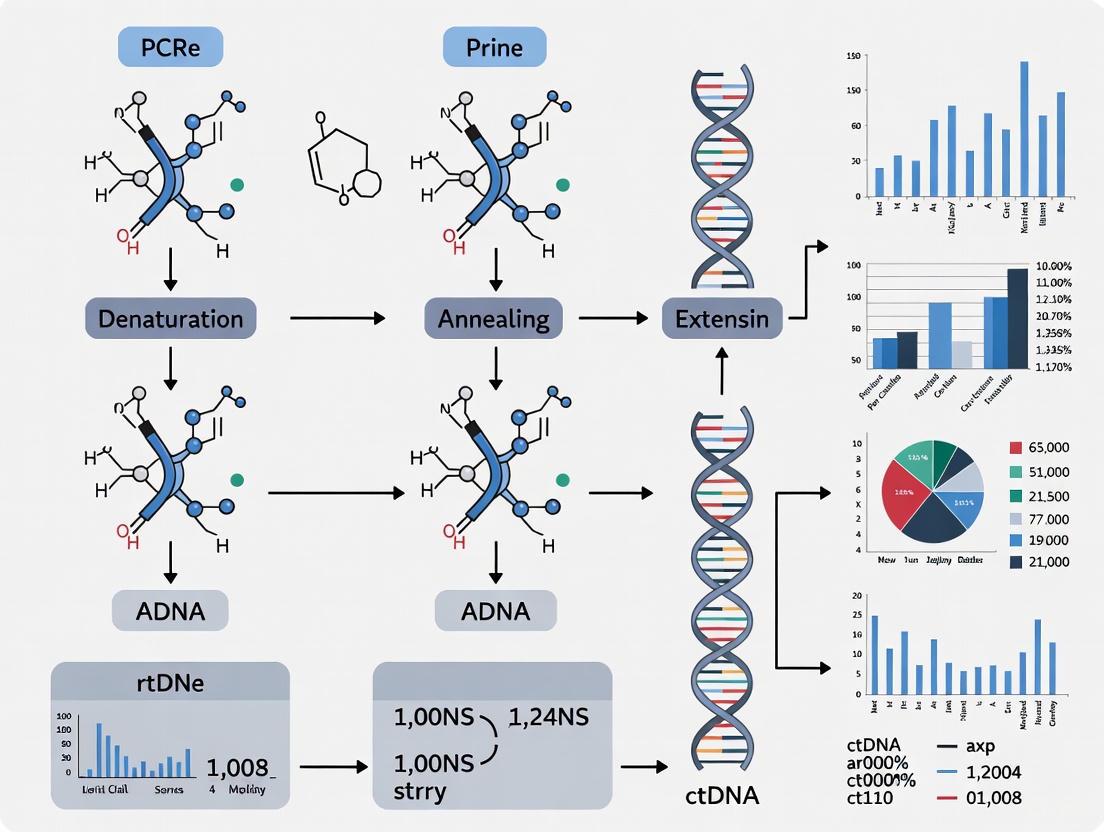
ctDNA Analysis Workflow
The following diagram illustrates the complete workflow for dPCR-based ctDNA analysis, from sample collection to clinical interpretation:
Clinical Decision Pathway
The clinical application of ctDNA monitoring results follows a structured decision pathway:
Current Challenges and Future Directions
Despite the considerable promise of dPCR-based ctDNA analysis, several technical challenges remain. Low ctDNA abundance in early-stage cancers and low-shedding tumors limits detection sensitivity [1] [6]. Pre-analytical variables including blood collection methods, sample processing delays, and extraction efficiencies can significantly impact results [7]. Additionally, clonal hematopoiesis can lead to false-positive calls when mutations originating from hematopoietic cells are mistaken for tumor-derived variants [2].
Future developments aim to enhance assay sensitivity through novel approaches such as fragmentomics analysis, which leverages the characteristic fragmentation patterns of ctDNA compared to normal cfDNA [1] [6]. Integration of multi-omic liquid biopsy analyses combining ctDNA with other biomarkers like circulating miRNAs and extracellular vesicles may provide complementary information [5]. Emerging technologies including CRISPR-based detection methods and nanomaterial-enhanced biosensors offer potential for point-of-care applications with attomolar sensitivity [6].
Standardization efforts through initiatives like the ctMoniTR Project and the Blood Profiling Atlas in Cancer are addressing key barriers to clinical implementation by establishing analytical validation standards and clinical interpretation guidelines [2]. As these advancements mature, dPCR-based ctDNA analysis is poised to become an increasingly integral component of precision oncology and drug development pipelines.
Liquid biopsy represents a transformative approach in oncology, shifting the diagnostic paradigm from traditional invasive tissue biopsies to minimally-invasive molecular analysis of tumor-derived components in bodily fluids [8]. This technology leverages the detection of circulating tumor DNA (ctDNA) and other biomarkers to provide a real-time snapshot of tumor dynamics, enabling personalized cancer management from early detection to treatment monitoring [9]. The core principle underpinning liquid biopsy is that tumors release biological materials into circulation throughout their lifecycle, creating an accessible window into the tumor's genetic landscape without requiring invasive tissue sampling [10]. The analysis of ctDNA, a minor fraction of the total cell-free DNA (cfDNA) in plasma, has emerged as a particularly powerful tool due to its short half-life (approximately 16 minutes to 2.5 hours), which allows for dynamic monitoring of treatment response and disease progression [10] [8].
The evolution of liquid biopsy from scientific concept to clinical tool spans more than a century of discovery, technological innovation, and clinical validation. This journey has accelerated dramatically in the past decade, with liquid biopsy now playing increasingly vital roles in companion diagnostics, minimal residual disease (MRD) monitoring, and therapy selection [11] [12]. As the field continues to mature, standardized protocols and reproducible methodologies have become essential for translating research findings into clinically actionable information, particularly in the context of digital PCR (dPCR) platforms that enable ultrasensitive detection of rare tumor-associated mutations in background of wild-type DNA [13] [14].
Historical Milestones in Liquid Biopsy Development
The development of liquid biopsy has progressed through four distinct eras, each marked by fundamental discoveries and technological breakthroughs that expanded its clinical potential [8]:
Table: Historical Timeline of Liquid Biopsy Development
| Time Period | Key Discoveries and Advancements | Clinical Impact |
|---|---|---|
| Scientific Exploration(Pre-1990s) | 1869: Thomas Ashworth discovers CTCs in patient blood1948: Mandel and Metais identify cfDNA in plasma1977: Leon et al. observe elevated cfDNA in cancer patients | Initial recognition of tumor-derived materials in circulation; foundation for future development |
| Scientific Development(1990s) | 1994: KRAS mutations detected in pancreatic cancer cfDNA1996: Raposo demonstrates biological activity of EVs1998: CTCs successfully isolated from blood | Establishment of correlation between molecular alterations in ctDNA and tumor tissue |
| Industrial Growth(2000-2010) | 2005: CTC count validated as independent prognostic factor in metastatic breast cancer2008: Diehl et al. monitor ctDNA changes during CRC treatment | Proof of clinical utility for prognosis and treatment monitoring; technological standardization begins |
| Industrial Outbreak(2010-Present) | 2014: EMA approves ctDNA for EGFR mutation testing in NSCLC2018: AJCC incorporates CTC testing for breast cancer prognosis2025: Focus on MRD detection and adaptive clinical trial design | Integration into clinical guidelines; expansion into therapy selection, MRD monitoring, and early detection |
This evolutionary pathway demonstrates how liquid biopsy has transitioned from observational science to clinical application, with regulatory approvals and guideline inclusions cementing its role in modern oncology [8]. The most recent phase has been characterized by explosive growth in both publications and clinical applications, with total publications in the past four years surpassing the cumulative output of the previous 36 years [15].
Current Clinical Applications and Quantitative Evidence
Liquid biopsy now plays multiple roles across the cancer care continuum, with distinct clinical applications supported by robust evidence. The following table summarizes key applications and their supporting data from recent studies:
Table: Current Clinical Applications of Liquid Biopsy with Supporting Evidence
| Clinical Application | Cancer Types | Key Evidence and Performance Metrics | Clinical Impact |
|---|---|---|---|
| Minimal Residual Disease (MRD) Monitoring | Colorectal Cancer (VICTORI study)Bladder Cancer (TOMBOLA trial)Non-Small Cell Lung Cancer [13] | 94.3% ctDNA positivity in treatment-naive patients (CRC)87% of recurrences preceded by ctDNA positivityctDNA detection 6+ months before radiographic recurrence [13] [16] | Enables early intervention before macroscopic recurrence; identifies patients for treatment de-escalation when ctDNA-negative |
| Therapy Selection & Companion Diagnostics | Metastatic Breast CancerNon-Small Cell Lung CancerMetastatic Prostate Cancer [12] | SERENA-6 trial: therapy modification based on ESR1 mutations73.5% sensitivity for actionable alterations in NSCLC49% concordance with tissue biopsy (ROME trial) [11] [13] | Guides targeted therapy; provides alternative when tissue biopsy is insufficient or infeasible |
| Early Detection & MCED | Multiple Solid TumorsPancreatic, Liver, Esophageal Cancers [13] | 98.5% specificity for MCED tests59.7% overall sensitivity (84.2% in late-stage)88.2% accuracy for Cancer Signal Origin prediction [13] | Potential for population screening; particularly valuable for cancers without standard screening |
| Response Monitoring & Resistance Detection | Various Solid TumorsNeuroblastomamPDAC [13] [14] | Correlation between ctDNA quantity and tumor volume (ρ=0.500 for liver mets)EV concentration higher in high-risk neuroblastomaCXCL11 elevation predicts immunotherapy toxicity [13] [14] | Real-time assessment of treatment efficacy; earlier detection of resistance than imaging |
The correlation between ctDNA levels and tumor volume has been quantitatively established in metastatic pancreatic ductal adenocarcinoma (mPDAC), where a liver metastases tumor volume threshold of 3.7 mL detected ctDNA with 85.1% sensitivity and 79.2% specificity [14]. This relationship enables ctDNA to serve as a quantitative surrogate for disease burden, with significant correlations observed between ctDNA quantity and both total tumor volume (Spearman's ρ=0.353, p=0.01) and liver metastasis volume (Spearman's ρ=0.500, p<0.001) [14].
Recent studies presented at AACR 2025 demonstrated that ctDNA-guided immunotherapy can effectively intercept cancer recurrence in mismatch repair-deficient (dMMR) solid tumors, with 86.4% (11/13) of ctDNA-positive patients clearing their disease and remaining recurrence-free at two years after pembrolizumab treatment [16]. This approach exemplifies the evolution of liquid biopsy from a passive monitoring tool to an active decision-making guide for treatment intervention.
Analytical Techniques: Focus on Digital PCR Methodologies
Technology Comparison and Selection Criteria
Multiple analytical platforms are available for ctDNA detection, each with distinct strengths and limitations. The selection of an appropriate methodology depends on the clinical context, required sensitivity, and available resources:
Table: Comparison of ctDNA Detection Methodologies
| Methodology | Sensitivity Range | Key Advantages | Key Limitations | Ideal Use Cases |
|---|---|---|---|---|
| Digital PCR (dPCR) | <0.1% (droplet digital PCR)2 parts per million (NeXT Personal assay) [16] | Absolute quantification without standards; high sensitivity; rapid turnaround; minimal sample requirements | Limited multiplexing capability; requires prior knowledge of target mutations; lower throughput | MRD monitoring; validation of NGS findings; low-frequency variant detection in known targets |
| Next-Generation Sequencing (NGS) | 0.1%-5% (varies by approach)0.1% for CAPP-Seq [10] | Comprehensive genomic profiling; discovery of novel alterations; high multiplexing capability | Higher DNA input requirements; complex bioinformatics; longer turnaround time; higher cost | Therapy selection; comprehensive genomic profiling; clinical trial screening |
| Tumor-Informed Approaches | 0.01% (Signatera, NeXT Personal) [16] | Highest sensitivity for MRD detection; reduced false positives; personalized tracking | Requires tumor tissue sequencing; longer development time; higher initial cost | Post-operative MRD monitoring; recurrence surveillance; therapy response assessment |
| Tumor-Naïve Approaches | 0.1%-1% [10] | No tumor tissue required; faster turnaround; fixed panels enable standardization | Lower sensitivity than tumor-informed; risk of false positives from clonal hematopoiesis | Therapy selection when tissue unavailable; initial molecular profiling |
Digital PCR Workflow for ctDNA Analysis
Digital PCR has emerged as a particularly valuable tool for ctDNA analysis due to its exceptional sensitivity, absolute quantification capability, and reproducibility. The following workflow diagram illustrates the key steps in dPCR-based ctDNA analysis:
Research Reagent Solutions for dPCR-based ctDNA Analysis
Table: Essential Research Reagents and Materials for dPCR-based ctDNA Analysis
| Reagent/Material | Specification | Function | Quality Control Considerations |
|---|---|---|---|
| Blood Collection Tubes | Streck Cell-Free DNA BCT or EDTA tubes | Preserves nucleated blood cells; prevents genomic DNA contamination | Time-to-processing validation; stability assessment for specific analytes |
| Nucleic Acid Extraction Kits | Silica membrane columns or magnetic beads | Isolation of high-quality cfDNA from plasma | Evaluation of yield, fragment size distribution, and inhibitor removal |
| dPCR Master Mix | Mutation-specific probes (FAM/HEX); polymerase with high fidelity | Enables partition-based amplification and detection of target mutations | Validation of limit of detection (LOD) and limit of blank (LOB) with reference materials |
| Reference Standards | Horizon Discovery, Seraseq, or custom reference materials | Assay validation; run-to-run performance monitoring | Allelic frequency verification; commutability with patient samples |
| Partitioning Oil/Reagents | Droplet generation oil (Bio-Rad) or chip-based partitioning | Creates thousands of individual reaction chambers | Lot-to-lot consistency; droplet stability and uniformity assessment |
Detailed Experimental Protocol: dPCR for MRD Monitoring
Pre-Analytical Phase: Sample Collection and Processing
Materials Required:
- Streck Cell-Free DNA BCT blood collection tubes
- Refrigerated centrifuge capable of 1600-2500 × g
- Microcentrifuge capable of ≥16,000 × g
- Plasma separation pipettes
- cfDNA extraction kit (e.g., QIAamp Circulating Nucleic Acid Kit)
Step-by-Step Protocol:
Blood Collection: Draw 10-20 mL of whole blood into Streck Cell-Free DNA BCT tubes using standard phlebotomy techniques. Invert tubes 8-10 times immediately after collection to ensure proper mixing with preservative.
Transport and Storage: Store blood tubes at 4-25°C if processing within 72 hours. For extended storage before processing, maintain at 4°C for up to 14 days. Avoid freeze-thaw cycles.
Plasma Separation:
- Centrifuge blood tubes at 1600-2500 × g for 10-20 minutes at 4°C within 72 hours of collection.
- Carefully transfer supernatant plasma to a sterile polypropylene tube using a plasma separation pipette, avoiding disturbance of the buffy coat.
- Perform a second centrifugation step at 16,000 × g for 10 minutes at 4°C to remove any remaining cellular debris.
- Transfer the clarified plasma to a fresh tube.
cfDNA Extraction:
- Process plasma within 24 hours of separation or freeze at -80°C for batch processing.
- Use column-based cfDNA extraction kits according to manufacturer's instructions, with the following modification: elute cfDNA in 20-50 μL of low-EDTA TE buffer or nuclease-free water to maximize concentration.
- Quantify cfDNA using fluorometric methods (e.g., Qubit dsDNA HS Assay) rather than spectrophotometry to accurately measure low concentrations.
Quality Assessment:
- Assess cfDNA fragment size distribution using Bioanalyzer or TapeStation to confirm expected profile (peak at ~167 bp).
- Document cfDNA concentration and volume for input calculations.
Analytical Phase: dPCR Assay Setup and Run
Materials Required:
- ddPCR Supermix for Probes (No dUTP)
- Mutation-specific FAM-labeled probes and WT-specific HEX-labeled probes
- Droplet generator and DG8 cartridges
- ddPCR droplet reader
- 96-well PCR plates and foil seals
Step-by-Step Protocol:
Reaction Setup:
- Prepare 20-22 μL reaction mix containing:
- 10 μL 2× ddPCR Supermix for Probes
- 1.8 μL each of forward and reverse primers (900 nM final concentration)
- 0.5 μL each of FAM-labeled mutation probe and HEX-labeled wild-type probe (250 nM final concentration)
- 5-20 ng of cfDNA extract
- Nuclease-free water to 20-22 μL total volume
- Include negative controls (no-template controls and wild-type DNA) and positive controls (synthetic reference standards with known mutation allele frequency) in each run.
- Prepare 20-22 μL reaction mix containing:
Droplet Generation:
- Transfer 20 μL of reaction mix to DG8 cartridge wells.
- Add 70 μL of droplet generation oil to appropriate wells.
- Place gasket on cartridge and generate droplets in the droplet generator.
- Carefully transfer 40 μL of generated droplets to a 96-well PCR plate.
- Seal the plate with a foil heat seal using a plate sealer at 180°C for 5 seconds.
PCR Amplification:
- Perform amplification using the following thermal cycling conditions:
- Enzyme activation: 95°C for 10 minutes
- 40 cycles of:
- Denaturation: 94°C for 30 seconds
- Annealing/Extension: 55-60°C (assay-specific) for 60 seconds
- Enzyme deactivation: 98°C for 10 minutes
- Hold at 4°C
- Use a ramp rate of 2°C/second for all steps.
- Perform amplification using the following thermal cycling conditions:
Droplet Reading:
- Transfer plate to droplet reader following manufacturer's instructions.
- Set well order and sample identification in the software.
- Run plate with appropriate settings for probe detection channels.
Post-Analytical Phase: Data Analysis and Interpretation
Software and Tools:
- QuantaSoft Software or equivalent
- Statistical analysis package (R, Python, or GraphPad Prism)
- Laboratory Information Management System (LIMS)
Step-by-Step Protocol:
Quality Assessment:
- Verify acceptable droplet count (≥10,000 droplets per well for optimal sensitivity).
- Confirm separation between positive and negative populations.
- Check control performance: NTCs should have minimal positive droplets; wild-type controls should show >99% wild-type signal; positive controls should recover expected allele frequency within acceptable range (±25% of expected).
Variant Calling:
- Use 2D amplitude plots to distinguish mutant-positive, wild-type-positive, and double-positive droplets.
- Apply manual thresholding if automated calling requires adjustment.
- Apply Poisson correction to calculate the absolute concentration of mutant and wild-type alleles (copies/μL).
Calculation of Variant Allele Frequency (VAF):
- Calculate VAF using the formula: VAF = [Mutant concentration / (Mutant concentration + Wild-type concentration)] × 100%
- For very low VAF samples (<0.1%), apply statistical confidence intervals using binomial or Poisson statistics.
Interpretation and Reporting:
- Compare results to validated clinical cutoffs (e.g., VAF ≥0.02% for MRD positivity in some assays).
- Correlate with clinical context, including prior results to assess trend.
- Generate report including:
- Sample quality metrics (cfDNA input, droplet count)
- Mutant allele frequency with confidence intervals
- Interpretation of clinical significance
- Technical limitations and potential interfering factors
Clinical Implementation and Decision Pathways
The integration of liquid biopsy results into clinical decision-making requires careful consideration of the clinical context and assay performance characteristics. The following pathway illustrates a standardized approach for implementing ctDNA testing in oncology practice:
This clinical decision pathway highlights how dPCR-based ctDNA analysis informs critical junctures in cancer management, from initial treatment selection to monitoring for recurrence. The actionable nature of results necessitates close collaboration between laboratory professionals and clinicians to ensure appropriate interpretation and implementation.
Liquid biopsy has completed its evolution from scientific curiosity to essential clinical tool, with dPCR playing a pivotal role in applications requiring high sensitivity and precise quantification. The technology's journey has been marked by key milestones: the initial discovery of circulating tumor-derived materials, the development of increasingly sensitive detection methods, validation of clinical utility across cancer types and clinical scenarios, and finally integration into routine practice and treatment guidelines.
Looking ahead, several emerging trends will shape the future of liquid biopsy. The SERENA-6 trial demonstrates the potential for therapy modification based on early detection of resistance mutations via serial ctDNA monitoring [11]. Fragmentomics - the analysis of cfDNA fragment patterns - represents another promising frontier that could complement mutation-based approaches, particularly for early detection applications [16]. The standardization of pre-analytical and analytical processes through initiatives like the European Liquid Biopsy Society (ELBS) will be crucial for ensuring reproducible results across laboratories [17].
As these technologies continue to mature, liquid biopsy is poised to become increasingly central to cancer management across the entire disease continuum - from screening and early detection through therapy selection and long-term surveillance. The ongoing refinement of dPCR methodologies will further enhance our ability to detect increasingly minute quantities of ctDNA, potentially enabling intervention at earlier timepoints when treatments may be more effective. Through these advances, liquid biopsy will continue to transform oncology practice, offering increasingly personalized, dynamic, and minimally-invasive approaches to cancer care.
Digital PCR (dPCR) represents a third-generation PCR technology that enables the absolute quantification of nucleic acid targets without the need for a standard curve. This method is based on the partitioning of a PCR reaction mixture into thousands to millions of discrete compartments, so that each contains either zero, one, or a few nucleic acid molecules [18]. Following PCR amplification, the fraction of positive partitions is counted using an end-point fluorescence measurement, and the absolute concentration of the target sequence is calculated using Poisson statistics [18] [19]. This partitioning approach minimizes competition between targets and allows for the detection of rare genetic events within a background of wild-type sequences, making it particularly valuable for applications in oncology, such as measuring circulating tumor DNA (ctDNA) in liquid biopsies [18] [20].
The fundamental principle that distinguishes dPCR from quantitative real-time PCR (qPCR) is its method of quantification. While qPCR relies on comparing amplification curves to standards of known concentration, dPCR provides direct, absolute quantification by counting individual molecules [21] [19]. This calibration-free approach offers powerful advantages including high sensitivity, absolute quantification, high accuracy and reproducibility, as well as a rapid turnaround time [18]. The technology has rapidly evolved since its conceptual origins in limiting dilution experiments, with modern implementations utilizing either water-in-oil droplet emulsification (droplet digital PCR or ddPCR) or microchamber-based systems [18].
Principles of Absolute Quantification
The Partitioning Principle and Poisson Statistics
The core principle of digital PCR involves sample partitioning, which allows for the transformation of a continuous measurement into a digital readout. The process begins with the distribution of a PCR reaction mixture containing the nucleic acid sample across thousands of individual partitions [18] [19]. This distribution follows Poisson statistics, meaning that the partitioning is random, and each compartment has an equal probability of receiving a target molecule [18]. Ideally, each partition contains either zero or one target molecule, though in practice, some partitions may contain more [18].
Following partitioning, PCR amplification is performed to endpoint, and each partition is analyzed for fluorescence. Partitions containing the target sequence (positive) fluoresce, while those without (negative) do not [19]. The ratio of positive to total partitions forms the basis for quantification using the Poisson distribution formula: λ = -ln(1 - p), where λ represents the average number of target molecules per partition and p is the fraction of positive partitions [19]. This calculation provides the absolute concentration of the target in the original sample, eliminating the need for external calibrators or reference genes [22] [19].
Comparison with Other PCR Technologies
dPCR vs. qPCR: Key Differences
| Feature | Digital PCR (dPCR) | Quantitative PCR (qPCR) |
|---|---|---|
| Quantification Method | Absolute, via Poisson statistics | Relative, via standard curve |
| Calibration Required | No | Yes |
| Sensitivity | Higher; detects rare targets | Lower |
| Precision | Higher; particularly at low target concentrations | Lower |
| Effect of Amplification Efficiency | Minimal impact | Significant impact on results |
| Data Output | Direct count of target molecules | Cycle threshold (Ct) value |
| Ideal Application | Rare mutation detection, copy number variation, liquid biopsy | Gene expression analysis, pathogen detection |
The partitioning approach makes dPCR particularly advantageous for detecting rare mutations and subtle copy number variations, as it effectively enriches low-abundance targets by separating them from the background [21] [19]. Studies have demonstrated that dPCR exhibits superior sensitivity and precision compared to qPCR, especially for quantifying low-level bacterial loads and copy number variations [23] [24]. This enhanced performance makes dPCR exceptionally suitable for liquid biopsy applications where ctDNA is often present in very low concentrations amidst a high background of wild-type DNA [25] [7].
dPCR Workflow and Experimental Design
Standard dPCR Workflow
The following diagram illustrates the core workflow of a digital PCR experiment, from sample preparation to final quantification:
Figure 1: Digital PCR Workflow. The process involves sample preparation, partitioning of the reaction mixture, endpoint PCR amplification, fluorescence detection, and data analysis using Poisson statistics for absolute quantification.
Essential Research Reagent Solutions
Key Reagents and Materials for dPCR Experiments
| Reagent/Material | Function | Application Notes |
|---|---|---|
| Specialized Blood Collection Tubes | Preserve ctDNA integrity by preventing white blood cell lysis during storage/transport | Examples: cfDNA BCT (Streck), PAXgene Blood ccfDNA (Qiagen) [7] |
| Microfluidic Chips/Plates | Create nanoliter-scale reaction chambers for sample partitioning | Fixed well arrays (e.g., QIAcuity Nanoplate) or droplet generators [18] [24] |
| Restriction Enzymes | Digest large genomic DNA fragments to prevent partitioning bias | Recommended for >75 ng gDNA input (e.g., Anza 52 PvuII) [19] [24] |
| TaqMan Probes | Provide sequence-specific fluorescence detection | Enable multiplexing with different fluorophores [21] [24] |
| Digital PCR Master Mix | Optimized buffer system for partitioning and amplification | Contains DNA polymerase, dNTPs, and stabilizers [24] |
Application in Circulating Tumor DNA Analysis
ctDNA Analysis Workflow in Liquid Biopsies
The application of dPCR for ctDNA analysis in liquid biopsy research involves specific considerations for sample handling and assay design:
Figure 2: ctDNA Analysis Workflow for Liquid Biopsies. The process from blood collection to dPCR analysis requires specialized sample processing to preserve the integrity of fragile ctDNA molecules.
Detailed Protocol: ctDNA Mutation Detection via ddPCR
Objective: Absolute quantification of tumor-specific mutations in plasma circulating tumor DNA.
Sample Preparation:
- Blood Collection and Processing: Collect 2×10 mL of blood into cell-free DNA BCT tubes (e.g., Streck cfDNA BCT) [7]. Process within 2-6 hours if using EDTA tubes, or within 3-7 days if using specialized BCT tubes. Perform double centrifugation: first at 1,600×g for 10 minutes at 4°C to separate plasma, then transfer supernatant and centrifuge at 16,000×g for 10 minutes to remove remaining cellular debris [7].
- cfDNA Extraction: Extract cfDNA from 4-10 mL plasma using commercially available cfDNA extraction kits (e.g., QIAamp DNA Mini kit) following manufacturer's instructions [7] [24]. Elute in 20-50 μL elution buffer. Quantify using fluorescence-based methods suitable for low-concentration samples.
- DNA Digestion (Optional): For inputs >75 ng genomic DNA, perform restriction digestion to fragment DNA and prevent partitioning bias [19].
Droplet Digital PCR Setup:
- Reaction Preparation: Prepare 40 μL reaction mixture containing:
- 10 μL sample DNA (or appropriate volume for 75 ng total DNA)
- 1× ddPCR Supermix
- 0.4 μM forward and reverse primers
- 0.2 μM FAM-labeled probe for mutant allele
- 0.2 μM HEX-labeled probe for wild-type allele
- Droplet Generation: Transfer reaction mixture to droplet generator cartridges. Generate droplets according to manufacturer's protocol (typically 20,000 droplets per sample) [18] [19].
- PCR Amplification: Transfer droplets to 96-well PCR plate. Seal plate and run with following thermal cycling conditions:
- Enzyme activation: 95°C for 10 minutes
- 45 cycles of: 94°C for 30 seconds, 55-60°C (assay-specific) for 60 seconds
- Enzyme deactivation: 98°C for 10 minutes
- Hold at 4°C
- Droplet Reading: Place plate in droplet reader. Measure fluorescence in FAM and HEX channels for each droplet.
- Data Analysis: Use manufacturer's software to identify positive and negative droplets for mutant and wild-type alleles. Apply Poisson correction to calculate absolute concentration of mutant alleles in copies/μL.
Quality Control:
- Include no-template controls (NTC) to detect contamination
- Use positive controls with known mutation frequency
- Set minimum threshold of 3 positive partitions for calling positive results [24]
- Apply volume precision factor for accurate concentration calculation if supported by platform [24]
Performance Characteristics and Validation
Analytical Performance of dPCR Platforms
Performance Comparison of dPCR with Other Technologies
| Parameter | Digital PCR | Quantitative PCR | Next-Generation Sequencing |
|---|---|---|---|
| Limit of Detection | 0.001% for mutant alleles | 1-5% for mutant alleles | 0.1-1% for mutant alleles |
| Precision (CV%) | 4.5% median intra-assay variability [24] | Higher variability, especially at low concentrations | Variable based on sequencing depth |
| Quantification Type | Absolute | Relative | Relative or absolute |
| Multiplexing Capacity | Moderate (2-6 plex) | Limited (typically 1-3 plex) | High (dozens to thousands) |
| Turnaround Time | 4-8 hours | 2-3 hours | Days to weeks |
| Cost per Sample | Moderate | Low | High |
| Ideal Application | Rare variant detection, absolute copy number | Gene expression, high abundance targets | Discovery, unknown variants, comprehensive profiling |
Technical Validation and Troubleshooting
Assay Validation Parameters:
- Linearity: Demonstrate R² > 0.99 across expected concentration range [24]
- Accuracy: Compare to orthogonal methods (e.g., PFGE for copy number variations) [23]
- Precision: Determine intra-assay and inter-assay coefficients of variation
- Specificity: Verify no cross-reactivity with similar sequences
Common Issues and Solutions:
- Low Positive Partitions: Increase input DNA quantity; verify DNA quality; optimize partitioning efficiency
- Rain Effect (Intermediate Partitions): Optimize annealing temperature; improve probe design; adjust fluorescence thresholds
- Inhibitors: Dilute sample; use inhibitor-resistant polymerases; purify DNA more thoroughly
- Droplet Coalescence: Ensure proper storage conditions; use fresh oil and surfactants [18]
Advanced Applications in Liquid Biopsy Research
Digital PCR has become an indispensable tool in liquid biopsy research, particularly in the context of cancer management. Its exceptional sensitivity and precision enable several advanced applications in ctDNA analysis:
Minimal Residual Disease (MRD) Detection: dPCR can detect ctDNA at very low variant allele frequencies (0.01% or lower), making it suitable for monitoring MRD after surgery or during treatment [20] [7]. Studies have shown that ctDNA clearance after treatment correlates with improved outcomes, while persistence or reappearance of ctDNA signals recurrence [25] [20].
Treatment Response Monitoring: The absolute quantification capability of dPCR allows for precise tracking of ctDNA dynamics during therapy [25] [20]. Rising ctDNA levels may indicate treatment resistance, while decreasing levels typically correspond to positive response. The high precision of dPCR enables detection of statistically significant changes in ctDNA burden earlier than imaging methods [7].
Tumor Heterogeneity Assessment: Multiplex dPCR assays can simultaneously track multiple mutations, providing insights into tumor heterogeneity and evolution under therapeutic pressure [18] [20]. This application benefits from dPCR's ability to precisely quantify multiple targets without cross-reactivity.
The implementation of dPCR in liquid biopsy workflows continues to evolve, with emerging approaches including integrated analysis of fragmentomics patterns and methylation status to improve detection sensitivity for early-stage cancers [25] [26]. As ctDNA testing becomes increasingly incorporated into clinical trial designs, particularly for guiding adjuvant treatment decisions, dPCR remains a key enabling technology for its robust performance and quantitative accuracy [20].
Why dPCR is Uniquely Suited for Low-Abundance ctDNA Detection
Circulating tumor DNA (ctDNA) has emerged as a pivotal biomarker in oncology, enabling non-invasive monitoring of tumor dynamics through liquid biopsy. However, a significant challenge persists: in early-stage cancers and minimal residual disease (MRD), ctDNA can be exceptionally scarce, often constituting less than 0.1% of the total cell-free DNA (cfDNA) in circulation [27] [1]. Detecting these rare mutations against an overwhelming background of wild-type DNA demands a technology with exceptional sensitivity and precision. Among available methodologies, Droplet Digital PCR (dPCR) stands out as uniquely suited for this task. Its ability to provide absolute quantification and reliably detect variant alleles at frequencies as low as 0.003% makes it an indispensable tool for researchers and clinicians aiming to translate liquid biopsy into clinical practice [28]. This application note details the fundamental principles, performance data, and experimental protocols that underpin dPCR's superior capability in low-abundance ctDNA detection.
Fundamental Technological Principles of dPCR
The core power of dPCR lies in its paradigm shift from relative to absolute quantification through sample partitioning.
Partitioning and Poisson Statistics
Unlike quantitative PCR (qPCR), which relies on the kinetics of amplification relative to a standard curve, dPCR partitions a single PCR reaction into thousands to millions of discrete, parallel reactions [18]. In droplet digital PCR (ddPCR), this is achieved by creating thousands of nanoliter-sized water-in-oil emulsion droplets, effectively turning a single sample into a vast array of individual experiments [27]. Following end-point PCR amplification, each partition is analyzed for fluorescence. Partitions containing the target sequence fluoresce, while those without it do not. This binary readout (positive or negative) is the "digital" aspect of the technology.
The absolute concentration of the target nucleic acid in the original sample is then calculated using Poisson statistics, which accounts for the random distribution of molecules across the partitions [27] [18]. This method provides a direct count of target molecules without the need for a standard curve, eliminating a major source of inaccuracy and variability [27].
Overcoming PCR Inhibition and Background Noise
The partitioning principle confers remarkable robustness. PCR inhibitors present in the sample are similarly diluted across the thousands of partitions, minimizing their impact on amplification efficiency within any single droplet [27]. Furthermore, the physical separation of target molecules prevents the dominant amplification of wild-type sequences from masking the signal of rare mutants, a common limitation in bulk PCR reactions [27]. This effectively lowers the background noise and enhances the signal-to-noise ratio, which is critical for distinguishing low-frequency variants.
Performance Data: dPCR vs. NGS in ctDNA Detection
Direct comparisons in clinical studies highlight dPCR's superior sensitivity for ctDNA detection, particularly in localized cancers.
Table 1: Performance Comparison of dPCR and NGS in Rectal Cancer ctDNA Detection [29]
| Parameter | Droplet Digital PCR (ddPCR) | Next-Generation Sequencing (NGS) |
|---|---|---|
| Detection Rate (Baseline Plasma) | 58.5% (24/41 patients) | 36.6% (15/41 patients) |
| Statistical Significance | p = 0.00075 | |
| Variant Allele Frequency (VAF) Sensitivity | Can detect down to 0.01% [29] | Threshold lowered to 0.01% for study comparison |
| Key Advantage | Higher sensitivity for low-abundance targets | Broader, untargeted mutation profiling |
A study on early breast cancer further demonstrates the extreme sensitivity achievable with optimized dPCR protocols. Using larger plasma volumes (20-40 mL) and highly partitioned assays, researchers detected ctDNA at a minimum variant allele frequency (VAF) of 0.003% and circulating tumor cells (CTCs) in patient blood samples. This approach enabled the detection of residual disease before surgery in all patients who did not achieve a complete pathological response [28].
Table 2: Ultrasensitive dPCR Detection in Early Breast Cancer [28]
| Analytic | Pre-Treatment Detection Rate | Minimum VAF or Concentration Detected |
|---|---|---|
| ctDNA | 90.5% (19/21 patients) | VAF: 0.01% |
| CTCs | 63.2% (12/19 patients) | 0.30 CTCs/mL blood |
| Combined (ctDNA and/or CTCs) | 100% (20/20 patients) | - |
Detailed Experimental Protocol for ctDNA Detection via ddPCR
The following protocol is adapted from methodologies used in recent clinical studies [29] [28] [30].
Pre-Analytical Phase: Sample Collection and cfDNA Extraction
- Blood Collection: Collect patient blood (recommended: 3 × 9 mL or larger volumes for increased sensitivity [28]) into Streck Cell-Free DNA BCT tubes or equivalent to preserve cfDNA.
- Plasma Isolation: Process blood within 6 hours of collection. Centrifuge using a double-spin protocol (e.g., 1,600 × g for 20 min, then 16,000 × g for 10 min) to isolate platelet-poor plasma.
- cfDNA Extraction: Extract cfDNA from plasma using commercially available kits (e.g., QIAamp Circulating Nucleic Acid Kit). Elute in a low-EDTA TE buffer or nuclease-free water. Quantify cfDNA using a fluorescence-based assay sensitive to low DNA concentrations.
Assay Design and Optimization
- Tumor-Informed Assay Design: For maximal sensitivity, design patient-specific ddPCR assays.
- Perform next-generation sequencing (e.g., Whole Exome Sequencing or a hotspot panel like the Ion AmpliSeq Cancer Hotspot Panel v2) on the patient's tumor tissue to identify truncal somatic mutations [29] [28].
- Select 1-2 mutations with the highest variant allele frequency in the tumor tissue for ddPCR probe design [29].
- Probe Design: Design and validate FAM-labeled mutant probes and HEX/VIC-labeled wild-type probes using the TaqMan chemistry.
ddPCR Reaction Setup and Partitioning
- Prepare Reaction Mix:
- 1X ddPCR Supermix for Probes (no dUTP)
- 900 nM of each primer
- 250 nM of each probe (FAM and HEX/VIC)
- Approximately 10-100 ng of extracted cfDNA (the maximum volume recommended per reaction)
- Nuclease-free water to a final volume of 20-22 µL.
- Partitioning: Load the reaction mix into a DG8 cartridge along with droplet generation oil. Use a QX200 Droplet Generator to create approximately 20,000 nanoliter-sized droplets per sample.
PCR Amplification and Droplet Reading
- Thermal Cycling: Transfer the emulsified samples to a 96-well plate and run PCR on a thermal cycler using optimized conditions. A standard two-step protocol is often used:
- 95°C for 10 minutes (enzyme activation)
- 40 cycles of: 94°C for 30 seconds (denaturation) and 55-60°C for 60 seconds (annealing/extension)
- 98°C for 10 minutes (enzyme deactivation)
- 4°C hold.
- Droplet Reading: Place the plate in a QX200 Droplet Reader. The reader flows the droplets sequentially past a two-color optical detection system that counts the number of FAM-positive (mutant), HEX/VIC-positive (wild-type), double-positive, and negative droplets.
Data Analysis and Interpretation
- Threshold Setting: Use the instrument's software (e.g., QuantaSoft) to set fluorescence amplitude thresholds for positive and negative droplets based on negative controls (no-template control and germline DNA control) and positive controls.
- Concentration Calculation: The software uses Poisson statistics to calculate the absolute concentration (copies/µL) of mutant and wild-type DNA in the original reaction.
- VAF Calculation:
- Variant Allele Frequency (VAF) = [Mutant concentration (copies/µL) / (Mutant + Wild-type concentration (copies/µL))] × 100.
- Limit of Detection (LOD): Establish the assay's LOD using titration experiments with synthetic or cell-line-derived mutant DNA diluted into wild-type DNA. The LOD is typically defined as a concentration where the mutant signal is statistically significant from the negative control in ≥95% of replicates.
The Scientist's Toolkit: Essential Research Reagents and Materials
Table 3: Key Reagent Solutions for dPCR-based ctDNA Detection
| Product Category | Example | Function & Critical Notes |
|---|---|---|
| Blood Collection Tubes | Streck Cell-Free DNA BCT | Preserves cfDNA by stabilizing nucleated blood cells, preventing genomic DNA contamination and enabling sample stability for up to 72-96 hours post-draw. |
| cfDNA Extraction Kits | QIAamp Circulating Nucleic Acid Kit | Optimized for low-abundance nucleic acids from large-volume plasma samples, providing high purity and recovery crucial for sensitivity. |
| ddPCR Supermix | Bio-Rad ddPCR Supermix for Probes | A homogeneous, optimized master mix for probe-based assays, formulated for robust amplification within droplets. |
| TaqMan Assays | Custom-designed FAM/HEX probes | Hydrolysis probes designed to specifically hybridize to mutant and wild-type alleles, providing the sequence specificity for accurate mutation calling. |
| Droplet Generation Oil | Bio-Rad Droplet Generation Oil for Probes | Creates a stable water-in-oil emulsion essential for consistent and reliable droplet formation during partitioning. |
| Reference Materials | Horizon Discovery Multiplex I cfDNA Reference Standard | Commercially available synthetic cfDNA containing known mutations at defined VAFs; used for assay validation, calibration, and inter-laboratory reproducibility. |
Droplet Digital PCR's unique combination of sample partitioning, absolute quantification via Poisson statistics, and exceptional resistance to PCR inhibitors renders it the gold-standard technology for detecting low-abundance ctDNA [27] [18]. Its demonstrated ability to identify MRD and predict disease relapse months before clinical recurrence offers a powerful tool for guiding adjuvant therapy and improving patient outcomes in oncology research and drug development [28] [30]. As liquid biopsy continues to reshape precision oncology, dPCR remains an indispensable, highly sensitive, and reliable method for quantifying the molecular footprints of cancer.
Circulating tumor DNA (ctDNA), a subset of cell-free DNA shed by tumor cells into the bloodstream, has emerged as a transformative biomarker in clinical oncology [6] [1]. These DNA fragments carry tumor-specific genetic alterations, enabling non-invasive access to the molecular landscape of malignancies [2]. The analysis of ctDNA via liquid biopsy provides a powerful alternative to traditional tissue biopsies, offering a comprehensive view of tumor heterogeneity while overcoming sampling biases associated with single-site tissue specimens [6] [1]. The half-life of ctDNA is remarkably short (16 minutes to several hours), allowing for real-time monitoring of dynamic changes in tumor burden and genomic evolution during treatment [1].
The integration of digital PCR (dPCR) technologies has been pivotal in advancing ctDNA analysis, particularly for applications requiring high sensitivity and precise quantification [18]. dPCR achieves unprecedented sensitivity by partitioning samples into thousands of individual reactions, enabling the detection of rare mutant molecules amid a background of wild-type DNA [18]. This technical advancement has opened new avenues for three critical clinical applications: minimal residual disease (MRD) assessment, therapy response monitoring, and tracking treatment resistance [18] [2]. This article delineates protocol frameworks and application notes for each domain, specifically contextualized within dPCR-based research methodologies.
Application 1: Minimal Residual Disease (MRD) Detection
Clinical Rationale and Definition
Minimal residual disease (MRD) refers to the presence of trace amounts of tumor DNA in patients after curative-intent treatment, such as complete surgical resection [31]. These occult tumor cells, while undetectable by standard imaging or clinical examination, serve as the source for subsequent recurrence and metastasis [31]. Traditional imaging techniques like CT and MRI are limited by their resolution and inability to detect microscopic disease burden [1]. ctDNA analysis has demonstrated superior sensitivity for MRD detection, often identifying molecular relapse months before clinical or radiographic recurrence [6] [32] [31].
Key Evidence and Clinical Validation
Evidence supporting ctDNA for MRD detection spans multiple cancer types. In colorectal cancer (CRC), a landmark study of 230 stage II patients demonstrated that postoperative ctDNA positivity identified patients with a significantly higher risk of recurrence (79% in ctDNA-positive vs. 9.8% in ctDNA-negative patients not receiving adjuvant chemotherapy) [31]. Similarly, in stage III CRC, patients with detectable ctDNA after chemotherapy showed only a 30% 3-year recurrence-free survival compared to 77% for those with undetectable ctDNA [31]. A prospective multicenter study utilizing the tumor-informed Signatera assay showed that CRC patients with positive MRD 30 days after surgery had a 7.2-fold increased recurrence risk, and this risk escalated to 17.5-fold if ctDNA remained positive after adjuvant chemotherapy [31]. The median lead time for ctDNA detection ahead of radiological recurrence was 8.7 months [31].
In breast cancer, structural variant-based ctDNA assays detected molecular relapse more than a year before clinical evidence emerged, allowing for potential early intervention strategies [6]. Similar promising data exist for lung, lymphoid, and gastroesophageal cancers [6].
Table 1: Clinical Performance of ctDNA-Based MRD Detection in Selected Studies
| Cancer Type | Study Design | ctDNA Assay | Key Finding | Lead Time Over Imaging |
|---|---|---|---|---|
| Stage II/III Colorectal Cancer | Prospective cohort (n=230) [31] | Safe-SeqS | Post-op ctDNA+: 79% recurrence; ctDNA-: 9.8% recurrence | Not specified |
| Stage I-III Colon Cancer | Prospective multicenter (n=130) [31] | Signatera (tumor-informed) | MRD+ post-surgery: 7.2x higher recurrence risk; post-chemotherapy: 17.5x higher risk | 8.7 months (median) |
| Locally Advanced Rectal Cancer [32] | Retrospective case series (n=28) | Signatera | 67% (6/9) ctDNA+ patients required surgery vs. 21% (4/19) ctDNA- patients | Not specified |
| Early-Stage Breast Cancer [6] | Cohort study | SV-based assay | ctDNA detected recurrence >1 year before clinical evidence | >12 months |
Digital PCR Experimental Protocol for MRD Detection
Principle: This protocol utilizes a tumor-informed approach (also known as patient-specific), where mutations identified in the tumor tissue are tracked in plasma using dPCR with extreme sensitivity [31].
Workflow:
Pre-Analytical Phase: Sample Collection and Processing
- Blood Collection: Collect 10-20 mL of peripheral blood into cell-free DNA blood collection tubes (e.g., Streck Cell-Free DNA BCT) [32]. Invert gently 8-10 times. Blood must be processed within 48-96 hours of collection for optimal yield.
- Plasma Isolation: Centrifuge blood at 1600-2000 × g for 10-20 minutes at 4°C. Transfer the supernatant (plasma) to a fresh tube without disturbing the buffy coat. Perform a second high-speed centrifugation at 16,000 × g for 10 minutes to remove residual cells and platelets.
- Cell-free DNA (cfDNA) Extraction: Extract cfDNA from plasma using commercially available silica-membrane or magnetic bead-based kits. Elute in a low-EDTA TE buffer or nuclease-free water. Quantify cfDNA using a fluorescence-based method (e.g., Qubit dsDNA HS Assay).
Assay Design
- Tumor Sequencing: Sequence the patient's tumor tissue (e.g., from formalin-fixed paraffin-embedded, FFPE, block) via Whole Exome Sequencing (WES) or a large targeted panel to identify 1-16 somatic, clonal, single-nucleotide variants (SNVs) [31].
- dPCR Assay Design: Design TaqMan-style probe-based dPCR assays for the 1-3 highest quality, patient-specific mutations identified. A wild-type reference assay for the same genomic region must be included to calculate variant allele frequency (VAF).
Digital PCR Setup and Run
- Partitioning: Prepare the dPCR reaction mix containing the extracted cfDNA, mutation-specific primers/probes, and dPCR supermix. Partition the reaction mixture into 20,000+ nanoliter-sized droplets or microwells using an automated droplet generator or chip-based system (e.g., Bio-Rad QX200, Qiagen QIAcuity) [18].
- Amplification: Perform PCR amplification on a thermal cycler with optimized cycling conditions for the specific assays.
- Reading: Transfer the plate/droplets to a droplet reader or chip reader that counts each partition as positive (mutant), positive (wild-type), or negative (no template) based on fluorescence amplitude [18].
Data Analysis and Interpretation
- Absolute Quantification: Use the system's software to apply Poisson statistics to the count of positive and negative partitions, providing an absolute concentration of mutant and wild-type DNA fragments (copies/μL) [18].
- VAF Calculation: Calculate the VAF as (mutant concentration / (mutant + wild-type concentration)) × 100%.
- MRD Calling: For tumor-informed approaches, a sample is typically called MRD-positive if at least 2 distinct tumor-specific mutations are detected above a predefined threshold (e.g., VAF ≥ 0.01%) [31]. A sample is MRD-negative if no mutations are detected above this limit of detection.
Diagram 1: Tumor-informed dPCR workflow for MRD detection.
Application 2: Therapy Response Monitoring
Clinical Rationale
Monitoring dynamic changes in ctDNA levels during treatment provides a powerful, real-time measure of therapeutic efficacy [1] [2]. Unlike traditional imaging, which assesses anatomical changes at discrete time points, ctDNA offers a quantitative molecular response that can be measured frequently and with minimal invasiveness [2]. A decline in ctDNA levels often correlates with reducing tumor burden and predicts radiographic response, while a persistent or rising level may indicate ineffective therapy [6] [1]. The ctDNA half-life of hours allows for rapid assessment of treatment effect, potentially enabling earlier adaptation of treatment strategies [1].
Key Evidence and Clinical Validation
The utility of ctDNA for therapy monitoring has been demonstrated across various cancers and treatment modalities. In advanced non-small cell lung cancer (NSCLC) treated with tyrosine kinase inhibitors (TKIs), the ctMoniTR project (a multi-assay, multi-study consortium) found that patients whose ctDNA levels dropped to undetectable within 10 weeks had significantly better overall survival and progression-free survival [2]. In NSCLC, a decline in ctDNA levels predicted radiographic response more accurately than follow-up imaging [6]. Similarly, in aggressive B-cell lymphoma, ctDNA-based MRD assays proved more sensitive and informative than standard PET or CT imaging for monitoring response to immunochemotherapy [6].
Table 2: ctDNA for Therapy Monitoring in Selected Cancers and Treatments
| Cancer Type | Therapy | Monitoring Paradigm | Clinical Utility |
|---|---|---|---|
| Advanced NSCLC [2] | Tyrosine Kinase Inhibitors (TKIs) | ctDNA levels at 10 weeks | Undetectable ctDNA at 10 weeks correlated with improved OS and PFS. |
| Various Solid Tumors [6] | Chemotherapy, Targeted Therapy | Longitudinal ctDNA level trends | ctDNA decline predicted radiographic response more accurately than imaging. |
| Aggressive B-cell Lymphoma [6] | Immunochemotherapy | ctDNA-MRD vs. PET/CT | ctDNA more sensitive and informative than standard imaging. |
| Colorectal Cancer [6] | Adjuvant Chemotherapy | ctDNA clearance during/after treatment | ctDNA monitoring enabled precision treatment intensification/de-escalation. |
Digital PCR Experimental Protocol for Therapy Monitoring
Principle: This protocol involves the longitudinal tracking of a known, tumor-derived mutation (or a small set of mutations) throughout treatment to quantify molecular response.
Workflow:
Baseline Assessment
- Pre-Treatment Blood Draw: Collect a baseline blood sample before the initiation of therapy.
- Identify Tracking Mutation(s): If a prior tumor tissue test has identified a key driver mutation (e.g., EGFR p.T790M in NSCLC, KRAS in CRC, ESR1 in breast cancer), design a dPCR assay for it [2]. Alternatively, use a tumor-informed approach as in Section 2.3 to select patient-specific mutations.
- Baseline dPCR: Run the baseline plasma cfDNA sample with the selected dPCR assay to establish the pre-treatment VAF.
Longitudinal Monitoring
- Scheduling: Schedule subsequent blood draws at predefined, clinically relevant timepoints (e.g., after 1-2 cycles of therapy, at the time of mid-treatment radiological assessment, and at the end of treatment) [2].
- Sample Processing: Process all longitudinal samples identically to the baseline sample to minimize technical variability.
- dPCR Analysis: Run each sample with the same dPCR assay(s) used at baseline.
Data Analysis and Response Criteria
- Absolute Concentration Tracking: Plot the concentration (copies/μL) of the mutant allele over time.
- Molecular Response Definitions:
- Molecular Response: A significant decrease (e.g., >50% or >90%) in mutant concentration from baseline.
- Complete Molecular Response: The mutant allele becomes undetectable (VAF below the assay's limit of detection).
- Molecular Progression: A significant increase in mutant concentration from the nadir, or the emergence of a new resistance mutation (see Section 4).
Diagram 2: Longitudinal therapy response monitoring via dPCR.
Application 3: Tracking Treatment Resistance
Clinical Rationale
Tumors evolve under the selective pressure of therapy, leading to the outgrowth of resistant clones [1]. These clones often harbor specific genetic alterations that confer resistance, such as secondary mutations in the drug target or activation of bypass signaling pathways [1] [2]. ctDNA analysis enables the non-invasive and early detection of these resistance mechanisms, often weeks or months before clinical progression is evident [6] [2]. This early warning provides a critical window for clinicians to switch or modify treatment strategies proactively.
Key Evidence and Clinical Validation
The canonical example is in EGFR-mutant NSCLC, where treatment with first- or second-generation EGFR TKIs inevitably leads to resistance, frequently mediated by the EGFR T790M mutation [6] [1]. ctDNA monitoring can detect the emergence of the T790M mutation, allowing for a timely switch to a third-generation TKI without the need for a repeat tissue biopsy [6]. Resistance mutations to targeted therapies (e.g., in KRAS, ESR1, BRAF) and chemotherapy can also be tracked in plasma [2]. The ability of ctDNA to capture the entire landscape of resistant subclones, which may be spatially heterogeneous within the tumor, provides a more comprehensive picture than a single-site biopsy [1].
Digital PCR Experimental Protocol for Resistance Mutation Detection
Principle: This protocol uses dPCR to screen for known, recurrent resistance mutations in a specific gene during treatment with a targeted agent.
Workflow:
Pre-Analytical Phase: Follow the same sample collection, processing, and cfDNA extraction steps as in Section 2.3.
Assay Selection for Resistance
- Targeted Approach: Based on the known resistance mechanisms to the administered therapy, select commercially available or custom-designed dPCR assays for specific resistance mutations. Examples include:
- EGFR T790M for patients on 1st/2nd gen EGFR TKIs.
- EGFR C797S for patients on 3rd gen EGFR TKIs (Osimertinib).
- ESR1 mutations for breast cancer patients on aromatase inhibitors.
- Multiplexing: Utilize the multiplexing capability of dPCR (e.g., with different fluorescent probes) to screen for several common resistance mutations in a single reaction, conserving precious cfDNA.
- Targeted Approach: Based on the known resistance mechanisms to the administered therapy, select commercially available or custom-designed dPCR assays for specific resistance mutations. Examples include:
Digital PCR Setup and Run
- Prepare the dPCR reaction mix with the cfDNA sample and the multiplexed probe assay for the resistance mutation(s) and a reference control.
- Partition, amplify, and read the reaction as described in Section 2.3.
Data Analysis and Interpretation
- The dPCR software will automatically classify droplets based on fluorescence and provide absolute counts for each target (e.g., wild-type, T790M mutant).
- A sample is considered positive for the resistance mutation if the mutant concentration is statistically significantly above the false-positive threshold of the assay (typically determined using healthy donor plasma controls).
- The VAF of the resistance mutation can be calculated, providing insight into the clonal abundance of the resistant population.
Diagram 3: dPCR workflow for detecting acquired resistance mutations.
The Scientist's Toolkit: Essential Reagents and Materials
Table 3: Key Research Reagent Solutions for dPCR-based ctDNA Analysis
| Item | Function/Description | Example Products/Notes |
|---|---|---|
| cfDNA Blood Collection Tubes | Stabilizes nucleated cells and prevents cfDNA release during transport, ensuring pre-analytical quality. | Streck Cell-Free DNA BCT, Roche Cell-Free DNA Collection Tubes [32] |
| cfDNA Extraction Kits | Isolation of high-purity, short-fragment cfDNA from plasma. Silica-membrane or magnetic bead-based. | QIAamp Circulating Nucleic Acid Kit, MagMAX Cell-Free DNA Isolation Kit, Circulating DNA Extraction Kit [32] |
| Droplet Digital PCR Systems | Instrumentation for partitioning, thermocycling, and droplet reading. The core dPCR platform. | Bio-Rad QX200/QX600, Qiagen QIAcuity, Stilla Naica System [18] [2] |
| dPCR Supermix | Optimized PCR master mix containing polymerase, dNTPs, and buffer, formulated for droplet stability and robust amplification. | ddPCR Supermix for Probes (Bio-Rad), QIAcuity Probe PCR Master Mix (Qiagen) [18] |
| Assays for Resistance Mutations | Pre-designed, validated probe-based assays for specific, high-interest resistance mutations. | dPCR Mutation Assays (e.g., for EGFR T790M, KRAS G12C, PIK3CA E545K), Custom TaqMan Assays [2] |
The integration of digital PCR into the ctDNA analysis workflow has robustly enabled three critical applications in modern oncology: MRD assessment, therapy response monitoring, and tracking treatment resistance. The exceptional sensitivity and absolute quantification capabilities of dPCR make it ideally suited for detecting the rare mutant molecules characteristic of MRD and early resistance [18] [2]. As the field progresses, the standardization of protocols and analytical thresholds, combined with data from ongoing prospective clinical trials like CIRCULATE-NORTH AMERICA [33], will solidify the role of dPCR-based ctDNA analysis in routine clinical practice and drug development, ultimately advancing the goals of precision oncology.
From Bench to Bedside: dPCR Workflows and Clinical Applications in Oncology
The analysis of circulating tumor DNA (ctDNA) from liquid biopsies represents a transformative approach in oncology, enabling non-invasive tumor genotyping, monitoring of treatment response, and detection of minimal residual disease (MRD) [25] [8]. This application note delineates a standardized clinical workflow from blood collection to ctDNA analysis, with particular emphasis on integration with digital PCR (dPCR) platforms. dPCR offers exceptional sensitivity for quantifying rare mutant alleles in a background of wild-type DNA, making it particularly suited for liquid biopsy applications where ctDNA can constitute as little as 0.01% of total cell-free DNA (cfDNA) [34] [35]. When properly executed, this workflow supports the reliable detection of low-frequency variants essential for early cancer detection and monitoring.
Pre-Analytical Phase: Blood Collection and Processing
The pre-analytical phase is critical, as approximately 60-70% of errors in liquid biopsy analysis originate from improper sample collection, storage, or processing [36]. Standardizing this phase is essential for ensuring analytical accuracy.
Blood Collection Considerations
Table 1: Key Considerations for Blood Collection for ctDNA Analysis
| Parameter | Recommendation | Rationale | Evidence Grade |
|---|---|---|---|
| Timing | Avoid during active therapy; for MRD, collect ≥1-2 weeks post-surgery [37] [36]. | Prevents false negatives from low ctDNA and false positives from treatment-related cfDNA release [37]. | B [37] |
| Collection Tube | Use K2/K3-EDTA tubes (process within 4-6 hrs) or dedicated cell preservation tubes (e.g., Cell3 Preserver, Streck) [37] [36]. | EDTA inhibits DNase; preservation tubes prevent leukocyte lysis, reducing background wild-type gDNA contamination [37] [36]. | A [37] |
| Blood Volume | Collect a minimum of 8-10 mL whole blood; more may be needed for high-sensitivity MRD assays [37] [36]. | Input plasma volume directly correlates with ctDNA yield and assay sensitivity [37] [8]. | B [37] |
| Transport | Avoid agitation and temperature fluctuations. For EDTA tubes, transport at 4°C or room temperature and process rapidly [37]. | Prevents hemolysis and cellular degradation, which compromise sample integrity [37]. | B [37] |
Plasma Preparation Protocol
Plasma, not serum, is the preferred sample matrix due to lower background DNA from leukocyte lysis during clotting [37]. A two-step centrifugation protocol is recommended to obtain cell-free plasma.
- Step 1: Initial Separation. Centrifuge whole blood at 800–1,600×g for 10 minutes at 4°C. Carefully transfer the supernatant (plasma) to a new tube, avoiding the buffy coat layer containing white blood cells [37] [36].
- Step 2: Clearing Debris. Centrifuge the initial plasma supernatant at a higher force of 14,000–16,000×g for 10 minutes at 4°C. This step pellets any remaining cellular debris and platelets [37] [36].
- Quality Control. Visually inspect the final plasma. Pink/red discoloration indicates hemolysis, which can interfere with analysis [37].
Plasma and cfDNA Storage
- Plasma Storage: For short-term storage (weeks), freeze plasma at -20°C. For long-term storage (months to years), store at -80°C. Avoid repeated freeze-thaw cycles [37] [36].
- cfDNA Extraction and Storage: Extract cfDNA as soon as possible after plasma separation to minimize fragment degradation. Once extracted, store cfDNA at -20°C [37].
The following workflow diagram summarizes the key pre-analytical steps:
Digital PCR Analysis of ctDNA
Digital PCR (dPCR) enables absolute quantification of nucleic acids by partitioning a sample into thousands of individual reactions, allowing for the detection of rare mutant alleles with high sensitivity and precision [35].
dPCR Experimental Workflow
Table 2: Key Steps in a dPCR Experiment for ctDNA Analysis
| Step | Description | Critical Parameters |
|---|---|---|
| Assay Design | Design mutation-specific probes (e.g., TaqMan). Tumor-informed assays increase sensitivity [38]. | Optimal annealing temperature, primer specificity, clear discrimination of mutant vs. wild-type. |
| Partitioning | The dPCR reaction mix is partitioned into 20,000+ nanoreactions [34]. | Reaction volume consistency is critical for Poisson statistics [39]. |
| Amplification | Endpoint PCR is run in each partition. | Optimized cycle number to maximize amplification while minimizing "rain" [39]. |
| Imaging & Analysis | Fluorescence in each partition is read. Partitions are classified as positive (mutant), positive (wild-type), or negative. | Thresholding must clearly discriminate positive from negative clusters [39]. |
dPCR Data Analysis and Interpretation
The quantification is based on the Poisson distribution, which models the random distribution of target molecules across partitions [39]. The fundamental calculation is:
( Concentration = \frac{-ln(1 - p)}{V} )
Where p is the fraction of positive partitions and V is the volume per partition.
To ensure publication-quality results, adhere to the dMIQE (Minimum Information for Publication of Quantitative Digital PCR Experiments) guidelines [39]. Key considerations include:
- Number of Partitions: A higher number of partitions (e.g., >10,000) improves statistical confidence and reduces relative uncertainty [39].
- Clear Thresholding: The instrument software must clearly discriminate between positive and negative droplet clusters to minimize "rain" and reduce interpretation errors [39].
- False-Positive Control: Include wild-type-only (WT-only) control samples to estimate the false-positive rate and set a statistically valid threshold for calling a sample positive [34].
Essential Research Reagent Solutions
Table 3: Key Reagents and Kits for the ctDNA Workflow
| Product Category | Example Product | Primary Function |
|---|---|---|
| Blood Collection Tubes with Stabilizer | Nonacus Cell3 Preserver, Streck tubes [36] | Prevents white blood cell lysis during storage/transport, preserving cfDNA profile and reducing gDNA contamination. |
| cfDNA Extraction Kits | QIAamp Circulating Nucleic Acid Kit (Qiagen) [34], Nonacus Bead Xtract/Cell3 Xtract [36] | Isolate and purify short-fragment cfDNA from plasma; optimized for low-abundance targets. |
| dPCR Master Mix | Supermix for Probes (no dUTP) (Bio-Rad) [34] | Provides reagents for efficient probe-based PCR amplification in partitioned reactions. |
| Custom Assay Design | Thermo Scientific Custom TaqMan SNP Genotyping Assays [38] | Allows for the design of patient- or mutation-specific assays for targeted ctDNA detection. |
A meticulously controlled workflow from blood collection to final dPCR analysis is fundamental for the reliable detection and quantification of ctDNA. The extreme rarity of ctDNA fragments in plasma demands rigorous standardization of pre-analytical steps to avoid sample degradation and contamination, which are the most significant sources of error. When optimized and validated according to established guidelines, dPCR provides the sensitivity, precision, and absolute quantification required for transformative cancer research and clinical applications, including therapy selection, response monitoring, and the detection of minimal residual disease.
Predicting Recurrence via Minimal Residual Disease (MRD) Detection
Minimal Residual Disease (MRD) refers to the presence of a small number of cancer cells that persist in a patient after treatment, which are undetectable by conventional imaging or standard laboratory methods but can lead to eventual relapse [40]. The rapid adaptation of this novel monitoring approach is intuitively appealing as it offers significantly greater sensitivity than traditional methods for detecting the small population of malignant cells that persist after treatment [40]. Circulating tumor DNA (ctDNA), as a component of liquid biopsy, has emerged as one of the most promising non-invasive biomarkers for detecting MRD, providing a highly sensitive marker of cancer that can detect signs of disease earlier than standard testing methods [41] [42].
In the context of solid tumors, MRD detection using ctDNA represents a paradigm shift in cancer surveillance and adjuvant treatment decisions [41]. ctDNA consists of small fragments of DNA released by tumor cells into the bloodstream, carrying tumor-specific genetic and epigenetic alterations [1]. The half-life of cfDNA in circulation is estimated between 16 minutes and several hours, enabling real-time monitoring of tumor heterogeneity and subclonal changes [1]. Because cfDNA is released largely as a result of cell death, it can provide early information on treatment response, with recent data suggesting that early ctDNA release may reflect outcomes across various tumor types [1].
Digital PCR (dPCR) represents the third generation of PCR technology, after conventional PCR and real-time quantitative PCR, and has become particularly valuable for MRD detection due to its exceptional sensitivity and absolute quantification capabilities without the need for standard curves [18]. dPCR operates by partitioning a PCR mixture supplemented with the sample into a large number of parallel reactions so that each partition contains either 0, 1, or a few nucleic acid targets, according to a Poisson distribution [18]. Following PCR amplification, the fraction of positive partitions is extracted from an end-point measurement, allowing the computation of the target concentration [18]. This calibration-free technology presents powerful advantages including high sensitivity, absolute quantification, high accuracy and reproducibility, as well as rapid turnaround time [18].
Key Applications and Clinical Utility of MRD Detection
Prognostic Stratification
MRD status has near-universal prognostic significance across hematologic malignancies and solid tumors [40]. In colorectal cancer (CRC), emerging data demonstrate the value of ctDNA assays as both a prognostic biomarker and a tool for decision making [41]. The Beta-CORRECT study presented at the 2025 ASCO Annual Meeting linked MRD positivity to higher recurrence risk in stages II, III and resectable IV CRC, reinforcing the test's role as both a prognostic marker and a tool for guiding treatment decisions [41]. In acute myeloid leukemia (AML), both adult and pediatric patients who achieve complete remission (CR) show significantly different 5-year overall survival (OS) rates based on their MRD status—68% if MRD-negative versus 34% if MRD-positive [40].
Table 1: Prognostic Value of MRD Across Hematologic Malignancies
| Malignancy | Prognostic Impact | Evidence Level |
|---|---|---|
| Acute Lymphoblastic Leukemia (ALL) | MRD positivity is the strongest predictor of relapse | Meta-analysis of 39 studies (>13,000 patients) |
| Acute Myeloid Leukemia (AML) | 5-year OS: 68% if MRD-negative vs 34% if MRD-positive | Clinical studies |
| Chronic Lymphocytic Leukemia (CLL) | 72% reduction in risk of progression or death with undetectable MRD | Systematic review & meta-analysis (2,765 patients) |
| Multiple Myeloma | Superior PFS (HR 0.33) and OS (HR 0.45) with MRD negativity | Meta-analyses |
Therapeutic Decision-Making
MRD testing is increasingly being used to guide adjuvant therapy decisions, particularly in cancers such as breast and colorectal cancer where the value of adjuvant chemotherapy can often be uncertain, especially in early stages [41]. When MRD is undetectable, some patients may be spared additional treatment, while when MRD is detected, clinicians may intervene earlier, even before signs of recurrence appear on imaging [41]. This approach allows a more patient-specific strategy that is grounded in each patient's tumor type [41]. In the context of clinical trials, MRD serves as a surrogate endpoint for more clinically meaningful outcomes such as survival in some malignancies, and as a biomarker in early-phase drug development [40].
Monitoring Treatment Response
ctDNA enables real-time monitoring of treatment response and emergence of resistance mechanisms [1]. Assessing molecular response using ctDNA involves evaluating ctDNA clearance after treatment, percent change from baseline, and other quantitative measures [1]. Emerging alterations such as acquired resistance mutations to targeted therapies or chemotherapy are invaluable as biomarkers to gauge treatment response and to select/modify treatment approaches [1]. In gastrointestinal stromal cancer, for example, it is possible to track the sensitivity to tyrosine kinase inhibitors (TKIs) by tracking mutations of KIT (S821F) and PDGFRA (D842V) [42].
Table 2: MRD Detection Performance Across Platforms
| Technology | Sensitivity | Key Features | Best Applications |
|---|---|---|---|
| dPCR/ddPCR | Varies by platform; can detect down to 0.005% mutant allele frequency [41] | Absolute quantification, high sensitivity, rapid turnaround | Targeted mutation monitoring, longitudinal tracking |
| NGS | Varies by approach (0.01%-1%) | Broad genomic coverage, multiplexing capability | Comprehensive profiling, novel alteration discovery |
| Flow Cytometry | 10⁻⁴ to 10⁻⁵ | Rapid, protein-based detection | Hematological malignancies |
| BEAMing | High sensitivity for targeted mutations | Combines PCR with flow cytometry | Single or few well-characterized mutations |
Experimental Protocols for MRD Detection Using dPCR
Sample Collection and Processing Protocol
Proper sample collection and processing is critical for reliable MRD detection. The following protocol outlines the standardized approach for blood collection and plasma separation:
Blood Collection: Collect blood into anticoagulant-containing tubes (K₂EDTA or Streck Cell-Free DNA BCT). For CRC patients, samples are typically collected pre-operation on the day before surgery and post-operation on days 5-7 after operation [43].
Plasma Isolation: Conduct plasma isolation within 3 hours of collection. First centrifugation: 1,600 × g for 10 minutes at 4°C to separate plasma from blood cells. Carefully collect the upper plasma layer without disturbing the buffy coat [43].
Second Centrifugation: Re-centrifuge the plasma at 16,000 × g for 10 minutes at 4°C to eliminate any remaining cellular debris [43].
Storage: Aliquot plasma and store at -80°C. Preservation with TRIzol LS Reagent may be used for RNA applications [43].
For ctDNA analysis from liquid biopsies, the sample matrix should be considered, as ctDNA is typically short fragmented DNA (approximately 166 base pairs) [42], requiring specialized handling compared to intact genomic DNA.
ctDNA Extraction and Quantification
Extraction of cell-free nucleic acids follows these steps:
Extraction Method: Use commercially available kits specifically designed for cell-free DNA extraction, such as the miRNeasy Serum/Plasma Kit for cfRNA or equivalent cfDNA extraction kits [43].
Elution Volume: Elute in a minimal volume (20-50 μL) of elution buffer to maximize concentration.
Quantification: Measure DNA quantity using fluorescence-based methods sensitive to low concentrations, such as Qubit dsDNA HS Assay. Quality control can include fragment analysis using Bioanalyzer or TapeStation to verify the typical cfDNA size distribution (~166 bp) [43].
dPCR Assay Design and Optimization
The design and optimization of dPCR assays for MRD detection requires careful consideration of several factors:
Assay Design: Design primers and probes to target known tumor-specific mutations (e.g., KRAS, PIK3CA, EGFR). For multiplex assays, carefully design primers and probes to avoid interactions using in silico tools [44].
Single-Plex Validation: Before multiplexing, validate each primer/probe set individually in single-plex format to ensure proper amplification and lack of non-specific amplification [44].
Temperature Optimization: Evaluate a range of elongation temperatures to determine the optimal reaction temperature that provides good separability between positive and negative populations without non-specific amplification [44].
Multiplexing: When combining assays, use specially formulated multiplex PCR mixes designed for dPCR. Ensure fluorescent dyes have distinct emission spectra and are compatible with the detection system [44].
For Crystal Digital PCR, the separability score provided by Crystal Miner software should be used as a metric to determine the optimal elongation temperature common to all probes in a multiplex assay [44].
dPCR Setup and Run Conditions
The general workflow for dPCR-based MRD detection includes:
Reaction Preparation: Prepare dPCR reactions according to manufacturer's specifications. A typical 20-25 μL reaction may contain 1X dPCR supermix, primers and probes at optimized concentrations, and 2-10 μL of extracted ctDNA.
Partitioning: Depending on the dPCR system, this may involve:
- Droplet generation for ddPCR systems
- Chip-based partitioning for systems like Naica or QIAcuity
- Microchamber-based partitioning for systems like QuantStudio
Thermal Cycling: Perform PCR amplification with tumor-specific conditions. A typical protocol includes:
- Initial denaturation: 95°C for 10 minutes
- 40-50 cycles of: Denaturation at 95°C for 30 seconds, Annealing/extension at optimized temperature (55-65°C) for 1 minute
- Final extension: 72°C for 5-10 minutes
- Signal stabilization (if required by the system)
Signal Detection and Analysis: Read partitions using the appropriate detection system (e.g., droplet reader, chip imager). Analyze data using the manufacturer's software to determine the concentration of mutant and wild-type alleles in copies/μL.
Research Reagent Solutions for dPCR-Based MRD Detection
Table 3: Essential Research Reagents for dPCR-Based MRD Detection
| Reagent Category | Specific Products | Function | Considerations |
|---|---|---|---|
| Nucleic Acid Extraction | miRNeasy Serum/Plasma Kit, QIAamp Circulating Nucleic Acid Kit | Isolation of high-quality ctDNA from plasma | Maximize yield of short fragments; minimize wild-type DNA contamination |
| dPCR Master Mixes | ddPCR Supermix for Probes, naica Multiplex PCR MIX | Provides optimized buffer for amplification | Formulated for partition stability; multiplex-compatible |
| Assay Design | PrimeTime qPCR Assays, Custom TaqMan Assays | Target-specific detection | HPLC-purified probes; specificity validation required |
| Reference Assays | Reference genes (e.g., GAPDH, Albumin) | Quality control and normalization | Assess total cfDNA input; DNA extraction efficiency |
| Partitioning Oil/Reagents | Droplet Generation Oil, Partitioning Plates/Chips | Create individual reaction compartments | Ensure partition integrity during thermal cycling |
| Positive Controls | Synthetic mutant DNA fragments, Reference cell lines | Assay validation and run QC | Mimic fragmented ctDNA; known variant allele frequency |
Analytical Considerations and Data Interpretation
Sensitivity and Specificity Requirements
The analytical performance of MRD assays must be carefully considered. For clinical utility, MRD tests require high sensitivity, with some commercial tests like the Oncodetect test capable of detecting ctDNA at levels as low as 0.005% (equivalent to one molecule of ctDNA among 20,000 molecules) [41]. The required sensitivity depends on the clinical context, with different thresholds for hematologic malignancies versus solid tumors [40]. The limit of detection (LOD) and limit of blank (LOB) should be established for each assay through rigorous validation using dilution series of positive controls [44].
Tumor-Informed vs. Tumor-Agnostic Approaches
Two main approaches exist for MRD assay design:
Tumor-Informed Approach: Sequencing of the primary tumor tissue identifies patient-specific mutations, which are then targeted in the dPCR assay. This approach offers higher sensitivity but requires tumor tissue and custom assay design [1].
Tumor-Agnostic Approach: Using predefined panels of common mutations in specific cancer types (e.g., KRAS in colorectal cancer, PIK3CA in breast cancer). This approach is more practical for widespread implementation but may miss patient-specific mutations [1].
Quantitative Interpretation and Thresholds
Data interpretation for MRD detection requires establishing clear thresholds:
MRD Positivity Threshold: Determine the variant allele frequency (VAF) cutoff that defines MRD positivity. This should be based on the assay's validated LOD and clinical correlation studies [40].
Dynamic Range: Establish the quantitative range of the assay, noting that dPCR provides absolute quantification but may have upper limits due to partition saturation [18].
Longitudinal Monitoring: When monitoring MRD over time, significant changes (e.g., 2-5 fold increases) may be clinically relevant even if values remain below the positivity threshold [1].
The application of dPCR for MRD detection via ctDNA analysis represents a significant advancement in precision oncology. The exceptional sensitivity and absolute quantification capabilities of dPCR make it ideally suited for detecting the low levels of ctDNA that characterize MRD. As the field evolves, standardization of assays, interpretation criteria, and clinical response pathways will be essential for broader implementation.
Future developments will likely include increased multiplexing capabilities, improved sensitivity through technological innovations, and greater integration of dPCR-based MRD monitoring into routine clinical practice and clinical trial endpoints. The ongoing refinement of MRD technologies and their clinical applications promises to further personalize cancer care and improve patient outcomes through earlier intervention and more precise monitoring.
Real-Time Monitoring of Therapeutic Efficacy
Circulating tumor DNA (ctDNA) refers to short fragments of cell-free DNA shed into the bloodstream by tumor cells through apoptosis or necrosis [45]. As a component of liquid biopsy, ctDNA analysis provides a minimally invasive method for obtaining tumor genetic information, enabling repeated sampling to monitor dynamic changes throughout treatment [46] [8]. The short half-life of ctDNA (approximately 1-2 hours) allows it to serve as a nearly real-time indicator of tumor burden, reflecting therapeutic response or disease progression much earlier than conventional imaging methods [45].
The principle of real-time therapeutic monitoring involves tracking quantitative changes in ctDNA levels or specific mutation variant allele frequencies (VAF) between baseline (pre-treatment) and early on-treatment timepoints [45]. A significant decrease in ctDNA levels indicates positive treatment response, while persistence or increase may suggest resistance or inadequate therapy [47] [45]. Digital PCR (dPCR), particularly droplet digital PCR (ddPCR), has emerged as a cornerstone technology for this application due to its absolute quantification capabilities, high sensitivity, and exceptional precision for detecting rare mutations in a background of wild-type DNA [48] [47] [49].
Key Advantages of Digital PCR for ctDNA Analysis
Technical Superiority for Liquid Biopsies
Digital PCR offers several distinct advantages for ctDNA analysis in therapeutic monitoring compared to other molecular detection methods. Unlike quantitative PCR (qPCR), dPCR provides absolute quantification without requiring standard curves, enabling more reproducible results across laboratories [49] [50]. Through sample partitioning into thousands of individual reactions, dPCR achieves superior sensitivity for rare mutations by effectively enriching the target sequence and reducing background interference [49]. This partitioning technology also enhances resistance to PCR inhibitors that may be present in clinical samples, ensuring more reliable results from complex biological fluids like plasma [49] [50].
The following table compares dPCR with other common PCR methods for ctDNA analysis:
Table 1: Comparison of PCR Technologies for ctDNA Analysis
| Feature | Digital PCR (dPCR) | Quantitative PCR (qPCR) | Endpoint PCR |
|---|---|---|---|
| Quantification Method | Absolute via Poisson statistics | Relative via standard curves | Qualitative/Semi-quantitative |
| Precision | High (low coefficient of variation) | Moderate | Low |
| Sensitivity | Excellent for rare variants (<0.1%) | Good (~1-5%) | Poor |
| Tolerance to Inhibitors | High | Moderate | Low |
| Throughput | Moderate | High | Moderate |
| Multiplexing Capacity | High (up to 6 colors) | Limited | Limited |
| Cost Considerations | Higher instrument cost, no standards needed | Lower instrument cost, requires standards | Lowest cost |
Clinical Applications in Monitoring Treatment Response
The high sensitivity and precision of dPCR make it particularly valuable for tracking minimal residual disease (MRD) and early treatment response across various malignancies [46] [45]. In pancreatic cancer, where tissue biopsy is challenging, dPCR-based detection of KRAS mutations in ctDNA has shown promise for monitoring tumor dynamics and treatment efficacy [48]. Similarly, in pediatric high-grade gliomas and diffuse midline gliomas, ddPCR has successfully detected tumor-specific mutations (H3F3AK27M, BRAFV600E) in cerebrospinal fluid, correlating with disease progression and treatment response [47].
For targeted therapies, dPCR enables monitoring of specific driver mutations, such as EGFR mutations in non-small cell lung cancer (NSCLC) during EGFR TKI therapy [45]. Studies demonstrate that early clearance of EGFR mutations (within 2-8 weeks of treatment initiation) correlates significantly with prolonged progression-free survival [45]. This "molecular response" assessment provides clinicians with valuable insights for treatment decisions potentially months before radiographic changes become apparent.
Experimental Design and Protocols
Sample Collection and Processing Protocol
Blood Collection and Plasma Separation
- Collect peripheral blood (recommended: 10-20 mL) in cell-free DNA collection tubes (e.g., Streck tubes) to preserve sample integrity [47].
- Process samples within 2-6 hours of collection with a double centrifugation protocol:
- First centrifugation: 1,600 × g for 10 minutes at room temperature to separate plasma from blood cells
- Second centrifugation: 16,000 × g for 10 minutes to remove remaining cellular debris [47]
- Aliquot cleared plasma and store at -80°C until cfDNA extraction
cfDNA Extraction
- Use specialized circulating nucleic acid extraction kits (e.g., QIAamp Circulating Nucleic Acid Kit) following manufacturer's protocols [47]
- Elute cfDNA in low TE buffer or nuclease-free water
- Quantify cfDNA using fluorometric methods (e.g., Qubit dsDNA HS Assay); typical yields range from 0-50 ng cfDNA per mL plasma [47]
- Assess DNA fragmentation profile using microfluidic capillary electrophoresis (e.g., Agilent TapeStation); ctDNA typically appears as a peak at ~160-170 bp [45]
Table 2: Troubleshooting Sample Collection and Processing
| Issue | Potential Cause | Solution |
|---|---|---|
| Low cfDNA yield | Insufficient blood volume, delayed processing | Collect ≥10 mL blood; process within 4 hours |
| High genomic DNA contamination | Incomplete centrifugation | Increase g-force/time for second centrifugation |
| Degraded DNA | Improper storage | Use specialized collection tubes; freeze plasma promptly |
| Inhibitors in extraction | Inefficient washing | Add extra wash steps; ensure complete buffer removal |
Digital PCR Assay Design and Optimization
Assay Design Considerations
- Design primers and probes to target short amplicons (60-100 bp) compatible with fragmented ctDNA [51]
- For multiplex assays, carefully manage fluorescence spectra to minimize spillover; Crystal Miner software provides separability scores for optimization [51]
- Include wild-type controls in each run to establish background signal and define negative populations
- Validate assay sensitivity and specificity using synthetic oligonucleotides or cell line DNA with known mutations [51]
Assay Optimization Steps
- Perform initial testing of each primer/probe set in single-plex format before multiplexing [51]
- Optimize annealing/extension temperature by testing a range (e.g., 55-65°C) to maximize separation between positive and negative populations [51]
- Titrate primer and probe concentrations (typically 0.125-1 μM); start with lower concentrations for multiplex assays [51]
- Evaluate primer-dimers and non-specific amplification using no-template controls
- For challenging targets, consider incorporating modified bases (LNA, MGB) to increase probe Tm while maintaining short length [51]
dPCR Setup and Data Analysis
Reaction Setup and Partitioning
- Prepare reaction mix according to manufacturer's specifications for the dPCR system
- Include reference dyes for normalization if required by the platform
- For rare mutation detection, use sufficient input cfDNA (recommended: 5-20 ng per reaction) to ensure adequate sampling of mutant molecules
- Create partitions according to system specifications (e.g., droplets for ddPCR, chips for crystal digital PCR)
- Amplify with optimized thermal cycling conditions (typically 40-45 cycles) [51]
Data Acquisition and Analysis
- Measure endpoint fluorescence in all partitions
- Apply quality thresholds to exclude poor-quality partitions
- Use platform-specific software (e.g., Crystal Miner, QuantStudio Analysis Suite) to classify partitions as positive or negative for target sequences
- Apply Poisson statistics to calculate absolute concentration of target molecules in the original sample
- For multiplex assays, apply fluorescence spillover compensation using single-color controls to generate a compensation matrix [51]
Data Interpretation and Molecular Response Assessment
Calculating Molecular Response
Multiple methodologies exist for quantifying molecular response using ctDNA data. The most common approaches include:
ctDNA Clearance
- Binary assessment of detectable vs. non-detectable ctDNA at early on-treatment timepoints
- Example: Clearance of EGFR driver mutations in NSCLC after 3 weeks of TKI therapy correlates with improved PFS (19.8 vs. 11.3 months) [45]
Variant Allele Frequency (VAF) Dynamics
- Delta VAF (dVAF): Difference in VAF between baseline and on-treatment timepoints
- Ratio VAF: Proportional change calculated as (mean on-treatment VAF)/(mean baseline VAF) [45]
Table 3: Molecular Response Calculation Methods
| Method | Calculation | Advantages | Limitations |
|---|---|---|---|
| ctDNA Clearance | Binary (detectable/undetectable) | Simple, clinically actionable | Does not capture partial responses |
| Delta VAF | dVAF = VAF~on-treatment~ - VAF~baseline~ | Captures direction of change | Does not account for residual disease burden |
| Ratio VAF | Ratio = VAF~on-treatment~/VAF~baseline~ | Accounts for both change and residual disease | More complex calculation |
| Molecular Response Ratio | MR = (mean VAF~ratio~ for all alterations) | Reduces sampling bias; comprehensive | Requires multiple mutations to track |
The following workflow diagram illustrates the experimental process from sample collection to molecular response interpretation:
Clinical Correlation and Decision Points
The timing of on-treatment sampling significantly impacts molecular response assessment. Most protocols recommend early timepoints between 2-8 weeks after treatment initiation [45]. The optimal interval depends on the treatment mechanism:
- Targeted therapies: 2-4 weeks for initial assessment
- Cytotoxic chemotherapy: 3-6 weeks for initial assessment
- Immunotherapy: May require longer intervals (6-12 weeks) due to potential pseudo-progression
Molecular response should be interpreted in conjunction with other clinical data. The following decision pathways illustrate how ctDNA monitoring can guide therapeutic strategies:
Research Reagent Solutions and Essential Materials
Table 4: Essential Research Reagents and Materials for dPCR-based ctDNA Analysis
| Category | Specific Products/Solutions | Function/Purpose |
|---|---|---|
| Blood Collection | Cell-Free DNA Blood Collection Tubes (Streck) | Preserves blood sample integrity, prevents genomic DNA release |
| cfDNA Extraction | QIAamp Circulating Nucleic Acid Kit (Qiagen) | Isolation of high-quality cfDNA from plasma |
| dPCR Master Mix | ddPCR Supermix for Probes (Bio-Rad); naica Multiplex PCR Mix (Stilla) | Optimized reaction buffer for partitioned PCR |
| Assay Design | TaqMan Assay Design Tools (Thermo Fisher); Custom LNA/DNA probes | Design of specific primers and probes for mutation detection |
| Reference Materials | Synthetic oligonucleotides with known mutations; Reference genomic DNA | Assay validation and quality control |
| Partitioning | DG8 Cartridges (Bio-Rad); MAP16 Plates (Thermo Fisher) | Creation of nanoliter-scale reaction chambers |
| Quantification | Qubit dsDNA HS Assay (Thermo Fisher); TapeStation (Agilent) | Precise measurement of DNA concentration and quality |
| Analysis Software | Crystal Miner (Stilla); QuantStudio Analysis Suite (Thermo Fisher) | Data interpretation and molecular response calculation |
Digital PCR represents a transformative technology for real-time monitoring of therapeutic efficacy through ctDNA analysis in liquid biopsies. Its exceptional sensitivity, absolute quantification capabilities, and robustness make it ideally suited for tracking minimal residual disease and early treatment response. The protocols outlined herein provide researchers with a comprehensive framework for implementing dPCR-based therapeutic monitoring, from proper sample collection through data interpretation. As the field advances, standardization of molecular response criteria and integration with other biomarker data will further enhance the clinical utility of this approach, ultimately enabling more personalized and dynamic cancer treatment strategies.
The analysis of circulating tumor DNA (ctDNA) from liquid biopsies has revolutionized oncology by enabling non-invasive, real-time molecular profiling of cancer. Among the technologies facilitating this advance, digital PCR (dPCR), particularly droplet digital PCR (ddPCR), has emerged as a cornerstone methodology for precise ctDNA quantification. ddPCR operates by partitioning a nucleic acid sample into thousands of nanoliter-sized droplets, effectively creating individual reaction chambers where PCR amplification occurs. This process allows for absolute quantification of target molecules without the need for standard curves, offering superior sensitivity, precision, and tolerance to PCR inhibitors compared to quantitative PCR (qPCR) [52] [53].
This application note details the implementation of ddPCR across three major malignancies: colorectal cancer (CRC), breast cancer, and melanoma. We present quantitative case study data, detailed experimental protocols for detecting key cancer-specific biomarkers, and visual workflows to support researchers and drug development professionals in deploying this powerful technology for ctDNA analysis in precision oncology.
Colorectal Cancer: Monitoring Methylated SEPT9
Case Study and Performance Data
Methylated septin 9 (SEPT9) is a well-established epigenetic biomarker for non-invasive colorectal cancer detection. A 2021 study demonstrated the application of a ddPCR assay to quantify plasma-methylated SEPT9 for CRC detection and early post-operative monitoring [52]. The study enrolled 103 CRC patients and 32 normal controls.
Table 1: Performance of ddPCR Assays for Methylated SEPT9 in CRC Detection
| Parameter | Methylated Ratio | Methylated Abundance |
|---|---|---|
| Area Under the Curve (AUC) | 0.707 | 0.710 |
| Level in CRC vs. Controls | Significantly higher (P < 0.05) | Significantly higher (P < 0.05) |
| Post-Operative Change (3-month) | Decreased (P = 0.053) | Significantly decreased (P = 0.005) |
| Key Monitoring Utility | --- | Pronounced decrease in Stage III and distal cancer (P < 0.01) |
The data highlights that methylated abundance, in particular, serves as a sensitive metric for monitoring treatment response, showing a significant decline following curative surgery [52].
Detailed Experimental Protocol
Protocol: Detection of Plasma-Methylated SEPT9 via ddPCR
1. Plasma Collection and DNA Extraction:
- Collect peripheral blood in cell-free DNA collection tubes (e.g., Streck tubes).
- Centrifuge samples twice: first at 1,600 × g for 10 minutes, then at 16,000 × g for 10 minutes to remove cellular debris.
- Extract genomic DNA from 250 μL of plasma using the QIAamp DNA Mini Kit (Qiagen). Elute in 25 μL of buffer.
- Immediately treat 22.5 μL of extracted DNA for bisulfite conversion using the EZ DNA Methylation Kit (Zymo Research). The conversion program includes incubation at 37°C for 15 minutes and dark incubation at 50°C for 12-16 hours. Recover 10 μL of bisulfite-converted DNA [52].
2. ddPCR Reaction Setup:
- Prepare a 25 μL reaction mixture containing:
- 8 μL of bisulfite-converted DNA
- 12.5 μL of 1x ddPCR Supermix for Probes (no dUTP) (Bio-Rad)
- Forward and reverse primers (final concentration 800 nM each). Sequences: Forward: 5'-AGAGAATTTTGTTTGGTTGTTTAAATATAG-3', Reverse: 5'-AAAAAAAATTCCTCCCCTTCC-3'
- FAM-labeled probe for methylated SEPT9 and HEX-labeled probe for unmethylated SEPT9 (final concentration 400 nM each) [52].
3. Droplet Generation and PCR Amplification:
- Generate droplets using the QX200 Droplet Generator (Bio-Rad) and DG8 Cartridges.
- Perform PCR amplification on a thermal cycler with the following profile:
- Enzyme activation: 95°C for 10 minutes
- 40 cycles of:
- Denaturation: 94°C for 30 seconds
- Annealing/Extension: 52°C for 1 minute
- Enzyme deactivation: 98°C for 10 minutes
- Hold at 4°C [52].
4. Data Analysis:
- Read the plate using the QX200 Droplet Reader (Bio-Rad).
- Analyze data with QuantaSoft Analysis Pro Software (Bio-Rad) to determine the concentration (copies/μL) of methylated and unmethylated SEPT9. Calculate the methylated ratio and abundance for downstream analysis [52].
Melanoma: Tracking the miRatio Biomarker
Case Study and Performance Data
In metastatic melanoma, a novel duplex ddPCR assay was developed in 2025 to simultaneously quantify two circulating miRNAs (cmiRNAs)—miR-4488 (oncomiR) and miR-579-3p (tumor suppressor)—in patient serum. The expression ratio between them, termed the miRatio, demonstrates significant prognostic value for monitoring response to MAPK inhibitor (MAPKi) therapy [54].
Table 2: Performance of Duplex ddPCR miRatio in Metastatic Melanoma
| Parameter | Finding |
|---|---|
| Technology | Duplex ddPCR (single-reaction) |
| Targets | miR-4488 (FAM) and miR-579-3p (HEX) |
| Analytical Performance | Superior sensitivity vs. qPCR, especially for low-abundance miR-4488 |
| Clinical Utility | miRatio predicts disease outcome at baseline (pre-treatment) |
| Monitoring Value | Dynamic miRatio changes correlate with treatment response |
| Key Advantage | Reduced sample/reagent use vs. singleplex, scalable for clinical use |
This duplex assay provides a technically robust platform for longitudinal monitoring, capturing the dynamic balance between oncogenic and tumor-suppressive pathways in real-time [54].
Detailed Experimental Protocol
Protocol: Duplex ddPCR for miR-4488 and miR-579-3p in Serum
1. Serum RNA Extraction:
- Isolate total RNA from 200 μL of serum using the miRNeasy Mini Kit (Qiagen). Include a DNase digestion step to remove genomic DNA contamination.
- Elute RNA in 20 μL of nuclease-free water. Note that concentration may be below standard detection limits, so use a fixed input volume for reverse transcription [54].
2. Reverse Transcription and Preamplification:
- Reverse transcribe RNA using the TaqMan Advanced miRNA cDNA Synthesis Kit. Use a fixed volume of 2 μL of total RNA input per reaction as per manufacturer's guidelines.
- Proceed with the miRNA preamplification step included in the kit to enrich target sequences [54].
3. Duplex ddPCR Setup:
- Prepare a 20-25 μL reaction mix containing:
- 1-2 μL of diluted preamplification product
- 10 μL of ddPCR Supermix for Probes (Bio-Rad)
- 1 μL of TaqMan Advanced Assay for miR-4488 (FAM-labeled)
- 1 μL of TaqMan Advanced Assay for miR-579-3p (HEX-labeled)
- Generate droplets using the QX200 Droplet Generator [54].
4. PCR Amplification and Analysis:
- Run the PCR with the following conditions:
- 95°C for 10 minutes
- 45 cycles of: 94°C for 30 seconds and 55°C for 1 minute
- 98°C for 10 minutes
- 4°C hold
- Read the plate on the QX200 Droplet Reader.
- Use QuantaSoft software to obtain absolute copies/μL for each miRNA and calculate the miRatio (miR-4488 / miR-579-3p) [54].
Breast Cancer: The Context of Pathologic Complete Response (pCR)
In breast cancer, the role of dPCR in monitoring ctDNA is intrinsically linked to the clinically significant endpoint of pathologic complete response (pCR)—the complete absence of invasive cancer in the breast and lymph nodes after neoadjuvant systemic therapy (NAST) [55].
Achieving pCR is a strong prognostic factor associated with excellent long-term outcomes, particularly in aggressive subtypes like HER2-positive breast cancer and triple-negative breast cancer (TNBC). dPCR excels in this setting by enabling ultra-sensitive detection of minimal residual disease (MRD) via ctDNA both during and after treatment, providing an early and quantitative readout of therapeutic efficacy that precedes radiographic changes [55] [1].
Table 3: pCR Rates and Prognostic Value by Breast Cancer Subtype
| Cancer Subtype | pCR Rate (%) | Correlation with Improved Overall Survival |
|---|---|---|
| Luminal A | 6.4 | Not significant |
| Luminal B (HER2-) | 11.2 | Significant (P = 0.005) |
| Luminal B (HER2+) | 16.8 | Not significant |
| HER2-Positive (non-luminal) | 30.5 | Significant (P < 0.001) |
| Triple-Negative (TNBC) | 31.0 | Significant (P < 0.001) |
Data adapted from a pooled analysis of 7 German trials (n=4,193 patients) [55].
The variation in pCR rates and their differential association with survival across subtypes underscores the necessity for precise biomarkers. dPCR assays targeting subtype-specific mutations (e.g., in ESR1, PIK3CA) or methylation patterns can be deployed to track tumor burden dynamics, allowing researchers to link molecular response with the ultimate goal of achieving pCR [55] [1].
The Scientist's Toolkit: Essential Reagent Solutions
Table 4: Key Research Reagents for dPCR-based ctDNA Analysis
| Reagent / Kit | Manufacturer | Function in Workflow |
|---|---|---|
| QIAamp DNA Blood Mini Kit / cNA Kit | Qiagen | Extraction of cell-free DNA from plasma or serum |
| miRNeasy Serum/Plasma Kit | Qiagen | Extraction of total RNA, including miRNAs, from serum/plasma |
| EZ DNA Methylation Kit | Zymo Research | Bisulfite conversion of DNA for methylation analysis |
| ddPCR Supermix for Probes | Bio-Rad | Ready-to-use reaction mix for probe-based ddPCR |
| TaqMan Advanced miRNA cDNA Kit | Thermo Fisher | Reverse transcription and preamplification of miRNA targets |
| Custom TaqMan Assays | Integrated DNA Technologies / Thermo Fisher | Target-specific primers and probes for mutation detection |
ddPCR has firmly established itself as a critical enabling technology for ctDNA analysis in liquid biopsy research. Its superior sensitivity, precision, and absolute quantification capabilities make it ideally suited for detecting low-frequency mutations, monitoring dynamic biomarkers like the miRatio in melanoma, tracking epigenetic changes such as SEPT9 methylation in colorectal cancer, and assessing MRD in the context of pCR in breast cancer. The structured protocols and data presented herein provide a framework for researchers to implement these assays, accelerating biomarker discovery and therapeutic development in precision oncology.
Optimizing dPCR Assays: Navigating Pre-Analytical and Technical Challenges
The analysis of circulating tumor DNA (ctDNA) via digital PCR (dPCR) represents a paradigm shift in non-invasive cancer monitoring and personalized medicine. dPCR's ability to absolutely quantify rare mutations requires the highest sample quality, as the technology is often tasked with detecting mutant allele frequencies as low as 0.1% [56]. The pre-analytical phase—encompassing sample collection, tube selection, processing, and storage—is a critical source of variability. Errors introduced during these initial steps can compromise nucleic acid integrity, leading to inaccurate quantification and potentially erroneous clinical conclusions [57]. This application note details standardized protocols to safeguard sample integrity from collection to analysis, ensuring the reliability of dPCR-based liquid biopsy data.
Blood Collection Tubes: Additives and Applications
The choice of blood collection tube is the first and one of the most decisive factors in the pre-analytical workflow. Different tubes contain specific additives designed to stabilize particular analytes, and their improper use is a major source of pre-analytical error [58].
Tube Types and Their Characteristics
The following table summarizes the common blood collection tubes, their additives, and their primary uses in molecular analyses.
Table 1: Characteristics of Common Blood Collection Tubes
| Tube Cap Color | Additive | Primary Use / Target Analyte | Key Considerations for ctDNA Analysis |
|---|---|---|---|
| Cell-Free DNA Tubes | Novel preservatives | Stabilize cfDNA/ctDNA | Gold standard. Prevents genomic DNA contamination and ctDNA degradation; allows stable transport at room temperature for several days. |
| EDTA (Lavender/Pink) | Ethylenediaminetetraacetic acid | Hematology, plasma preparation | Requires rapid processing (ideally <2 hours) to prevent cell lysis and release of wild-type genomic DNA, which dilutes mutant allele frequency. |
| Citrate (Light Blue) | Sodium citrate | Coagulation studies | Less common for ctDNA; similar handling requirements to EDTA tubes. |
| Heparin (Green) | Lithium/Sodium Heparin | Plasma chemistry | Not recommended. Heparin can inhibit PCR enzymes, leading to false-negative dPCR results. |
| Serum (Red) | Clot activator (no anticoagulant) | Serum chemistry, serology | Not recommended. The clotting process can release genomic DNA from blood cells, increasing background and reducing assay sensitivity. |
| Gray | Sodium Fluoride/Potassium Oxalate | Glucose/lactate testing | Not suitable for ctDNA analysis. |
For ctDNA analysis, dedicated cell-free DNA blood collection tubes are strongly recommended. These tubes contain proprietary preservatives that stabilize nucleated blood cells, preventing them from lysing and releasing their genomic DNA into the plasma fraction over time [57]. This is paramount for maintaining the low background of wild-type DNA essential for detecting rare ctDNA mutations. Using EDTA tubes is acceptable only if plasma can be separated from the cellular blood components within a short timeframe (typically within 2 to 6 hours of collection) [57]. Tubes containing heparin or those that yield serum (red tops) should be avoided due to their significant interference with downstream molecular applications [58] [57].
Tube Material and Its Impact
Blood collection tubes are typically made from either glass or plastic (polypropylene). While the tube material itself is less impactful than the additive, it is a consideration for sample integrity.
- Glass Tubes: Traditionally used, glass is less likely to interact with chemicals in the blood sample. However, they are fragile, prone to breakage, and can pose a safety hazard [59].
- Plastic Tubes: Modern plastic tubes are durable, shatterproof, and cost-effective. They are suitable for a wide range of tests, though some studies indicate a potential for chemical leaching that could theoretically affect sensitive assays [59]. For most applications, including ctDNA, high-quality plastic tubes are the preferred choice.
Sample Processing Protocols for Optimal ctDNA Recovery
Adherence to a standardized processing protocol immediately after blood draw is essential to preserve the quality of the ctDNA fraction.
Centrifugation Protocols
A double-centrifugation protocol is the consensus method for obtaining high-purity plasma suitable for ctDNA analysis. The workflow is designed to efficiently remove cells and cell debris.
Diagram 1: Plasma Processing Workflow
Detailed Procedure:
- Initial Centrifugation: Perform the first centrifugation step within 2-6 hours of blood collection if using EDTA tubes, or within the manufacturer's specified timeframe for cfDNA tubes (often up to 7 days). Centrifuge at 800-1600 RCF (Relative Centrifugal Force) for 10-20 minutes at 4°C [57] [60]. Using a refrigerated centrifuge is critical to minimize cell degradation.
- Intermediate Plasma Transfer: Carefully transfer the supernatant (plasma) into a new conical centrifuge tube using a sterile pipette. Exercise extreme caution to avoid disturbing the buffy coat (the white layer of white blood cells) or the red blood cell pellet at the bottom, as this is a primary source of contamination with wild-type genomic DNA.
- Second Centrifugation: Centrifuge the transferred plasma a second time at a higher speed of 16,000 RCF for 10-20 minutes at 4°C. This "clearing spin" pellets any remaining cellular debris and platelets.
- Final Plasma Storage: Transfer the final, cell-free plasma into cryovials for immediate cfDNA extraction or long-term storage at -80°C [57].
Sample Storage and Stability
Proper storage conditions are vital for preserving ctDNA until analysis. The following table provides evidence-based guidelines for sample stability.
Table 2: Stability of Cell-Free DNA in Plasma Under Various Conditions [57]
| Storage Temperature | Maximum Recommended Duration | Notes |
|---|---|---|
| Room Temperature | Up to 24 hours | Acceptable for cfDNA tubes; not recommended for EDTA tubes without processing. |
| 2-8°C (Refrigerated) | Up to 5 days | Optimal short-term storage for plasma in cfDNA/EDTA tubes. |
| -20°C | Longer than 5 days | Acceptable for interim storage; freeze-thaw cycles should be minimized. |
| -80°C | 9 to 41 months | Gold standard for long-term preservation of plasma or extracted cfDNA. |
Connecting Pre-Analytics to Digital PCR Performance
The quality of the pre-analytical phase has a direct and measurable impact on the performance of dPCR assays. dPCR works by partitioning a sample into thousands of nanoreactions, allowing for the absolute quantification of target DNA molecules [18]. The integrity of the input material is therefore foundational.
- Inhibition: Contaminants from tube additives (e.g., heparin) or from the sample itself can inhibit the PCR reaction, leading to false negatives and an underestimation of the mutant allele frequency [58].
- Background Genomic DNA: Improper processing or the use of serum tubes leads to a high background of wild-type DNA. This "dilutes" the rare ctDNA molecules, effectively reducing the mutant allele frequency and pushing it below the limit of detection of the dPCR assay [60]. The pre-analytical steps are designed to enrich for ctDNA by minimizing this background.
- cfDNA Fragmentation: ctDNA is highly fragmented, typically around 166-250 base pairs [60]. Prolonged exposure to improper fixation or storage conditions can lead to further, uncontrolled fragmentation, potentially affecting primer and probe binding efficiency in dPCR.
The Scientist's Toolkit: Essential Reagents and Materials
The following table lists key materials and reagents required for the robust collection and processing of blood for ctDNA analysis.
Table 3: Essential Research Reagent Solutions for ctDNA Analysis
| Item | Function | Recommendation |
|---|---|---|
| cfDNA BCTs | Blood collection & stabilization | Use manufacturer-validated cell-free DNA blood collection tubes (e.g., Streck, Roche). |
| EDTA Tubes | Blood collection & anticoagulation | Use K2EDTA or K3EDTA tubes if processing within 2-6 hours. |
| Pipettes & Tips | Liquid handling | Use sterile, aerosol-resistant tips to prevent cross-contamination. |
| Conical Tubes | Plasma processing | Use high-quality polypropylene tubes. |
| Refrigerated Centrifuge | Plasma separation | Essential for maintaining 4°C during spins to preserve sample integrity. |
| Cryovials | Plasma/DNA storage | Use sterile, DNase-free tubes designed for low-temperature storage. |
| dPCR Plates/Tubes | Partitioning & amplification | Use thin-wall, polypropylene plates for optimal thermal conductivity [61]. |
| dPCR Master Mix | Amplification | Use a robust master mix compatible with your probe chemistry (e.g., TaqMan). |
In the context of dPCR for liquid biopsy, the pre-analytical phase is not merely a preliminary step but an integral part of the assay itself. Rigorous standardization of blood collection tubes and processing protocols is a prerequisite for generating accurate, reproducible, and clinically meaningful ctDNA data. By implementing the guidelines and protocols outlined in this document, researchers can significantly reduce pre-analytical variability, thereby unlocking the full potential of dPCR's sensitivity for cancer monitoring and drug development.
The analysis of circulating tumor DNA (ctDNA) via liquid biopsy represents a transformative approach in precision oncology, enabling non-invasive tumor genotyping, monitoring of treatment response, and detection of minimal residual disease (MRD) [60]. The reliable quantification of these rare alleles in a high background of wild-type circulating cell-free DNA (cfDNA) often depends on highly sensitive methods like digital PCR (dPCR) [62]. However, the analytical sensitivity of any downstream application is fundamentally constrained by the pre-analytical phase, particularly the efficiency and reproducibility of the cfDNA extraction method [63] [64] [65]. The yield, fragment size profile, and purity of the isolated cfDNA are critical determinants for the success of subsequent dPCR assays. This application note evaluates the performance of various cfDNA extraction technologies, providing structured data and detailed protocols to guide researchers in optimizing this crucial step for robust dPCR-based ctDNA measurement.
Comparative Performance of cfDNA Extraction Methods
The choice of extraction methodology significantly impacts cfDNA yield, fragment size distribution, and suitability for sensitive downstream applications like dPCR. The following tables synthesize quantitative and qualitative findings from comparative studies.
Table 1: Quantitative Comparison of Automated and Semi-Automated cfDNA Extraction Systems. Data were derived from a study comparing four (semi)-automated systems using matched plasma samples, with cfDNA quantified via multiple methods [63].
| Extraction System (Vendor) | Mean cfDNA Yield (Qubit HS) | Mean cfDNA Yield (ddPCR) | Dominant Fragment Size Profile | Performance in Clinical Applications |
|---|---|---|---|---|
| IDEAL (IDSolution) | High | Not Significant | 74% ± 8% fragments <239 bp | Efficient detection of RHD cffDNA; lower exogenous DNA Ct values |
| LABTurbo 24 (Taigen) | High | Not Significant | 74% ± 8% fragments <239 bp | Reliable chimerism quantification by NGS; efficient RHD cffDNA detection |
| MagNA Pure 24 (Roche) | Lower | Not Significant | 90% ± 9% fragments <239 bp | Detected RHD cffDNA; yielded significantly smaller cfDNA fragments |
| Chemagic 360 (Perkin Elmer) | Lower | Not Significant | 74% ± 8% fragments <239 bp | Detected RHD cffDNA; higher exogenous DNA Ct values |
Table 2: Performance of Manual cfDNA Extraction Kits in Sequencing Applications. A separate study compared manual kits, highlighting their impact on next-generation sequencing (NGS), which shares a need for high-quality input with dPCR [65] [66].
| Extraction Kit (Vendor) | Technology | Average Insert Size (NGS) | Key Performance Notes |
|---|---|---|---|
| QIAamp Circulating Nucleic Acid Kit (Qiagen) | Silica-based column | 168-169 bp | High yield; considered a "gold standard"; less amenable to automation [66] |
| MagMAX Cell-Free DNA Kit (Thermo Fisher) | Magnetic beads | 168-169 bp | Yield and sequencing performance comparable to column methods; amenable to automation [66] |
| cfPure Cell-Free DNA Kit (BioChain) | Magnetic beads | 168-169 bp | Scalable and automatable; consistent performance in WGS metrics [66] |
| Promega, Stratec Kits | Not Specified | Varies | Showed variable performance in yield and read length profiles compared to Qiagen [65] |
The data in Table 1 reveal that while absolute yield measurements can vary depending on the quantification method (e.g., Qubit vs. ddPCR), the extraction system can introduce a bias in the fragment size profile recovered. The MagNA Pure 24 system isolated a significantly higher proportion of smaller fragments compared to other methods [63]. Furthermore, the performance in specific clinical assays, such as chimerism quantification or fetal RHD detection, was not uniform across systems, underscoring the need for application-specific validation [63].
Detailed Experimental Protocols
Protocol: Comparative Evaluation of cfDNA Extraction Methods
This protocol outlines a standardized procedure for comparing different cfDNA extraction methods from plasma, using spike-in controls and dPCR quantification to assess yield and efficiency [63] [64].
I. Materials and Reagents
- Blood Collection Tubes: K₂EDTA tubes or preservative tubes (e.g., Streck, PAXgene).
- Plasma Preparation: Centrifuges (refrigerated capable of 1600-2500 × g and 16,000 × g).
- cfDNA Extraction Kits: Kits/Methods for evaluation (e.g., QIAamp Circulating Nucleic Acid Kit, MagMAX cfDNA Kit, and automated system reagents).
- Spike-in Control: Synthetic, non-human DNA construct (e.g., CEREBIS, 180 bp fragment) [64].
- Quantification Instruments: Fluorometer (e.g., Qubit with HS dsDNA kit), digital PCR system.
- ddPCR Reagents: ddPCR supermix, primer/probe assays for a reference gene (e.g., AGO1) and for the spike-in control.
II. Methodology
Step 1: Plasma Sample Preparation
- Collect whole blood into appropriate collection tubes.
- For K₂EDTA tubes, perform a first centrifugation at 1600-2500 × g for 10-20 minutes at 4°C within 1-2 hours of draw. For preservative tubes, follow manufacturer's instructions (can be 48-168 hours post-draw) [67].
- Transfer the supernatant (plasma) to a fresh tube without disturbing the buffy coat.
- Perform a second, high-speed centrifugation at 16,000 × g for 10 minutes at 4°C to remove any residual cells or debris.
- Aliquot and store the final cleared plasma at -80°C until extraction.
Step 2: Spike-in Addition and cfDNA Extraction
- Thaw plasma aliquots on ice or in a refrigerator.
- Spike each plasma sample with a known quantity of the CEREBIS (or similar) control DNA before extraction to monitor extraction efficiency [64].
- Extract cfDNA from a fixed volume of plasma (e.g., 1-5 mL) using the different methods under evaluation, strictly adhering to the respective manufacturer's protocols. Include a negative control (water) during extraction.
- Elute all samples in an identical, small volume of elution buffer or nuclease-free water (e.g., 50-100 µL) to maximize concentration.
Step 3: Quantification and Efficiency Calculation
- Quantify the total double-stranded DNA concentration in each eluate using a fluorometric method (Qubit).
- Quantify the amount of human cfDNA and the recovered spike-in DNA using duplex ddPCR.
- Prepare a ddPCR reaction mix containing supermix and primer/probe sets for a human reference gene (e.g., PDGFRA, AGO1) and the spike-in sequence.
- Partition the reaction into droplets, perform PCR amplification, and read the droplet fluorescence on a droplet reader.
- Use Poisson correction to determine the absolute concentration (copies/µL) of the human target and the spike-in target in the eluate.
- Calculate the extraction efficiency for each method:
Extraction Efficiency (%) = (Measured Spike-in Concentration / Initial Spike-in Concentration) × 100
Step 4: Data Analysis
- Compare the total cfDNA yield (ng/mL plasma) and the absolute human genome copies (copies/mL plasma) across methods.
- Compare the extraction efficiencies to identify method-specific biases and losses.
- Perform statistical analysis (e.g., paired t-tests, ANOVA) to determine if observed differences in yield and efficiency are significant.
Workflow: Automated cfDNA Extraction and dPCR Analysis
The following diagram illustrates the logical workflow for evaluating cfDNA extraction methods and analyzing ctDNA using dPCR, integrating key decision points and quality control checks.
The Scientist's Toolkit: Essential Research Reagents
Table 3: Key Reagents and Kits for cfDNA Extraction and Evaluation. This table catalogs essential materials for conducting the experiments described in this note.
| Item Name | Vendor Examples | Function and Application Notes |
|---|---|---|
| Cell-Free DNA BCT | Streck | Preservative blood collection tube; stabilizes nucleated blood cells for up to 14 days, preventing gDNA contamination and enabling extended sample transport [67]. |
| QIAamp Circulating Nucleic Acid Kit | Qiagen | Silica-membrane column-based extraction; widely referenced "gold standard" for manual cfDNA isolation from up to 5 mL plasma [65] [66]. |
| MagMAX Cell-Free DNA Isolation Kit | Thermo Fisher | Magnetic bead-based extraction; scalable and easily automated, offering performance comparable to column methods [66]. |
| CEREBIS Spike-In Control | Synthesized | Synthetic non-human DNA fragment (~180 bp); used to precisely calculate sample-specific cfDNA extraction efficiency [64]. |
| Qubit dsDNA HS Assay Kit | Thermo Fisher | Fluorometric quantification; highly sensitive for determining double-stranded DNA concentration in low-yield cfDNA eluates [63] [67]. |
| ddPCR Supermix for Probes (No dUTP) | Bio-Rad | Reaction mix for digital PCR; enables absolute quantification of target sequences without a standard curve, ideal for low-abundance ctDNA [62]. |
Maximizing cfDNA yield and preserving its integrity through optimized extraction is a cornerstone of reliable dPCR-based liquid biopsy research. The data and protocols presented herein demonstrate that extraction method choice directly influences critical performance metrics, including yield, fragment size bias, and technical variability. Magnetic bead-based systems offer advantages in automation and scalability, while specific silica-column methods remain robust benchmarks. The implementation of standardized protocols, including the use of spike-in controls for efficiency monitoring, is paramount for generating comparable and reproducible data across studies. By carefully evaluating and validating pre-analytical workflows, researchers can significantly enhance the sensitivity and accuracy of ctDNA detection, thereby unlocking the full potential of liquid biopsies in oncology research and drug development.
Addressing the Challenge of Low ctDNA Fraction in Early-Stage Disease
The analysis of circulating tumor DNA (ctDNA) in liquid biopsies represents a paradigm shift in oncology, offering a non-invasive means for cancer detection, monitoring, and treatment selection. However, a significant challenge impedes its application in early-stage disease: the vanishingly low abundance of ctDNA in circulation during initial cancer stages. In early-stage malignancies, ctDNA can constitute less than 0.1% of the total cell-free DNA (cfDNA), placing it near the detection limit of conventional genomic analyses [68] [8]. This low tumor fraction severely challenges the sensitivity of liquid biopsy assays, creating a risk of false negatives and limiting clinical utility for early detection.
The clinical imperative to overcome this barrier is starkly illustrated by oncology statistics. For example, approximately 50% of lung cancer cases are detected at stage 4 when curative treatment is often no longer feasible, with a corresponding 5-year survival rate of only about 15% [68]. Enhancing the sensitivity of ctDNA detection for early-stage cancers could fundamentally transform this trajectory, enabling earlier intervention and potentially improving survival outcomes. Within this context, digital PCR (dPCR) has emerged as a powerful technology for rare mutation detection due to its exceptional sensitivity and absolute quantification capabilities without need for standard curves [69].
Understanding ctDNA Biology and Detection Challenges
Biological Characteristics of ctDNA in Early-Stage Disease
Circulating tumor DNA consists of short, fragmented DNA molecules released into the bloodstream through various mechanisms, including apoptosis, necrosis, and active secretion from tumor cells [8] [70]. These fragments typically range from 70–200 base pairs in length and carry tumor-specific genomic alterations, including single-nucleotide variants (SNVs), copy number alterations (CNAs), and epigenetic modifications such as DNA methylation changes [71].
In early-stage disease, several biological factors contribute to the characteristically low ctDNA fraction:
- Limited tumor burden: Smaller tumors simply release less ctDNA into circulation
- Reduced cellular turnover: Early-stage tumors may exhibit lower rates of apoptosis and necrosis
- Efficient clearance mechanisms: The liver and spleen effectively clear cfDNA from circulation
- Short half-life: ctDNA has an estimated half-life of between 16 minutes to several hours, meaning it is rapidly cleared from the bloodstream [1]
These biological constraints mean that ctDNA levels in early-stage cancers can be present at frequencies as low as 0.01% of total cfDNA, creating an extraordinary technical challenge for detection assays that must distinguish these rare mutant molecules against a background of wild-type DNA [48] [70].
Technical Hurdles in Low ctDNA Fraction Analysis
The primary technical challenges in detecting low-frequency ctDNA variants include:
- Background noise: Errors introduced during PCR amplification can create false-positive signals
- Limited input material: The total amount of cfDNA obtainable from standard blood draws is restricted
- Pre-analytical variables: Blood collection tubes, processing time, and DNA extraction methods significantly impact DNA quality and yield [72]
- Clonal hematopoiesis: Age-related mutations in blood cells can create confounding signals that mimic tumor-derived mutations [20]
These challenges necessitate both technical innovations in detection methodologies and optimized pre-analytical protocols to maximize the signal-to-noise ratio for reliable ctDNA detection in early-stage disease contexts.
Advanced Methodological Approaches for Enhanced Detection
Digital PCR: A Key Technology for Rare Variant Detection
Digital PCR (dPCR) has emerged as a particularly powerful technology for detecting low-frequency ctDNA variants due to its exceptional sensitivity and absolute quantification capabilities. The fundamental principle underlying dPCR's enhanced sensitivity is sample partitioning, where each reaction is divided into thousands of individual nanoliter-scale reactions, effectively enriching rare targets and enabling detection of mutation allele frequencies (MAFs) as low as 0.1% [69].
Key advantages of dPCR for low-abundance ctDNA analysis include:
- High sensitivity: Ability to detect rare mutations present at frequencies as low as 0.1% MAF
- Absolute quantification: Does not require standard curves for quantification
- Superior precision: Excellent reproducibility for monitoring minimal residual disease
- Robustness: Tolerates PCR inhibitors better than conventional qPCR
- Rapid turnaround: Results can be obtained in as little as 90 minutes with systems like the QuantStudio Absolute Q Digital PCR System [69]
These characteristics make dPCR particularly well-suited for tumor-informed mutation tracking, where specific mutations identified in tumor tissue are monitored in plasma using highly sensitive, patient-specific assays.
Multi-Analyte Approaches to Overcome Sensitivity Limitations
Given the limitations of any single detection method, a multi-analyte approach that combines several analytical techniques provides the most promising path forward for enhancing sensitivity in early-stage cancer detection. The following table summarizes the key methodologies being employed to address the challenge of low ctDNA fractions:
Table 1: Comparative Analysis of Methodologies for Low ctDNA Fraction Detection
| Methodology | Analytical Target | Reported Sensitivity | Key Advantages | Limitations |
|---|---|---|---|---|
| Digital PCR | Known mutations | 0.1% MAF | High sensitivity for known variants, rapid turnaround, quantitative | Limited to targeted mutations, requires prior knowledge of mutations |
| Whole-Genome Methylation Profiling (MeD-Seq) | Genome-wide methylation patterns | 57.5% detection rate in early breast cancer [72] | Epigenetic changes are early events in carcinogenesis, tissue-of-origin identification | Complex data analysis, requires specialized expertise |
| Shallow Whole Genome Sequencing | Copy number alterations | 7.7% detection rate in early breast cancer [72] | Genome-wide coverage, identifies chromosomal instability | Lower sensitivity for early-stage disease |
| Fragmentomics | Fragment size patterns | Not quantified | Utilizes inherent biological characteristics, no prior knowledge of mutations required | Emerging technology, requires validation |
| Multi-analyte Integration | Combined genomic/epigenomic features | 65% detection rate when combined [72] | Leverages complementary strengths of multiple approaches | Increased cost and complexity |
The integration of multiple approaches appears particularly promising. A comparative study of four ctDNA assays demonstrated that while individual tumor-agnostic methods had limited sensitivity (ranging from 7.7% to 57.5%), when combined they could detect ctDNA in 65% of early breast cancer patients [72]. This suggests that a multi-modal approach substantially enhances detection capabilities for early-stage disease.
Experimental Protocols for Optimal ctDNA Analysis
Pre-Analytical Phase: Blood Collection and Plasma Processing
Optimal pre-analytical procedures are critical for maximizing the detection of low-frequency ctDNA variants. The following protocol has been demonstrated to preserve ctDNA integrity and maximize yield:
Table 2: Research Reagent Solutions for ctDNA Analysis
| Reagent/Consumable | Function | Application Notes |
|---|---|---|
| CellSave or Streck Blood Collection Tubes | Preserves nucleated blood cells and ctDNA | Enables plasma isolation within 96 hours of collection [72] |
| QiaAmp Circulating Nucleic Acid Kit | cfDNA extraction from plasma | Optimized for low-abundance nucleic acids |
| Quant-IT dsDNA High-Sensitivity Assay | Quantification of extracted cfDNA | Essential for normalizing input DNA across samples |
| Absolute Q Liquid Biopsy dPCR Assays | Detection of known somatic mutations | Pre-optimized for 0.1% VAF detection; available for common cancer genes [69] |
| LpnPI Restriction Enzyme | Methylation-sensitive digestion for MeD-Seq | Enables genome-wide methylation profiling [72] |
Step-by-Step Protocol:
Blood Collection: Draw 10-20 mL of whole blood into CellSave or Streck tubes to preserve cell integrity and prevent cfDNA release from blood cells during storage.
Plasma Isolation:
- Centrifuge blood samples at 1711 × g for 10 minutes at room temperature to separate plasma from cellular components.
- Transfer the supernatant to a fresh tube and perform a second centrifugation at 12,000 × g for 10 minutes at 4°C to remove remaining cellular debris.
- Aliquot cleared plasma and store at -80°C until DNA extraction.
cfDNA Extraction:
- Use the QiaAmp Circulating Nucleic Acid Kit according to manufacturer's instructions.
- Elute DNA in 20-50 μL of elution buffer to maximize concentration.
- Quantify DNA using the Quant-IT dsDNA High-Sensitivity Assay on a Qubit Fluorometer.
Quality Control:
- Assess DNA fragment size distribution using a Bioanalyzer or TapeStation.
- Verify that the majority of fragments are in the 70-200 bp range, characteristic of cfDNA.
Analytical Phase: dPCR Setup and Analysis
For the detection of rare variants using dPCR, the following protocol is recommended:
dPCR Reaction Setup:
Reaction Preparation:
- Prepare dPCR reactions using 10 ng of cfDNA as input material.
- Use pre-optimized Absolute Q Liquid Biopsy dPCR assays or design custom assays targeting patient-specific mutations identified through tumor tissue sequencing.
- Include positive controls (synthetic mutant DNA fragments) and negative controls (wild-type DNA) in each run.
Partitioning and Amplification:
- Load reactions onto the dPCR chip or plate according to manufacturer's instructions.
- Perform PCR amplification with the following typical cycling conditions:
- Initial denaturation: 95°C for 10 minutes
- 40 cycles of: 95°C for 30 seconds, 60°C for 1 minute (annealing/extension)
- Final extension: 60°C for 5 minutes
Data Analysis:
- Use manufacturer's software to analyze fluorescence amplitude data and call positive and negative partitions.
- Calculate variant allele frequency using the formula: VAF = (Number of mutant partitions / Total number of partitions) × 100%
- Establish a limit of detection (LOD) for each assay using statistical methods such as Poisson confidence intervals.
The following workflow diagram illustrates the complete process from sample collection to data analysis:
Integrated Workflows and Future Directions
Multi-Modal Assay Integration
Given the limitations of any single detection method, the most promising approach for enhancing sensitivity in early-stage cancer detection involves integrating multiple analytical techniques. The following diagram illustrates how a multi-modal approach synergistically enhances detection capabilities:
This integrated approach leverages the complementary strengths of different methodologies. For instance, while dPCR provides exceptional sensitivity for known mutations, methylation profiling can detect epigenetic alterations that are early events in carcinogenesis and may be present in a larger proportion of tumor cells [68] [72]. Fragmentomics, which analyzes the size distribution and fragmentation patterns of cfDNA, can further enhance detection specificity by leveraging the observation that ctDNA fragments tend to be shorter than non-cancer cfDNA [1] [70].
Emerging Technologies and Future Prospects
The field of ctDNA analysis for early-stage cancer detection is rapidly evolving, with several promising technological advancements on the horizon:
Enhanced sequencing technologies: Methods like Concatenating Original Duplex for Error Correction (CODEC) allow for 1000-fold higher accuracy than conventional NGS while using up to 100-fold fewer reads than duplex sequencing [1]
Multi-omic liquid biopsy approaches: Simultaneous analysis of ctDNA, proteins, and other biomarkers in a single liquid biopsy sample may further enhance sensitivity and specificity [73]
Artificial intelligence applications: Machine learning algorithms applied to multi-dimensional liquid biopsy data can identify subtle patterns indicative of early-stage malignancy
Novel pre-analytical enrichment techniques: Methods to selectively enrich for tumor-derived DNA fragments could dramatically improve the signal-to-noise ratio
As these technologies mature and become integrated into standardized workflows, the potential for liquid biopsy to transform early cancer detection and monitoring continues to grow. The ongoing challenge of low ctDNA fraction in early-stage disease is being systematically addressed through technical innovations, methodological refinements, and integrative approaches that leverage the complementary strengths of multiple analytical techniques.
The Impact of Pre-Amplification on Variant Allele Frequency Accuracy
The analysis of circulating tumor DNA (ctDNA) from liquid biopsies represents a transformative approach in oncology, enabling non-invasive cancer detection, therapy monitoring, and assessment of minimal residual disease [56]. A critical analytical challenge in this field is the accurate quantification of variant allele frequency (VAF)—the proportion of DNA molecules harboring a specific mutation within a biological sample. Precise VAF measurement is essential for detecting rare mutant alleles amidst an abundant background of wild-type DNA, a scenario typified by the low concentration of ctDNA in early-stage cancer patients [56].
Digital PCR (dPCR) has emerged as a leading technology for rare allele detection due to its exceptional sensitivity and absolute quantification capabilities without requiring a standard curve [18]. This technique operates by partitioning a PCR reaction into thousands of nanoscale reactions, enabling the detection of single DNA molecules. The clinical utility of dPCR for VAF quantification is well-established for mutations in genes such as JAK2 V617F in myeloproliferative neoplasms, where it achieves a limit of quantification (LoQ) as low as 0.01% [74], and for KRAS and GNAS mutations in pancreatic cancer precursors [75].
Pre-amplification—a limited-cycle PCR step performed prior to the main dPCR reaction—is sometimes employed to increase the quantity of target DNA, particularly when analyzing samples with extremely low DNA concentrations, such as liquid biopsies. However, this preparatory step has the potential to introduce significant biases in VAF measurement due to amplification efficiency differences between variant and wild-type alleles. This application note systematically evaluates the impact of pre-amplification on VAF accuracy within the context of ctDNA analysis, providing validated protocols and analytical frameworks to ensure data reliability.
Theoretical Background
Principles of Digital PCR and VAF Quantification
Digital PCR enables absolute nucleic acid quantification by dividing a sample into numerous partitions such that each contains zero, one, or a few target molecules [18]. Following PCR amplification, partitions are analyzed for fluorescence, and the fraction of positive partitions is used to calculate the absolute target concentration via Poisson statistics. The fundamental equation for concentration calculation is:
[ C = -\frac{1}{V} \times \ln(1 - p) ]
Where C is the target concentration (copies/μL), V is the partition volume, and p is the fraction of positive partitions. Variant allele frequency is subsequently calculated as:
[ VAF = \frac{C{mutant}}{C{mutant} + C_{wild-type}} \times 100\% ]
This partitioning approach provides dPCR with significant advantages for rare mutation detection, including enhanced resistance to PCR inhibitors and the ability to detect mutant alleles at frequencies as low as 0.1% or lower, depending on the total number of partitions analyzed [74] [56].
The Pre-Amplification Challenge in VAF Analysis
Pre-amplification involves a limited number of PCR cycles (typically 5-15) performed on bulk DNA before dPCR analysis. While this step can increase the amount of available template—particularly beneficial for low-input samples like liquid biopsies—it introduces specific risks for VAF distortion:
- Differential Amplification Efficiencies: Sequence variations between wild-type and mutant alleles can lead to different amplification rates during pre-amplification, potentially skewing the original VAF [18].
- Stochastic Effects: At very low template concentrations, random sampling effects during pre-amplification setup can lead to over- or under-representation of rare alleles.
- PCR Bottlenecking: When the number of pre-amplification cycles is excessive, the initial stochastic distribution of molecules can create artificial ceilings for rare allele detection.
The following diagram illustrates the potential impact of pre-amplification on VAF accuracy throughout the dPCR workflow:
Materials and Methods
Research Reagent Solutions
The following table details essential reagents and materials required for dPCR analysis of VAF with pre-amplification:
Table 1: Essential Research Reagents for dPCR VAF Analysis
| Reagent/Material | Function | Example Specifications |
|---|---|---|
| DNA Extraction Kit | Isolation of high-quality cfDNA from plasma samples | QIAamp DNA Mini Kit [74] or similar silica-membrane based systems |
| dPCR Supermix | PCR reaction mixture containing polymerase, dNTPs, and buffer | 2× ddPCR Supermix for Probes (Bio-Rad) [74] |
| Sequence-Specific Primers | Amplification of target genomic region | 450 nM final concentration, HPLC-purified [74] |
| TaqMan MGB Probes | Allele-specific detection with wild-type and mutant discrimination | 250 nM final concentration, different fluorophores (FAM/VIC) [74] |
| Pre-Amplification Master Mix | Limited-cycle amplification prior to dPCR | Same polymerase as dPCR supermix for consistency |
| Partitioning Oil/Surfactant | Generation of stable droplets in ddPCR | ddPCR Droplet Generation Oil for Probes (Bio-Rad) [18] |
| Reference DNA Standards | Assay validation and quality control | WHO International Reference Panel (e.g., JAK2 V617F 16/120) [74] |
Experimental Protocol: Assessing Pre-Amplification Bias
Sample Preparation and DNA Extraction
- Blood Collection and Processing: Collect peripheral blood into EDTA or specialized cfDNA collection tubes. Process within 2-4 hours of collection with double centrifugation (e.g., 800 × g for 10 minutes, then 16,000 × g for 10 minutes) to obtain platelet-poor plasma.
- cfDNA Extraction: Extract cfDNA from 1-5 mL plasma using the QIAamp DNA Mini Kit according to manufacturer's instructions, with final elution in 20-50 μL TE buffer [74].
- DNA Quantification: Measure DNA concentration using fluorometric methods (e.g., Qubit dsDNA HS Assay), as spectrophotometric approaches may overestimate concentration due to potential RNA contamination.
Pre-Amplification Optimization
- Reaction Setup: Prepare pre-amplification reactions containing:
- 1× PCR buffer (compatible with downstream dPCR chemistry)
- 200 nM each forward and reverse primer
- 200 μM dNTPs
- 0.5 U/μL DNA polymerase
- 2-10 ng cfDNA template
- Nuclease-free water to final volume
- Thermal Cycling: Perform limited cycles (5-15) using the same annealing temperature as the subsequent dPCR assay:
- Initial denaturation: 95°C for 2 minutes
- Cycling: Denature at 95°C for 15 seconds, anneal/extend at assay-specific temperature (e.g., 57-60°C) for 60 seconds
- Final hold: 4°C
- Product Dilution: Dilute pre-amplified products 5-20 fold in nuclease-free water to reduce carryover of primers and dNTPs into the dPCR reaction.
Digital PCR Setup and Analysis
- Reaction Assembly: Prepare 20 μL dPCR reactions containing:
- 1× ddPCR Supermix for Probes
- 450 nM forward and reverse primers
- 250 nM each wild-type and mutant probe
- 2-5 μL pre-amplified DNA or equivalent amount of non-pre-amplified DNA
- Nuclease-free water to volume [74]
- Droplet Generation: Generate droplets using the QX200 Droplet Generator according to manufacturer's instructions.
- PCR Amplification: Perform amplification with the following protocol:
- 95°C for 10 minutes (enzyme activation)
- 40 cycles of: 95°C for 15 seconds (denaturation) and 57-60°C for 60 seconds (annealing/extension)
- 98°C for 10 minutes (enzyme deactivation)
- 12°C hold [74]
- Droplet Reading and Analysis: Read droplets using the QX200 Droplet Reader and analyze with appropriate software (e.g., QuantaSoft). Set thresholds between positive and negative clusters based on controls.
Validation Experiments for VAF Accuracy
To systematically evaluate the impact of pre-amplification on VAF accuracy, implement the following validation scheme:
- Reference Material Titration: Create dilution series of commercially available reference DNA with known VAF (e.g., 0.1%, 0.5%, 1%, 5%, 10%, 50%) and compare measured VAF with and without pre-amplification.
- Limit of Blank (LoB) Assessment: Analyze 40 replicates of no-template controls (NTC) and 40 replicates of wild-type genomic DNA to establish background signal using the protocol: LoB = μₙ + cₚσₙ, where μₙ is the mean of negative samples, σₙ is the standard deviation, and cₚ is the 95th percentile multiplier [74].
- Precision Evaluation: Assess intra-assay, inter-assay, and total precision by testing samples with varying VAF levels in triplicate across multiple days and operators, calculating mean, standard deviation, and coefficient of variation (CV) [74].
Results and Data Analysis
Quantitative Performance Metrics
The following table summarizes typical performance characteristics of dPCR with and without pre-amplification for VAF analysis, based on validation data:
Table 2: Performance Comparison of dPCR with and without Pre-Amplification
| Performance Parameter | Without Pre-Amplification | With Pre-Amplification (Optimized) | Acceptance Criteria |
|---|---|---|---|
| Limit of Detection (LOD) | 0.01% VAF [74] | 0.02-0.05% VAF | ≤0.1% VAF |
| Limit of Quantification (LOQ) | 0.01% VAF (CV≈76%) [74] | 0.1% VAF (CV≤25%) | CV ≤25% at LOQ |
| Linear Range | 0.01%-100% VAF [74] | 0.1%-100% VAF | R² ≥0.98 |
| Intra-Assay Precision (CV) | 5-15% (at LOQ) [74] | 8-20% (at LOQ) | ≤25% |
| Inter-Assay Precision (CV) | 10-20% (at LOQ) [74] | 15-30% (at LOQ) | ≤35% |
| Mean Bias at 1% VAF | ±10% | ±15-25% | ≤30% |
| Required Input DNA | 10-100 ng | 1-10 ng | Sample dependent |
Impact Assessment of Pre-Amplification Cycles
The relationship between pre-amplification cycle number and VAF measurement accuracy can be visualized as follows:
Discussion
Strategic Implementation of Pre-Amplification
The data presented indicate that pre-amplification introduces measurable effects on VAF accuracy, yet remains a valuable tool when strategically implemented. The decision to employ pre-amplification should be guided by sample characteristics and analytical requirements:
- Appropriate Use Cases: Pre-amplification is justified when DNA input is severely limited (<5 ng total DNA) and the target VAF is above 0.5%, where the benefits of increased template availability outweigh potential accuracy trade-offs.
- Cycle Number Optimization: Our results demonstrate that limiting pre-amplification to 10 or fewer cycles minimizes VAF bias while still providing sufficient template for robust dPCR analysis. Each additional cycle beyond this threshold progressively increases measurement error.
- Validation Requirements: Each pre-amplification protocol requires rigorous validation using reference materials spanning the expected VAF range, with particular attention to the lower limit of quantification needed for clinical or research applications.
Mitigation Strategies for Pre-Amplification Bias
Several approaches can minimize the impact of pre-amplification on VAF accuracy:
- Uniform Reaction Conditions: Maintain identical polymerase formulations and buffer compositions between pre-amplification and dPCR steps to reduce amplification efficiency disparities.
- Cycle Number Titration: Establish the minimum number of pre-amplification cycles needed to detect the target VAF with acceptable precision, typically through empirical testing with dilution series of reference materials.
- Duplicate Analysis: When sample volume permits, analyze specimens both with and without pre-amplification to identify potential bias in clinical or research specimens.
- Probe and Primer Optimization: MGB (Minor Groove Binder) probes, as used in the JAK2 V617F assay [74], can improve allele discrimination and reduce pre-amplification bias through enhanced binding specificity.
Application in Liquid Biopsy Research
In liquid biopsy applications for oncology, the careful implementation of pre-amplification extends the utility of dPCR across challenging scenarios:
- Longitudinal Monitoring: For tracking minimal residual disease or treatment response, consistency in pre-amplification parameters across timepoints is more critical than absolute VAF accuracy, enabling reliable assessment of dynamic changes.
- Multiplex Detection: Highly multiplexed dPCR assays, such as the 14-plex assay for pancreatic cancer precursors detecting KRAS and GNAS mutations [75], may benefit from targeted pre-amplification to ensure adequate template for all targets, though each target may require individual bias assessment.
- Low-Input Applications: In contexts with extremely limited sample availability, such as pediatric oncology or serial sampling from responders in clinical trials, pre-amplification enables analysis that would otherwise be impossible despite introducing quantifiable bias.
Pre-amplification represents a double-edged sword in digital PCR-based VAF analysis for liquid biopsy research. While it enables analysis of low-input samples that would otherwise be insufficient for dPCR, it introduces measurable biases that can impact VAF accuracy, particularly at low allele frequencies and with increasing cycle numbers. Through rigorous optimization and validation using reference materials, researchers can establish conditions where pre-amplification provides net analytical benefit while maintaining acceptable measurement uncertainty. The protocols and data presented herein provide a framework for implementing pre-amplification in dPCR workflows while safeguarding the analytical integrity essential for sensitive ctDNA detection in cancer research and clinical applications.
Strategies for Standardizing dPCR Protocols Across Laboratories
The analysis of circulating tumor DNA (ctDNA) from liquid biopsies has emerged as a transformative approach in oncology, enabling non-invasive tumor genotyping, therapy selection, and disease monitoring [8]. Within this field, digital PCR (dPCR) has established itself as a particularly powerful technology for ctDNA analysis due to its exceptional sensitivity and absolute quantification capabilities without the need for external standard curves [18] [76]. Unlike quantitative PCR (qPCR), which provides relative quantification dependent on reference standards, dPCR enables absolute quantification by partitioning samples into thousands of individual reactions, allowing precise counting of target DNA molecules through Poisson statistics [76] [77].
The standardization of dPCR protocols across different laboratories represents a critical challenge that must be addressed to ensure the reproducibility and reliability of ctDNA measurements in multi-center clinical trials and routine diagnostic applications. Consistent protocol implementation is essential for generating comparable data across sites, establishing uniform quality control measures, and ultimately validating clinically relevant thresholds for tumor DNA detection [18] [47]. This application note outlines evidence-based strategies for standardizing dPCR workflows specifically for ctDNA analysis, addressing pre-analytical, analytical, and post-analytical variables through structured protocols and quality control frameworks.
Critical Factors in dPCR Standardization
Pre-Analytical Variables
The pre-analytical phase introduces significant variability in ctDNA analysis, particularly concerning sample collection, processing, and DNA extraction. Research demonstrates that ctDNA presents at significantly different concentrations across various biofluid types, with cerebrospinal fluid (CSF) showing substantially higher levels compared to plasma and/or serum in pediatric high-grade glioma studies [47]. This highlights the necessity of standardizing both sample type and collection volume.
For blood-based liquid biopsies, the use of specialized collection tubes containing cell-stabilizing preservatives is recommended to prevent leukocyte degradation and the subsequent release of genomic DNA that would dilute the tumor-derived fraction. Standardized centrifugation protocols should be implemented to efficiently separate plasma from cellular components while minimizing cell lysis – typically involving an initial centrifugation at 1600 × g for 10 minutes followed by a higher-speed centrifugation at 16,000 × g to remove remaining debris [47]. The cfDNA extraction method must be consistently applied across laboratories, with silica membrane-based methods (such as the QIAamp circulating nucleic acid kit) demonstrating reliable performance for ctDNA isolation [47]. Extraction yields should be quantitatively monitored using fluorometric methods (e.g., Qubit fluorometer) rather than spectrophotometric approaches, which are less accurate for low-concentration samples [47].
Analytical Variables
The analytical phase of dPCR introduces multiple variables requiring standardization, including platform selection, assay design, and reaction optimization. Different dPCR systems employ distinct partitioning technologies, with droplet-based dPCR (ddPCR) and chip-based systems representing the two predominant approaches [18]. Each platform has characteristic partition numbers and volumes, directly influencing the theoretical dynamic range and detection sensitivity – critical parameters for rare variant detection in ctDNA applications.
Assay design must prioritize optimization for specificity and efficiency, particularly when detecting single-nucleotide variants present at very low allele frequencies in ctDNA. TaqMan hydrolysis probes are predominantly used in dPCR assays due to their high specificity, with careful attention to avoiding primer-dimer formations and ensuring optimal probe hybridization conditions [78]. The limit of detection (LoD) for each assay must be empirically determined through serial dilution studies of mutant DNA in wild-type DNA (e.g., 1:10 to 1:10,000 dilutions), establishing the lowest allele frequency that can be reliably detected above background [47]. Furthermore, template quality and quantity must be carefully controlled, with recommendations to use 100 ng gDNA in 20 μL reactions when targeting single-copy genes in ddPCR systems [78]. For ctDNA analysis, input DNA amounts may need adjustment based on expected target concentration to ensure optimal partitioning according to Poisson distribution requirements.
Table 1: Key Performance Characteristics of dPCR Platforms for ctDNA Analysis
| Platform Characteristic | Droplet Digital PCR | Chip-Based dPCR |
|---|---|---|
| Number of Partitions | ~20,000 (Bio-Rad QX100) [78] | Thousands to millions (varies by system) [18] |
| Partition Volume | Picoliter to nanoliter range [18] | Nanoliter range [18] |
| Throughput | 96-well plate compatibility [78] | Varies by platform [18] |
| Detection Method | In-line droplet flow cytometry [18] | Planar fluorescence imaging [18] |
| Advantages for ctDNA | High scalability, cost-effectiveness [18] | Higher reproducibility, ease of automation [18] |
Post-Analytical Variables
The post-analytical phase encompasses data analysis, interpretation, and reporting, where consistent application of analytical frameworks is essential for cross-laboratory comparability. The fundamental principle of dPCR quantification relies on Poisson statistics to account for the random distribution of target molecules across partitions, calculating the initial template concentration based on the fraction of positive and negative partitions [18] [77]. The implementation of standardized threshold setting methods for distinguishing positive from negative partitions is crucial, particularly for low-abundance targets where background fluorescence may approach signal intensity.
Data normalization approaches must be consistently applied, with particular attention to factors that may affect partition volume consistency across runs. For ctDNA analysis, reporting standards should include both absolute concentration (copies per mL of plasma) and variant allele frequency when applicable, along with confidence intervals derived from Poisson statistics [18]. The establishment of quality control metrics including acceptable ranges for total partition numbers, template loading efficiency, and positive/negative control performance ensures consistent assay performance across laboratories and over time.
Standardized Experimental Protocol for ctDNA Analysis
Sample Preparation and cfDNA Extraction
- Blood Collection and Processing: Collect peripheral blood (8-10 mL recommended) into Cell-Free DNA Collection Tubes (e.g., Streck tubes) [47]. Process within specified time windows (typically within 6 hours of collection) with double centrifugation: first at 1600 × g for 10 minutes at room temperature to separate plasma from cellular components, followed by a second centrifugation at 16,000 × g for 10 minutes to remove residual debris [47]. Aliquot cleared plasma and store at -80°C if not extracting immediately.
- cfDNA Extraction: Use the QIAamp circulating nucleic acid kit or equivalent silica-membrane based method according to manufacturer's instructions [47]. Elute in a consistent volume (typically 50-100 μL) of the provided elution buffer. Quantify extracted cfDNA using fluorometric methods (e.g., Qubit dsDNA HS Assay) rather than spectrophotometry due to superior accuracy for low-concentration samples [47].
- DNA Quality Assessment: Analyze DNA fragment size distribution using microfluidic capillary electrophoresis systems (e.g., Agilent TapeStation with Genomic DNA ScreenTape) to verify expected cfDNA size profile (~160-200 bp) [47].
dPCR Reaction Setup and Partitioning
- Reaction Master Mix Preparation: Prepare reactions in a total volume of 20-25 μL containing 2× ddPCR supermix, primers and probes at optimized concentrations (typically 900 nM and 250 nM final concentrations, respectively, though this requires empirical optimization), and template DNA (typically 1-100 ng cfDNA depending on expected target abundance) [78]. Include no-template controls (NTC) and positive controls with known target concentrations in each run.
- Droplet Generation: For droplet-based systems (e.g., Bio-Rad QX100), load 20 μL of the prepared reaction mix into designated wells of an 8-chamber cartridge, followed by 70 μL of droplet generation oil in adjacent wells [78]. Place a rubber gasket over the cartridge and generate droplets using the droplet generator according to manufacturer's instructions, typically producing ~20,000 droplets per sample [78].
- Transfer and Sealing: Carefully transfer 40 μL of generated droplets to a semi-skirted 96-well PCR plate using a 50 μL multichannel pipette [78]. Seal the plate with pierceable foil heat seal using a plate sealer at the recommended temperature (usually 180°C for 5 seconds).
Thermal Cycling and Data Acquisition
- PCR Amplification: Perform amplification on a thermal cycler with a 96-well fast reaction module using the following standard conditions: initial enzyme activation at 95°C for 10 minutes; 40 cycles of denaturation at 94°C for 30 seconds and combined annealing/extension at a primer-specific temperature (typically 55-60°C) for 60 seconds; final enzyme deactivation at 98°C for 10 minutes [78]. Use a ramp rate of 3°C/second.
- Droplet Reading: Following amplification, place the plate in the droplet reader and analyze according to manufacturer's specifications. The reader will measure end-point fluorescence in each droplet (typically FAM and HEX channels for multiplex assays) [78].
- Data Analysis: Use manufacturer-provided software (e.g., Quantasoft for Bio-Rad systems) to apply amplitude thresholds distinguishing positive from negative droplets. The software automatically applies Poisson correction to calculate absolute target concentration in copies/μL of the original reaction.
Reagent and Material Standardization
The consistent performance of dPCR assays across laboratories depends heavily on standardizing the quality and sources of key reagents. The following table outlines essential materials and their specifications for ctDNA analysis.
Table 2: Essential Research Reagent Solutions for Standardized dPCR
| Reagent/Material | Function/Specification | Standardization Recommendations |
|---|---|---|
| dPCR Supermix | Provides DNA polymerase, dNTPs, buffers, and Mg²⁺ optimized for digital PCR [78] | Use the same commercial master mix across laboratories; validate lot-to-lot consistency |
| Primers and Probes | Target-specific amplification and detection [78] | Use HPLC-purified primers and dual-labeled probes with the same sequences and modifications |
| Droplet Generation Oil | Creates water-in-oil emulsion for partitioning [78] | Use manufacturer-recommended oil with appropriate surfactants to prevent coalescence |
| cfDNA Extraction Kit | Isolation of cell-free DNA from plasma [47] | Standardize on a single silica membrane-based method across sites |
| Digital Plates/Cartridges | Sample accommodation and partitioning [78] | Use the same manufacturer-supplied plates/cartridges to ensure consistent partition characteristics |
Workflow Visualization
The following diagram illustrates the complete standardized dPCR workflow for ctDNA analysis, from sample collection to data interpretation:
Standardization of dPCR protocols across laboratories is an achievable but multifaceted endeavor requiring meticulous attention to pre-analytical, analytical, and post-analytical variables. The strategies outlined in this application note provide a framework for implementing consistent dPCR workflows specifically optimized for ctDNA analysis in liquid biopsy research. By adopting these standardized approaches, research consortia and diagnostic networks can generate directly comparable data, accelerate biomarker validation, and ultimately advance the clinical implementation of liquid biopsy technologies.
Future developments in dPCR standardization will likely include reference materials specifically designed for ctDNA applications, interlaboratory proficiency testing programs, and automated platforms that further reduce technical variability. As the technology continues to evolve, maintaining focus on standardization will be essential for realizing the full potential of dPCR in precision oncology and molecular diagnostics.
dPCR Platform Comparison and Clinical Validation in Prospective Trials
The analysis of circulating tumor DNA (ctDNA) presents a significant technical challenge in precision oncology. ctDNA often constitutes less than 0.1% of the total cell-free DNA (cfDNA) in circulation, particularly in early-stage cancers or for monitoring minimal residual disease (MRD) [6]. This demands analytical methods of exceptional sensitivity and specificity.
Digital PCR (dPCR) addresses this need by leveraging a simple but powerful principle: limiting dilution and end-point PCR. The reaction mixture is partitioned into thousands of individual reactions, such that a partition contains either one or zero target DNA molecules. Following PCR amplification, the number of positive partitions is counted, allowing for the absolute quantification of the target sequence without the need for a standard curve [79]. This technique is especially valuable for detecting low-frequency mutations in a high background of wild-type DNA.
Two main dPCR platforms dominate the research landscape: Droplet Digital PCR (ddPCR) and Plate-Based Digital PCR (pdPCR), also known as nanoplate-based dPCR. This application note provides a detailed, head-to-head comparison of these two technologies for ctDNA analysis, offering structured data and actionable protocols for researchers in the field.
Technology Comparison: ddPCR vs. pdPCR
The core difference between ddPCR and pdPCR lies in their method of partition generation. ddPCR uses a water-oil emulsion to generate thousands of nanoliter-sized droplets, while pdPCR employs microfluidic chips or nanostructured plates to create physically isolated reaction chambers [79] [80].
Table 1: Technical Comparison of ddPCR and pdPCR Platforms
| Feature | Droplet Digital PCR (ddPCR) | Plate-Based Digital PCR (pdPCR) |
|---|---|---|
| Partitioning Mechanism | Water-in-oil emulsion droplets [79] | Nanoscale wells on a silicon chip [80] |
| Typical Partition Number | ~20,000 droplets per sample [29] | Up to 30,000 partitions or more, depending on the chip [80] |
| Reaction Volume | ~20 µL total; partitions in nL range [80] | ~40 µL total; partitions in nL range [80] |
| Detection Method | Flow-based droplet reading with a fluorescent detector [80] | In-situ imaging of the entire nanoplate [80] |
| Key Strength | High tolerance to PCR inhibitors [79] | Consistent partition volume, potentially higher precision [80] |
| Throughput | Medium; often requires manual droplet generation | High; integrated, automated partition formation |
| Mutation Detection Sensitivity | Can detect mutant allele frequencies as low as 0.001% [79] | Comparable high sensitivity, suitable for low-frequency variants [80] |
Table 2: Performance Comparison for ctDNA Analysis
| Performance Metric | Droplet Digital PCR (ddPCR) | Plate-Based Digital PCR (pdPCR) | Context & Notes |
|---|---|---|---|
| Limit of Detection (LOD) | ~0.17 copies/µL input [80] | ~0.39 copies/µL input [80] | LOD is platform and assay-dependent. |
| Limit of Quantification (LOQ) | ~4.26 copies/µL input [80] | ~1.35 copies/µL input [80] | LOQ is the lowest concentration measurable with acceptable precision. |
| Precision (CV) with EcoRI | 2.5% - 62.1% (high variability) [80] | 0.6% - 27.7% [80] | CV varies with target concentration; tested with Paramecium DNA. |
| Precision (CV) with HaeIII | < 5% (all samples) [80] | 1.6% - 14.6% [80] | Restriction enzyme choice significantly impacts precision, especially for ddPCR. |
| Cost per Sample | 5–8.5-fold lower than NGS [29] | Information not explicitly stated in search results | ddPCR is generally a low-cost assay, though custom probes can be expensive [29]. |
| ctDNA Detection Rate | 58.5% (24/41) in baseline plasma [29] | Information not explicitly stated for pdPCR in search results | In a rectal cancer study, ddPCR showed a higher detection rate than a 36.6% rate for NGS [29]. |
Experimental Workflows
The following diagrams illustrate the core procedural differences between the two platforms, from sample to analysis.
ddPCR Workflow
pdPCR Workflow
Detailed Application Protocol: KRAS G12D Mutation Detection
This protocol provides a methodology for detecting a specific point mutation (KRAS G12D) in plasma cfDNA from colorectal cancer patients, adaptable to both ddPCR and pdPCR systems.
Pre-Analytical Phase: Plasma cfDNA Extraction
Materials & Reagents:
- Streck Cell-Free DNA BCT Tubes: Used for blood collection, they prevent leukocyte lysis and preserve cfDNA integrity for up to 7 days at room temperature [7].
- Double-Spin Centrifugation Protocol: First spin at 1,600 × g for 10 min at 4°C to isolate plasma, followed by a second spin at 16,000 × g for 10 min to remove residual cells [29] [7].
- cfDNA Extraction Kit: Use a commercially available cfDNA isolation kit (e.g., QIAamp Circulating Nucleic Acid Kit from QIAGEN) following the manufacturer's instructions.
- Fluorometer: For quantifying and assessing the quality of extracted cfDNA (e.g., Qubit dsDNA HS Assay Kit).
Procedure:
- Collect 2 x 10 mL of venous blood into Streck BCT tubes [7].
- Process tubes within 2-6 hours if using EDTA tubes, or within 7 days if using specialized BCT tubes [7].
- Perform double-spin centrifugation as described to obtain cell-free plasma.
- Extract cfDNA from 3-5 mL of plasma using the selected kit. Elute in a volume of 20-50 µL.
- Quantify the cfDNA using a fluorometer. A minimum of 1-10 ng/µL is typically required for dPCR analysis.
dPCR Assay Setup
Research Reagent Solutions:
Table 3: Essential Reagents for dPCR ctDNA Analysis
| Reagent | Function | Example Product & Notes |
|---|---|---|
| dPCR Supermix | Provides optimized buffer, enzymes, and dNTPs for digital PCR. | Bio-Rad ddPCR Supermix for Probes; QIAcuity PCR Master Mix. Must be suitable for the platform. |
| Mutation-Specific Assay | Fluorescently-labeled probes and primers for specific target detection. | Bio-Rad ddPCR Mutation Assay for KRAS G12D; or custom-designed TaqMan assays. |
| Restriction Enzyme | Can be used to digest wild-type sequences or improve DNA accessibility, enhancing precision [80]. | HaeIII or EcoRI. Choice of enzyme can significantly impact results [80]. |
| Cartridge/Plate & Seal | Platform-specific consumable for creating partitions. | Bio-Rad DG8 Cartridge and Droplet Generator Oil for ddPCR; QIAcuity Nanoplate for pdPCR. |
Procedure for ddPCR (using Bio-Rad QX200 System):
- Prepare Reaction Mix (20 µL total volume):
- 10 µL of 2x ddPCR Supermix for Probes (no dUTP)
- 1 µL of 20x KRAS G12D Mutation Assay (FAM-labeled)
- 1 µL of 20x KRAS Wild-Type Reference Assay (HEX-labeled)
- Up to 8 µL of cfDNA template (typically 1-10 ng)
- Nuclease-free water to 20 µL
- Generate Droplets: Transfer the 20 µL reaction mix to a DG8 cartridge. Follow the manufacturer's protocol using the Droplet Generator to create ~20,000 droplets.
- PCR Amplification: Carefully transfer 40 µL of the generated droplets to a 96-well PCR plate. Seal the plate and run on a thermal cycler using the following recommended cycling conditions:
- 95°C for 10 minutes (enzyme activation)
- 40 cycles of:
- 94°C for 30 seconds (denaturation)
- 55°C for 60 seconds (annealing/extension)
- 98°C for 10 minutes (enzyme deactivation)
- 4°C hold
- Read Droplets: Place the plate in the QX200 Droplet Reader. The reader will flow droplets one-by-one past a two-color (FAM/HEX) optical detector.
Procedure for pdPCR (using QIAGEN QIAcuity System):
- Prepare Reaction Mix:
- The components are similar to the ddPCR mix. Use the QIAcuity PCR Master Mix and the same probe assays.
- The total reaction volume is typically 40 µL, which includes the cfDNA template.
- Load Nanoplate: Pipette the reaction mix into the designated wells of the QIAcuity Nanoplate.
- Seal and Run: Place the provided seal on the nanoplate and load the entire plate into the QIAcuity Instrument.
- Partition, Amplify, and Image: The instrument automatically performs partitioning into nanoscale wells, PCR amplification using a built-in thermal cycler, and final fluorescence imaging. The cycling conditions can be set to match those used for ddPCR.
Data Analysis
- Threshold Setting: Use the instrument's software (QuantaSoft for Bio-Rad, QIAcuity Suite for QIAGEN) to set fluorescence amplitude thresholds to distinguish positive and negative partitions for each channel (FAM and HEX).
- Quality Control: Check that the number of total partitions meets the expected yield. For ddPCR, a count below 10,000 may indicate a problem with droplet generation.
- Interpretation:
- The software will calculate the concentration of the mutant target (copies/µL) and the wild-type target (copies/µL) in the original reaction mix based on Poisson statistics.
- Calculate Variant Allele Frequency (VAF):
VAF (%) = [Mutant concentration / (Mutant concentration + Wild-type concentration)] * 100 - A sample is considered positive for the mutation if the number of mutant-positive partitions is statistically significant above the false-positive threshold determined from negative controls (e.g., no-template controls and wild-type-only controls).
The choice between ddPCR and pdPCR is nuanced and depends on the specific requirements of the research project.
- ddPCR is a well-established, robust technology known for its high sensitivity and tolerance to PCR inhibitors. It is particularly suited for projects requiring the detection of very rare mutations or when analyzing samples of variable quality [79]. The findings of a recent 2025 study in rectal cancer, which demonstrated a 58.5% ctDNA detection rate at baseline using ddPCR, underscore its clinical applicability [29].
- pdPCR offers a more integrated and automated workflow, which can reduce hands-on time and potential for user error. Recent comparative studies suggest that pdPCR can achieve comparable, and in some cases superior, precision, especially when optimized with appropriate restriction enzymes [80]. The consistent partition volume in a nanoplate may contribute to more reproducible quantification.
A critical finding from recent research is the impact of restriction enzyme selection on data precision, particularly for ddPCR. Using HaeIII instead of EcoRI was shown to dramatically reduce the coefficient of variation (CV) for ddPCR, bringing it below 5% across all tested samples [80]. This highlights a key optimization step that researchers should consider for their assays, regardless of the platform chosen.
In conclusion, both ddPCR and pdPCR are powerful techniques for absolute quantification of ctDNA. ddPCR currently has a longer track record in published ctDNA research, while pdPCR represents an automated, high-throughput alternative with significant promise. The decision should be guided by factors such as required throughput, the need for automation, budget, and the specific performance metrics for the target of interest.
The analysis of circulating tumor DNA (ctDNA) from liquid biopsies represents a paradigm shift in precision oncology, enabling non-invasive monitoring of tumor dynamics, treatment response, and minimal residual disease (MRD). Digital PCR (dPCR) has emerged as a cornerstone technology in this field, offering the precision and sensitivity required to detect rare tumor-derived mutations amidst a high background of wild-type DNA. This application note provides a structured comparison of dPCR performance metrics, detailing its advantages in clinical research contexts where accurate quantification of low-abundance targets is paramount. We frame this discussion within the broader thesis that dPCR is an indispensable tool for advancing liquid biopsy applications in cancer research and drug development.
The fundamental challenge in liquid biopsy analysis stems from the low abundance of ctDNA, which often constitutes less than 0.01% of total cell-free DNA (cfDNA) in patients with early-stage disease or low-shedding tumors [48]. This technical hurdle necessitates detection methods with exceptional sensitivity and specificity. While next-generation sequencing (NGS) offers broad genomic coverage, dPCR provides superior sensitivity for tracking known mutations, making it ideally suited for longitudinal monitoring of specific oncogenic drivers or resistance mutations during therapy [1].
Performance Metrics: dPCR in Liquid Biopsy
Extensive validation studies have demonstrated that dPCR consistently outperforms traditional quantitative PCR (qPCR) in key analytical parameters, particularly for low-frequency variant detection. The table below summarizes comparative performance data for dPCR across various applications.
Table 1: Comparative Analytical Performance of dPCR
| Application / Target | Sensitivity (LoD) | Specificity | Concordance with NGS | Key Performance Notes |
|---|---|---|---|---|
| NSCLC Multiplex DNA Panel [81] | 0.1% - 0.9% Mutant Allele Fraction (MAF) | Established using pathology-negative FFPE curls | >97% | Sensitivity reported at 7.5-20 ng DNA input; Covers 15 variants in 9 genes |
| NSCLC RNA Fusion Targets [81] | 23-101 detected counts with 5 ng RNA input | N/A | >97% | Detects ALK, RET, ROS, NTRK1/2/3 fusions, MET Exon 14 skipping |
| Periodontal Pathobionts [24] | Superior detection of low bacterial loads | High specificity confirmed | High concordance for F. nucleatum | Lower intra-assay variability (median CV: 4.5%) vs qPCR; 5-fold better detection of A. actinomycetemcomitans |
| Respiratory Viruses [82] | Superior accuracy for high viral loads | High | High overall | Demonstrated greater consistency and precision than RT-PCR, especially for intermediate viral loads |
The partitioning technology underlying dPCR enables absolute quantification of nucleic acids without standard curves, eliminating a major source of variability in qPCR assays [83] [84]. This feature is particularly valuable in ctDNA analysis, where precise measurement of allele frequency changes over time can provide early indicators of treatment response or emerging resistance.
For non-small cell lung cancer (NSCLC) applications, a highly multiplexed dPCR panel demonstrated robust performance with greater than 97% concordance with next-generation sequencing (NGS) methods [81]. This high concordance, combined with a significantly faster turnaround time (under 4 hours excluding extraction), positions dPCR as a compelling solution for clinical research settings requiring rapid biomarker results.
Experimental Protocols for ctDNA Analysis
This section provides detailed methodologies for implementing dPCR in liquid biopsy research, from sample preparation to data analysis.
Protocol: Multiplex dPCR for NSCLC Variant Detection
The following protocol adapts the methodology from ChromaCode's HDPCR NSCLC Panel for use on the QIAcuity platform [81]:
- Sample Preparation: Extract DNA and RNA from FFPE tissue sections or plasma-derived cfDNA using standardized kits (e.g., Maxwell HT FFPE DNA Isolation System). Quantify nucleic acids using fluorometry.
- Assay Principle: The HDPCR NSCLC Panel utilizes dPCR with amplitude modulation and multi-spectral encoding, allowing multiple targets to be detected per well by producing unique endpoint fluorescent intensities [81].
- Reaction Setup:
- DNA Wells: Combine 10.5 µL QIAcuity Probe Master Mix, 8.4 µL HDPCR Mix, 2.1 µL nuclease-free water, and 21 µL DNA sample (7.5-40 ng total input).
- RNA Well: Combine 10.5 µL QIAcuity OneStep Advance Probe Master Mix, 0.45 µL OneStep RT Mix, 8.4 µL HDPCR Mix, 1.65 µL nuclease-free water, and 21 µL RNA sample (5 ng input).
- Partitioning and Thermocycling:
- Load 39 µL of reaction mix into a QIAcuity Nanoplate 26K.
- Perform thermocycling: Reverse transcription at 50°C for 40 min (RNA only), preheat at 95°C for 2 min, then 35 cycles of denaturation (95°C, 30 s) and annealing/extension (58°C, 1 min).
- Data Analysis:
- Acquire terminal fluorescence intensity data in all five color channels.
- Analyze using cloud-based software (ChromaCode Cloud) that reports detected targets, estimated mutant allele frequency (MAF) for DNA, and positive partition counts for RNA targets.
- Calculate estimated MAF as (target counts / internal control counts) × 100.
Protocol: Methylation-Specific ddPCR for Lung Cancer Detection
For detection of tumor-specific methylation patterns in ctDNA [85]:
- Sample Collection: Collect plasma from EDTA blood tubes within 4 hours of venipuncture. Centrifuge at 2,000 × g for 10 minutes to isolate plasma.
- cfDNA Extraction: Extract cfDNA from 4 mL plasma using the DSP Circulating DNA Kit on QIAsymphony SP. Elute in 60 µL elution buffer.
- Bisulfite Conversion: Concentrate DNA to 20 µL using Amicon Ultra-0.5 Centrifugal Filter units. Convert using EZ DNA Methylation-Lightning Kit. Elute in 15 µL M-Elution Buffer.
- ddPCR Setup:
- Prepare reaction mix containing ddPCR Supermix for Probes, bisulfite-converted DNA, and primer-probe mixes for five methylation markers (identified through in silico analysis of Illumina 450K arrays).
- Generate droplets using a droplet generator.
- Perform PCR amplification with optimized cycling conditions.
- Droplet Reading and Analysis:
- Read droplets on a droplet reader.
- Analyze using manufacturer's software to determine methylation status based on positive droplet counts.
- Apply Poisson statistics to calculate absolute concentration of methylated targets.
Workflow Comparison: dPCR vs. qPCR
The following diagram illustrates the key procedural differences between dPCR and qPCR workflows, highlighting steps that contribute to their differential performance in sensitivity and precision.
Figure 1: Comparative Workflows of qPCR and dPCR Technologies. The dPCR workflow incorporates physical partitioning of samples, enabling absolute quantification through binary endpoint detection and Poisson statistics, unlike the bulk reaction and relative quantification of qPCR. [83] [24] [84]
The Scientist's Toolkit: Essential Research Reagent Solutions
Successful implementation of dPCR in liquid biopsy research requires carefully selected reagents and platforms. The following table outlines key solutions for developing robust dPCR assays.
Table 2: Essential Research Reagent Solutions for dPCR-based Liquid Biopsy
| Category | Specific Product/Platform | Research Application | Key Features |
|---|---|---|---|
| dPCR Platforms | QIAcuity (Qiagen) | Multiplex dPCR analysis | Nanowell-based partitioning; 5-plex capability; Integrated thermocycler and imaging [81] [82] |
| dPCR Platforms | Crystal Digital PCR (Stilla) | High-sensitivity ctDNA detection | 6-plex capability; End-to-end workflow solutions [86] |
| Nucleic Acid Extraction | Maxwell RSC Instruments (Promega) | Automated nucleic acid extraction | Integration with dPCR platforms; Suitable for FFPE and plasma samples [85] [86] |
| Extraction Kits | DSP Circulating DNA Kit (Qiagen) | cfDNA extraction from plasma | Optimized for low-abundance targets; High recovery efficiency [85] |
| Specialized Kits | HDPCR NSCLC Panel (ChromaCode) | NSCLC variant detection | Detects 15 variants in 9 genes; DNA and RNA targets; Cloud-based analysis [81] |
| Sample Preparation | EZ DNA Methylation-Lightning Kit (Zymo Research) | Bisulfite conversion for methylation studies | Rapid conversion (90 minutes); High conversion efficiency [85] |
Digital PCR represents a significant advancement in liquid biopsy research, offering unparalleled sensitivity, specificity, and precision for ctDNA analysis. The technology's ability to provide absolute quantification of rare mutations without standard curves makes it particularly valuable for monitoring treatment response, detecting minimal residual disease, and tracking resistance mutations in oncology research. While NGS offers broader genomic coverage for discovery applications, dPCR provides a more sensitive and cost-effective solution for validating and monitoring known biomarkers. As dPCR platforms continue to evolve with improved multiplexing capabilities and streamlined workflows, their adoption in cancer research and drug development is poised to expand, accelerating the translation of liquid biopsy into clinical practice.
The analysis of circulating tumor DNA (ctDNA) through liquid biopsy has emerged as a transformative approach in clinical oncology, providing a non-invasive method for tumor genotyping and disease monitoring. ctDNA consists of short DNA fragments (approximately 166 base pairs) released into the bloodstream through tumor cell apoptosis, necrosis, or active secretion [48] [42]. As a component of cell-free DNA (cfDNA), ctDNA carries the genetic and epigenetic signatures of the tumor from which it originates, making it a highly specific biomarker for cancer management [48]. Within the context of digital PCR (dPCR) and its advanced form droplet digital PCR (ddPCR), researchers can achieve ultra-sensitive detection and absolute quantification of rare mutant alleles within a background of wild-type DNA, with demonstrated sensitivity for detecting mutant allelic fractions as low as 0.01% to 0.1% [48] [87]. This technical capability is critical for leveraging ctDNA as a prognostic tool in clinical trials, where it provides a real-time snapshot of tumor dynamics, enabling assessment of minimal residual disease, early detection of recurrence, and evaluation of therapeutic efficacy [48] [42] [88].
The prognostic validation of ctDNA within prospective trials represents a cornerstone for its integration into clinical decision-making. Trials such as COMBI-AD in melanoma and studies in triple-negative breast cancer (TNBC) have systematically evaluated the correlation between ctDNA detection and traditional survival endpoints like relapse-free survival (RFS) and overall survival (OS) [88] [89]. This application note details the clinical validation data and provides standardized protocols for assessing the prognostic performance of ctDNA using ddPCR, framed within a broader thesis on advancing liquid biopsy research.
Key Clinical Trial Data and Prognostic Performance
Data from recent prospective trials consistently demonstrate that the presence of ctDNA following curative-intent therapy is a powerful, independent prognostic factor for disease recurrence and survival across multiple cancer types.
Prognostic Validation in Triple-Negative Breast Cancer
A 2023 prospective, multisite registry study of TNBC patients with residual disease after neoadjuvant systemic therapy (NAST) provided compelling evidence for ctDNA as a prognostic biomarker [88]. The study analyzed end-of-treatment (EOT) ctDNA status in 80 patients using a next-generation sequencing-based approach and correlated findings with residual cancer burden (RCB) class and survival outcomes.
Table 1: ctDNA Status and Survival in TNBC with Residual Disease [88]
| Patient Group | ctDNA Positive Rate | 3-Year Event-Free Survival (EFS) | 3-Year Overall Survival (OS) | Statistical Significance (P-value) |
|---|---|---|---|---|
| All ctDNA+ Patients (n=26) | 33% | 48% | 50% | EFS: <0.001; OS: 0.002 |
| All ctDNA- Patients (n=54) | — | 82% | 86% | — |
| RCB-I, ctDNA+ | 14% | Not Reached | Not Reached | — |
| RCB-II, ctDNA+ | 31% | 65% | Not Reported | EFS: 0.044 |
| RCB-III, ctDNA+ | 57% | 13% | Not Reported | Trend for inferior EFS (P=0.081) |
The study found that ctDNA status and RCB class were independently prognostic on multivariate analysis, with ctDNA positivity conferring a hazard ratio (HR) of 3.71 for event-free survival [88]. This confirms that ctDNA provides complementary prognostic information to standard pathological assessment.
Prognostic and Clinical Insights from the COMBI-AD Trial
The COMBI-AD trial was a randomized, placebo-controlled, phase 3 study that evaluated the adjuvant use of dabrafenib plus trametinib in patients with resected stage III BRAF V600E/K-mutant melanoma [89]. While the primary analysis demonstrated a significant improvement in relapse-free survival (RFS) with the targeted combination (3-year RFS of 58% vs. 39% with placebo; HR=0.47), the patient-reported outcomes (PROs) provided an additional layer of clinical validation [89].
Table 2: Key Outcomes from the COMBI-AD Trial in Melanoma [89]
| Parameter | Dabrafenib + Trametinib (n=438) | Placebo (n=432) | Clinical Significance |
|---|---|---|---|
| 3-Year Relapse-Free Survival | 58% | 39% | HR 0.47, P<0.001 |
| Treatment Discontinuation due to AEs | 26% | 3% | — |
| Most Common Adverse Events | Pyrexia (63%), Fatigue (47%), Nausea (40%) | — | — |
| Health-Related Quality of Life (EQ-5D-3L VAS) | No substantial effect during treatment or long-term follow-up | No substantial effect during treatment or long-term follow-up | Supports maintained patient well-being during effective therapy |
The PRO analysis revealed that despite a higher rate of treatment discontinuation due to adverse events, adjuvant therapy with dabrafenib plus trametinib did not substantially affect patients' self-reported health status during the 12-month treatment period or long-term follow-up [89]. This is a critical finding, as it underscores that effective adjuvant therapy can be delivered without compromising quality of life. Furthermore, a pronounced decrease in health-related quality of life was observed after disease recurrence, highlighting the intrinsic value of effective adjuvant treatment in preserving patient well-being by preventing relapse [89].
Experimental Protocols for ctDNA Analysis in Prognostic Studies
This section provides a detailed methodological framework for analyzing ctDNA in the context of prospective clinical trials, based on techniques validated in the cited studies.
Sample Collection and Processing Protocol
Principle: Pre-analytical variables significantly impact ctDNA analysis quality. Standardized collection and processing are critical for reproducible results [42].
Materials:
- Collection Tubes: Cell-free DNA blood collection tubes (e.g., Streck, PAXgene)
- Centrifuge: Capable of refrigeration (2-8°C)
- Microcentrifuge
- Plasma Extraction Kit
- Cell-free DNA Extraction Kit (Silica-membrane or magnetic bead-based)
- Spectrophotometer/Fluorometer (e.g., NanoDrop, Qubit)
Workflow:
- Blood Collection: Draw 10-20 mL of peripheral venous blood into cell-free DNA stabilization tubes. Invert gently 8-10 times for mixing.
- Transport and Storage: Store tubes at 2-8°C. Process within 4-6 hours of collection for optimal yield.
- Plasma Separation:
- Centrifuge at 1600-2000 x g for 10-20 minutes at 4°C to separate cellular components from plasma.
- Carefully transfer the supernatant (plasma) to a new sterile tube without disturbing the buffy coat.
- Perform a second, high-speed centrifugation at 16,000 x g for 10 minutes at 4°C to remove any remaining cells and debris.
- Transfer the clarified plasma to a new tube.
- cfDNA Extraction: Extract cfDNA from 1-5 mL of plasma using a commercially available kit, following the manufacturer's instructions. Elute in a low-EDTA TE buffer or nuclease-free water (e.g., 50-100 µL).
- DNA Quantification and Quality Control: Quantify cfDNA using a fluorometer (Qubit dsDNA HS Assay). Assess fragment size distribution using a bioanalyzer (e.g., Agilent 2100); a peak at ~166 bp indicates high-quality cfDNA.
ddPCR Assay Design and Execution for Mutant Detection
Principle: ddPCR partitions a DNA sample into thousands of nanoliter-sized droplets, allowing for absolute quantification of target DNA sequences through end-point PCR and Poisson statistics [48] [87]. It is ideal for detecting low-frequency mutations (e.g., KRAS, BRAF, PIK3CA) in a high background of wild-type DNA.
Materials:
- ddPCR System (e.g., Bio-Rad QX200)
- Droplet Generator
- PCR Plate Heat Sealer
- Thermal Cycler
- Droplet Reader
- ddPCR Supermix for Probes (no dUTP)
- Mutation-Specific Assays: Fam-labeled mutant probe, Hex-labeled wild-type probe.
Workflow:
- Reaction Mixture Setup:
- Prepare a 20-22 µL reaction mix per sample:
- 10 µL of 2x ddPCR Supermix
- 1 µL of 20x Mutation-Specific Assay (Fam-labeled)
- 1 µL of 20x Reference Assay (Hex-labeled, wild-type)
- Up to 100 ng of extracted cfDNA
- Nuclease-free water to volume.
- Prepare a 20-22 µL reaction mix per sample:
- Droplet Generation:
- Transfer the reaction mix to a DG8 cartridge.
- Add 70 µL of Droplet Generation Oil for Probes.
- Place the cartridge in the Droplet Generator. Typically, ~20,000 droplets per sample are generated.
- PCR Amplification:
- Carefully transfer the emulsified droplets to a 96-well PCR plate.
- Seal the plate with a foil heat seal.
- Run PCR on a thermal cycler with optimized cycling conditions for the target.
- Example Cycling Conditions:
- 95°C for 10 minutes (enzyme activation)
- 40 cycles of: 94°C for 30 seconds (denaturation) and 55-60°C for 60 seconds (annealing/extension)
- 98°C for 10 minutes (enzyme deactivation)
- 4°C hold.
- Droplet Reading and Analysis:
- Place the PCR plate in the Droplet Reader.
- The reader streams droplets one-by-one and measures Fam and Hex fluorescence.
- Analyze data using the manufacturer's software (e.g., QuantaSoft).
- Set thresholds to distinguish mutant-positive, wild-type-positive, double-positive, and negative droplets.
- Quantification and Interpretation:
- The software calculates the concentration (copies/µL) of mutant and wild-type DNA based on the fraction of positive droplets and Poisson statistics.
- Mutant Allele Frequency (MAF) is calculated as: [Mutant concentration / (Mutant + Wild-type concentration)] x 100%.
- A sample is typically called "ctDNA positive" if the mutant concentration is statistically above the false-positive threshold determined from negative controls (e.g., water and plasma from healthy donors).
The Scientist's Toolkit: Essential Research Reagents and Materials
Table 3: Key Research Reagent Solutions for ctDNA Analysis via ddPCR
| Item | Function/Benefit | Example Application in Protocol |
|---|---|---|
| cfDNA Stabilization Blood Tubes | Preserves blood sample integrity by preventing white blood cell lysis and release of genomic DNA, stabilizing ctDNA for up to several days at room temperature. | Used during initial blood draw for all prospective patient samples in clinical trials [42]. |
| Silica-Membrane cfDNA Extraction Kits | Efficiently isolates short-fragment cfDNA from large-volume plasma samples while removing PCR inhibitors like heparin and hemoglobin. | Critical pre-processing step before ddPCR to ensure pure, amplifiable template DNA [42]. |
| ddPCR Supermix for Probes | Optimized reagent mix for digital PCR applications, containing DNA polymerase, dNTPs, and buffers suitable for probe-based assays. | Forms the base of the reaction mixture for mutation detection with TaqMan chemistry [87]. |
| Custom TaqMan SNP Genotyping Assays | Fam and Hex/VIC-labeled probes designed to specifically bind and detect single nucleotide variants (e.g., KRAS G12D, BRAF V600E) with high specificity. | Enable sensitive discrimination between mutant and wild-type alleles in partitioned droplets [48] [87]. |
| Droplet Generation Oil & DG8 Cartridges | Consumables for generating a stable water-in-oil emulsion, partitioning the PCR reaction into tens of thousands of individual nanoliter-sized reactions. | Essential for the "digital" aspect of the assay, enabling absolute quantification of target molecules [48]. |
| Bio-Rad QX200 Droplet Digital PCR System | Integrated platform providing droplet generation, thermal cycling, and droplet reading capabilities, with user-friendly software for data analysis. | The core instrumentation system for executing the entire ddPCR workflow from sample to result [48] [87]. |
Integrated Signaling Pathways and Clinical Decision Logic
The clinical utility of ctDNA detection hinges on its position within the broader context of tumor biology and cancer management pathways. The following diagram integrates the molecular origin of ctDNA with key clinical decision points validated in trials like COMBI-AD and TNBC studies.
Data from prospective trials including COMBI-AD and TNBC studies provide robust clinical validation for ctDNA as a prognostic biomarker. The integration of sensitive detection technologies like ddPCR allows for the precise quantification of ctDNA, which correlates strongly with survival outcomes such as RFS and OS [88] [89]. The detailed protocols and toolkit outlined in this document provide a framework for researchers to implement these analyses, contributing to the advancement of liquid biopsy applications in clinical trials and, ultimately, more personalized and proactive cancer patient management.
The analysis of circulating tumor DNA (ctDNA) has emerged as a cornerstone of liquid biopsy research, enabling non-invasive tumor genotyping and monitoring of treatment response. Two predominant technologies have advanced this field: droplet digital PCR (dPCR) and next-generation sequencing (NGS). Each offers distinct advantages and limitations in the critical performance metrics of cost, turnaround time, and sensitivity. This application note provides a structured comparison of these platforms, framed within the context of ctDNA analysis, to guide researchers and drug development professionals in selecting the optimal methodology for their specific applications. The choice between dPCR and NGS involves a fundamental trade-off between the highly sensitive, cost-effective, rapid quantification of predefined targets and the broad, hypothesis-free exploration of the genomic landscape.
Comparative Performance Analysis
The selection between dPCR and NGS is multifaceted, requiring a clear understanding of their operational and performance characteristics. The table below summarizes a direct, quantitative comparison based on recent clinical and research studies.
Table 1: Direct Comparison of dPCR and NGS for ctDNA Analysis
| Parameter | Droplet Digital PCR (dPCR) | Next-Generation Sequencing (NGS) |
|---|---|---|
| Sensitivity (VAF) | Can detect variants down to 0.01% VAF [29] [6] | Typically ~0.1% VAF for standard panels; can be lower with advanced error-correction [6] [90] |
| Detection Type | Mutation-specific; requires a priori knowledge of the target [29] | Comprehensive; can detect known/unknown SNVs, indels, CNVs, and fusions in a single assay [91] [92] |
| Analytical Concordance | 95-100% with validated assays [93] [94] | ~71-100%, depends on gene and ctDNA burden [91] [90] |
| Operational Cost | 5 to 8.5-fold lower than NGS per sample [29] | High, due to reagents, sequencing, and bioinformatics [29] |
| Typical Turnaround Time (TAT) | ~3 hours to 5 days [95] [94] | ~8 to 20 days [95] [90] [92] |
| Multiplexing Capability | Limited (low-plex); optimal for 1-5 targets per reaction [96] | High (high-plex); can simultaneously interrogate dozens to hundreds of genes [91] [92] |
| Ideal Application | High-sensitivity tracking of known mutations (e.g., MRD, resistance) [29] [96] | Discovery, comprehensive profiling, and detection of novel or rare variants [6] [92] |
Abbreviations: VAF, Variant Allele Frequency; SNV, Single Nucleotide Variant; Indel, Insertion-Deletion; CNV, Copy Number Variation; MRD, Minimal Residual Disease.
Experimental Protocols for ctDNA Analysis
Protocol 1: Tumor-Informed ctDNA Detection using dPCR
This protocol is optimized for monitoring minimal residual disease (MRD) or therapy response for a known mutation [29].
Workflow Diagram: Tumor-Informed dPCR Analysis
Step-by-Step Procedure:
Primary Tumor Genotyping:
- Isolate DNA from formalin-fixed, paraffin-embedded (FFPE) tumor tissue or a pre-therapy biopsy.
- Perform targeted NGS using a panel like the Ion AmpliSeq Cancer Hotspot Panel v2 to identify somatic mutations present in the tumor. This panel covers hotspot regions in 50 genes and has a theoretical detection rate of 98% down to 5% VAF with ~2000x coverage [29].
Assay Design:
- Select 1-2 somatic mutations with the highest variant allele frequency (VAF) from the NGS report for tracking.
- Design and validate custom TaqMan dPCR assays (FAM/HEX probes) for the selected mutations.
Plasma Processing and cfDNA Isolation:
- Collect peripheral blood (e.g., 3x9 mL) in cell-free DNA blood collection tubes (e.g., Streck BCTs).
- Process plasma within 5 days through a double-centrifugation protocol (10 min at 1,600 × g, then 10 min at 16,000 × g) to remove cellular debris.
- Extract cfDNA from plasma using the QIAamp Circulating Nucleic Acid Kit (Qiagen), eluting in a 50 µL volume [29] [91]. Quantify using a Qubit dsDNA HS Assay.
dPCR Setup and Run:
- Prepare a 20-25 µL reaction mix containing:
- 2-9 µL of extracted cfDNA.
- 1x ddPCR Supermix for Probes.
- 900 nM of each primer.
- 250 nM of each probe.
- Generate droplets using a QX200 Droplet Generator.
- Transfer the emulsified sample to a 96-well plate and seal.
- Perform PCR amplification on a thermal cycler with standard conditions.
- Read the plate on a QX200 Droplet Reader.
- Prepare a 20-25 µL reaction mix containing:
Data Analysis:
- Use Poisson statistics to calculate the absolute concentration (copies/µL) of mutant and wild-type DNA fragments from the positive and negative droplets.
- Determine the variant allele frequency (VAF) as [mutant concentration / (mutant + wild-type concentration)].
Protocol 2: Broad-Panel Genotyping by Liquid Biopsy NGS
This protocol is designed for the comprehensive identification of actionable mutations from plasma in patients with advanced cancer [91] [92].
Workflow Diagram: Liquid Biopsy NGS Analysis
Step-by-Step Procedure:
Sample Collection and Processing:
- Collect peripheral blood in cell-stabilizing tubes (e.g., Roche Cell-Free DNA or Streck BCTs).
- Centrifuge for 10 min at 1,600 × g to separate plasma.
- Transfer the supernatant to a new tube and perform a second centrifugation at 16,000 × g for 10 min to ensure complete platelet removal.
- Aliquot and store plasma at -80°C until use.
cfDNA Isolation and Quantification:
- Extract cfDNA from 2-5 mL of plasma using the QIAamp Circulating Nucleic Acid Kit (Qiagen), following the manufacturer's instructions [91].
- Elute in a volume of 50-60 µL. Quantify the yield using a fluorescence-based method like the Qubit dsDNA HS Assay.
Library Preparation and Target Enrichment:
- Use 10-50 ng of cfDNA (or all available material if below this amount) for library preparation.
- Construct sequencing libraries using a kit such as the Twist Library Preparation Kit. Incorporate Unique Molecular Identifiers (UMIs) during adapter ligation to correct for PCR errors and sequencing artifacts [91].
- Perform hybrid-capture-based enrichment using a custom panel (e.g., a 117 kb panel covering hotspot regions of 45 genes) [91].
Sequencing:
- Sequence the enriched libraries on a high-throughput platform (e.g., Illumina NovaSeq 6000) to achieve a minimum median deduplicated read depth of 4,000x [91].
Bioinformatic Analysis:
- Primary Analysis: Demultiplex sequencing data and align reads to the reference genome (e.g., Hg19).
- UMI Processing: Group reads by their UMI families to generate consensus sequences and remove duplicates.
- Variant Calling: Use a specialized caller (e.g., GATK Mutect2) to identify somatic variants.
- Filtering: Apply stringent filters:
- Remove variants with a population frequency >0.1% (e.g., in gnomAD).
- Require a minimum number of supporting variant reads (e.g., ≥5).
- Filter out variants not significantly above the background noise level found in healthy control cfDNA samples.
- Annotation and Reporting: Annotate filtered variants and classify them based on clinical actionability (e.g., using AMP/ACMG guidelines) [91].
The Scientist's Toolkit: Essential Research Reagents
Successful ctDNA analysis relies on a suite of specialized reagents and kits. The following table details the essential components for the workflows described above.
Table 2: Key Research Reagent Solutions for ctDNA Analysis
| Reagent/Kits | Function | Example Product |
|---|---|---|
| Cell-Free DNA Blood Collection Tubes | Stabilizes nucleated blood cells to prevent genomic DNA contamination during shipment and storage. | Streck Cell-Free DNA BCT [29], Roche Cell-Free DNA Collection Tubes [91] |
| cfDNA Extraction Kits | Isolate short, low-concentration cfDNA from plasma with high efficiency and purity. | QIAamp Circulating Nucleic Acid Kit (Qiagen) [91] [14] |
| dPCR Supermix & Assays | Enzymes and buffers optimized for partitioning and endpoint PCR amplification for absolute quantification. | ddPCR Supermix for Probes (Bio-Rad) [29], Custom TaqMan Assays |
| NGS Library Prep Kits | Fragment DNA, add sequencing adapters, and incorporate indices for sample multiplexing. | Twist Library Preparation Kit [91] |
| Hybrid-Capture Panels | Biotinylated probes to enrich sequencing libraries for genomic regions of interest. | Custom Panels (e.g., Twist Biosciences) [91] |
| UMI Adapters | Unique barcodes added to each original DNA molecule pre-PCR to enable error correction. | xGEN UMI Adapters (IDT) [91] |
dPCR and NGS are complementary, not competing, technologies in the liquid biopsy toolkit. dPCR excels in ultra-sensitive, rapid, and cost-effective monitoring of known mutations, making it ideal for applications like MRD assessment and resistance mutation tracking. In contrast, NGS provides a broad, unbiased view of the tumor genome, which is indispensable for initial molecular profiling and discovery. The optimal choice is dictated by the specific research question, with the emerging paradigm being a combined approach: using NGS for initial discovery and dPCR for longitudinal, high-sensitivity monitoring.
The digital PCR (dPCR) market is experiencing robust growth, driven by its superior sensitivity and precision in quantifying nucleic acids. This technology has become indispensable in life science research, clinical diagnostics, and biotechnology, particularly for applications requiring absolute quantification of rare targets such as circulating tumor DNA (ctDNA) in liquid biopsies.
Market Size and Growth Projections
The global dPCR market is demonstrating strong expansion, with varying projections from different industry analysts, as summarized in Table 1.
Table 1: Digital PCR Market Size and Growth Projections
| Source | Base Year/Value | Projection Year/Value | Compound Annual Growth Rate (CAGR) |
|---|---|---|---|
| Fortune Business Insights [97] | USD 857.2 million (2025) | USD 3,678.8 million (2032) | 23.1% |
| LinkedIn/Market Research Intellect [98] | USD 10.13 billion (2025) | USD 20.51 billion (2033) | 12.48% |
| Towards Healthcare [99] | Part of combined qPCR/dPCR market | Combined market to reach USD 21.87 billion (2034) | 7.97% (for combined market) |
This growth is fueled by several key factors, including the rising prevalence of cancer and infectious diseases, successful product introductions and acquisitions, and the increasing adoption of dPCR in clinical laboratories [97]. The technology's critical advantage in detecting rare genetic mutations within a background of wild-type genes has paved the way for transformative applications in oncology and liquid biopsy [18].
Key Market Players and Strategic Developments
The dPCR market is characterized by the presence of established life science giants and innovative emerging companies. Their recent activities highlight the dynamic nature of this field, as detailed in Table 2.
Table 2: Key Players and Strategic Developments in the dPCR Market (2024-2025)
| Company | Key Developments and Focus Areas |
|---|---|
| Bio-Rad Laboratories, Inc. | Leader in droplet digital PCR (ddPCR); launched QX600 Droplet Digital PCR System; expanded oncology-focused assays for ctDNA; announced acquisition of Stilla Technologies in February 2025 [100]. |
| Thermo Fisher Scientific Inc. | Major player driven by strategic acquisitions; acquired Combinati in 2024, adding high-resolution counting technology; launched integrated AI-powered software for workflow automation [100]. |
| QIAGEN N.V. | Focused on expanding its QIAcuity digital PCR system; increased multiplexing capabilities in January 2025; strengthened infectious disease testing applications [100]. |
| Stilla Technologies | Known for its Crystal Digital PCR platform; closed USD 26.5 million Series C round (Feb 2024); entered U.S. distribution partnership with Avantor (July 2024); focus on multiplexing innovation [100]. |
| Other Notable Players | Sysmex Corporation, Merck KGaA (leading reagent supplier), JN Medsys (cost-effective platforms for emerging markets), Takara Bio (portfolio expansion) [100]. |
The competitive landscape is further enriched by emerging startups such as Precigenome (low-cost systems) and Oncocyte (dPCR-based liquid biopsy diagnostics), which are introducing further innovation and specialization [100].
Technological Innovations in Digital PCR
Continuous technological advancement is a hallmark of the dPCR market, enhancing its performance, accessibility, and application range.
Core Technology Segmentation and Trends
dPCR technology is primarily segmented into partitioning methods, with Droplet Digital PCR (ddPCR) and Chip-based Digital PCR being the two dominant types.
- Droplet Digital PCR (ddPCR): This segment held the largest market share in 2024 [97]. It works by dispersing the sample into thousands of nanoliter-sized water-in-oil droplets. A major driver of its dominance is the strong foundation of peer-reviewed validation studies and continuous product launches from key players like Bio-Rad [101].
- Chip-Based Digital PCR: This technology uses an array of thousands of microscopic wells or chambers embedded in a solid chip. A significant market trend is the shift from droplet to chip-based systems in some diagnostic settings. Chip-based methods offer advantages such as reduced risk of cross-contamination (as droplets run in closed channels), more rapid and technically simple workflows, and cost-effective screening [97]. Innovations in nanomaterials are improving amplification efficiency in these systems [101].
Beyond partitioning methods, a key technological shift involves the integration of Artificial Intelligence (AI). AI-powered software enhances workflow automation, data accuracy, and predictive analytics, which is crucial for both clinical diagnostics and complex research data interpretation [100].
Application in Circulating Tumor DNA (ctDNA) Analysis
The analysis of ctDNA via liquid biopsy is one of the most impactful clinical applications of dPCR. ctDNA consists of short DNA fragments shed by tumor cells into the bloodstream, carrying tumor-specific genetic alterations [79]. dPCR is exceptionally suited for this application due to its:
- High Sensitivity and Absolute Quantification: It can detect mutant allele frequencies as low as 0.001%, enabling the identification of a single mutant molecule among 100,000 wild-type molecules [79]. This is critical because ctDNA often constitutes less than 1-10% of the total cell-free DNA (cfDNA) in a sample [79].
- Tolerance to PCR Inhibitors: The partitioning step in dPCR dilutes out PCR inhibitors present in biological samples, reducing their impact and improving the reliability of results in complex matrices like plasma [79].
The following workflow diagram illustrates the core steps of a dPCR-based ctDNA analysis, from sample collection to data analysis for cancer management.
dPCR ctDNA Analysis Workflow
Detailed Experimental Protocol: dPCR for ctDNA Detection
This protocol provides a step-by-step methodology for detecting and quantifying a specific tumor-derived mutation in plasma ctDNA using a probe-based droplet digital PCR (ddPCR) assay.
Principle
The protocol leverages the high sensitivity of ddPCR to detect a known point mutation (e.g., KRAS G12D) in ctDNA. The sample is partitioned into approximately 20,000 droplets, following a Poisson distribution. After endpoint PCR amplification, droplets are analyzed for fluorescence. The concentration of mutant and wild-type DNA molecules is then absolutely quantified using Poisson statistics, allowing for the calculation of mutant allele frequency (MAF) [79] [18].
Materials and Equipment
Table 3: Research Reagent Solutions and Essential Materials
| Item | Function/Description | Example (Supplier Specific) |
|---|---|---|
| ddPCR System | Instrument platform for droplet generation, thermal cycling, and droplet reading. | Bio-Rad QX600; Stilla Technologies' Crystal Digital PCR; Thermo Fisher Absolute Q [100]. |
| Supermix for Probes | PCR reaction mix optimized for droplet formation, containing DNA polymerase, dNTPs, and stabilizers. | ddPCR Supermix for Probes (Bio-Rad). |
| Mutation-Specific Assay | Fluorescently labeled probes and primers designed to specifically detect the target mutant sequence. | TaqMan SNP Genotyping Assay (Thermo Fisher). |
| Wild-Type Assay | Fluorescently labeled probes and primers to detect the corresponding wild-type sequence. | TaqMan SNP Genotyping Assay (Thermo Fisher). |
| DG Cartridges and Gaskets | Microfluidic consumables for generating uniform nanodroplets. | DG8 Cartridges and Gaskets (Bio-Rad). |
| ddPCR Plate | A semi-skirted 96-well plate used for setting up reactions. | -- |
| Pierceable Foil Heat Seal | Seal used to prevent well-to-well contamination and evaporation during PCR. | -- |
| Plate Sealer | Instrument to apply foil heat seals to the plate. | -- |
| Thermal Cycler | Instrument for performing PCR amplification. | -- |
| Droplet Reader | Instrument for flowing droplets and reading fluorescence in each droplet. | Integrated in full ddPCR systems. |
Step-by-Step Procedure
Sample Preparation and cfDNA Extraction
- Collect peripheral blood into EDTA or Streck Cell-Free DNA BCT tubes.
- Centrifuge blood within a few hours to separate plasma (e.g., 1600 × g for 10 min at 4°C). Transfer the supernatant and perform a second, higher-speed centrifugation (e.g., 16,000 × g for 10 min) to remove residual cells [79].
- Extract cell-free DNA (cfDNA) from the clarified plasma using a commercially available circulating nucleic acid kit, following the manufacturer's instructions. Elute the cfDNA in a low-EDTA TE buffer or nuclease-free water. Quantify the cfDNA using a fluorometer.
ddPCR Reaction Setup
- Prepare the reaction mix on ice in a total volume of 22 μL per sample. A sample master mix for a single reaction is:
- 11 μL of ddPCR Supermix for Probes (2X)
- 1.1 μL of Mutation-Specific Assay (20X)
- 1.1 μL of Wild-Type Assay (20X)
- X μL of cfDNA template (recommended 1-10 ng)
- Nuclease-free water to 22 μL
- Gently mix the reaction by pipetting. Avoid creating bubbles.
- Prepare the reaction mix on ice in a total volume of 22 μL per sample. A sample master mix for a single reaction is:
Droplet Generation
- Transfer 20 μL of the reaction mix from each well into the sample wells of a DG8 cartridge.
- Carefully pipette 70 μL of Droplet Generation Oil into the oil wells of the same cartridge.
- Place a DG8 gasket over the cartridge.
- Place the cartridge-gasket set into the droplet generator. The instrument will automatically generate the droplets, which will accumulate in the droplet collection well.
- Using a multi-channel pipette with wide-bore tips, carefully transfer the emulsified droplets (~40 μL) to a 96-well ddPCR plate.
PCR Amplification
- Seal the plate with a pierceable foil heat seal using a plate sealer (recommended: 180°C for 5 seconds).
- Place the sealed plate in a thermal cycler and run the following optimized cycling protocol:
- Step 1: Enzyme activation at 95°C for 10 minutes.
- Step 2: 40 cycles of:
- Denaturation: 94°C for 30 seconds.
- Annealing/Extension: 55-60°C (assay-specific) for 60 seconds.
- Step 3: Enzyme deactivation at 98°C for 10 minutes.
- Step 4: Hold at 4°C.
- After amplification, the plate can be stored short-term at 4°C or protected from light until reading.
Droplet Reading and Data Analysis
- Place the plate into the droplet reader.
- The instrument will aspirate the droplets from each well, flow them past a two-color (FAM/HEX) optical detector, and count the number of positive and negative droplets for each channel.
- Use the associated software to analyze the data. The software will apply a fluorescence amplitude threshold to classify droplets as positive or negative for the mutant and wild-type sequences.
- The software automatically calculates the absolute concentration (copies/μL) of mutant and wild-type targets in the original reaction mix, applying Poisson correction to account for the possibility of multiple targets per droplet [18].
- Calculate the Mutant Allele Frequency (MAF) using the formula: MAF (%) = [Concentration of Mutant DNA / (Concentration of Mutant DNA + Concentration of Wild-Type DNA)] × 100
Concluding Remarks
The dPCR market is poised for continued expansion and deeper integration into clinical practice. Key trends shaping its future include mainstream clinical adoption, particularly in liquid biopsy for oncology; the advancement of multiplex testing capabilities to interrogate multiple biomarkers simultaneously; and the increasing use of AI-powered diagnostics to enhance data analysis and interpretation [100]. The ongoing innovation from both established players and agile startups ensures that dPCR will remain at the forefront of precise nucleic acid quantification, solidifying its role in personalized medicine and advanced molecular diagnostics.
Conclusion
Digital PCR has firmly established itself as a robust, sensitive, and clinically actionable technology for ctDNA analysis, particularly for minimal residual disease detection and therapy monitoring. Its advantages include a short turnaround time, cost-effectiveness compared to NGS, and high sensitivity for detecting low-frequency variants, making it a strong candidate for widespread clinical implementation. Future directions should focus on the standardization of pre-analytical protocols, the development of large, prospective multi-center trials to further demonstrate clinical utility, and technological innovations that integrate dPCR with artificial intelligence and point-of-care testing. As the field of liquid biopsy continues to mature, dPCR is poised to play a pivotal role in advancing personalized oncology and improving patient outcomes.
References
- 1. Circulating tumor DNA to monitor treatment response in ... [https://www.nature.com/articles/s41698-025-00876-y]
- 2. Drug Discovery News — ctDNA monitoring is providing a ... [https://friendsofcancerresearch.org/news/drug-discovery-news-ctdna-monitoring-is-providing-a-smarter-way-to-track-and-treat-cancer/]
- 3. Circulating Tumor DNA–Guided Risk Stratification in ... [https://ascopost.com/issues/june-10-2025/circulating-tumor-dna-guided-risk-stratification-in-colorectal-cancer-evolving-evidence-and-future-utility/]
- 4. Clinical validation of droplet digital PCR assays in ... [https://pubmed.ncbi.nlm.nih.gov/40250457/]
- 5. Development of an innovative duplex digital PCR assay for ... [https://translational-medicine.biomedcentral.com/articles/10.1186/s12967-025-06941-1]
- 6. Monitoring and Assessment of Circulating Tumor DNA in ... [https://pmc.ncbi.nlm.nih.gov/articles/PMC12550271/]
- 7. Improvement of the sensitivity of circulating tumor DNA- ... [https://www.explorationpub.com/Journals/etat/Article/1002333]
- 8. Liquid biopsy in cancer: current status, challenges and ... [https://www.nature.com/articles/s41392-024-02021-w]
- 9. Liquid biopsy- A pivotal test to help navigate clinical ... [https://www.sciencedirect.com/science/article/pii/S2950195425000396]
- 10. Circulating Tumor DNA detection in cancer - PubMed Central [https://pmc.ncbi.nlm.nih.gov/articles/PMC12450317/]
- 11. SABCS 2025 to highlight dynamic landscape of liquid biopsy [https://www.sabcsmeetingnews.org/sabcs-2025-to-highlight-dynamic-landscape-of-liquid-biopsy/]
- 12. Cell-free DNA Testing (Liquid Biopsy) for the Management of ... [https://guidelines.carelonmedicalbenefitsmanagement.com/cell-free-dna-testing-liquid-biopsy-for-the-management-of-cancer-2024-11-17-updated-2025-07-01/]
- 13. Cancer in a Drop: Liquid biopsy insights from AACR 2025 [https://pmc.ncbi.nlm.nih.gov/articles/PMC12223554/]
- 14. Correlation between circulating tumor DNA quantity ... [https://www.nature.com/articles/s41598-025-13160-7]
- 15. Tracing the history of clinical practice of liquid biopsy [https://www.frontiersin.org/journals/immunology/articles/10.3389/fimmu.2025.1574736/full]
- 16. Leveraging Liquid Biopsy to Improve Cancer Care | Blog [https://www.aacr.org/blog/2025/05/21/breakthroughs-in-the-blood-leveraging-liquid-biopsy-to-improve-cancer-care/]
- 17. Fostering the implementation of liquid biopsy in clinical practice [https://pmc.ncbi.nlm.nih.gov/articles/PMC12100835/]
- 18. Digital PCR: from early developments to its future application ... [https://pubs.rsc.org/en/content/articlehtml/2025/lc/d5lc00055f]
- 19. Entering the Digital Age for Quantitative PCR Analysis [https://bitesizebio.com/27227/entering-the-digital-age-for-quantitative-pcr-analysis-digital-pcr/]
- 20. Clinical applications of cell-free DNA-based liquid biopsy ... [https://www.sciencedirect.com/science/article/pii/S1936523325002505]
- 21. Digital PCR | Thermo Fisher Scientific - US [https://www.thermofisher.com/us/en/home/life-science/pcr/digital-pcr.html]
- 22. Digital PCR Principles - Download Brochure [https://info.stillatechnologies.com/digital-pcr-principles]
- 23. Digital droplet PCR is an accurate and precise method to ... [https://www.nature.com/articles/s41598-025-20944-4]
- 24. Digital PCR outperforms quantitative real-time PCR for the ... [https://pmc.ncbi.nlm.nih.gov/articles/PMC12288185/]
- 25. A Review of Circulating Tumor DNA (ctDNA) and the Liquid ... [https://pmc.ncbi.nlm.nih.gov/articles/PMC12032849/]
- 26. a roadmap for DNA methylation biomarkers in liquid biopsies [https://www.nature.com/articles/s41388-025-03624-5]
- 27. Droplet Digital PCR: Advantages and Applications in ... [https://www.labx.com/resources/droplet-digital-pcr-advantages-and-applications-in-molecular-diagnostics/5581]
- 28. Increased blood draws for ultrasensitive ctDNA and CTCs ... [https://www.nature.com/articles/s41523-024-00642-6]
- 29. Performance Comparison of Droplet Digital PCR and Next ... [https://pmc.ncbi.nlm.nih.gov/articles/PMC12062871/]
- 30. Articles Clinical validation of droplet digital PCR assays ... [https://www.sciencedirect.com/science/article/abs/pii/S1470204525001391]
- 31. Circulating tumor DNA for MRD detection in colorectal cancer [https://biomarkerres.biomedcentral.com/articles/10.1186/s40364-025-00796-w]
- 32. Circulating Tumor DNA Detects Minimal Residual Disease ... [https://pmc.ncbi.nlm.nih.gov/articles/PMC12346188/]
- 33. Testing ctDNA as a Biomarker for Adjuvant Therapy ... [https://www.nrgoncology.org/Home/News/Post/testing-ctdna-as-a-biomarker-for-adjuvant-therapy-intensity-in-colon-cancer-nrg-gi008-circulate-north-america-study/]
- 34. Droplet digital PCR for detection and quantification of circulating tumor DNA in plasma of head and neck cancer patients | BMC Cancer | Full Text [https://bmccancer.biomedcentral.com/articles/10.1186/s12885-017-3424-0]
- 35. Following the Molecular Fingerprints of Cancer with Digital PCR [https://www.thermofisher.com/blog/behindthebench/early-molecular-relapse-detection-dpcr]
- 36. An optimized pre-analytical workflow is essential for liquid ... [https://nonacus.com/blog-the-importance-of-an-optimized-pre-analytical-workflow-for-liquid-biopsy-analysis/]
- 37. Clinical Practice Guideline for Blood-based Circulating Tumor ... [https://pmc.ncbi.nlm.nih.gov/articles/PMC10813828/]
- 38. Comparison of Circulating Tumor DNA Assays for Molecular ... [https://pmc.ncbi.nlm.nih.gov/articles/PMC10870111/]
- 39. Mastering dMIQE Guidelines for the Publication of dPCR ... [https://www.thermofisher.com/tr/en/home/life-science/pcr/digital-pcr/education/basic-research/mastering-dmiqe-guidelines.html]
- 40. Measurable residual disease in hematologic malignancies [https://pmc.ncbi.nlm.nih.gov/articles/PMC12257026/]
- 41. Can MRD testing reshape adjuvant and surveillance ... [https://www.statnews.com/sponsor/2025/10/06/can-mrd-testing-reshape-adjuvant-and-surveillance-decisions-in-cancer-care/]
- 42. Circulating tumor DNA in cancer diagnosis, monitoring, and ... [https://jenci.springeropen.com/articles/10.1186/s43046-022-00109-4]
- 43. Validation of Prognostic Circulating Cell-Free RNA ... [https://pmc.ncbi.nlm.nih.gov/articles/PMC12293443/]
- 44. for 3- Guidelines multiplex assay design for... | Stilla Technologies color [https://www.stillatechnologies.com/digital-pcr/naica-system-analysis/guidelines-for-3-color-multiplex-assay-design-for-optimized-performance-with-crystal-digital-pcr/]
- 45. Molecular response assessment using circulating tumor ... [https://www.nature.com/articles/s41416-023-02445-1]
- 46. Liquid biopsy for human cancer: cancer screening, monitoring ... [https://pmc.ncbi.nlm.nih.gov/articles/PMC11130638/]
- 47. Droplet digital PCR-based detection of circulating tumor DNA ... [https://pmc.ncbi.nlm.nih.gov/articles/PMC8218704/]
- 48. Circulating Tumor DNA Detection by Digital-Droplet PCR in ... [https://pmc.ncbi.nlm.nih.gov/articles/PMC7956845/]
- 49. dPCR vs qPCR vs end-point PCR [https://www.qiagen.com/us/knowledge-and-support/knowledge-hub/bench-guide/pcr/digital-pcr/digital-pcr-vs-quantitative-pcr-vs-end-point-pcr]
- 50. Digital PCR - Methods and Protocols - PMC [https://pmc.ncbi.nlm.nih.gov/articles/PMC6802450/]
- 51. Guidelines for 6-color multiplex assay design for optimized ... [https://www.stillatechnologies.com/digital-pcr/naica-system-analysis/guidelines-for-6-color-multiplex-assay-design-for-optimized-performance-with-crystal-digital-pcr/]
- 52. Application of droplet digital polymerase chain reaction ... [https://www.nature.com/articles/s41598-021-02879-8]
- 53. Droplet Digital PCR: An Emerging Technology for ... [https://www.mdpi.com/2673-9976/7/1/20]
- 54. Development of an innovative duplex digital PCR assay for ... [https://pmc.ncbi.nlm.nih.gov/articles/PMC12351995/]
- 55. Changing the role of pCR in breast cancer treatment [https://pmc.ncbi.nlm.nih.gov/articles/PMC10391828/]
- 56. Digital PCR for Rare Mutation Detection [https://www.thermofisher.com/dpcr-liquidbiopsy]
- 57. Pre-analytical Practices in the Molecular Diagnostic Tests ... [https://pmc.ncbi.nlm.nih.gov/articles/PMC7691716/]
- 58. Laboratory Tube Collection - StatPearls - NCBI Bookshelf - NIH [https://www.ncbi.nlm.nih.gov/books/NBK555991/]
- 59. Materials Used in Blood Collection Tubes: Glass, Plastic ... [https://www.needle.tube/product-articles-9/materials-used-in-blood-collection-tubes-glass-plastic-and-rubber-stoppers-impact-on-clinical-practice]
- 60. Techniques of using circulating tumor DNA as a liquid ... [https://pmc.ncbi.nlm.nih.gov/articles/PMC6197739/]
- 61. PCR/qPCR Plates, Tubes, and Seals Education [https://www.thermofisher.com/us/en/home/life-science/cloning/cloning-learning-center/invitrogen-school-of-molecular-biology/pcr-education/pcr-qpcr-plastics.html]
- 62. How Digital PCR Enables Early Molecular Relapse Detection [https://www.thermofisher.com/blog/behindthebench/early-molecular-relapse-detection-dpcr/]
- 63. Qualitative and quantitative of cell-free DNA and cell-free... comparison [https://translational-medicine.biomedcentral.com/articles/10.1186/s12967-020-02671-8]
- 64. Evaluation of variability in cell-free DNA extraction ... [https://www.nature.com/articles/s41598-025-06563-z]
- 65. Comparison of different semi-automated cfDNA extraction ... [https://pmc.ncbi.nlm.nih.gov/articles/PMC6779285/]
- 66. Comparison of three commercially available circulating cell ... [https://www.biochain.com/blog/comparison-of-three-commercially-available-circulating-cell-free-dna-ccfdna-extraction-methods-through-low-coverage-whole-genome-sequencing/]
- 67. Evaluation of automatic cell free DNA extraction metrics ... [https://pmc.ncbi.nlm.nih.gov/articles/PMC12134210/]
- 68. Advances and challenges in circulating tumor DNA-based ... [https://pmc.ncbi.nlm.nih.gov/articles/PMC12605629/]
- 69. Digital PCR for Rare Mutation Detection [https://www.thermofisher.com/us/en/home/life-science/pcr/digital-pcr/mutation-detection.html]
- 70. Circulating tumor DNA: a promising biomarker in the liquid ... [https://pmc.ncbi.nlm.nih.gov/articles/PMC5217053/]
- 71. Clinical applications of circulating tumor DNA profiling in ... [https://pmc.ncbi.nlm.nih.gov/articles/PMC11567053/]
- 72. A comparative study of four cell-free DNA assays for ... [https://pmc.ncbi.nlm.nih.gov/articles/PMC12217890/]
- 73. The growing field of liquid biopsy and its Snowball effect on ... [https://www.sciencedirect.com/science/article/pii/S2950195425000098]
- 74. Laboratory-developed Droplet Digital PCR Assay for ... [https://pmc.ncbi.nlm.nih.gov/articles/PMC10994690/]
- 75. Highly multiplexed digital PCR assay for simultaneous ... [https://pubmed.ncbi.nlm.nih.gov/40077847/]
- 76. Absolute vs. Relative Quantification for qPCR [https://www.thermofisher.com/us/en/home/life-science/pcr/real-time-pcr/real-time-pcr-learning-center/real-time-pcr-basics/absolute-vs-relative-quantification-real-time-pcr.html]
- 77. What are the differences between PCR, RT-PCR, qPCR ... [https://www.enzo.com/note/what-are-the-differences-between-pcr-rt-pcr-qpcr-and-rt-qpcr/]
- 78. dPCR Protocol [https://www.sigmaaldrich.com/US/en/technical-documents/protocol/genomics/qpcr/dpcr?srsltid=AfmBOoqjQXjRWy0xfP_8hiU1Y6i8rSBWmIgo5MdRkxD35LzG3H-ZDppC]
- 79. Revolution of Circulating Tumor DNA [https://pmc.ncbi.nlm.nih.gov/articles/PMC12192262/]
- 80. Comparing the precision of two digital PCR applications for ... [https://www.nature.com/articles/s41598-025-13143-8]
- 81. Analytical Performance and Concordance with Next ... [https://pmc.ncbi.nlm.nih.gov/articles/PMC10650055/]
- 82. Comparative Performance of Digital PCR and Real-Time RT ... [https://pmc.ncbi.nlm.nih.gov/articles/PMC12474457/]
- 83. Digital PCR: from early developments to its future application ... [https://pmc.ncbi.nlm.nih.gov/articles/PMC12278255/]
- 84. Quantitative or digital PCR? A comparative analysis for ... [https://www.sciencedirect.com/science/article/abs/pii/S0165993624001584]
- 85. Development and validation of a methylation-specific ... [https://www.nature.com/articles/s41598-025-15228-w]
- 86. Liquid Biopsy Test Through Digital PCR [https://www.stillatechnologies.com/pcr-applications/liquid-biopsy-test/]
- 87. Therapeutic Monitoring of Circulating DNA Mutations in ... [https://www.sciencedirect.com/science/article/pii/S1525157819304337]
- 88. ctDNA and residual cancer burden are prognostic in triple ... [https://www.nature.com/articles/s41523-023-00512-7]
- 89. Articles Patient-reported outcomes in patients with resected ... [https://www.sciencedirect.com/science/article/abs/pii/S1470204518309409]
- 90. Diagnostic accuracy of next-generation sequencing (NGS ... [https://pubmed.ncbi.nlm.nih.gov/40944810/]
- 91. Clinical utility of liquid biopsy next-generation sequencing ... [https://www.nature.com/articles/s41598-025-13667-z]
- 92. Next-Generation Sequencing in NSCLC: Liquid Biopsy ... [https://esmed.org/next-generation-sequencing-in-nsclc-liquid-biopsy-insights/]
- 93. Comparative performance of a targeted next-generation ... [https://www.sciencedirect.com/science/article/abs/pii/S1525157825002375]
- 94. A Novel Digital PCR Assay for Accurate Detection and ... [https://www.mdpi.com/2072-6694/17/5/811]
- 95. Evaluation of turnaround times and performance of in- ... [https://pubmed.ncbi.nlm.nih.gov/40328657/]
- 96. Multiplex digital PCR enables sensitive detection of ... [https://link.springer.com/article/10.1007/s00277-025-06200-9]
- 97. Digital PCR Market Share, Growth Analysis | Forecast [2032] [https://www.fortunebusinessinsights.com/digital-pcr-market-102014]
- 98. Digital Pcr Dpcr Market Latest Report | Emerging Trends & ... [https://www.linkedin.com/pulse/digital-pcr-dpcr-market-latest-report-tivqf/]
- 99. Digital PCR (dPCR) and Real-time PCR (qPCR) Market to ... [https://www.towardshealthcare.com/insights/digital-pcr-dcpr-and-real-time-pcr-qpcr-market-sizing]
- 100. Top Companies Shaping the Digital PCR dPCR Industry ... [https://www.intellectualmarketinsights.com/blogs/top-companies-shaping-the-digital-pcr-dpcr-industry-in-2025]
- 101. Digital PCR Market Size, Forecast Report [https://www.mordorintelligence.com/industry-reports/digital-pcr-market]