Dissecting the Tumor Immune Microenvironment with Single-Cell RNA Sequencing: From Cellular Heterogeneity to Clinical Translation
Single-cell RNA sequencing (scRNA-seq) has revolutionized our understanding of the tumor immune microenvironment (TIME) by enabling unprecedented resolution of cellular heterogeneity, functional states, and intercellular communication networks.
Dissecting the Tumor Immune Microenvironment with Single-Cell RNA Sequencing: From Cellular Heterogeneity to Clinical Translation
Abstract
Single-cell RNA sequencing (scRNA-seq) has revolutionized our understanding of the tumor immune microenvironment (TIME) by enabling unprecedented resolution of cellular heterogeneity, functional states, and intercellular communication networks. This comprehensive review explores how scRNA-seq technologies are transforming cancer immunology and drug development, from foundational discoveries of novel immune cell subsets and dysfunctional states to clinical applications in biomarker discovery, patient stratification, and therapy prediction. We examine methodological frameworks for scRNA-seq data analysis, address critical challenges in standardization and integration, and highlight emerging applications in validating therapeutic targets and comparing treatment responses across cancer types. For researchers and drug development professionals, this synthesis provides both technical guidance and strategic insights into how single-cell technologies are advancing personalized cancer immunotherapy.
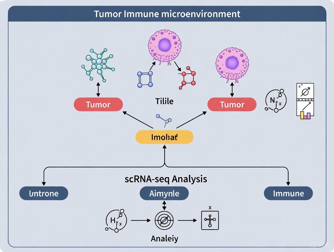
Unraveling TIME Complexity: scRNA-seq Reveals Cellular Heterogeneity and Novel Immune Dynamics
The tumor microenvironment (TME) is a highly complex and heterogeneous ecosystem comprising malignant cells, diverse immune cell populations, and various stromal components that collectively influence tumor genesis, development, metastasis, and therapeutic resistance [1] [2]. The cellular components of the TME include cancer-associated fibroblasts (CAFs), mesenchymal stem cells (MSCs), tumor-associated adipocytes (CAAs), tumor endothelial cells (TECs), pericytes, and a multitude of immune cells including T cells, B cells, natural killer (NK) cells, and tumor-associated macrophages (TAMs) [1] [2]. Understanding the precise composition and interactions of these cellular subpopulations is critical for advancing cancer biology and developing more effective therapeutic strategies.
Single-cell RNA sequencing (scRNA-seq) has emerged as a transformative technology for dissecting this complexity, offering unprecedented resolution into cellular heterogeneity and functional diversity within tumors [3] [4] [5]. This technical guide provides a comprehensive framework for resolving immune and stromal cell subpopulations within the TME using scRNA-seq methodologies, with specific protocols, analytical workflows, and visualization strategies tailored for researchers, scientists, and drug development professionals working in cancer immunology.
Computational Workflow for scRNA-seq Analysis
The analytical workflow for scRNA-seq data involves multiple critical steps from raw data processing to biological interpretation. The following diagram illustrates the standard pipeline for processing single-cell RNA sequencing data to resolve cellular diversity:
Quality Control and Data Preprocessing
Quality control (QC) represents the foundational step in scRNA-seq analysis, ensuring that only high-quality cells proceed through the analytical pipeline. The QC process involves:
- Filtering Parameters: Remove cells with ≤100 or ≥6000 expressed genes, ≤200 UMIs, and ≥10% mitochondrial gene content [6]. These thresholds may vary based on tissue type, disease state, and experimental conditions.
- QC Metrics Visualization: Utilize violin plots, density plots, or histograms to visualize distributions of genes per cell, UMI counts per cell, and percentage of mitochondrial genes [6].
- Data Normalization: Apply the
NormalizeDatafunction (Seurat) to scale datasets and regress out mitochondrial gene effects [3]. - Batch Effect Correction: Implement the
Harmonypackage to correct for technical variations between samples, experiments, or sequencing batches [3].
Dimensionality Reduction and Clustering
Following quality control, the normalized data undergoes dimensionality reduction to visualize and identify cell subpopulations:
- Highly Variable Gene Selection: Use the
FindVariableFeaturesfunction to identify genes with high cell-to-cell variation [3]. - Principal Component Analysis (PCA): Apply linear dimensionality reduction to identify primary sources of transcriptional variation [3] [7].
- Non-linear Dimensionality Reduction: Employ UMAP (Uniform Manifold Approximation and Projection) and t-SNE (t-distributed Stochastic Neighbor Embedding) for unsupervised cell clustering and two-dimensional visualization [3] [6]. UMAP preserves both local and global data structure, while t-SNE emphasizes local relationships [7].
- Cell Clustering: Utilize graph-based clustering algorithms implemented in Seurat to partition cells into distinct subpopulations based on transcriptional similarities [3].
Key Cellular Players in the Tumor Microenvironment
Immune Cell Subpopulations
The tumor immune microenvironment contains diverse immune cell types that play critical roles in anti-tumor immunity and immunotherapy response:
- T cells and Exhaustion States: CD8+ T cells often enter a state of exhaustion characterized by impaired effector activity and sustained inhibitory receptor expression, representing a significant barrier to durable immunotherapy responses [5]. Recent studies using TCR sequencing have revealed that higher T-cell diversity in peripheral blood is associated with superior response to dual immune checkpoint inhibitor therapy in metastatic NSCLC [8].
- Tumor-Associated Macrophages (TAMs): TAMs exhibit remarkable plasticity and functional diversity. Single-cell studies in gastric cancer have identified TAM subpopulations with elevated activity in P53, Wnt, and JAK-STAT3 signaling pathways [3]. In hepatocellular carcinoma (HCC), SPP1+ macrophages have been identified as key mediators of immune suppression through their ability to suppress CD8+ T cell proliferation [5].
- Monocyte Subsets: An interferon-stimulated gene-high (ISGhigh) monocyte subset has been shown to be significantly enriched in syngeneic mouse models responsive to anti-PD-1 therapy, suggesting its potential role in treatment response [9].
- Neutrophils: The role of neutrophils in the TME appears context-dependent. Neutrophil depletion experiments using anti-Ly6G antibodies resulted in variable antitumor effects across different models but failed to consistently enhance the efficacy of PD-1 blockade [9].
Stromal Cell Subpopulations
Stromal cells constitute a major cellular component of the TME and play crucial roles in tumor progression, immune modulation, and therapeutic resistance:
- Cancer-Associated Fibroblasts (CAFs): CAFs are the most abundant stromal cell type in many solid tumors, particularly in breast, prostate, pancreatic, and gastric cancers [1]. They exhibit both tumor-promoting and tumor-suppressing phenotypes, with the former representing the majority of CAF populations. Single-cell studies have revealed extensive CAF heterogeneity, with identified subtypes including:
- Myofibroblastic CAFs (myCAFs) with tumor-inhibitory effects
- Inflammatory CAFs (iCAFs) that secrete IL-6, LIF, and CXCL1 to promote tumor progression
- Antigen-presenting CAFs (apCAFs) with immune-modulating capabilities [1]
- Mesenchymal Stem Cells (MSCs): MSCs can be recruited to the TME and differentiate into various stromal cell types, contributing to tumor growth and metastasis [1].
- Tumor-Associated Adipocytes (CAAs): CAAs support tumor metabolism through lipid transfer and secretion of adipokines that promote cancer cell proliferation and invasion [1] [2].
- Tumor Endothelial Cells (TECs) and Pericytes: These vascular components facilitate angiogenesis and regulate immune cell trafficking within the TME [1].
Table 1: Key Stromal Cell Types in the Tumor Microenvironment
| Cell Type | Key Markers | Primary Functions | Therapeutic Implications |
|---|---|---|---|
| Cancer-Associated Fibroblasts (CAFs) | α-SMA, FAP, FSP1, PDGFR-α/β | ECM remodeling, immune suppression, cytokine secretion | CAF depletion, FAP-targeting |
| Mesenchymal Stem Cells (MSCs) | CD44, CD73, CD90, CD105 | Differentiation into stromal cells, immunomodulation | Inhibition of MSC recruitment |
| Tumor-Associated Adipocytes (CAAs) | PLIN1, PLIN2, FABP4 | Lipid transfer, adipokine secretion, metabolic reprogramming | Lipid metabolism inhibition |
| Tumor Endothelial Cells (TECs) | CD31, VEGFR2, Endoglin | Angiogenesis, immune cell trafficking | Anti-angiogenic therapies |
| Pericytes | NG2, PDGFR-β, α-SMA | Vessel stabilization, metastasis regulation | Vascular normalization |
Advanced Analytical Methods for Cellular Resolution
Differential Expression and Pathway Analysis
Identifying differentially expressed genes (DEGs) across cell subpopulations is crucial for understanding their functional states:
- Differential Expression Testing: Use the
FindAllMarkersfunction with Wilcoxon rank sum test, setting log2FC threshold to 0.25 and minimum gene expression ratio to 0.25 [3]. - Volcano Plots: Visualize DEGs using scatter plots that display log2 fold change versus statistical significance (-log10 p-value) [6].
- Pathway Enrichment Analysis: Perform Gene Ontology (GO), Kyoto Encyclopedia of Genes and Genomes (KEGG), and Gene Set Variation Analysis (GSVA) to identify enriched biological pathways in specific cell clusters [3].
- Gene Set Enrichment Analysis (GSEA): Utilize tools like GSEA to identify enriched or depleted pathways using multiple gene sets from Reactome, Wikipathways, and Hallmark collections [7].
Cell-Cell Communication Analysis
Understanding signaling networks between immune and stromal subpopulations provides critical insights into TME dynamics:
- Ligand-Receptor Interaction Mapping: Employ the
CellChatpackage to identify hyper-variable ligand-receptor pairs and their mutual signaling pathways [3]. - Communication Probability Calculation: Use the
netPslot in CellChat to calculate communication probabilities and aggregate networks at the signaling pathway level [3]. - Visualization Methods: Represent cell-cell communication networks using circos plots (for direction and flow of signaling) and heatmaps (for quantitative comparison of interaction strength) [6].
- Key Pathways: In gastric cancer, the CCL5-CCR1 ligand-receptor signaling axis has been identified as a potential immune checkpoint, with elevated expression in TAMs and mast cells associated with poor long-term survival [3].
The following diagram illustrates the complex interplay between major cellular components in the tumor microenvironment:
Trajectory Inference and Cellular Dynamics
Pseudotemporal ordering methods reconstruct cellular differentiation trajectories and state transitions:
- Pseudotime Analysis: Utilize Monocle3 to order cells along differentiation trajectories using advanced machine learning techniques [3].
- Differentiation Potential Assessment: Apply CytoTRACE to predict cellular differentiation states based on transcriptomic diversity [3].
- Gene Expression Dynamics: Monitor how gene expression changes along reconstructed trajectories to identify drivers of cell state transitions [3].
- Application Example: In gastric cancer, pseudotemporal analysis has demonstrated the differentiation potential of TAMs into mast cells, with APOC1, C1QB, FCN1, FTL, S100A9, CD1C, CD1E, and FCER1A identified as the top genes driving this process [3].
Machine Learning for Predictive Biomarker Discovery
Advanced computational methods enable the identification of predictive signatures from scRNA-seq data:
- Feature Selection: Implement Boruta algorithm for robust feature selection, identifying gene signatures predictive of treatment response across cancer types [4].
- Predictive Modeling: Apply XGBoost (eXtreme Gradient Boosting) to predict immunotherapy response while maintaining single-cell resolution [4].
- Model Interpretation: Utilize SHAP (Shapley Additive exPlanations) values to dissect the contribution of selected genes and identify complex, non-linear gene-pair interactions [4].
- Cell-Based Signatures: Develop reinforcement learning models to identify the most informative single cells for predicting patient response to therapy [4].
Table 2: Computational Tools for scRNA-seq Analysis of TME
| Tool Category | Software Package | Primary Function | Key Applications |
|---|---|---|---|
| Data Preprocessing | Seurat | Quality control, normalization, integration | Batch effect correction, data scaling |
| Dimensionality Reduction | Harmony | Batch effect correction | Integration of multiple datasets |
| Cell Communication | CellChat | Ligand-receptor interaction analysis | Inference of signaling networks |
| Trajectory Analysis | Monocle3 | Pseudotemporal ordering | Cell differentiation, state transitions |
| Machine Learning | PRECISE/XGBoost | Response prediction, biomarker discovery | Immunotherapy response prediction |
| Differential Expression | DESeq2, edgeR | Statistical testing for DEGs | Identification of marker genes |
Successful resolution of immune and stromal cell subpopulations requires carefully selected research reagents and computational resources:
Table 3: Essential Research Reagents and Resources for scRNA-seq TME Studies
| Category | Reagent/Resource | Specification | Application/Function |
|---|---|---|---|
| Wet Lab Reagents | Single Cell 3' Library Kit (10x Genomics) | v3 chemistry | Droplet-based scRNA-seq library preparation |
| Enzyme D, R, A (Miltenyi Biotec) | 130-096-730 | Tissue dissociation for single-cell suspension | |
| Anti-CD45 Antibodies | BD Biosciences 550994 | Immune cell isolation and sorting | |
| Fixable Viability Stain | BD Biosciences 562247 | Exclusion of non-viable cells | |
| Computational Tools | R Version | 4.4.1 or higher | Statistical computing environment |
| Seurat Package | Version 5 | Single-cell data analysis and visualization | |
| CellChat | Latest version | Cell-cell communication analysis | |
| Monocle3 | 3.22 or higher | Pseudotime and trajectory analysis | |
| Reference Databases | CellMarker | http://xteam.xbio.top/CellMarker/ | Cell type marker gene database |
| Enrichr | https://maayanlab.cloud/Enrichr/ | Gene set enrichment analysis | |
| GEO Database | https://www.ncbi.nlm.nih.gov/geo/ | Repository for scRNA-seq data |
The resolution of immune and stromal cell subpopulations within the tumor microenvironment using scRNA-seq technologies has fundamentally advanced our understanding of cancer biology and therapeutic resistance. The methodologies outlined in this technical guide provide a comprehensive framework for researchers to characterize cellular diversity, identify novel biomarkers, and uncover potential therapeutic targets. As these technologies continue to evolve, standardization of both experimental and computational pipelines will be essential for improving reproducibility and enabling meaningful comparisons across studies [5]. The integration of scRNA-seq with spatial transcriptomics, proteomics, and computational modeling approaches promises to further unravel the complexity of the TME, ultimately guiding the development of more effective and personalized cancer immunotherapies.
Characterizing T Cell Exhaustion States and Functional Plasticity
T cell exhaustion represents a critical dysfunctional state of T lymphocytes that arises during chronic antigen exposure, such as in cancer and persistent viral infections. This state is characterized by progressive loss of effector functions, sustained expression of inhibitory receptors, and altered transcriptional programming. Within the tumor immune microenvironment (TIME), exhausted T cells (TEX) demonstrate impaired cytokine production, reduced cytotoxic capacity, and poor proliferative potential, which collectively undermine effective anti-tumor immunity [10] [11]. The development of T cell exhaustion is now recognized as a major mechanism of immune evasion by tumors and a significant contributor to resistance against immunotherapies, including immune checkpoint blockade.
Single-cell RNA sequencing (scRNA-seq) has revolutionized our understanding of the TIME by enabling high-resolution profiling of its cellular composition at the transcriptional level. This technology has revealed remarkable heterogeneity within the TME, identifying novel or rare immune cell subsets and delineating their dynamic functional states [12]. In particular, scRNA-seq has illuminated the complex intercellular signaling networks and temporal cell-state transitions that drive tumor progression and immune evasion. The application of scRNA-seq to dissect T cell exhaustion states provides unprecedented insights into the transcriptional programs and cellular ecosystems associated with this dysfunctional state, offering new avenues for therapeutic intervention.
Defining T Cell Exhaustion: Markers, Subsets, and Functional States
Core Characteristics of Exhausted T Cells
T cell exhaustion develops progressively through a continuum of differentiation states rather than representing a single, uniform population. According to recent nomenclature guidelines, exhausted T cells (TEX) are defined as "T cells that arise during chronic infections and cancer, characterized by a hierarchical loss of effector functions, expression of multiple inhibitory receptors, altered transcriptional regulation, and impaired homeostatic self-renewal" [10]. This definition distinguishes TEX from other T cell differentiation states such as naive, effector, and memory T cells based on specific functional and molecular criteria.
The functional hallmarks of T cell exhaustion include:
- Hierarchical loss of effector functions: Production of interleukin-2 (IL-2) is lost first, followed by impaired cytotoxicity and tumor necrosis factor-alpha (TNF-α) secretion, with interferon-gamma (IFN-γ) production being the most resistant to loss
- Proliferative impairment: Reduced expansion capacity in response to antigenic stimulation
- Altered metabolic programs: Shift from oxidative phosphorylation to glycolysis with mitochondrial dysfunction
- Sustained inhibitory receptor expression: Concurrent expression of multiple checkpoint molecules including PD-1, TIM-3, LAG-3, CTLA-4, and TIGIT
- Transcriptional reprogramming: Expression of specific transcription factors such as TOX, NR4A, and EOMES that enforce the exhausted state
Key Surface Markers and Soluble Mediators
The phenotypic identification of exhausted T cells relies on both surface markers and soluble mediators that can be detected in plasma or serum. Table 1 summarizes the primary markers used to characterize T cell exhaustion states in human and mouse systems, along with their detection methods and biological significance.
Table 1: Key Markers for Identifying and Characterizing T Cell Exhaustion
| Marker | Full Name | Detection Method | Expression Pattern | Biological Significance |
|---|---|---|---|---|
| PD-1 | Programmed cell death protein 1 | Flow cytometry, IHC, scRNA-seq | Sustained high expression on TEX | Primary inhibitory receptor; target of checkpoint inhibitors |
| TIM-3 | T cell immunoglobulin and mucin domain 3 | Flow cytometry, ELISA (soluble), scRNA-seq | Upregulated on TEX; correlates with severity of exhaustion | Marker of terminal exhaustion; regulates macrophage activation |
| LAG-3 | Lymphocyte-activation gene 3 | Flow cytometry, ELISA (soluble), scRNA-seq | Co-expressed with PD-1 on TEX | Inhibitory receptor that binds MHC class II; impairs T cell function |
| sCD25 | Soluble CD25 (IL-2Rα) | ELISA | Elevated in chronic inflammation | Marker of T cell activation; associated with persistent symptoms in PCC |
| CTLA-4 | Cytotoxic T-lymphocyte-associated protein 4 | Flow cytometry, IHC | Upregulated on TEX, especially Tregs | Early inhibitory receptor; regulates early stages of T cell activation |
| TIGIT | T cell immunoreceptor with Ig and ITIM domains | Flow cytometry, scRNA-seq | Co-expressed with other inhibitory receptors on TEX | Inhibits T cell function through multiple mechanisms |
Recent studies have validated the utility of measuring soluble forms of these markers as potential biomarkers for disease progression and treatment response. For instance, elevated plasma levels of sTIM-3 and sLAG-3 have been associated with persistent symptomatology in chronic conditions, suggesting their potential as biomarkers for T cell dysfunction in cancer settings [13].
Heterogeneity Within the Exhausted T Cell Compartment
scRNA-seq analyses have revealed substantial heterogeneity within the exhausted T cell compartment, identifying multiple subsets with distinct functional capacities and differentiation states. Two major subsets have been characterized:
Progenitor-exhausted T cells (TEX-prog): These cells retain some capacity for self-renewal and differentiation potential, express the transcription factor TCF-1, and respond better to immune checkpoint blockade therapy.
Terminally exhausted T cells (TEX-term): This population demonstrates severe functional impairment, expresses high levels of multiple inhibitory receptors, and shows minimal responsiveness to immunotherapy.
The balance between these subsets within tumors has emerged as a critical determinant of response to immunotherapy, with a higher proportion of progenitor-exhausted T cells correlating with improved clinical outcomes [14] [11].
Experimental Models for Studying T Cell Exhaustion
In Vitro Model of Chronic T Cell Stimulation
A robust in vitro model for generating exhausted CD8+ T cells has been developed that recapitulates critical hallmarks of exhaustion observed in vivo. The protocol involves repeated stimulation of T cells with their cognate antigen, followed by comprehensive temporal phenotypic characterization [11].
Detailed Experimental Protocol:
T Cell Isolation and Initial Activation:
- Isolate naive CD8+ T cells from mouse spleen or human peripheral blood using magnetic bead separation or fluorescence-activated cell sorting (FACS)
- Culture cells in RPMI-1640 medium supplemented with 10% FBS, 2 mM L-glutamine, 1 mM sodium pyruvate, and 50 μM β-mercaptoethanol
- Activate cells with plate-bound anti-CD3 (5 μg/mL) and soluble anti-CD28 (2 μg/mL) antibodies for 48 hours
Chronic Antigen Stimulation Phase:
- After initial activation, wash cells and resuspend in complete medium containing IL-2 (100 U/mL)
- Restimulate cells every 3-4 days with fresh antigen-presenting cells loaded with cognate peptide antigen (1 μM) or with plate-bound anti-CD3 antibody
- Maintain cells at a density of 0.5-1 × 10^6 cells/mL with regular medium changes
Phenotypic Characterization:
- Analyze surface marker expression (PD-1, TIM-3, LAG-3, etc.) by flow cytometry at weekly intervals
- Assess functional capacity through cytokine production (IFN-γ, TNF-α, IL-2) upon restimulation
- Evaluate cytotoxic potential using granzyme B and perforin staining or killing assays
- Measure proliferative capacity by CFSE dilution or Ki67 expression
This model successfully recapitulates key features of T cell exhaustion, including impaired proliferation, reduced cytokine production, decreased cytotoxic granule release, metabolic alterations, and progressive expression of inhibitory receptors [11]. The resulting exhausted T cells exhibit a gene signature shared with in vivo exhausted states and tumor-infiltrating T cells from multiple human tumor types, validating the translational potential of this model for discovering new therapies.
In Vivo Validation Models
The in vitro findings require validation using in vivo models that more closely mimic the complex tumor microenvironment. Two primary approaches are commonly employed:
Chronic Infection Models:
- Lymphocytic choriomeningitis virus (LCMV) clone 13 infection in mice
- Provides a well-characterized system for studying T cell exhaustion in a physiological context
- Allows tracking of antigen-specific T cells using tetramers
Syngeneic Tumor Models:
- Implantation of tumor cell lines into genetically identical mouse strains
- Enables evaluation of T cell exhaustion in authentic tumor microenvironments
- Permits assessment of responses to immunotherapeutic interventions
These validation models have confirmed that the gene signature derived from the in vitro exhaustion model is observed in tumor-infiltrating T cells from multiple human tumor types, supporting its relevance for human cancer biology [9] [11].
Single-Cell RNA Sequencing for Dissecting T Cell Exhaustion
Experimental Workflow for scRNA-seq of Tumor-Infiltrating T Cells
The application of scRNA-seq to characterize T cell exhaustion states in the TIME involves a multi-step process that requires careful experimental design and execution. Figure 1 illustrates the complete workflow from sample processing to data analysis.
Figure 1: Experimental workflow for scRNA-seq analysis of tumor-infiltrating T cells
Detailed Methodology:
Sample Collection and Processing:
- Obtain fresh tumor tissues from surgical specimens or biopsies
- Process tissues within 1-2 hours of collection to maintain cell viability
- Mechanically dissociate tissues using gentleMACS Octo Dissociator with Heaters
- Use enzyme cocktails containing Enzyme D, R, and A (Miltenyi Biotec) for optimal cell recovery
- Filter cell suspensions through 70μm mesh to remove debris
Immune Cell Enrichment and Sorting:
- Stain cells with PerCP-Cy5.5 anti-CD45 and Fixable Viability Stain
- Sort viable CD45+ cells using FACS with strict gating strategies
- Confirm >80% viability post-sorting through reanalysis
- Resuspend cells in PBS at 1×10^6 cells/mL for loading
Single-Cell Library Preparation and Sequencing:
- Load cells onto Chromium Controller (10x Genomics) for droplet-based encapsulation
- Use Single Cell 3' Library and Gel Bead Kit v3 (10x Genomics) according to manufacturer's protocol
- Assess library quality using Bioanalyzer or TapeStation
- Sequence libraries on Illumina platforms to achieve minimum depth of 50,000 reads per cell
This standardized protocol has been successfully applied to profile the immune microenvironment across multiple cancer types, including breast cancer, gastric cancer, and various syngeneic mouse models [14] [3] [9].
Computational Analysis Pipeline
The analysis of scRNA-seq data from tumor-infiltrating T cells involves multiple computational steps to extract biologically meaningful insights about T cell exhaustion states:
Data Processing and Quality Control:
- Process raw sequencing data using Cell Ranger (10x Genomics) to generate gene-cell matrices
- Filter cells based on quality metrics: number of genes detected (300-7000), UMI counts (>1000), and mitochondrial content (<20%)
- Normalize data using SCTransform or similar methods to remove technical variability
Integration and Batch Correction:
- Integrate multiple samples using Harmony or SCVI to correct for batch effects
- Incorporate sample identity as a covariate in integration models
- Perform biology-aware integration using tools like SCANVI and CellHint for improved annotation accuracy
Cell Clustering and Annotation:
- Perform principal component analysis followed by graph-based clustering
- Annotate T cell subsets using established marker genes: CD3D/E/G (pan-T), CD4 (helper), CD8A (cytotoxic), FOXP3 (Treg), and exhaustion markers (PDCD1, HAVCR2, LAG3)
- Validate annotations using reference datasets and automated tools like SingleR
Differential Expression and Pathway Analysis:
- Identify differentially expressed genes between conditions using Wilcoxon rank sum test
- Perform gene set enrichment analysis (GSEA) to identify enriched pathways in exhausted subsets
- Analyze copy number variations (CNV) in malignant cells using InferCNV with T cells as reference
Trajectory Inference and Cell-Cell Communication:
- Reconstruct differentiation trajectories using Monocle3 or Slingshot to model T cell exhaustion progression
- Infer cell-cell communication networks using CellChat to identify interactions between T cell subsets and other TIME components
This comprehensive analytical approach has revealed substantial transcriptional diversity within the T cell compartment and identified distinct exhaustion states in primary and metastatic tumors [14].
Key Signaling Pathways Regulating T Cell Exhaustion
T cell exhaustion is regulated by a complex network of signaling pathways that integrate external cues from the tumor microenvironment with intrinsic transcriptional and metabolic programs. Figure 2 illustrates the major signaling pathways and their interactions in regulating T cell exhaustion states.
Figure 2: Signaling pathways regulating T cell exhaustion
Inhibitory Receptor Signaling
The signaling pathways downstream of inhibitory receptors play a central role in establishing and maintaining T cell exhaustion:
PD-1 Signaling Pathway:
- PD-1 engagement by its ligands PD-L1/PD-L2 recruits SHP1 and SHP2 phosphatases to the immunological synapse
- These phosphatases dephosphorylate key signaling molecules in the TCR cascade, including CD3ζ, ZAP70, and PKCθ
- Downstream effects include reduced activation of RAS-MAPK, PI3K-AKT, and NF-κB pathways
- Ultimately leads to impaired T cell proliferation, survival, and effector function
TIM-3 Signaling Pathway:
- TIM-3 interacts with multiple ligands including galectin-9, CEACAM1, HMGB1, and phosphatidylserine
- Engagement with galectin-9 induces calcium flux and Th1 cell death
- Interaction with HMGB1 competitively inhibits its binding to DNA, reducing innate immune activation
- TIM-3 signaling disrupts Bat3-mediated protection of exhaustion-associated transcription factors
LAG-3 Signaling Pathway:
- LAG-3 binds MHC class II molecules with higher affinity than CD4
- This interaction interferes with CD4 coreceptor function and downstream TCR signaling
- LAG-3 signaling inhibits calcium flux and AKT phosphorylation
- The cytoplasmic KIEELE motif is essential for LAG-3 inhibitory function
Transcriptional Regulation of Exhaustion
The exhausted T cell state is enforced by a specific transcriptional network that differs from those governing effector and memory T cell differentiation:
NFAT-TOX Axis:
- Chronic TCR stimulation leads to sustained nuclear factor of activated T cells (NFAT) activation
- NFAT drives expression of thymocyte selection-associated high mobility group box protein (TOX)
- TOX reprograms the epigenetic landscape of exhausted T cells, promoting a stable dysfunctional state
- TOX expression is required for the development of exhaustion and maintenance of exhausted T cells
NR4A Transcription Factors:
- NR4A family members (NR4A1, NR4A2, NR4A3) are rapidly induced by TCR signaling
- In chronic stimulation, sustained NR4A expression promotes exhaustion by repressing effector gene expression
- NR4A factors compete with AP-1 for binding to composite NFAT-AP-1 sites, shifting the balance toward exhaustion
Epigenetic Regulation:
- Exhausted T cells display distinct chromatin accessibility patterns compared to effector and memory T cells
- Exhaustion-associated regions show enrichment for binding sites of exhaustion-related transcription factors
- These epigenetic changes create a barrier to reprogramming exhausted T cells back to functional states
Research Reagent Solutions for T Cell Exhaustion Studies
The experimental approaches described require specific reagents and tools carefully selected for their applicability to T cell exhaustion research. Table 2 provides a comprehensive list of essential research reagents with their specific applications in characterizing T cell exhaustion states.
Table 2: Essential Research Reagents for T Cell Exhaustion Studies
| Reagent Category | Specific Examples | Application in T Cell Exhaustion Research | Key Considerations |
|---|---|---|---|
| Antibodies for Flow Cytometry | Anti-PD-1 (CD279), Anti-TIM-3 (CD366), Anti-LAG-3 (CD223), Anti-CD3, Anti-CD8, Anti-CD4, Anti-CD45 | Phenotypic characterization of exhausted T cell subsets | Multicolor panels (10+ colors) required to resolve heterogeneous populations; include viability dyes |
| scRNA-seq Kits | 10x Genomics Single Cell 3' Reagent Kits, Parse Biosciences Single Cell RNA Sequencing Kit | Transcriptional profiling of T cell states at single-cell resolution | Consider cell throughput requirements; incorporate feature barcoding for surface protein detection |
| Cell Isolation Kits | CD8+ T Cell Isolation Kit (human/mouse), Pan T Cell Isolation Kit, CD45+ Selection Kits | Enrichment of specific T cell populations from tumor tissue | Minimize activation during isolation; maintain cell viability for functional assays |
| Cytokine ELISA Kits | IFN-γ, TNF-α, IL-2, IL-10, TGF-β ELISA kits | Quantification of cytokine production capacity | Use high-sensitivity kits; measure both supernatant and intracellular cytokines |
| Functional Assay Kits | CFSE Cell Division Tracker, Granzyme B Activity Assay, Mitochondrial Stress Test Kit | Assessment of proliferation, cytotoxicity, and metabolic function | Optimize assay conditions for exhausted T cells which may have limited responses |
| In Vivo Models | Syngeneic tumor models (CT26.WT, EMT6, MC38), PD-1/PD-L1 blockade antibodies, LCMV clone 13 | Validation of exhaustion mechanisms in physiological contexts | Select models based on research question; consider genetic background effects |
These reagents form the foundation for comprehensive studies of T cell exhaustion and should be selected based on the specific research questions and model systems being employed. Recent studies have highlighted the importance of using multiple complementary approaches to fully characterize the heterogeneous nature of exhausted T cell populations [9] [11].
Comparative Analysis of T Cell States Across Cancer Types
scRNA-seq studies have enabled systematic comparison of T cell exhaustion states across different cancer types, revealing both shared and cancer-specific features. Table 3 summarizes key findings from recent studies investigating T cell exhaustion in various human cancers and mouse models.
Table 3: Comparative Analysis of T Cell Exhaustion Across Cancer Types
| Cancer Type | Sample Source | Key Exhaustion Features | Response to Immunotherapy | References |
|---|---|---|---|---|
| ER+ Breast Cancer | Primary and metastatic tumors (23 patients) | Increased exhausted cytotoxic T cells and FOXP3+ Tregs in metastases; distinct CNV patterns in malignant cells | Reduced tumor-immune cell interactions in metastases; potential for TNF-α signaling targeting | [14] |
| Gastric Cancer with Peritoneal Metastasis | 20 scRNA-seq samples from GEO database | TAMs and mast cells show elevated CCL5-CCR1 axis; pseudotemporal analysis demonstrates TAM differentiation | CCL5-CCR1 pathway identified as potential immune checkpoint; associated with poor survival | [3] |
| Multiple Syngeneic Mouse Models | 10 syngeneic models across 7 cancer types | ISGhigh monocyte subset enriched in anti-PD-1 responsive models; neutrophil depletion shows context-dependent effects | ISGhigh monocytes as potential biomarker for PD-1 response; neutrophil role varies by model | [9] |
| Pan-Cancer Analysis | Integrated data from 77 scRNA-seq studies (1163 tumors) | Shared exhausted CD8+ T cell signature across cancer types; correlation between CD8+ cytotoxicity and macrophage interferon response | Identification of conserved resistance mechanisms; TLS signatures associated with better response | [12] |
This comparative analysis reveals that while certain features of T cell exhaustion are conserved across cancer types, there are also important differences that may influence responses to immunotherapy. These findings highlight the need for cancer-specific approaches to targeting T cell exhaustion and the importance of using appropriate model systems that recapitulate these differences.
The characterization of T cell exhaustion states using scRNA-seq has provided unprecedented insights into the complexity of the tumor immune microenvironment and the mechanisms underlying immune evasion. The experimental approaches and analytical frameworks described in this technical guide provide a foundation for comprehensive investigation of T cell dysfunction in cancer and other chronic conditions. As single-cell technologies continue to evolve, integrating transcriptomic data with epigenetic, proteomic, and spatial information will further enhance our understanding of T cell exhaustion and enable the development of more effective immunotherapeutic strategies.
Future directions in this field include the development of more sophisticated in vitro models that better recapitulate the complex cellular interactions within the TIME, the integration of single-cell multi-omics approaches to connect transcriptional states with functional potential, and the application of spatial transcriptomics to map exhausted T cell populations within the architectural context of tumors. Additionally, there is growing recognition of the importance of studying T cell exhaustion across diverse cancer types and patient populations to identify both universal and context-specific therapeutic targets. These advances will ultimately contribute to more effective strategies for reversing T cell exhaustion and restoring anti-tumor immunity in cancer patients.
The tumor immune microenvironment (TIME) is a critical orchestrator of cancer progression, immune evasion, and therapeutic resistance. It comprises malignant cells, stromal components, and diverse immune cell populations that collectively shape antitumor immunity [15]. Among these components, immunosuppressive cells—including myeloid-derived suppressor cells (MDSCs), regulatory T cells (Tregs), and tumor-associated macrophages (TAMs)—function as vital barriers to effective immune destruction of tumors [16]. The emergence of single-cell RNA sequencing (scRNA-seq) has revolutionized our capacity to dissect TIME heterogeneity at unprecedented resolution, revealing novel cellular subsets and molecular mechanisms driving immunosuppression [5]. This review focuses on two such mechanisms identified through scRNA-seq studies: the role of SPP1+ macrophages and HMGB2-mediated immune evasion, exploring their biology, functional significance, and therapeutic implications across multiple cancer types.
SPP1+ Macrophages: Prototypical Immunosuppressive Myeloid Cells
Identification and Characterization of SPP1+ Macrophages
Secreted phosphoprotein 1 (SPP1), also known as osteopontin (OPN), marks a distinct macrophage subpopulation within the TIME. ScRNA-seq studies across various cancers consistently identify SPP1+ macrophages (SPP1+ Macs) as tumor-specific TAMs that diverge from classical M1/M2 polarization paradigms [17]. In head and neck squamous cell carcinoma (HNSCC), scRNA-seq of paired tumor and normal tissues revealed that SPP1+ Macs constitute a myeloid subpopulation predominantly present in tumor tissue, with minimal presence in normal counterparts [17]. Similar findings emerge from hypopharyngeal squamous cell carcinoma (HSCC), where SPP1+ macrophages are significantly overexpressed in tumor tissues and lymphatic metastases compared to normal hypopharyngeal tissues, and are characterized as M2-type immunosuppressive macrophages [18].
Table 1: SPP1+ Macrophage Characteristics Across Cancer Types
| Cancer Type | Identification Method | Key Features | Clinical Correlation |
|---|---|---|---|
| Head and Neck Squamous Cell Carcinoma (HNSCC) | scRNA-seq (5 patient pairs) | TNF-α and IL-1β secretion via NF-κB pathway; promotes tumor proliferation | Positive correlation with poor prognosis [17] |
| Hypopharyngeal Squamous Cell Carcinoma (HSCC) | scRNA-seq (5 patients) | M2-like phenotype; enriched in tumor and lymphatic tissues | Associated with lymphatic metastasis [18] |
| Esophageal Squamous Cell Carcinoma (ESCC) | TCGA analysis + mouse models | Drives M2 polarization via CD44/PI3K/AKT signaling | Predicts poor prognosis; blockade inhibits tumor growth [19] |
| Hepatocellular Carcinoma (HCC) | scRNA-seq deconvolution | Mediates CD8+ T cell suppression | SPP1 inhibition reprograms macrophages to less suppressive state [5] |
Functional Mechanisms of SPP1+ Macrophages in Immune Suppression
SPP1+ Macs employ multifaceted mechanisms to foster an immunosuppressive TIME and promote tumor progression:
Cytokine-Mediated Immunosuppression: In HNSCC, SPP1+ Macs increase secretion of TNF-α and IL-1β via NF-κB pathway activation. These cytokines directly support tumor cell proliferation and migration while creating an inflammatory microenvironment conducive to cancer progression [17]. Functional experiments using SPP1-overexpressing (SPP1-OE) and SPP1-knockdown (SPP1-KD) macrophages demonstrated that SPP1+ Mac-derived TNF-α and IL-1β are critical for HNSCC malignant progression [17].
Recruitment and Polarization of Immunosuppressive Cells: In ESCC, SPP1 mediates crosstalk between cancer cells and TAMs by recruiting macrophages and promoting their M2 polarization through CD44/PI3K/AKT signaling activation. These polarized M2 TAMs subsequently secrete VEGFA and IL6 to sustain ESCC progression [19].
Metabolic Reprogramming of TIME: SPP1+ Mac-derived TNF-α and IL-1β promote the expression of OPN (the protein product of SPP1) in both tumor cells and adjacent macrophages, establishing a feed-forward amplification loop that sustains the immunosuppressive microenvironment [17].
CD8+ T Cell Suppression: In hepatocellular carcinoma (HCC), SPP1-expressing macrophages function as key mediators of immune suppression by directly suppressing CD8+ T cell proliferation, thereby limiting antitumor immunity [5].
Figure 1: SPP1+ Macrophage Signaling and Immunosuppressive Mechanisms. SPP1 activates multiple pathways including NF-κB-mediated cytokine production, CD44/PI3K/AKT-driven M2 polarization, and direct CD8+ T cell suppression.
HMGB2: A Novel Regulator of Phagocytosis and Immune Evasion
HMGB2 Biology and Expression in Cancer
High-mobility group box 2 (HMGB2) belongs to the HMGB family of DNA-binding proteins that regulate chromatin structure and function. Beyond its intracellular nuclear roles, HMGB2 can function as an extracellular damage-associated molecular pattern (DAMP) that contributes to immune responses and tumor development [20]. A pan-cancer analysis of female-specific cancers (breast, cervical, ovarian, and endometrial) revealed that HMGB2 exhibits differential expression across various cancers and plays significant roles in modulating tumor progression [21]. In hepatocellular carcinoma (HCC), elevated HMGB2 expression is linked to poor prognosis and fosters immune evasion by promoting T cell exhaustion [5].
Functional Mechanisms of HMGB2 in Immune Evasion
HMGB2 mediates immunosuppression through several distinct mechanisms:
Phagocytosis Regulation: HMGB2 knockdown in macrophages leads to significant impairment in phagocytosis of breast, cervical, ovarian, and endometrial cancer cells. This positions HMGB2 as a critical regulator of the phagocytic process in multiple female-specific cancers [21].
T Cell Exhaustion Promotion: In HCC, HMGB2 fosters immune evasion by promoting T cell exhaustion, a dysfunctional state characterized by impaired effector activity and sustained expression of inhibitory receptors that presents a significant barrier to effective immunotherapy [5].
Integration with Key Oncogenic Pathways: HMGB2 expression correlates with activation of multiple cancer-associated pathways. When HMGB2 knockdown is combined with Palbociclib (a CDK4/6 inhibitor) treatment, a significant decrease in tumor cell proliferation is observed across multiple cancer models, suggesting synergistic therapeutic potential [21].
Table 2: HMGB2-Mediated Mechanisms Across Cancer Types
| Cancer Type | Experimental Approach | Key Findings | Functional Outcome |
|---|---|---|---|
| Female-Specific Cancers (Breast, Cervical, Ovarian, Endometrial) | Pan-cancer analysis + in vitro validation | HMGB2 knockdown impairs macrophage phagocytosis | Enables immune evasion by reducing cancer cell clearance [21] |
| Hepatocellular Carcinoma (HCC) | Multi-omics integration (scRNA-seq, bulk RNA-seq, spatial transcriptomics) | Promotes T cell exhaustion | Creates immunosuppressive microenvironment [5] |
| Multiple Cancer Models | Drug combination studies | HMGB2 knockdown + Palbociclib synergistically decreases proliferation | Suggests combination therapy potential [21] |
Experimental Approaches for Studying Immunosuppressive Mechanisms
ScRNA-seq Workflow for TIME Dissection
ScRNA-seq provides an powerful tool for identifying novel immunosuppressive cell populations like SPP1+ macrophages and elucidating HMGB2 functions. A standardized workflow includes:
Tissue Processing and Single-Cell Suspension: Fresh tumor samples are washed in ice-cold storage buffer (RPMI-1640 + 0.04% BSA), cut into small pieces (0.5 mm³), and digested with a human Tumor Dissociation Kit according to manufacturer protocols. The lysates are filtered through 40μm cell strainers, centrifuged, and treated with red blood cell lysis buffer before final resuspension [17].
scRNA-seq Library Preparation and Sequencing: Cell suspensions are used to construct cDNA libraries with 10× Genomics Chromium Next GEM Single Cell 3′ Reagent Kits, followed by sequencing on Illumina platforms (e.g., NovaSeq 6000 in PE150 mode) [17].
Bioinformatic Analysis: The Cell Ranger software pipeline processes sequencing data for barcode demultiplexing. Subsequent analyses include cell type identification (Loupe Browser), cell-cell communication analysis (CellChat), trajectory inference (Monocle2), and copy number variation estimation (inferCNV) [17] [22].
Figure 2: Experimental Workflow for Identifying Immunosuppressive Mechanisms. From tumor tissue dissociation to functional validation of SPP1+ macrophages and HMGB2.
Functional Validation Approaches
In Vitro Macrophage Polarization and Co-culture: THP-1 monocytes are differentiated into macrophages using PMA, followed by construction of SPP1-overexpressing (SPP1-OE) and SPP1-knockdown (SPP1-KD) macrophages. These macrophages are co-cultured with HNSCC cell lines to assess their impact on tumor cell proliferation and migration. Cytokine secretion is measured via Luminex liquid suspension chip detection assay [17].
In Vivo Therapeutic Assessment: Mouse xenograft models are employed to verify SPP1+ Mac functions in HNSCC progression. The inhibitor VGX-1027, which targets macrophage-derived TNF-α and IL-1β, is used to confirm the roles of these cytokines. In ESCC models, SPP1 blockade with RNA aptamer significantly inhibits tumor growth and M2 TAM infiltration [17] [19].
HMGB2 Functional Assays: HMGB2 knockdown is performed in both cancer cells and macrophages to evaluate impacts on cancer cell proliferation, migration, invasion, and macrophage phagocytosis. Combination therapies with drugs like Palbociclib are tested for synergistic effects [21].
Therapeutic Targeting Strategies and Research Reagents
Targeting SPP1+ Macrophages and HMGB2
Several therapeutic approaches have emerged for targeting these immunosuppressive mechanisms:
SPP1-Directed Therapeutics: RNA aptamers against SPP1 significantly inhibit tumor growth and M2 TAM infiltration in ESCC xenograft models [19]. In HCC, SPP1 inhibition can reprogram macrophages toward a less suppressive phenotype [5].
Cytokine Pathway Inhibition: VGX-1027, an inhibitor of macrophage-derived TNF-α and IL-1β, confirmed that SPP1+ Mac-derived cytokines promote HNSCC progression in both in vitro and in vivo experiments [17].
HMGB2 Targeting: HMGB2 knockdown significantly inhibits cancer cell proliferation, migration, and invasion in female-specific cancers. When combined with Palbociclib treatment, HMGB2 depletion causes a significant decrease in tumor cell proliferation across multiple cancer models [21].
Table 3: Research Reagent Solutions for Studying Immunosuppressive Mechanisms
| Reagent/Assay | Application | Function/Utility | Reference |
|---|---|---|---|
| 10× Genomics Chromium | scRNA-seq library preparation | High-resolution cellular heterogeneity analysis | [17] |
| Luminex Liquid Suspension Chip | Cytokine detection | Multiplex measurement of macrophage-secreted factors (TNF-α, IL-1β) | [17] |
| VGX-1027 | Small molecule inhibitor | Blocks macrophage-derived TNF-α and IL-1β | [17] |
| SPP1 RNA Aptamer | SPP1 inhibition | Specifically blocks SPP1-mediated signaling | [19] |
| Anti-HMGB2 siRNA | Gene knockdown | Validates HMGB2 function in phagocytosis and immune evasion | [21] |
| CIBERSORTx | Computational deconvolution | Infers cell type abundance from bulk RNA-seq data | [17] |
The identification of SPP1+ macrophages and HMGB2-mediated immunosuppression represents significant advances in our understanding of tumor immune evasion. ScRNA-seq technologies have been instrumental in revealing these novel mechanisms, providing insights that could not be achieved through bulk sequencing approaches. Therapeutic targeting of these pathways holds promise for overcoming current limitations of cancer immunotherapy.
Future research directions should focus on: (1) Developing more specific inhibitors against SPP1 and HMGB2; (2) Exploring combination therapies that simultaneously target both pathways; (3) Investigating spatial relationships between SPP1+ macrophages, HMGB2-expressing cells, and T cell populations within the TIME using spatial transcriptomics; (4) Validating these targets in larger patient cohorts across diverse cancer types. As our understanding of these immunosuppressive mechanisms deepens, they may offer novel therapeutic opportunities to enhance the efficacy of existing immunotherapies and overcome treatment resistance.
The tumor immune microenvironment (TIME) is a complex ecosystem where the spatial relationships between immune, stromal, and cancer cells critically influence disease progression and therapeutic response [23] [24]. While single-cell RNA sequencing (scRNA-seq) has revolutionized our understanding of cellular heterogeneity, it inherently lacks spatial context due to tissue dissociation requirements [23]. The integration of scRNA-seq with spatially resolved technologies now enables researchers to preserve this architectural information, providing unprecedented insights into cellular organization, communication networks, and functional states within native tissue contexts [23] [24]. This technical guide explores current methodologies, analytical frameworks, and applications for integrating transcriptomic data with tissue architecture, with a specific focus on advancing TIME research within the broader thesis of dissecting tumor ecosystems using scRNA-seq.
Technological Foundations
Single-Cell RNA Sequencing Platforms
scRNA-seq enables high-resolution transcriptomic profiling at the individual-cell level, revealing cellular heterogeneity, identifying rare populations, and characterizing dynamic biological processes [25] [23]. A standard scRNA-seq workflow begins with tissue dissociation into single-cell suspensions, followed by cell isolation using fluorescence-activated cell sorting (FACS) or microfluidics, with droplet-based microfluidics being the most widely adopted high-throughput platform [25]. After isolation, cells undergo lysis, reverse transcription, cDNA amplification, library construction, and sequencing [25]. Critical analytical steps include quality control, normalization, batch correction, clustering, and cell type annotation using established computational tools [25].
Despite its transformative potential, scRNA-seq presents notable limitations including relatively low RNA capture efficiency per cell, high costs, technical challenges in sample processing, and most critically, the loss of native spatial relationships due to mandatory tissue dissociation [23]. This spatial information is crucial for understanding cell-cell interactions within intact tissue architectures [23].
Spatial Transcriptomics Technologies
Spatial transcriptomics (ST) encompasses emerging technologies that enable spatially resolved gene expression profiling within intact tissue sections, preserving native histological context [23] [24]. Current ST methodologies can be broadly classified into two categories: image-based (I-B) and barcode-based (B-B) approaches [23].
Image-based methods, such as in situ hybridization (ISH) and in situ sequencing (ISS), utilize fluorescently labeled probes to directly detect RNA transcripts within tissues, allowing visualization of gene expression patterns while maintaining spatial integrity [23]. These have evolved into high-plex RNA imaging (HPRI) techniques including multiplexed error-robust fluorescence in situ hybridization (MERFISH) and sequential fluorescence in situ hybridization (seqFISH) [23].
Barcode-based approaches rely on spatially encoded oligonucleotide barcodes to capture RNA transcripts. In solid-phase transcriptome capture, RNAs hybridize to immobilized barcoded probes on slides before sequencing [23]. Deterministic spatial barcoding assigns unique barcodes to each transcript, retaining positional information throughout sequencing [23]. The 10× Genomics Visium platform uses chips containing spatially barcoded oligo(dT) to capture mRNA from overlaid tissue, while Slide-seq transfers RNA onto DNA-barcoded beads with known positions [24]. High-Definition Spatial Transcriptomics (HDST) uses microwell-based fluorescence spatial indexing beads for higher resolution capture [24].
Table 1: Comparison of Spatial Transcriptomics Technologies
| Technology | Resolution | Methodology | Tissue Compatibility | Key Applications |
|---|---|---|---|---|
| 10× Genomics Visium | 55-100 μm | Spatial barcoding | FF, FFPE | Unbiased spatial transcriptomics |
| Slide-seq | 10 μm | DNA-barcoded beads | FF | High-resolution mapping |
| HDST | 2 μm | Microwell beads | FF | Near single-cell resolution |
| MERFISH/seqFISH | Subcellular | Multiplexed FISH | FFPE | High-plex RNA imaging |
| DBiT-seq | 10 μm | Microfluidic barcoding | FF | Integrated protein detection |
Spatial Proteomics and Multiplexed Imaging
Beyond transcriptomics, spatially resolved proteomic analysis provides crucial information about protein expression and post-translational modifications within tissue architecture. Multiplexed imaging technologies enable simultaneous detection of multiple protein markers in tissue sections [26]. These include multiplexed immunohistochemistry (IHC) and immunofluorescence (IF) methods, imaging mass cytometry (IMC) which combines metal-labeled antibodies with mass spectrometry, and multiplexed ion beam imaging (MIBI) [26] [24].
Imaging mass cytometry, for instance, applies metal-tagged antibodies to tissue sections followed by laser ablation and mass spectrometry detection, enabling simultaneous assessment of 35-40 markers while preserving spatial information [27]. This approach was used in a recent study of non-small cell lung cancer (NSCLC) that analyzed 204 histopathology images from 102 patients, revealing distinct spatial patterns of immune cell aggregation in lung adenocarcinoma (LUAD) versus lung squamous cell carcinoma (LUSC) [27].
Analytical Frameworks for Spatial Data
Spatial Pattern Analysis
The spatial distribution of immune cells within the TIME presents non-random patterns with significant prognostic implications [28] [26]. Analytical frameworks have been developed to quantify these spatial relationships, moving beyond simple cell density measurements to capture complex organizational patterns.
The Spatiopath framework provides a null-hypothesis approach to distinguish statistically significant immune cell associations from random distributions [28]. This method extends Ripley's K function to analyze both cell-cell and cell-tumor interactions using embedding functions to map cell contours and tumor regions [28]. The mathematical foundation generalizes spatial point processes to accommodate interactions between point patterns and closed shapes, enabling robust identification of significant spatial associations beyond fortuitous accumulations [28].
Cell-cell interaction analysis employs permutation testing strategies to statistically evaluate if specific cell types are likely to interact spatially, and whether these relationships differ between conditions [27]. For example, a NSCLC study revealed increased tendency for B cell-cancer cell interactions in LUAD versus LUSC, suggesting distinct functional relationships [27].
Neighborhood analysis identifies recurrent cellular communities within tissues, capturing co-occurrence patterns across multiple cell types [26]. These neighborhoods represent functional units within the TIME that may have clinical significance, with specific combinations associated with patient prognosis or treatment response [26].
Integration Methods for scRNA-seq and ST Data
The integration of scRNA-seq and ST data requires computational methods that leverage the strengths of each technology [23]. Several strategies have been developed for this purpose:
Deconvolution approaches use scRNA-seq data as a reference to infer cell type proportions and gene expression patterns within spatial spots containing multiple cells [29]. Tools like SPOTlight combine single-cell and spatial transcriptomics data to identify colocalization patterns of immune, stromal, and cancer cells in tumor sections [29].
Mapping methods project cell types or states identified from scRNA-seq onto spatial coordinates, preserving transcriptional heterogeneity while adding spatial context [23]. Multimodal intersection analysis (MIA) was introduced to integrate scRNA-seq and ST data, mapping spatial associations and cell-type relationships [23].
Integration frameworks like EcoTyper implement machine learning algorithms to discover and characterize cell states and ecosystem subtypes (ecotypes) from scRNA-seq data, which can then be recovered in spatial data or bulk RNA-seq cohorts [30]. This approach has identified ecotypes associated with improved immunotherapy responses across multiple cancer types [30].
Diagram Title: Workflow for Integrating scRNA-seq and Spatial Data
Experimental Protocols
Integrated scRNA-seq and Spatial Analysis Workflow
This protocol outlines a comprehensive approach for analyzing the spatial organization of immune cells in tumor tissues through integrated scRNA-seq and spatial transcriptomics.
Sample Preparation and Processing
- Collect fresh tumor tissues and divide into two portions: one for scRNA-seq (immediately processed or preserved in appropriate medium) and one for spatial analysis (optimally embedded in OCT for frozen sections or FFPE fixation)
- For scRNA-seq: dissociate tissue using enzymatic methods (e.g., collagenase-based solutions) optimized for the specific tumor type to generate single-cell suspensions while preserving cell viability
- For spatial transcriptomics: prepare cryosections (5-10 μm thickness) or FFPE sections (4-5 μm thickness) mounted on appropriate slides compatible with the chosen spatial technology
- Perform quality control assessments including RNA quality number (RQN) for scRNA-seq and morphological preservation for spatial sections
scRNA-seq Library Preparation and Sequencing
- Process single-cell suspensions using a preferred platform (e.g., 10× Genomics Chromium) following manufacturer protocols
- Perform cell capture, reverse transcription, cDNA amplification, and library construction with appropriate quality checks at each step
- Sequence libraries to a minimum depth of 20,000-50,000 reads per cell on an Illumina platform
Spatial Transcriptomics Processing
- Process spatial slides according to platform-specific protocols (e.g., 10× Visium, Slide-seq, or MERFISH)
- For sequencing-based methods: perform tissue permeabilization, cDNA synthesis, library preparation, and sequencing
- For imaging-based methods: perform multiple rounds of hybridization and imaging with appropriate fiducial markers for alignment
Computational Analysis Pipeline
- Process scRNA-seq data through standard preprocessing: quality control, normalization, feature selection, dimensionality reduction, clustering, and cell type annotation
- Process spatial data: alignment to histology images, spot/cell segmentation, expression quantification, and spatial coordinate registration
- Integrate datasets using reference mapping (e.g., Seurat anchoring) or deconvolution approaches (e.g., SPOTlight)
- Perform spatial analysis: cell-cell interaction testing, neighborhood identification, and spatial pattern quantification
Imaging Mass Cytometry for Spatial Proteomics
This protocol details the application of imaging mass cytometry for high-plex spatial protein analysis in tumor tissues, as utilized in recent NSCLC studies [27].
Panel Design and Antibody Validation
- Select 35-40 protein targets covering key immune lineages, functional markers, and structural proteins
- Conjugate purified antibodies with purified metal isotopes using MAXPAR X8 antibody labeling kits
- Validate antibody specificity and titers using cell lines or control tissues with known expression patterns
Tissue Staining and Data Acquisition
- Section FFPE tissues at 4-5 μm thickness and mount on glass slides
- Deparaffinize, rehydrate, and perform antigen retrieval using standard immunohistochemistry methods
- Incubate with metal-conjugated antibody panel overnight at 4°C
- Wash thoroughly and stain with DNA intercalator (e.g., Iridium) for cell segmentation
- Acquire data using Hyperion or Helios mass cytometer systems with laser ablation
- Export images as multi-channel TIFF files for downstream analysis
Image Processing and Cell Segmentation
- Preprocess images: correct for background, normalize signal intensities across samples
- Perform cell segmentation using nuclear markers (DNA intercalator) and expand to approximate full cell area
- Extract single-cell expression data for all protein markers
- Cell type assignment using canonical lineage markers and clustering approaches
Spatial Analysis
- Calculate cell-type frequencies and distributions within defined tissue regions
- Perform permutation-based interaction testing to identify significant cell-cell spatial relationships
- Identify cellular neighborhoods using clustering approaches on spatial proximity graphs
- Correlate spatial features with clinical outcomes
Key Research Findings and Data
Spatial Heterogeneity in NSCLC Subtypes
Recent research applying imaging mass cytometry to 204 histopathology images from 102 NSCLC patients revealed fundamental differences in the spatial immune architecture between LUAD and LUSC, even when patients were clinically matched for age, sex, stage, and smoking history [27].
Table 2: Immune Cell Frequencies in NSCLC Subtypes
| Cell Type | LUAD (% of total cells) | LUSC (% of total cells) | P-value | Functional Significance |
|---|---|---|---|---|
| Total immune cells | 45.3% | 36.7% | <0.05 | Higher overall immune infiltration in LUAD |
| Cancer cells | 36.0% | 45.1% | <0.05 | Higher tumor cellularity in LUSC |
| Neutrophils | 4.1% | 8.1% | <0.05 | NETs formation in LUSC |
| CD163- macrophages | 8.6% | 4.3% | <0.05 | Immunostimulatory phenotype |
| CD163+ macrophages | 2.8% | 1.0% | <0.05 | Immunosuppressive phenotype |
| CD4+ T cells | 12.5% | 7.4% | <0.05 | Enhanced T helper responses in LUAD |
| CD8+ T cells | No significant difference | No significant difference | NS | Similar cytotoxic potential |
| Tregs | 0.62% | 0.33% | <0.05 | Increased immunosuppression in LUAD |
| Endothelial cells | Significantly higher | Significantly lower | <0.05 | Enhanced vascularization in LUAD |
Beyond compositional differences, this study identified crucial distinctions in spatial organization:
- Macrophages exhibited distinct organization patterns that correlated with patient prognosis differentially in LUAD versus LUSC [27]
- LUAD showed enrichment in neutrophil-endothelial interactions, absent in LUSC despite higher neutrophil frequency [27]
- B cells demonstrated increased spatial interactions with cancer cells in LUAD compared to LUSC [27]
Pan-Cancer Ecotypes and Immunotherapy Response
Integrative analysis of pan-cancer single-cell data from 34 scRNA-seq datasets has revealed conserved ecosystem subtypes (ecotypes) associated with immunotherapy response [30]. Machine learning frameworks like EcoTyper have identified specific ecotypes enriched in responders to immune checkpoint inhibition across multiple cancer types [30].
A novel immunotherapy-responsive ecotype signature (IRE.Sig) was established and validated through analysis of pan-cancer data, successfully predicting immune checkpoint inhibitor responses in validation and testing cohorts with AUC values of 0.72 and 0.71, respectively [30]. This ecotype-based classification outperformed traditional biomarkers such as tumor mutational burden or PD-L1 expression alone in predicting treatment response [30].
Fibroblast Heterogeneity in Cervical Cancer
Spatial multi-omics analyses in cervical cancer have revealed six distinct fibroblast subtypes with specialized functional roles and spatial distributions [31]. The C0 MYH11+ fibroblast subtype demonstrated unique roles in stemness maintenance, metabolic activity, and immune regulation, with spatial enrichment in normal adjacent tissue compared to tumor zones [31].
Notably, these fibroblasts engaged in critical tumor-fibroblast crosstalk through the MDK-SDC1 signaling axis, with SDC1 knockdown significantly inhibiting cancer cell proliferation, migration, and invasion in functional experiments [31]. This highlights how spatial transcriptomics can identify targetable cellular interactions within the TIME.
Diagram Title: From Spatial Patterns to Clinical Insights
The Scientist's Toolkit: Research Reagent Solutions
Table 3: Essential Research Reagents for Spatial Transcriptomics
| Reagent/Category | Specific Examples | Function/Application | Technical Considerations |
|---|---|---|---|
| Tissue Preservation | RNAlater, OCT compound, Neutral buffered formalin | Preserve RNA/protein integrity and morphology | Compatibility with downstream applications; OCT for frozen sections, FFPE for long-term storage |
| Dissociation Kits | Miltenyi Tumor Dissociation kits, Worthington collagenase | Tissue dissociation for scRNA-seq | Optimization required for different tumor types to preserve viability and surface markers |
| Spatial Barcoding | 10× Visium slides, Slide-seq beads, MERFISH probes | Spatial localization of transcripts | Resolution varies by platform (Visium: 55μm, Slide-seq: 10μm, MERFISH: subcellular) |
| Antibody Panels | BioLegend TotalSeq, BD AbSeq, IMC metal-tagged antibodies | Multiplexed protein detection | Validation required for specific applications; conjugation with oligonucleotides or metals |
| Cell Segmentation | DAPI, Hoechst, DNA intercalator (Iridium) | Nuclear identification for cell segmentation | Concentration optimization for specific tissue types and thickness |
| Library Prep Kits | 10× Chromium kits, SMART-seq kits | scRNA-seq library preparation | Throughput and sensitivity considerations (droplet-based vs. plate-based) |
| Normalization Controls | Spike-in RNAs (ERCC), hashing antibodies | Technical variation control | Enable batch correction and multiplet identification |
The integration of transcriptomic data with tissue architecture represents a paradigm shift in cancer immunology research, moving beyond compositional analysis to spatially resolved ecosystem-level understanding. The methodologies and frameworks outlined in this technical guide provide researchers with powerful approaches to dissect the spatial organization of immune cells within the tumor microenvironment. As these technologies continue to evolve, they promise to uncover novel therapeutic targets, improve patient stratification strategies, and ultimately enhance precision oncology approaches through spatially informed biomarkers and treatment strategies. The consistent finding of spatially organized immune responses across cancer types highlights the fundamental importance of tissue architecture in tumor immunity and emphasizes the necessity of incorporating spatial context into single-cell analyses.
The tumor microenvironment (TME) represents a complex ecosystem composed of malignant cells surrounded by a diverse array of nonmalignant cell types, including immune cells, cancer-associated fibroblasts (CAFs), endothelial cells, and extracellular matrix (ECM) components [32]. These cellular and non-cellular elements engage in constant, dynamic communication through direct cell-to-cell contact and secreted signaling molecules, fundamentally influencing tumor initiation, progression, metastasis, and therapeutic resistance [33]. Central to these interactions are ligand-receptor (LR) pairs—specific molecular couplings where signaling molecules (ligands) bind to their cognate receptors on target cells, triggering intracellular signaling cascades that modulate cellular behavior.
The emergence of single-cell RNA sequencing (scRNA-seq) has revolutionized our ability to deconstruct this complexity, enabling researchers to profile gene expression at single-cell resolution and uncover the remarkable heterogeneity within both tumor and stromal compartments [22] [34]. When applied to the study of ligand-receptor interactions, scRNA-seq provides unprecedented insights into the precise cellular sources of ligands and the specific cell types expressing corresponding receptors, allowing researchers to reconstruct the intricate communication networks that underlie cancer biology [33] [35]. This technical guide explores the methodologies, analytical frameworks, and applications of mapping ligand-receptor communication networks within the tumor-immune microenvironment, providing a comprehensive resource for researchers aiming to leverage scRNA-seq data to unravel the complex social network of cancer tissues.
Core Principles of Ligand-Receptor Interaction Analysis
Defining Ligand-Receptor Pairs and Their Biological Significance
Ligand-receptor pairs represent specific molecular couplings where signaling molecules (ligands) bind to their cognate receptors on target cells. In the TME, these interactions facilitate crucial communication pathways between different cell types, regulating processes such as immune evasion, angiogenesis, metastasis, and drug resistance [33]. The functional repertoire of these interactions is diverse, encompassing:
- Paracrine Signaling: Ligands secreted by one cell type bind to receptors on neighboring distinct cell types, enabling stromal-tumor and immune-tumor cross-talk.
- Autocrine Signaling: Cells respond to signals they themselves produce, creating self-reinforcing loops that maintain cellular states.
- Juxtacrine Signaling: Membrane-bound ligands interact with receptors on adjacent cells, requiring direct cell-cell contact.
The systematic curation of known ligand-receptor pairs is foundational to this research. Publicly available databases such as CellPhoneDB and FANTOM5 provide curated lists of ligand-receptor pairs, with typical repositories containing >2,500 documented pairs [33] [34] [35]. These curated databases serve as essential references for inferring cell-cell communication from transcriptomic data.
Analytical Frameworks for Inferring Interactions from scRNA-seq Data
The inference of cell-cell communication from scRNA-seq data relies on a fundamental principle: the co-expression of a ligand and its cognate receptor across interacting cell types is necessary (though not sufficient) for functional communication to occur [33]. Computational methods typically follow these core analytical steps:
- Cell Type Identification: Clustering and annotation of cell types from scRNA-seq data using established marker genes.
- Ligand-Receptor Expression Mapping: Assessment of ligand and receptor expression across different cell populations.
- Interaction Scoring: Statistical evaluation of the significance and strength of potential interactions through permutation testing or correlation analyses.
- Network Construction: Integration of significant interactions into comprehensive communication networks.
This analytical framework has revealed critical insights into tumor biology, such as the identification of specific CAF subtypes at the tumor-stroma interface that correlate with long-term survival in ovarian cancer [36], and the discovery of mature regulatory dendritic cells (mregDCs) that promote immune tolerance in osteosarcoma by recruiting regulatory T cells [22].
Experimental and Computational Methodologies
Single-Cell RNA Sequencing Workflow
The standard scRNA-seq workflow for analyzing ligand-receptor interactions encompasses multiple stages from sample preparation to data interpretation, each with specific considerations for optimizing cell-cell communication studies.
Table 1: Key Steps in scRNA-seq Workflow for Ligand-Receptor Analysis
| Step | Description | Considerations for LR Analysis |
|---|---|---|
| Sample Preparation | Dissociation of fresh tumor tissues into single-cell suspensions | Optimization to preserve cell viability while minimizing stress-induced gene expression changes |
| Single-Cell Isolation & Barcoding | Partitioning individual cells with unique molecular identifiers (UMIs) | Sufficient cell number capture to ensure representation of rare but important stromal populations |
| Library Preparation & Sequencing | cDNA synthesis, amplification, and sequencing on high-throughput platforms | Sequencing depth sufficient to detect ligands and receptors, which may be expressed at lower levels |
| Quality Control | Filtering of low-quality cells based on UMI counts, gene detection, and mitochondrial content | Careful balance to exclude dying cells without introducing population biases |
| Cell Clustering & Annotation | Dimensionality reduction and clustering followed by cell type identification using marker genes | Detailed annotation of stromal subsets crucial for understanding communication networks |
| Ligand-Receptor Analysis | Application of computational tools to infer communication | Selection of appropriate LR database and statistical thresholds |
A critical quality control process involves filtering cells based on established criteria, typically retaining cells with 500-50,000 UMIs, 300-7,000 genes detected, and mitochondrial content below 25% [31]. Following quality control, data normalization and integration across multiple samples are performed using methods such as Harmony to correct for batch effects [22] [31]. Cell types are then annotated using reference databases such as CellMarker, with particular attention to distinguishing functionally distinct stromal subsets, such as inflammatory CAFs (iCAFs), myofibroblast-like CAFs (myCAFs), and antigen-presenting CAFs (apCAFs) [22] [32].
Computational Tools for Mapping Communication Networks
Several specialized computational tools have been developed to decipher cell-cell communication from scRNA-seq data, each with distinct methodologies and advantages:
- CellPhoneDB: A publicly available repository of ligands, receptors, and their interactions that incorporates subunit architecture of proteins and utilizes a permutation-based approach to identify statistically significant interactions [35] [31].
- CellChat: Employs pattern recognition of coordinated ligand-receptor expression and can model complex higher-order signaling interactions [22].
- NicheNet: Integrates ligand-receptor interactions with intracellular signaling pathways and gene regulatory networks to predict ligand-target links [37].
- sc2MeNetDrug: A comprehensive tool that not only identifies cell-cell communications but also predicts potential drugs that can disrupt these interactions [37].
These tools typically require a pre-processed single-cell expression matrix and cell type annotations as input, and generate output comprising statistically significant ligand-receptor pairs between cell populations, along with visualizations of communication networks.
Integration with Spatial Transcriptomics and Validation
While scRNA-seq provides unparalleled resolution of cellular heterogeneity, it loses spatial context crucial for understanding local cellular interactions. Spatial transcriptomics technologies address this limitation by mapping gene expression within tissue architecture, enabling validation of predicted ligand-receptor interactions in their native spatial context [36] [31]. For example, spatial transcriptomics analysis of ovarian cancer revealed increased APOE-LRP5 cross-talk specifically at the stroma-tumor interface in short-term survivors compared to long-term survivors [36].
Experimental validation of computationally predicted interactions typically employs:
- Multiplex Immunohistochemistry/Immunofluorescence: To visualize the spatial proximity of ligand-expressing and receptor-expressing cells [36] [22].
- Functional Co-culture Assays: Where candidate sender and receiver cells are cultured together to test the functional consequences of putative interactions [31].
- Knockdown Studies: siRNA or CRISPR-mediated gene silencing of identified ligands or receptors to assess functional impact on tumor cell behaviors [35] [31].
Key Signaling Networks in Cancer Biology
Clinically Significant Ligand-Receptor Interactions Across Cancers
The application of ligand-receptor analysis to scRNA-seq data has uncovered numerous clinically relevant communication networks across cancer types. These interactions frequently involve cross-talk between tumor cells and various stromal components, particularly cancer-associated fibroblasts and immune cells.
Table 2: Key Ligand-Receptor Interactions in Tumor Microenvironment
| Cancer Type | Ligand-Receptor Pair | Interacting Cells | Functional Role |
|---|---|---|---|
| Ovarian Cancer | APOE-LRP5 | Stroma-Tumor Interface | Associated with short-term survival; potential predictive biomarker [36] |
| Osteosarcoma | CCL17/CCL19/CCL22-CCR7 | mregDCs-T cells | Recruitment of regulatory T cells; promotes immune tolerance [22] |
| Triple-Negative Breast Cancer | CXCL9-CXCR3 | Immune-Tumor | Immune cell recruitment; knockdown inhibits proliferation and migration [35] |
| Cervical Cancer | MDK-SDC1 | CAF-Tumor | Promotes tumor proliferation, migration; inhibits apoptosis [31] |
| Lung Adenocarcinoma | Multiple identified | Macrophage-Tumor | Prognostic significance; basis for machine learning prediction models [34] |
| Glioma | Multiple identified | CSC-Macrophage | Prognostic significance; machine learning prediction [38] |
These interactions highlight recurring themes in tumor-stroma communication, including immune suppression through Treg recruitment, enhancement of tumor cell stemness and proliferation, and the creation of metastatic niches.
Cancer-Associated Fibroblast Heterogeneity and Communication
Cancer-associated fibroblasts represent a particularly plastic and heterogeneous stromal component that engages in extensive communication with tumor cells. Single-cell analyses have revealed multiple CAF subtypes with distinct functions:
- myCAFs (myofibroblast-like CAFs): Characterized by high expression of α-SMA (ACTA2), typically located near tumor cells, and associated with ECM remodeling [22] [32].
- iCAFs (inflammatory CAFs): Defined by expression of cytokines and inflammatory mediators such as IL-6, typically located in areas distant from tumor cells [22] [32].
- apCAFs (antigen-presenting CAFs): Express antigen-presenting genes and may play roles in immune modulation [32].
In cervical cancer, spatial and functional analyses revealed that MYH11+ fibroblasts play central roles in tumor-fibroblast interactions, particularly through the MDK-SDC1 signaling axis, promoting tumor cell proliferation, migration, and inhibition of apoptosis [31]. Similarly, in ovarian cancer, specific CAF subtypes and their spatial location relative to cancer cell subtypes correlated with long-term survival [36].
Advanced Applications and Therapeutic Implications
Prognostic Modeling and Drug Discovery
The systematic identification of ligand-receptor interactions has enabled the development of prognostic models and therapeutic strategies targeting these communications:
Machine Learning Prognostic Models: Studies in lung adenocarcinoma and glioma have employed machine learning algorithms (e.g., XGBoost) to construct predictive models of patient survival based on ligand-receptor pair expression [34] [38]. These models typically utilize expression data from bulk RNA-seq but are informed by scRNA-seq-derived interactions.
LR-Based Prognostic Scoring Systems: In triple-negative breast cancer, ligand-receptor-based scoring systems (LR.score) have been developed that correlate with overall survival and response to immune checkpoint inhibitors [35]. Patients with lower LR.scores showed increased sensitivity to immunotherapies.
Therapeutic Targeting of LR Interactions: Functional validation of targets through knockdown approaches has demonstrated the therapeutic potential of disrupting specific interactions. For example, silencing the CXCL9/CXCR3 axis in TNBC significantly diminished proliferation, colony formation, and migratory capabilities of cancer cells [35].
Table 3: Essential Research Reagents and Computational Tools for LR Analysis
| Category | Specific Tool/Reagent | Application/Function |
|---|---|---|
| Computational Tools | Seurat R package | scRNA-seq data preprocessing, normalization, clustering, and visualization [22] [31] |
| CellChat/CellPhoneDB | Dedicated cell-cell communication analysis from scRNA-seq data [35] [31] | |
| Harmony package | Batch effect correction across multiple samples [22] | |
| sc2MeNetDrug | Comprehensive tool for identifying communications and predicting disrupting drugs [37] | |
| Reference Databases | CellMarker | Database of cell type markers for cell annotation [31] |
| FANTOM5/CellPhoneDB | Curated ligand-receptor pair databases [34] [35] | |
| Experimental Reagents | Fluorescence-activated Cell Sorting (FACS) | Isolation of specific cell populations for validation [31] |
| siRNA/shRNA | Knockdown of identified ligands/receptors for functional validation [35] [31] | |
| Multiplex Immunofluorescence | Spatial validation of predicted interactions in tissue context [36] [31] |
The mapping of ligand-receptor communication networks using scRNA-seq represents a transformative approach to understanding the complex social dynamics of the tumor microenvironment. By moving beyond bulk tissue analysis to single-cell resolution, researchers can now identify specific cellular interactions that drive tumor progression, immune evasion, and therapeutic resistance. The integration of these findings with spatial transcriptomics and functional validation provides a powerful framework for discovering novel therapeutic targets and biomarkers.
Future directions in this field will likely include the increased integration of multi-omic single-cell technologies (including epigenomics and proteomics), the development of more sophisticated computational models that can predict dynamic changes in communication networks during therapy, and the application of these approaches to guide combination therapies that simultaneously target tumor cells and their supportive communication networks. As these methodologies continue to mature, mapping ligand-receptor interactions will play an increasingly central role in advancing our fundamental understanding of cancer biology and developing more effective therapeutic strategies.
From Data to Insights: scRNA-seq Workflows, Analytical Tools, and Therapeutic Applications
Single-cell RNA sequencing (scRNA-seq) has revolutionized our ability to dissect the tumor immune microenvironment (TIME) at unprecedented resolution. This technical guide provides a comprehensive workflow covering experimental design, library preparation, computational analysis, and clinical interpretation of scRNA-seq data, with specific emphasis on applications in cancer research and immunotherapy development. By integrating detailed methodologies, analytical frameworks, and practical considerations, this review serves as an essential resource for researchers and clinicians seeking to leverage scRNA-seq to unravel cellular heterogeneity, identify novel therapeutic targets, and advance personalized cancer treatment strategies.
The tumor immune microenvironment represents a complex ecosystem where malignant cells interact with diverse immune populations, stromal elements, and vascular components. Traditional bulk RNA sequencing approaches mask this cellular heterogeneity by providing averaged transcriptome profiles, potentially overlooking rare but functionally critical cell populations. scRNA-seq technology enables high-resolution dissection of this complexity by capturing transcriptome-wide gene expression data from individual cells, allowing researchers to identify novel cell subtypes, trace developmental trajectories, and characterize cell-cell communication networks that underlie tumor progression and treatment resistance [39] [40].
Since its conceptual breakthrough in 2009, scRNA-seq has evolved from a specialized technique to an accessible tool driving discoveries across biomedical research [39]. In oncology, applications include characterizing tumor heterogeneity, identifying rare cell populations such as cancer stem cells or pre-metastatic clones, understanding mechanisms of therapy resistance, and discovering novel immune checkpoints [3] [31]. The technology has become particularly valuable for profiling the TIME, revealing how immune cell composition, functional states, and spatial organization influence disease progression and response to immunotherapies [41] [9].
Experimental Design and Library Preparation
Strategic Planning and Sample Preparation
Successful scRNA-seq experiments begin with careful experimental design tailored to specific research questions in TIME analysis. Key considerations include:
Cell vs Nucleus Isolation: Single-cell RNA sequencing typically provides higher RNA content from cytoplasmic transcripts, while single-nucleus RNA sequencing (snRNA-seq) is preferable for tissues difficult to dissociate (e.g., brain, frozen samples) and minimizes dissociation-induced stress responses [39] [42]. snRNA-seq has been successfully applied to muscle, heart, kidney, lung, pancreas, and various tumor tissues [39].
Sample Preservation: Fresh samples generally yield highest quality data, but fixation methods (e.g., methanol fixation with DSP) enable sample preservation for later analysis while minimizing artifacts [42]. For frozen tissues, snRNA-seq is often preferred due to challenges in obtaining viable cell suspensions after thawing [43].
Cell Viability and Quality Control: High viability (>80%) is crucial, and viability stains (e.g., Calcein AM) combined with cell death markers (e.g., EthD-1) help ensure quality input material [44]. Tissue dissociation should be optimized to minimize artificial transcriptional stress responses, with evidence suggesting dissociation at 4°C rather than 37°C reduces stress gene expression [39].
Single-Cell Isolation and Capture Technologies
Multiple platforms are available for single-cell isolation, each with specific advantages for TIME studies:
Table 1: Comparison of Single-Cell Capture Platforms
| Platform | Technology | Throughput (Cells/Run) | Capture Efficiency | Max Cell Size | Sample Multiplexing |
|---|---|---|---|---|---|
| 10× Genomics Chromium | Microfluidic oil partitioning | 500-20,000 | 70-95% | 30 µm | 1-8 samples |
| BD Rhapsody | Microwell partitioning | 100-20,000 | 50-80% | 30 µm | 8 samples |
| Parse Evercode | Multiwell-plate | 1,000-1M | >90% | - | Up to 96 samples |
| Fluent/PIPseq (Illumina) | Vortex-based oil partitioning | 1,000-1M | >85% | - | 1 sample |
Droplet-based methods (e.g., 10× Genomics, Illumina PIPseq) generally offer higher throughput, while plate-based approaches provide greater flexibility for full-length transcript capture [40] [43]. For TIME studies requiring high immune cell recovery, droplet-based methods are often preferred due to their ability to process thousands of cells simultaneously.
Library Preparation Protocols
The core library preparation process involves several critical steps:
Cell Lysis and RNA Capture: Cells are lysed to release RNA, which is captured using poly(dT) primers targeting the poly-A tail of mRNA molecules [44] [40].
Reverse Transcription and cDNA Synthesis: Reverse transcription converts mRNA to cDNA using template-switching oligonucleotides. The SMARTer (Switching Mechanism at 5' End of RNA Template) technology utilizes terminal transferase activity to create full-length cDNA with universal priming sites on both ends [44].
cDNA Amplification: The minute amounts of cDNA are amplified using PCR or in vitro transcription (IVT). PCR-based amplification (used in SMART-seq, Smart-seq2, 10× Genomics) provides higher sensitivity for detecting low-abundance transcripts, while IVT (used in CEL-seq, MARS-seq) offers linear amplification but may introduce 3' coverage biases [39].
Library Construction and Barcoding: Amplified cDNA is fragmented and sequencing adapters are added. Critical to this process is incorporating Unique Molecular Identifiers (UMIs) that tag individual mRNA molecules to correct for amplification bias and enable accurate transcript quantification [39] [44]. Commercial kits such as Illumina's Nextera XT are commonly used for this final library preparation step [44].
The following diagram illustrates the complete experimental workflow from sample preparation to sequencing:
Computational Analysis Pipeline
Quality Control and Preprocessing
The initial computational phase focuses on data quality assessment and filtering:
- Raw Data Processing: Conversion of base calls to FASTQ files, followed by alignment to reference genomes using tools like STAR or HISAT2 [45].
- Quality Metrics: Filtering cells based on unique molecular identifiers (UMIs), genes detected per cell (nFeature), and mitochondrial percentage. Typical thresholds include: UMI counts >1000, genes detected between 300-7000 per cell, and mitochondrial gene percentage <25% [3] [31].
- Batch Effect Correction: Using tools like Harmony to correct for technical variations between samples or sequencing batches [3].
Core Analytical Steps for TIME Dissection
Table 2: Essential scRNA-seq Analysis Steps for TIME Characterization
| Analysis Step | Purpose | Common Tools | Key Output |
|---|---|---|---|
| Normalization & Scaling | Adjust for sequencing depth differences | SCTransform, LogNormalize | Comparable expression values |
| Feature Selection | Identify highly variable genes | FindVariableFeatures | Genes driving heterogeneity |
| Dimensionality Reduction | Visualize high-dimensional data | PCA, UMAP, t-SNE | 2D/3D cell distribution maps |
| Clustering | Identify cell populations | FindNeighbors, FindClusters | Cell type annotations |
| Differential Expression | Find marker genes | FindAllMarkers, Wilcox test | Cell type signatures |
| Cell-Cell Communication | Predict ligand-receptor interactions | CellChat, NicheNet | Interaction networks |
The analytical workflow progresses through interconnected stages that transform raw data into biological insights:
Advanced Analytical Approaches for TIME
Sophisticated analytical methods enable deeper investigation of TIME dynamics:
Trajectory Inference: Tools like Monocle, Slingshot, and CytoTRACE reconstruct cellular differentiation paths and developmental trajectories, useful for understanding immune cell maturation or tumor evolution [3] [31]. For example, pseudotime analysis has revealed differentiation potential of tumor-associated macrophages into mast cells in gastric cancer [3].
Cell-Cell Communication Analysis: Platforms like CellChat leverage ligand-receptor databases to infer intercellular signaling networks within TIME [3] [31]. Studies have identified key pathways such as CCL5-CCR1 axis in gastric cancer peritoneal metastasis [3].
Copy Number Variation Inference: Algorithms like InferCNV distinguish malignant from non-malignant cells based on chromosomal alterations, crucial for identifying cancer cell subpopulations within TIME [31].
Integrated Multi-omics Approaches: Combining scRNA-seq with spatial transcriptomics, ATAC-seq, or CITE-seq provides complementary layers of information about cellular states, epigenetic regulation, and spatial organization within tumors [31] [43].
Applications in Tumor Immune Microenvironment Research
Characterizing Cellular Heterogeneity
scRNA-seq has revealed previously unappreciated diversity within both tumor and immune compartments. In gastric cancer studies, researchers identified 13 distinct cell clusters, including rare populations with potential functional importance [3]. Similar approaches in cervical cancer uncovered six fibroblast subtypes, with C0 MYH11+ fibroblasts demonstrating unique roles in stemness maintenance and immune regulation [31].
Identifying Therapeutic Targets and Biomarkers
Differential expression analysis combined with survival data integration has identified potential therapeutic targets. In gastric cancer peritoneal metastasis, the CCL5-CCR1 pathway was identified as a potential immune checkpoint, with high expression of associated genes (APOC1, C1QB, FCN1, FTL, S100A9, CD1C, CD1E, FCER1A) correlating with poor survival [3]. Similarly, in cervical cancer, the MDK-SDC1 signaling axis between fibroblasts and tumor cells emerged as a promising therapeutic target [31].
Predicting and Monitoring Therapy Response
scRNA-seq enables investigation of therapy response mechanisms at single-cell resolution. Analysis across syngeneic mouse models revealed an interferon-stimulated gene-high (ISGhigh) monocyte subset enriched in anti-PD-1 responsive models, providing a potential predictive biomarker [9]. Neutrophil depletion experiments further demonstrated context-dependent effects on immunotherapy efficacy, highlighting the complexity of treatment response within TIME [9].
Analyzing Cell-Cell Communication Networks
Ligand-receptor analysis provides insights into how cellular crosstalk shapes the immunosuppressive microenvironment. Studies consistently show more robust cell communication in tumor groups compared to metastatic groups, with specific pathways such as CCL5-CCR1 signaling elevated in tumor-associated macrophages and mast cells [3]. The following diagram illustrates how scRNA-seq data enables reconstruction of communication networks within TIME:
Integration with Clinical Applications
Translational Potential and Biomarker Discovery
scRNA-seq facilitates translation of basic findings to clinical applications through several approaches:
Prognostic Model Construction: Integration with bulk RNA-seq data and clinical outcomes enables development of prognostic signatures. Studies have built models incorporating fibroblast-specific markers that demonstrate robust predictive power for patient survival [31].
Resistance Mechanism Elucidation: Tracking cellular dynamics during treatment reveals adaptation mechanisms. For example, pseudotime analysis has shown differentiation potential of immune populations in response to therapy pressure [3].
Novel Immunotherapy Target Identification: Comprehensive TIME profiling identifies potential targets beyond established immune checkpoints. The CCL5-CCR1 pathway represents one such target identified through scRNA-seq analysis of gastric cancer [3].
Technical and Analytical Considerations for Clinical Translation
Implementing scRNA-seq in clinical contexts requires addressing several challenges:
Sample Processing Artifacts: Dissociation-induced stress responses can alter transcriptional profiles. Mitigation strategies include cold dissociation protocols, rapid processing, or snRNA-seq approaches [39] [42].
Data Integration Frameworks: Harmonizing data across patients, platforms, and institutions requires standardized processing pipelines and batch correction methods [45].
Multi-omics Correlation: Integrating scRNA-seq with genomic, epigenomic, and spatial data provides more comprehensive biological insights but increases computational complexity [31] [43].
Successful scRNA-seq experiments require carefully selected reagents and tools throughout the workflow:
Table 3: Key Reagents and Resources for scRNA-seq Workflows
| Category | Specific Examples | Function/Purpose |
|---|---|---|
| Cell Isolation | Enzyme D, R, A (Miltenyi), Buffer TCL (Qiagen) | Tissue dissociation into single-cell suspensions |
| Viability Stains | Calcein AM, Fixable Viability Stain 450, EthD-1 | Live/dead cell discrimination |
| Library Preparation | SMARTer Ultra Low Input RNA Kit, Nextera XT DNA Sample Prep Kit | cDNA synthesis, amplification, library construction |
| Barcoding | Unique Molecular Identifiers (UMIs), Cell Barcodes | Sample multiplexing, PCR duplicate removal |
| Sequencing | Single Cell 3' Library Kit (10x Genomics), Illumina sequencing reagents | Platform-specific library preparation and sequencing |
| Data Analysis | Seurat, Scanpy, Monocle, CellChat | Computational analysis and visualization |
The comprehensive scRNA-seq workflow presented here—from experimental design through clinical interpretation—provides a powerful framework for dissecting the tumor immune microenvironment. As the technology continues to evolve with improvements in throughput, sensitivity, and multi-omics integration, its impact on cancer research and clinical translation will expand. Remaining challenges include standardization of analytical pipelines, reduction of technical artifacts, and development of computational methods for increasingly complex datasets. By addressing these challenges and leveraging the full potential of scRNA-seq, researchers can advance our understanding of tumor immunology and develop more effective, personalized cancer immunotherapies.
The dissection of the tumor immune microenvironment (TIME) represents a frontier in cancer research, with single-cell RNA sequencing (scRNA-seq) serving as a pivotal tool. This technology provides an unprecedented high-resolution view of cellular heterogeneity, enabling the direct measurement of transcriptional outputs from individual cells within complex tumor tissues [46]. However, the journey from raw sequencing data to biologically meaningful insights is fraught with technical challenges. The fidelity of downstream analyses—from identifying novel cell states to understanding cell-cell communication—is entirely dependent on a rigorously applied bioinformatics pipeline for quality control (QC), normalization, and batch effect correction. This guide details these foundational steps, framed within the context of TIME research, to ensure data robustness and biological validity.
Quality Control: Ensuring a High-Quality Single-Cell Dataset
Quality control is the first and most critical step in scRNA-seq data analysis. Its purpose is to distinguish authentic, intact single cells from artifacts such as dying cells, damaged cells, and doublets (multiple cells captured within a single droplet) [47].
Key QC Metrics and Thresholds
The following metrics are routinely examined for each cell barcode, with threshold selection being crucial and often context-dependent [46] [47].
Table 1: Key Quality Control Metrics and Interpretation
| QC Metric | Biological/Technical Meaning | Typical Threshold (Guideline) |
|---|---|---|
| Count Depth | Total number of UMIs (Unique Molecular Identifiers) per cell. | Lower limit: Varies by protocol/tissue. Upper limit: Excessively high counts may indicate doublets. |
| Number of Detected Genes | The number of genes with at least one count in a cell. | Lower limit: ~300-500 genes/cell. Upper limit: Very high gene counts often signal doublets. |
| Mitochondrial Read Fraction | Percentage of reads mapping to the mitochondrial genome. | Upper limit: Highly variable, but cells under stress or apoptosis exhibit significantly elevated fractions (e.g., >10-20%). |
| Ribosomal Read Fraction | Percentage of reads mapping to ribosomal genes. | Not a standard QC filter; high variation can be biologically meaningful. |
| Hemoglobin Gene Expression | Expression of genes like HBB and HBA1/2. | Upper limit: High expression in non-PBMC samples indicates red blood cell contamination. |
Experimental Protocols and Implementation
For TIME studies involving solid tumors, tissue specimens must be carefully processed. After biopsy, tissues are typically cut into small sections (~1 mm³), washed with PBS to remove necrotic areas and fat, and then dissociated into a single-cell suspension using enzymatic kits (e.g., Human Tumor Dissociation Kit) [46]. The resulting cell suspension is stained with trypan blue to confirm viability before proceeding to library preparation.
In computational pipelines, such as those in R with the Seurat package, QC is implemented by setting filters on these metrics. Researchers must inspect the joint distribution of these metrics to determine appropriate thresholds. For instance, cells with low UMI counts/gene numbers and high mitochondrial content are typically removed [47]. The scRNABatchQC tool can also facilitate quality assessment across multiple datasets [46].
Normalization: Correcting for Technical Biases
Normalization adjusts for cell-specific technical biases, primarily differences in sequencing depth (library size) and RNA capture efficiency, to make gene expression measurements comparable across cells [48].
Common Normalization Methods
Several methods are available, each with distinct strengths and limitations.
Table 2: Comparison of scRNA-seq Data Normalization Methods
| Method | Underlying Principle | Strengths | Limitations | Common Use Cases |
|---|---|---|---|---|
| Log Normalization | Counts are divided by total library size, scaled by a factor (e.g., 10,000), and log-transformed. | Simple; fast; default in Seurat/Scanpy [48]. | Assumes cells have similar RNA content; does not handle dropout events. | Standard workflows with homogeneous cell populations. |
| SCTransform | Uses regularized negative binomial regression to model technical noise. | Excellent variance stabilization; integrates normalization and feature selection in Seurat [48]. | Computationally intensive; relies on negative binomial distribution assumptions. | Recommended for complex datasets with high technical variability. |
| Scran Pooling | Employs a deconvolution strategy to estimate size factors by pooling cells. | Effective for datasets with highly diverse cell types [48]. | Can be slow for very large datasets. | Heterogeneous tissues like solid tumors. |
| CLR Normalization | Applies a centered log-ratio transformation to the data. | Designed for compositional data. | Rarely used for RNA counts in scRNA-seq. | Primarily for CITE-seq antibody-derived tag (ADT) data. |
Application in TIME Research
In a study of gastric cancer and peritoneal metastasis, researchers used the NormalizeData function in Seurat to normalize the raw count data, followed by scaling and regression of mitochondrial gene effects [3]. This step is a prerequisite for all downstream analyses, including the identification of highly variable genes and dimensionality reduction.
Batch Effect Correction: Unlocking Multi-Sample Integration
Batch effects are technical, non-biological variations introduced when samples are processed in different batches, sequencing runs, or by different personnel [49]. In TIME studies, which often combine samples from multiple patients, time points, or laboratories, these effects can confound true biological variation, leading to spurious conclusions.
Strategies and Computational Tools
Good experimental design is the first line of defense, including standardizing protocols and multiplexing libraries [49]. When batch effects remain, computational correction is essential. The field has developed numerous tools, each with a different approach.
Table 3: Evaluation of Common Batch Effect Correction Methods
| Tool | Correction Principle | Input Data | Output | Performance Notes |
|---|---|---|---|---|
| Harmony | Iterative clustering in PCA space with soft k-means and linear batch correction [48]. | Normalized count matrix. | Corrected low-dimensional embedding. | Recommended for its balance of batch mixing and biological preservation; computationally efficient [50] [51]. |
| Seurat Integration | Uses Canonical Correlation Analysis (CCA) and Mutual Nearest Neighbors (MNN) to align datasets [48] [49]. | Normalized count matrix. | Corrected count matrix and embedding. | High biological fidelity but can be computationally intensive for large datasets [50]. |
| scDML | Deep metric learning using triplet loss, guided by initial clusters and MNN information [51]. | Normalized count matrix. | Corrected low-dimensional embedding. | Excels at preserving rare cell types and improving clustering accuracy [51]. |
| BBKNN | Batch Balanced K-Nearest Neighbors; directly corrects the k-NN graph [48]. | k-NN graph. | Corrected k-NN graph. | Fast and lightweight but may be less effective for complex, non-linear batch effects [48]. |
| Scanorama | Finds mutual nearest neighbors across datasets in a panorama to guide integration [51]. | Normalized count matrix. | Corrected embedding. | Effective for large-scale integrations [51]. |
A 2025 benchmark study evaluating eight methods found that many introduce detectable artifacts. Harmony was the only method that consistently performed well across all tests, making it a highly recommended choice [50]. Another recent study highlighted scDML for its superior ability to preserve subtle and rare cell types, which is critical for identifying rare immune populations in the TIME [51].
Assessing Correction Quality and the Risk of Overcorrection
Evaluating the success of batch effect correction is paramount. Common metrics include:
- LISI (Local Inverse Simpson's Index): Measures batch mixing (higher is better) and cell type separation (higher is better) [48] [52].
- kBET (k-nearest neighbor Batch Effect Test): A statistical test for batch mixing within local neighborhoods [48].
A critical, often-overlooked risk is overcorrection, where true biological variation is erased alongside technical noise. A 2025 study introduced RBET, a reference-informed framework that is sensitive to overcorrection. RBET uses stabley expressed "reference genes" (e.g., housekeeping genes) to evaluate whether correction has altered biologically meaningful signal, providing a more fair assessment of BEC methods [52].
The Scientist's Toolkit: Essential Research Reagents & Computational Tools
Table 4: Key Resources for scRNA-seq in TIME Research
| Item / Reagent | Function / Purpose | Example / Note |
|---|---|---|
| Human Tumor Dissociation Kit | Enzymatically dissociates solid tumor tissue into viable single-cell suspensions. | Critical for sample preparation from biopsies [46]. |
| 10X Genomics Chromium | High-throughput microfluidic platform for capturing single cells and preparing barcoded libraries. | A widely used platform; offers high sensitivity [46]. |
| Trypan Blue | Dye used to assess cell viability prior to library preparation. | Distinguishes live from dead cells [46]. |
| Seurat | A comprehensive R toolkit for single-cell genomics data analysis. | Covers the entire workflow from QC to advanced analysis [48]. |
| Scanpy | A scalable Python toolkit for analyzing single-cell gene expression data. | Python's counterpart to Seurat; integrates with machine learning libraries [48]. |
| Harmony | Algorithm for integrating multiple scRNA-seq datasets to remove batch effects. | Noted for its speed, scalability, and reliable performance [50]. |
Visualizing the Bioinformatics Pipeline for TIME
The following workflow diagram outlines the core steps in a standard scRNA-seq analysis pipeline, from raw data to biological insights in the context of the tumor immune microenvironment.
Diagram 1: scRNA-seq Bioinformatics Pipeline for TIME Analysis. The workflow progresses from raw data through critical preprocessing (yellow), core computational steps (green), and finally to biologically-focused analyses (blue) that dissect the tumor immune microenvironment.
A rigorous and well-executed bioinformatics pipeline for quality control, normalization, and batch effect correction is the bedrock upon which reliable scRNA-seq findings are built. This is especially true for the complex and clinically relevant study of the tumor immune microenvironment. By adhering to best practices in QC, selecting appropriate normalization strategies, and employing robust, well-benchmarked batch integration tools like Harmony or scDML, researchers can confidently navigate technical variability. This ensures that the profound biological insights into cellular heterogeneity, immune cell states, and tumor-immune interactions revealed by scRNA-seq are both accurate and actionable, ultimately accelerating the development of novel diagnostic and therapeutic strategies.
Computational Tools for Cell Annotation, Trajectory Inference, and Cell-Cell Communication
The tumor immune microenvironment (TIME) is a complex ecosystem where dynamic interactions between malignant, immune, and stromal cells dictate tumor progression, therapy response, and clinical outcomes. Single-cell RNA sequencing (scRNA-seq) has emerged as a transformative technology for unraveling this cellular complexity and functional heterogeneity at unprecedented resolution [25]. This technical guide provides a comprehensive overview of current computational methodologies for the core analytical steps in TIME research: cell annotation, trajectory inference, and cell-cell communication analysis. By enabling high-resolution profiling of individual cells, scRNA-seq moves beyond bulk tissue analysis—described as the "lab equivalent of a fruit smoothie"—to precisely characterize cellular states, dynamic transitions, and intercellular signaling networks that underlie immune evasion and therapy resistance [53].
Computational Cell Annotation
Cell annotation is the foundational step of classifying individual cells into specific biological identities based on their transcriptomic profiles. Accurate annotation is crucial for mapping the cellular architecture of the TIME and identifying clinically relevant cell populations.
Methodological Approaches
Table 1: Core Methodologies for Cell Annotation
| Method Category | Representative Tools | Underlying Algorithm | Key Applications in TIME |
|---|---|---|---|
| Marker-Based Annotation | Seurat, Scanpy | PCA and clustering with manual annotation | Initial cell type identification using canonical markers (e.g., CD3D for T cells, CD68 for macrophages) [54] |
| Automated Reference-Based | SingleR | Spearman correlation to reference datasets | Rapid annotation of tumor-infiltrating immune cells against curated reference atlases [54] |
| Probabilistic Models | CellAssign, SCINA | Bayesian frameworks incorporating prior knowledge | Automated annotation of predefined cell types in large-scale datasets [54] |
| Integrated Annotation | CellHint | Multi-reference integration with biology-aware matching | Harmonizing annotations across multiple samples or datasets [14] |
Experimental Protocol for Robust Cell Annotation
A standardized workflow for cell annotation in TIME studies includes:
Quality Control & Preprocessing: Filter cells based on mitochondrial content, unique molecular identifier (UMI) counts, and gene detection thresholds. For primary and metastatic breast cancer samples, rigorous QC typically retains 40,000-60,000 cells per sample after removing doublets and low-quality cells [14].
Normalization & Batch Correction: Apply methods like SCTransform (Seurat) or Scanorama to address technical variability and integrate multiple samples. In multi-condition experiments, tools like Harmony or CONOS effectively correct batch effects while preserving biological signals [25].
Dimensionality Reduction & Clustering: Perform principal component analysis (PCA) followed by graph-based clustering (Louvain/Leiden algorithm) in reduced dimensions. Uniform Manifold Approximation and Projection (UMAP) is commonly used for visualization.
Differential Expression & Marker Identification: Use Wilcoxon rank-sum tests to identify cluster-defining genes. For TIME studies, this reveals expression of canonical markers (e.g., PTPRC for immune cells, COL1A1 for fibroblasts, PECAM1 for endothelial cells) [14].
Reference Mapping & Validation: Project clusters onto established reference atlases using tools like SingleR or Azimuth. Validation may include cross-referencing with copy number variation (CNV) analysis to distinguish malignant from non-malignant cells [14].
Figure 1: Cell Annotation Workflow. Standardized pipeline for annotating cell types in scRNA-seq data, from quality control to final validation.
Trajectory Inference in Dynamic TIME Processes
Trajectory inference (TI) methods model dynamic biological processes such as immune cell differentiation, tumor evolution, and drug resistance development by ordering cells along a pseudotemporal continuum.
Advanced TI Methods and Their Applications
Table 2: Trajectory Inference Methods for TIME Analysis
| Method | Algorithmic Approach | Topology Handling | TIME Applications |
|---|---|---|---|
| Slingshot | Minimum spanning trees + principal curves | Branching trajectories | T-cell exhaustion and differentiation paths in melanoma [55] |
| condiments | Statistical framework for multiple conditions | Differential topology, progression, and fate selection | Comparing immune cell dynamics across treatment conditions [55] |
| TICCI | Integration of cell-cell interaction information | Complex branching with CCI enhancement | Developmental trajectories with intercellular communication [56] |
| Palantir | Diffusion maps + absorbing Markov chains | Branching probabilities | Hematopoietic differentiation and cancer plasticity [57] |
| dandelionR | VDJ feature space + diffusion maps | Lymphocyte development | T-cell and B-cell maturation integrating V(D)J recombination [57] |
Experimental Protocol for Multi-Condition Trajectory Analysis
The condiments workflow provides a robust framework for analyzing trajectories across different experimental conditions (e.g., treated vs. control, primary vs. metastatic):
Trajectory Topology Assessment: Perform differential topology test to determine whether to infer a common trajectory or condition-specific trajectories. The null hypothesis tests if the underlying developmental process is fundamentally different between conditions [55].
Global Difference Testing:
- Differential Progression: Test whether cells from different conditions progress at different rates along shared lineages.
- Differential Fate Selection: Test whether cells from different conditions show preference for specific lineage fates at branch points.
Gene-Level Differential Analysis: Identify genes exhibiting different expression patterns between conditions along the inferred trajectories, moving beyond static cluster-based comparisons.
Visualization and Interpretation: Project trajectory structures onto low-dimensional embeddings and color cells by condition, pseudotime, and lineage probabilities to facilitate biological interpretation.
Figure 2: Multi-Condition Trajectory Analysis. The condiments workflow for comparing trajectories across experimental conditions.
Inferring Cell-Cell Communication Networks
Cell-cell communication (CCC) analysis predicts intercellular signaling events from scRNA-seq data, revealing how different cell types in the TIME coordinate through ligand-receptor interactions.
Comprehensive CCC Methodologies
Table 3: Cell-Cell Communication Inference Tools
| Method | Ligand-Receptor Database | Scoring Approach | Unique Features for TIME |
|---|---|---|---|
| CellChat | Curated DB with multimeric complexes and cofactors | Law of mass action with statistical testing | Identifies coordinated signaling roles of cell populations; characterizes conserved and context-specific pathways [58] |
| CellPhoneDB | Curated including heteromeric complexes | Mean expression with permutation testing | Considers subunit stoichiometry of ligand-receptor complexes [59] |
| NicheNet | Literature-based prior knowledge | Personalised PageRank on ligand-target links | Predicts ligand-to-target signaling networks and downstream responses [59] |
| scTensor | Manually curated interactions | Tensor decomposition for higher-order interactions | Models many-to-many communications involving multiple cell clusters [59] |
| CytoTalk | Integrated network of interactions | Mutual information-based network construction | Constructs intercellular and intracellular gene-gene interaction networks [59] |
Experimental Protocol for CCC Analysis
A comprehensive CCC analysis protocol includes:
Database Selection and Curation: Select an appropriate ligand-receptor database (e.g., CellChatDB containing 2,021 validated molecular interactions with 60% paracrine/autocrine signaling). Methods like CellChat account for heteromeric complexes and co-factors (agonists, antagonists), which is crucial for accurately modeling pathways like TGF-β signaling that involve multi-subunit receptors [58].
Communication Probability Calculation: Compute interaction probabilities using method-specific approaches. CellChat applies the law of mass action based on average expression of ligands and receptors, combined with their cofactors, then identifies significant interactions through permutation testing [58].
Network Analysis and Visualization: Apply graph theory metrics (out-degree, in-degree, betweenness centrality) to identify key signaling sources, targets, and mediators. For example, in skin wound healing data, centrality analysis revealed specific myeloid populations as dominant sources and mediators of TGF-β signaling [58].
Pattern Recognition and Comparative Analysis: Use unsupervised learning (non-negative matrix factorization, manifold learning) to identify conserved communication patterns across datasets and context-specific signaling in different biological conditions (e.g., primary vs. metastatic tumors) [58].
Integration with Spatial and Functional Data: Enhance predictions by integrating with spatial transcriptomics when available, or validate through downstream functional assays targeting predicted key interactions.
Figure 3: Cell-Cell Communication Inference. Conceptual framework for predicting communication events from scRNA-seq data.
Integrated Analysis of Primary and Metastatic Breast Cancer TIME
To illustrate the application of these computational tools, we highlight a comprehensive study comparing the TIME of primary and metastatic ER+ breast cancer using scRNA-seq data from 23 patients [14].
Experimental Workflow and Key Findings
The integrated analytical approach included:
Cell Annotation and Composition Analysis: After processing 99,197 high-quality cells, researchers identified seven main cell types (malignant cells, myeloid cells, T cells, NK cells, B cells, endothelial cells, fibroblasts) but found distinct subtype distributions between primary and metastatic samples. Metastatic samples showed enrichment for CCL2+ and SPP1+ pro-tumorigenic macrophages, while primary tumors had more FOLR2+ and CXCR3+ inflammatory macrophages [14].
Malignant Cell Characterization with CNV Analysis: Using InferCNV and CaSpER with T cells as reference, researchers identified higher genomic instability in metastatic cells and specific CNV regions (chr7q34-q36, chr2p11-q11, chr16q13-q24) more frequent in metastases. These regions encompass cancer-associated genes including MSH2, MSH6, and MYCN [14].
Cell-Cell Communication Alterations: CCC analysis revealed marked decrease in tumor-immune cell interactions in metastatic tissues, suggesting an immunosuppressive microenvironment. In contrast, primary samples showed increased TNF-α signaling via NF-κB as a potential therapeutic target [14].
Research Reagent Solutions
Table 4: Essential Research Reagents for scRNA-seq TIME Studies
| Reagent Category | Specific Examples | Function in Experimental Workflow |
|---|---|---|
| Tissue Dissociation Kits | Tumor Dissociation Kits (commercial) | Generation of single-cell suspensions from tumor biopsies while preserving cell viability and RNA integrity [14] |
| Cell Viability Stains | Propidium iodide, DAPI | Identification and removal of dead cells during quality control steps before sequencing [14] |
| scRNA-seq Library Prep Kits | 10x Genomics Chromium Single Cell 3' | Barcoding and library preparation for high-throughput single-cell transcriptomics [14] |
| Reference Databases | CellChatDB, CellMarker, Human Cell Atlas | Prior knowledge for cell annotation and cell-cell communication inference [58] [54] |
| Validation Antibodies | Anti-FOLR2, Anti-CCL2, Anti-CXCR3 | Immunohistochemical validation of computationally identified cell subtypes and their spatial distribution [14] |
The integrated application of computational tools for cell annotation, trajectory inference, and cell-cell communication analysis has dramatically advanced our understanding of the tumor immune microenvironment. As these methods continue to evolve—particularly through the integration of multi-omics data, spatial information, and artificial intelligence—they promise to uncover novel therapeutic targets and predictive biomarkers, ultimately accelerating the development of personalized cancer immunotherapies. Future directions will likely focus on improving scalability for large-scale clinical applications, enhancing integration of time-series and perturbation data, and developing more sophisticated models of cellular crosstalk dynamics in response to therapy.
The tumor microenvironment (TME) is not a mere collection of malignant cells but a complex ecosystem composed of immune cells, cancer-associated fibroblasts, endothelial cells, and extracellular matrix components [60]. Traditionally, bulk transcriptomic analyses obscured this cellular heterogeneity, masking critical cell-type-specific disease mechanisms. The advent of single-cell RNA sequencing (scRNA-seq) has revolutionized our capacity to dissect this complexity, enabling the precise identification of cellular subpopulations, their functional states, and their intricate communication networks [61] [60]. This technical guide outlines the advanced methodologies and analytical frameworks for linking gene expression within specific cell types to disease mechanisms in cancer, with a particular focus on the TME. By moving beyond bulk-level analyses, researchers can uncover novel therapeutic targets and biomarkers with unprecedented cellular precision, ultimately advancing the development of more effective and personalized oncology treatments.
Analytical Frameworks for Cell-Type-Resolved Mechanism Discovery
Differential Gene Coordination Network Analysis (dGCNA)
The dGCNA framework moves beyond simple differential expression to identify networks of genes whose coordination is altered by disease [62]. This method is particularly powerful for detecting perturbations in biological pathways even when individual gene expression levels do not change significantly.
Experimental Protocol for dGCNA [62]:
- Data Generation: Perform high-depth scRNA-seq (e.g., Smart-seq2) on samples from disease and control cohorts. The cited study used islets from 16 T2D and 16 non-T2D individuals.
- Cell Type Identification: Use integration and clustering tools (e.g., Conos) to identify distinct cell types based on known markers.
- Correlation Calculation: For each cell type, calculate gene-pair correlation coefficients across cells.
- Statistical Modeling: Employ a linear mixed-effect model to compare correlation coefficients between disease and control states, accounting for donor-specific effects.
- Network Construction: Apply a dynamic bootstrap-based threshold to identify robust links, creating a Robust Differential Network (RDN).
- Module Identification: Perform topological analysis and clustering on the RDN to identify Networks of Differentially Coordinated Genes (NDCGs).
- Functional Annotation: Annotate NDCGs using Gene Ontology (GO) terms and pathway analysis to infer biological processes affected by the disease.
In an application to type 2 diabetes, dGCNA in beta cells revealed eleven distinct NDCGs, including modules for mitochondrial electron transport, glycolysis, and unfolded protein response, which were de-coordinated in disease, while exocytosis and lysosomal programs were hyper-coordinated [62].
Reference-Based and De Novo Network Inference
Network biology provides a systems-level perspective for prioritizing disease genes. Two complementary approaches exist for constructing cell-type-specific gene networks (CGNs) from scRNA-seq data [63].
- Reference-Based (Top-Down) Approach (e.g., scHumanNet): This method uses a prior interactome (e.g., HumanNet) as a scaffold. The SCINET algorithm calculates a gene activity score from scRNA-seq data and tests each gene pair in the reference interactome for its likelihood of existing within the user-defined cell type. This results in high-confidence, cell-type-specific subnetworks [63].
- De Novo (Bottom-Up) Approach: This data-driven method infers gene associations directly from the scRNA-seq expression matrix. To overcome the challenges of data sparsity, imputation and Bayesian filtering are often used to control false positives. This approach can discover novel interactions not present in existing databases [63].
Table 1: Comparison of Network Analysis Methods for scRNA-seq Data
| Method | Principle | Advantages | Limitations | Primary Use Case |
|---|---|---|---|---|
| dGCNA [62] | Identifies changes in gene-gene coordination between states. | Uncovers pathway dysregulation beyond differential expression. | Computationally intensive; requires matched case-control data. | Identifying disease-perturbed pathways within a specific cell type. |
| Reference-Based CGNs [63] | Filters a global interactome using cell-type-specific expression. | High confidence in interactions; leverages prior knowledge. | Limited to known interactions; cannot make novel discoveries. | Contextualizing known interactions within a specific cell type. |
| De Novo CGNs [63] | Infers gene associations directly from scRNA-seq data. | Enables novel discovery of cell-type-specific interactions. | High false-positive rate requires careful filtering and validation. | Discovering novel, cell-type-specific gene interactions and targets. |
Integrating Single-Cell Genetics for Causal Inference
The integration of scRNA-seq with single-cell expression quantitative trait loci (sc-eQTL) mapping allows researchers to move from correlation to causation. Large-scale projects, such as the TenK10K project which profiled over 5 million peripheral blood mononuclear cells from 1,925 individuals, generate vast catalogs of cell-type-specific causal effects of gene expression on diseases [64]. This approach can pinpoint the specific cell types through which genetic risk variants operate, providing a powerful foundation for identifying and validating therapeutic targets.
Key Signaling Pathways in the Tumor Microenvironment
ScRNA-seq studies have elucidated critical pathways that drive immune evasion and tumor progression within the TME. The following diagram synthesizes key signaling pathways discovered through these analyses, particularly in gastric cancer (GC) and hepatocellular carcinoma (HCC).
Diagram 1: Key immune-suppressive pathways in the TME revealed by scRNA-seq. SPP1 and HMGB2 pathways drive T-cell suppression and exhaustion in HCC [61], while the CCL5-CCR1 axis facilitates pro-metastatic communication in gastric cancer [3].
Pathway-Specific Experimental Insights:
- SPP1 Signaling in HCC: Jin et al. used scRNA-seq data to deconvolute bulk RNA-seq profiles, quantifying immune cell abundance and establishing SPP1+ macrophages as key mediators of CD8+ T cell suppression and poor prognosis. Therapeutically, SPP1 inhibition was shown to reprogram macrophages toward a less suppressive phenotype [61].
- HMGB2 in HCC: Chen et al. employed a multi-omics approach (scRNA-seq, bulk RNA-seq, spatial transcriptomics) to demonstrate that high HMGB2 expression fosters immune evasion by promoting T cell exhaustion, highlighting its dual potential as a prognostic marker and therapeutic target [61].
- CCL5-CCR1 in Gastric Cancer: A comprehensive analysis of GC and peritoneal metastasis (PM) samples using the CellChat tool revealed robust communication via the CCL5-CCR1 ligand-receptor pair between tumor-associated macrophages (TAMs) and mast cells. This pathway was associated with poor long-term survival, nominating it as a potential immune checkpoint [3].
Integrated Workflow: From Single-Cell Data to Target Validation
The path from raw single-cell data to a validated therapeutic target involves a multi-stage process, integrating wet-lab and computational biology. The following diagram outlines a comprehensive workflow.
Diagram 2: An integrated workflow for target discovery and validation, from single-cell data generation through computational analysis and target prioritization to experimental functional validation.
Detailed Methodologies for Key Workflow Stages:
scRNA-seq Data Generation & Preprocessing [3]:
- Quality Control: Filter cells based on unique molecular identifier (UMI) counts (>1000), genes detected per cell (300-7000), and mitochondrial gene content.
- Normalization & Scaling: Use functions like
NormalizeDataandScaleDatain Seurat, regressing out sources of noise like mitochondrial gene effects. - Batch Correction: Apply integration algorithms (e.g., Harmony) to merge datasets from multiple samples and remove batch effects.
- Cell Type Annotation: Use a combination of automated tools (e.g.,
SingleR) and manual curation with reference databases (e.g., CellMarker) to assign cell identities based on marker genes.
Computational Analysis & Target Prioritization:
- Differential Expression & Enrichment: Use
FindAllMarkers(Wilcoxon rank-sum test) to identify marker genes for clusters or differentially expressed genes between conditions. Perform GO and KEGG pathway enrichment analyses on the results [3]. - Cell-Cell Communication: Utilize tools like CellChat to infer and analyze ligand-receptor interactions across cell populations within the TME [3].
- Pseudotemporal Ordering: Apply tools like Monocle3 to reconstruct cellular differentiation trajectories or state transitions, identifying genes that drive these processes [3].
- Survival Analysis: Integrate key gene candidates with clinical data from repositories like TCGA using platforms such as GEPIA2 to assess their prognostic significance [3].
- Differential Expression & Enrichment: Use
Experimental Validation:
- Functional Assays: As demonstrated in the dGCNA study, predictions require experimental validation. This can include gene knockdown/overexpression in cell lines followed by functional assays (e.g., measuring proliferation, apoptosis, or microfilament organization) to confirm the role of a predicted target like TMEM176A/B [62].
- Spatial Validation: Confirm the spatial co-localization of cell populations and key targets identified by scRNA-seq using multiplex immunohistochemistry or spatial transcriptomics [61].
Table 2: Key Research Reagent Solutions for scRNA-seq TME Studies
| Reagent / Resource | Function | Example Use Case |
|---|---|---|
| Smart-seq2 / 10x Genomics | High-depth / high-throughput scRNA-seq platform. | Generating single-cell transcriptomes from dissociated tumor tissue [62] [3]. |
| Commercial Human Islets/ Cells | Source of primary cells for study. | Procuring pancreatic islets from non-T2D and T2D donors for metabolic disease research [62]. |
| Seurat V5 / R 4.4.1 | Comprehensive software environment for scRNA-seq data analysis. | Data integration, clustering, and differential expression analysis [3]. |
| CellChat | R toolkit for inference and analysis of cell-cell communication. | Identifying dysregulated ligand-receptor pairs (e.g., CCL5-CCR1) in the TME [3]. |
| HumanNet / Interactome DBs | Reference database of protein-protein interactions. | Serving as a scaffold for reference-based construction of cell-type-specific gene networks [63]. |
| Monocle3 | R package for pseudotime trajectory analysis. | Reconstructing the differentiation path of TAMs into mast cells in gastric cancer [3]. |
| GEPIA2 | Online tool for gene expression and survival analysis. | Correlating expression of key targets (e.g., APOC1, C1QB) with patient survival using TCGA data [3]. |
| Harmony | Algorithm for integrating multiple scRNA-seq datasets. | Removing batch effects from 20 GC and PM samples to enable joint analysis [3]. |
Single-cell RNA sequencing has fundamentally altered the landscape of target identification by providing an unbiased, high-resolution view of the cellular and molecular architecture of diseases like cancer. By employing advanced analytical frameworks—including differential network analysis, cell-type-resolved genetics, and integrated multi-omics—researchers can now move from descriptive cellular catalogs to a mechanistic understanding of disease pathways within specific cell types. The iterative cycle of computational discovery and experimental validation, as outlined in this guide, provides a robust pipeline for pinpointing novel therapeutic targets and biomarkers. As these technologies and methods continue to mature, they hold the promise of ushering in a new era of precision oncology, where therapies are directed against the precise cellular mechanisms driving an individual's disease.
The tumor immune microenvironment (TIME) is a complex ecosystem where malignant cells co-evolve with diverse immune and stromal components, profoundly influencing cancer progression and therapeutic response [5] [65] [66]. Traditional bulk RNA sequencing methods obscure this cellular heterogeneity by providing averaged transcriptome profiles from mixed cell populations. The advent of single-cell RNA sequencing (scRNA-seq) has revolutionized our ability to dissect this complexity at unprecedented resolution, enabling the detailed characterization of cellular diversity, functional states, and intercellular communication networks within tumors [67] [68].
In drug discovery, scRNA-seq has emerged as a transformative tool for mechanism of action (MOA) studies and perturbation screening, allowing researchers to directly observe how therapeutic compounds reshape the transcriptional landscape of individual cells [69]. This technical guide explores the established protocols, analytical frameworks, and applications of scRNA-seq in drug screening, with a specific focus on its power to elucidate compound effects within the intricate context of the tumor immune microenvironment.
Technological Foundations of scRNA-seq in Drug Screening
The application of scRNA-seq to drug screening builds upon core technological advancements that enable high-throughput, multiplexed profiling of drug responses. The fundamental workflow begins with the preparation of a high-quality single-cell suspension from tumor tissue, a step that is critical for preserving cellular integrity and RNA content [70]. Following dissociation, individual cells are captured using microfluidic systems (e.g., droplet-based or microwell platforms) where each cell is lysed and its mRNA is barcoded with a unique cellular identifier (cell barcode) and molecular identifier (UMI) to track individual transcripts [67] [68]. After reverse transcription, amplification, and library construction, next-generation sequencing generates a digital expression matrix that captures the transcriptome of thousands of individual cells simultaneously [70].
For perturbation screening, a key innovation is live-cell barcoding using antibody-oligonucleotide conjugates. This approach, exemplified in a 2025 Nature Chemical Biology study, allows researchers to pool multiple drug-treated samples before scRNA-seq processing, significantly enhancing throughput and reducing batch effects [69]. In this method, cells from different treatment conditions are labeled with unique hashtag oligos (HTOs) targeting ubiquitous surface markers like β2-microglobulin (B2M) and CD298. The pooled cells undergo scRNA-seq, and bioinformatic demultiplexing is used to assign each cell to its original treatment condition based on the HTO readouts [69]. This multiplexed framework enables the systematic profiling of dozens to hundreds of drug conditions in a single experiment, making large-scale pharmacotranscriptomic screening feasible.
Experimental Design and Protocols for Perturbation Screening
Multiplexed scRNA-seq Pharmacotranscriptomic Pipeline
A state-of-the-art protocol for perturbation screening involves several meticulously optimized steps [69]:
Cell Model Selection and Drug Treatment: The process begins with selecting appropriate cellular models, which can include cancer cell lines or, more translationally relevant, patient-derived cancer cells (PDCs) cultured ex vivo to maintain phenotypic fidelity. Cells are treated with a library of compounds representing diverse mechanisms of action (e.g., kinase inhibitors, epigenetic modifiers, apoptosis inducers) across a range of concentrations, typically for 24 hours to capture early transcriptional responses. A dimethyl sulfoxide (DMSO) vehicle serves as the control.
Live-Cell Barcoding and Pooling: After treatment, cells from each well of a 96-well plate are stained with a unique pair of antibody-oligonucleotide conjugates (Hashtag Oligos, HTOs). For instance, a combination of 12 column-specific and 8 row-specific HTOs can uniquely tag 96 conditions. The labeled cells are then pooled into a single suspension.
Single-Cell Library Preparation and Sequencing: The pooled cell suspension is loaded onto a droplet-based scRNA-seq platform (e.g., 10x Genomics). Within the droplets, individual cells are co-encapsulated with barcoded beads, cells are lysed, and mRNA transcripts are hybridized to the beads. The resulting libraries, containing both gene expression (GEX) and hashtag oligo (HTO) information, are sequenced.
Bioinformatic Demultiplexing and Analysis: The sequencing data is processed using pipelines like Cell Ranger to align reads and generate a feature-barcode matrix. Bioinformatics tools (e.g., the
HTODemuxfunction in Seurat) are then used to assign each cell to its original treatment condition based on the HTO signals. Subsequent analyses—including clustering, differential expression, and pathway analysis—are performed on the demultiplexed data to characterize drug-specific transcriptional responses.
Key Research Reagent Solutions
The table below summarizes essential reagents and their functions in a typical multiplexed scRNA-seq drug screen.
Table 1: Key Research Reagent Solutions for Multiplexed scRNA-seq Screening
| Reagent / Solution | Function | Application Notes |
|---|---|---|
| Antibody-Oligo Conjugates (HTOs) | Unique barcoding of live cells from different drug treatments via surface proteins (e.g., B2M, CD298). | Enables sample multiplexing and pooling; requires titration to optimize staining [69]. |
| Single-Cell 3' RNA Kit | Library preparation for transcriptome capture, barcoding, and sequencing. | Standard for droplet-based platforms; determines sensitivity and gene detection rates [69] [70]. |
| Tissue Dissociation Kit | Enzymatic and mechanical breakdown of solid tissues or tumor samples into single-cell suspensions. | Critical step; must be optimized per tissue type to maximize viability and minimize stress responses [65] [70]. |
| Cell Staining Buffer | Base buffer for antibody-oligo conjugate staining steps. | Typically PBS with low BSA concentration to prevent non-specific binding. |
| Viability Stain | Distinguishes live from dead cells during quality control. | e.g., Trypan Blue; used with fluorescence cell analyzer to assess suspension quality pre-loading [66]. |
Analytical Framework for Deciphering Drug Mechanisms
The analysis of scRNA-seq data from perturbation screens involves a multi-layered bioinformatic workflow to extract meaningful biological insights.
Data Preprocessing and Quality Control: Raw sequencing data undergoes alignment to a reference genome, and a count matrix is generated. Quality control is performed to remove low-quality cells, which are often defined by a high percentage of mitochondrial reads (suggesting apoptosis or compromised membranes) or an unusually low number of detected genes. The standard filtering threshold often excludes cells with >25% mitochondrial UMIs or <500 detected genes [65] [71].
Data Normalization, Integration, and Clustering: The filtered count data is normalized and log-transformed. To compare cells across different drug treatments and correct for technical batch effects, integration algorithms such as Harmony are applied [65] [71]. Dimensionality reduction is performed using principal component analysis (PCA), followed by graph-based clustering (e.g., Louvain algorithm) on the top principal components to group transcriptionally similar cells [71]. Cells are visualized in two dimensions using UMAP (Uniform Manifold Approximation and Projection).
Differential Expression and Functional Analysis: For each drug treatment, differentially expressed genes (DEGs) are identified against control cells using statistical tests. These gene signatures are then subjected to functional enrichment analysis using tools like Gene Set Variation Analysis (GSVA) to uncover activated or suppressed biological pathways (e.g., GO, KEGG) [69] [71]. This step is crucial for formulating hypotheses about a drug's MOA.
Drug Response Prediction: Computational frameworks like CaDRReS-Sc can be employed to predict the sensitivity of specific cell clusters to drugs. These models leverage pre-trained machine learning algorithms on large-scale drug response databases (e.g., GDSC, PRISM) to estimate metrics like the half-maximal inhibitory concentration (IC50) for cell subpopulations based on their transcriptomic profiles [71].
The following diagram illustrates the core logical workflow from experimental setup to mechanistic insight.
Application Insights: Dissecting the TIME and Drug Response
The power of scRNA-seq in drug screening is demonstrated by its application in uncovering novel biology within the TIME. Key insights include:
Uncovering Cell-Type Specific Mechanisms: In hepatocellular carcinoma (HCC), scRNA-seq revealed that a subset of PI3K/AKT/mTOR inhibitors induced a drug resistance feedback loop by upregulating caveolin 1 (CAV1), leading to activation of receptor tyrosine kinases like EGFR. This finding, which would be masked in bulk sequencing, suggested a rational combination therapy targeting both PI3K–AKT–mTOR and EGFR pathways [69].
Identifying Key Mediators of Immune Suppression: Analysis of the TIME in lung adenocarcinoma (LUAD) ground-glass nodules identified distinct tumor-associated macrophage (TAM) subsets—CXCL9+ and TREM2+ macrophages—that dynamically shape tumor progression and response. CXCL9+ TAMs were associated with a stronger immune response and interaction with CD8+ T cells, while TREM2+ TAMs promoted tumor progression [65]. Similarly, in hypopharyngeal squamous cell carcinoma (HSCC), SPP1+ macrophages were significantly overexpressed in tumor and lymphatic tissues and identified as M2-type, pro-tumoral macrophages [66]. Such findings highlight potential cellular targets for new immunotherapies.
Elucidating Resistance Pathways: In osteosarcoma, scRNA-seq characterized a population of mature regulatory dendritic cells (mregDCs) that specifically recruit regulatory T cells (Tregs) to foster an immunosuppressive microenvironment. This population was nearly absent in normal tissues, presenting a myeloid-targeted strategy to overcome immune tolerance [22].
Mapping Heterogeneous Treatment Responses: The ability to profile thousands of cells post-treatment captures the spectrum of cellular responses, from apoptosis and cell cycle arrest in malignant cells to the activation or exhaustion states in immune cells like CD8+ T cells [5] [66]. This allows for the identification of drug-resistant subpopulations and the design of subsequent targeting strategies.
Table 2: Representative Findings from scRNA-seq Drug Perturbation Studies in Cancer
| Cancer Type | Key Finding | Implication for Therapy |
|---|---|---|
| High-Grade Serous Ovarian Cancer (HGSOC) [69] | PI3K/AKT/mTOR inhibitors trigger a CAV1-mediated feedback loop activating EGFR. | Suggests efficacy of combination therapy (PI3K–AKT–mTOR + EGFR inhibitors). |
| Lung Adenocarcinoma (LUAD) [65] | TREM2+/SPP1+ tumor-associated macrophages (TAMs) are enriched in part-solid nodules and promote progression. | TREM2+ TAMs are a potential therapeutic target for modulating the TIME. |
| Hepatocellular Carcinoma (HCC) [5] | SPP1+ macrophages suppress CD8+ T cell proliferation, and HMGB2 expression fosters T cell exhaustion. | SPP1 or HMGB2 inhibition presents a strategy to reverse immune suppression. |
| Osteosarcoma (OS) [22] | Tumor-specific mregDCs recruit regulatory T cells (Tregs), shaping an immunosuppressive niche. | Myeloid-targeted immunotherapy could be a promising approach. |
Challenges and Future Directions
Despite its transformative potential, the application of scRNA-seq in drug screening faces several challenges. Technical variability in tissue dissociation protocols, sequencing platforms, and computational parameters can affect reproducibility and data integration across studies [5]. The high cost of scRNA-seq, though decreasing, and the limited availability of clinical samples remain practical constraints for large-scale screening [69] [70]. Furthermore, analytical complexity necessitates specialized bioinformatic support, as a definitive "gold-standard" analytical platform is still lacking [67].
Future progress will hinge on the standardization of experimental and computational pipelines [5]. The integration of scRNA-seq with other single-cell omics technologies—such as ATAC-seq for chromatin accessibility, CITE-seq for surface protein expression, and TCR-seq for immune repertoire—into a multi-omics framework will provide a more holistic view of drug-induced changes [68]. Finally, the incorporation of artificial intelligence and machine learning into data analysis workflows promises to enhance drug response prediction and uncover deeper biological patterns from these rich datasets, ultimately accelerating the development of personalized cancer therapies [67] [71].
The advent of cancer immunotherapy, particularly immune checkpoint inhibitors (ICIs), has revolutionized oncology by offering durable responses for a subset of patients across multiple malignancies [72] [73]. However, the fundamental challenge remains that only a fraction of patients derive clinical benefit, with response rates varying considerably across cancer types [72] [4]. This variability underscores the critical need for robust predictive biomarkers to guide patient selection and optimize therapeutic outcomes [72] [74].
The tumor immune microenvironment (TIME) plays a pivotal role in determining response to immunotherapy, functioning as a complex ecosystem where immune cells, stromal components, and tumor cells interact through intricate signaling networks [3] [41]. The emergence of single-cell RNA sequencing (scRNA-seq) has revolutionized our ability to deconstruct this complexity, enabling researchers to profile cellular heterogeneity, identify novel cell states, and characterize cell-cell communication at unprecedented resolution [75] [41]. This technical guide explores how scRNA-seq is driving biomarker discovery for immunotherapy response within the broader context of dissecting the tumor immune microenvironment.
Established and Emerging Biomarkers in Immunotherapy
Clinically Validated Biomarkers
Currently, three primary biomarkers have received regulatory approval for guiding immunotherapy decisions, though each possesses significant limitations [72] [73] [76].
- Programmed Death-Ligand 1 (PD-L1) Expression: Measured via immunohistochemistry, PD-L1 expression on tumor or immune cells represents the most widely used biomarker [72] [73]. Its predictive value is well-established in certain cancers, such as non-small cell lung cancer (NSCLC), where the KEYNOTE-024 trial demonstrated significantly improved overall survival with pembrolizumab versus chemotherapy in patients with PD-L1 expression ≥50% [72]. However, PD-L1 testing faces challenges including assay variability, temporal and spatial heterogeneity in expression, and conflicting results in some trials like CheckMate-026 [72] [73].
- Microsatellite Instability-High (MSI-H) / Mismatch Repair Deficiency (dMMR): MSI-H/dMMR status reflects a hypermutated tumor phenotype with increased neoantigen load, leading to enhanced immune recognition [72]. This biomarker was the first to receive tissue-agnostic approval for pembrolizumab based on trials showing a 39.6% overall response rate with durable responses in 78% of cases [72]. Despite its predictive power, MSI-H is limited to a small subset of patients across cancer types [72].
- Tumor Mutational Burden (TMB): Defined as the number of somatic mutations per megabase, TMB reflects tumor neoantigen burden [72] [73]. The KEYNOTE-158 trial demonstrated that TMB ≥10 mutations/Mb was associated with a 29% objective response rate to pembrolizumab versus 6% in low-TMB tumors [72]. However, TMB assessment lacks standardization in sequencing panels and cutoff values, limiting its broad clinical utility [73].
Emerging Biomarker Candidates
Beyond these established biomarkers, several promising candidates are under investigation:
- Tumor-Infiltrating Lymphocytes (TILs): The presence and density of TILs, particularly cytotoxic T cells, correlates with improved responses to ICIs in multiple cancer types, including triple-negative breast cancer and melanoma [72]. TIL assessment offers advantages of being low-cost and reproducible, though standardized scoring systems are needed [72].
- Circulating Tumor DNA (ctDNA): This non-invasive biomarker shows promise for monitoring treatment response. Studies indicate that ≥50% reduction in ctDNA levels within 6-16 weeks after starting ICI therapy correlates with improved progression-free and overall survival [72].
- Multi-Omics Signatures: Integrating genomic, transcriptomic, and proteomic data with machine learning approaches has demonstrated improved predictive accuracy compared to single biomarkers. One study reported approximately 15% improvement in predictive accuracy using multi-omics approaches [72].
Table 1: Clinically Utilized Predictive Biomarkers for Immunotherapy
| Biomarker | Mechanism | Clinical Utility | Limitations |
|---|---|---|---|
| PD-L1 Expression | Reflects pre-existing immune engagement; target for ICIs | Predictive for anti-PD-1/PD-L1 in NSCLC, melanoma, others [72] | Assay variability, tumor heterogeneity, dynamic expression [72] [73] |
| MSI-H/dMMR | Defective DNA repair → high neoantigen load | Tissue-agnostic predictor for anti-PD-1 response [72] | Limited to small patient subsets [72] |
| Tumor Mutational Burden (TMB) | High mutation load → increased neoantigens | Predictive across multiple cancer types [72] [73] | Lack of standardization; cutoff variability [73] |
| Tumor-Infiltrating Lymphocytes (TILs) | Indicates pre-existing anti-tumor immunity | Prognostic in TNBC, melanoma; predictive for immunotherapy [72] | No universal scoring standards [72] |
The Role of scRNA-seq in Dissecting the Tumor Immune Microenvironment
Technical Advantages of scRNA-seq
Single-cell RNA sequencing provides unparalleled resolution for analyzing the TIME by capturing transcriptomic profiles of individual cells rather than bulk tissue averages [75] [41]. This approach enables: (1) comprehensive mapping of cellular heterogeneity and rare cell populations; (2) identification of novel cell states and transitional phenotypes; (3) reconstruction of cellular differentiation trajectories; and (4) characterization of complex cell-cell communication networks [75] [41]. These capabilities are particularly valuable for understanding the complexity of immunotherapy responses, which involve coordinated interactions between multiple immune cell subsets, tumor cells, and stromal components [75].
The application of scRNA-seq in immuno-oncology is rapidly expanding, with 79 registered cancer treatment clinical trials currently utilizing this technology to identify tumor-specific molecular markers, explore TIME composition, and investigate mechanisms of ICI efficacy and resistance [75].
Key Cellular Signatures Revealed by scRNA-seq
scRNA-seq studies have identified specific immune cell populations and states that correlate with immunotherapy response:
- Tumor-Associated Macrophages (TAMs): In gastric cancer, scRNA-seq analysis of 26,594 peritoneal metastasis cells and 17,894 gastric cancer cells revealed TAMs with elevated activity in P53, Wnt, and JAK-STAT3 pathways, contributing to an immunosuppressive microenvironment [3]. These TAMs showed robust communication through the CCL5-CCR1 ligand-receptor axis, and pseudotemporal analysis demonstrated their differentiation potential into mast cells, with high expression of driver genes (APOC1, C1QB, FCN1, FTL, S100A9, CD1C, CD1E, FCER1A) associated with poor survival [3].
- Interferon-Stimulated Gene-high (ISGhigh) Monocytes: A cross-species analysis of syngeneic murine models identified an ISGhigh monocyte subset significantly enriched in tumors responsive to anti-PD-1 therapy, suggesting its potential role in promoting treatment efficacy [9].
- Cancer-Associated Fibroblasts (CAFs): In cervical cancer, scRNA-seq integrated with spatial transcriptomics identified six distinct fibroblast subtypes, with the C0 MYH11+ CAF subset promoting tumor progression through the MDK-SDC1 signaling axis [31]. This subpopulation was spatially regulated, with enrichment in normal zones compared to tumor areas, indicating dynamic stromal remodeling during cancer progression [31].
Table 2: Experimentally-Defined Cellular Biomarkers from scRNA-seq Studies
| Cell Type | Cancer Type | Predictive Value | Key Identified Features |
|---|---|---|---|
| TAMs | Gastric Cancer | Poor response [3] | CCL5-CCR1 axis; P53/Wnt/JAK-STAT3 pathway activity; differentiation to mast cells [3] |
| ISGhigh Monocytes | Multiple (Murine Models) | Response to anti-PD-1 [9] | Interferon-stimulated gene signature [9] |
| C0 MYH11+ CAFs | Cervical Cancer | Poor prognosis [31] | MDK-SDC1 signaling; stemness maintenance; spatial zoning [31] |
| T cell subsets | Melanoma | Response to ICI [4] | 11-gene signature predictive across cancer types [4] |
Experimental and Computational Workflows
Standardized scRNA-seq Wet-Lab Protocol
A typical scRNA-seq workflow for TIME analysis involves these critical steps [3] [31]:
Sample Preparation and Single-Cell Suspension: Fresh tumor tissues are dissociated using enzymatic cocktails (e.g., Enzyme D, R, and A from Miltenyi Biotec) with mechanical dissociation on systems like the gentleMACS Octo Dissociator with Heaters [9]. The resulting cell suspension is filtered through a 70μm mesh.
Cell Viability and Immune Cell Enrichment: Cells are stained with viability dyes (e.g., Fixable Viability Stain 450) and immune cell markers (e.g., anti-CD45 antibodies) for fluorescence-activated cell sorting (FACS) [9]. Viable CD45+ cells are sorted to enrich for immune populations, with post-sort viability typically exceeding 80% [9].
Single-Cell Library Preparation and Sequencing: Single-cell suspensions are loaded onto microfluidic platforms such as the 10x Genomics Chromium Controller using kits like the Single Cell 3' Library and Gel Bead Kit v3 [9]. This step encapsulates individual cells in droplets with barcoded beads for reverse transcription before library preparation and sequencing.
Quality Control Parameters: Critical quality thresholds include [3] [31]:
- Unique Molecular Identifier (UMI) counts: >1000 per cell
- Genes detected: 300-7000 per cell
- Mitochondrial gene content: <25% of total counts
- Removal of potential doublets using tools like DoubletFinder
Computational Analysis Pipeline
The computational workflow for analyzing scRNA-seq data involves several standardized steps implemented primarily in R and Python environments [3] [4] [31]:
Data Preprocessing and Integration: Raw sequencing data is processed using tools like Cell Ranger (10x Genomics) to generate feature-barcode matrices. Subsequent analysis typically utilizes the Seurat package in R, which includes data normalization, identification of highly variable genes, and principal component analysis [3] [31]. Batch effects between samples are corrected using algorithms like Harmony [3].
Cell Clustering and Annotation: Cells are clustered using graph-based methods (e.g., FindNeighbors and FindClusters in Seurat) and visualized in two dimensions with UMAP or t-SNE [3]. Cell types are annotated using reference databases (CellMarker, CellChatDB) and automated annotation tools (SingleR) [3] [31].
Differential Expression and Pathway Analysis: Differentially expressed genes between clusters or conditions are identified using Wilcoxon rank sum tests [3] [31]. Functional enrichment analysis (GO, KEGG) is performed with clusterProfiler or similar tools [31].
Advanced Analytical Modules:
- Cell-Cell Communication: Tools like CellChat infer intercellular signaling networks from scRNA-seq data by mapping ligand-receptor interactions [3] [31].
- Pseudotemporal Ordering: Monocle3 or Slingshot reconstruct cellular differentiation trajectories [3] [31].
- Gene Regulatory Networks: pySCENIC infers transcription factor activities and regulatory networks [31].
Machine Learning Approaches for Predictive Modeling
Integration of scRNA-seq Data with Machine Learning
The high-dimensional nature of scRNA-seq data makes it particularly amenable to machine learning approaches for predicting immunotherapy response [4]. The PRECISE (Predicting therapy Response through Extraction of Cells and genes from Immune Single-cell Expression data) framework exemplifies this integration, utilizing XGBoost (eXtreme Gradient Boosting) to predict patient response from scRNA-seq data of immune cells [4].
In this approach, individual cells are labeled according to their sample of origin's response status (responder vs. non-responder). The model is trained at the single-cell level, then predictions are aggregated to generate a sample-level score representing the proportion of cells predicted as "responders" [4]. This method achieved an AUC of 0.84 in predicting response to immune checkpoint inhibitors in melanoma, which improved to 0.89 following Boruta feature selection that identified an 11-gene signature predictive across cancer types [4].
Key Computational Tools and Their Applications
Table 3: Essential Computational Tools for scRNA-seq Analysis in Immunotherapy
| Tool | Function | Application in Immunotherapy |
|---|---|---|
| Seurat | Single-cell data analysis and integration | Cell clustering, visualization, and differential expression [3] [31] |
| Monocle3 | Pseudotemporal trajectory analysis | Reconstruction of T cell exhaustion or macrophage polarization trajectories [3] [31] |
| CellChat | Cell-cell communication inference | Mapping ligand-receptor interactions in TIME [3] [31] |
| XGBoost | Machine learning prediction | Response prediction from single-cell features [4] |
| Harmony | Batch effect correction | Integration of multiple samples and datasets [3] |
Table 4: Key Research Reagent Solutions for scRNA-seq Biomarker Discovery
| Reagent/Kit | Manufacturer | Function in Workflow |
|---|---|---|
| Single Cell 3' Library & Gel Bead Kit | 10x Genomics | Droplet-based single-cell RNA library preparation [9] |
| Tissue Dissociation Kit (Enzymes D, R, A) | Miltenyi Biotec | Gentle tissue dissociation to viable single-cell suspension [9] |
| Fixable Viability Stain 450 | BD Biosciences | Discrimination of live/dead cells during FACS sorting [9] |
| Anti-mouse/human CD45 Antibodies | Multiple (BD, BioLegend) | Pan-immune cell marker for immune population enrichment [9] |
| Chromium Controller | 10x Genomics | Microfluidic platform for single-cell partitioning [9] |
The integration of scRNA-seq technologies with advanced computational approaches is fundamentally advancing our ability to discover predictive biomarkers for immunotherapy response. By enabling comprehensive dissection of the tumor immune microenvironment at single-cell resolution, these methods are revealing unprecedented insights into cellular heterogeneity, signaling networks, and spatial relationships that govern treatment outcomes. The continued refinement of experimental workflows, computational tools, and machine learning models promises to accelerate the development of clinically applicable biomarkers that will ultimately improve patient selection and therapeutic strategies in immuno-oncology.
Navigating Technical Challenges: Standardization, Integration, and Reproducibility in scRNA-seq Studies
The successful dissection of the tumor immune microenvironment (TIME) using single-cell RNA sequencing (scRNA-seq) hinges on the initial process of creating high-quality single-cell suspensions. Tissue dissociation stands as a pivotal first step, whose quality directly influences all downstream data. Technical variability introduced at this stage can create artifacts that obscure true biological signals, compromise cell viability, and skew the apparent cellular composition of the TIME [77]. This technical guide addresses the core challenges and solutions in tissue dissociation protocol optimization and platform selection, providing a structured framework for researchers aiming to generate robust, reproducible, and high-fidelity single-cell data for cancer immunology and drug development.
The Challenge of Technical Variability in Single-Cell Workflows
A primary obstacle in single-cell research is the lack of standardized, validated systems for tissue dissociation. Conventional methods often face significant challenges regarding cell viability, yield, processing time, and the introduction of artifacts that can distort downstream analyses [77]. This technical variability is particularly problematic for the TIME, where preserving the native state of delicate immune cells—such as T cells and macrophages—is essential for accurately characterizing their function and exhaustion states [5] [14].
The inconsistency across studies begins at the pre-analytical phase. As noted in a review of scRNA-seq in endometrial cancer, factors like inconsistent tissue dissociation protocols, sequencing platforms, and parameter settings significantly impact results and hinder cross-study comparisons [5]. Furthermore, computational variability in data processing, from quality control to cell annotation, exacerbates these issues. This underscores the necessity for standardized experimental and computational pipelines to improve reproducibility [5].
Comparative Analysis of Tissue Dissociation Methodologies
Tissue dissociation methodologies can be broadly categorized into traditional and emerging technologies. The table below summarizes the performance characteristics of these different approaches based on current literature.
Table 1: Performance Comparison of Tissue Dissociation Technologies
| Technology | Dissociation Type | Example Tissue Type | Key Efficacy Metrics | Viability | Time |
|---|---|---|---|---|---|
| Optimized Chemical-Mechanical Workflow [77] | Enzymatic & Mechanical | Bovine Liver Tissue, Breast Cancer Cells | 92% ± 8% (vs. 37%-42% enzymatic only) | >90% | 15 min |
| Protocol for Skin Biopsies [78] | Mechanical & Enzymatic | Human Skin Biopsy | ~24,000 cells/4 mm punch biopsy | 92.75% | ~3 hours |
| Automated Mechanical Device [77] | Mechanical & Enzymatic | Mouse Lung, Kidney, Heart | 1x10^5 to 1.5x10^6 cells (depending on tissue) | 50%-80% | ~1 hour |
| Mixed Modal Microfluidic Platform [77] | Microfluidic, Mechanical & Enzymatic | Mouse Kidney, Breast Tumor, Liver, Heart | Thousands of cells/mg tissue (varies by cell type) | 50%-95% (varies by cell type) | 1-60 min |
| Electric Field Dissociation [77] | Electrical | Bovine Liver, Glioblastoma | 95% ± 4%; >5x higher than traditional (GBM) | 80% - 90% ± 8% | 5 min |
| Ultrasound Sonication [77] | Ultrasound & Enzymatic | Bovine Liver Tissue | 72% ± 10% (with enzyme) vs. 53% ± 8% (sonication only) | 91%-98% | 30 min |
Traditional Enzymatic and Mechanical Dissociation
Traditional dissociation relies on a combination of mechanical mincing and enzymatic digestion to break down the extracellular matrix (ECM) and cell-cell junctions. Commonly used enzymes include collagenase, dispase, trypsin, papain, and hyaluronidase, often supplemented with the chelating agent EDTA [77].
While widely used, these methods have inherent drawbacks:
- Extended Processing Times: Protocols can require hours or even overnight digestion, increasing the risk of contamination and RNA degradation [77] [78].
- Cellular Damage: Enzymes can damage cell surface proteins, reduce viability, and destroy the very cells researchers aim to study [77].
- Stress-Induced Artifacts: The prolonged process can induce stress responses, altering transcriptional profiles and skewing the representation of cell types in the TIME [14].
Optimized protocols for specific tissues have been developed to mitigate these issues. For instance, an optimized protocol for fresh human skin biopsies achieved high viability (92.75%) and consistent cell yields by carefully controlling digestion time and enzyme composition [78]. The protocol emphasized that minimizing exposure to stress factors is crucial for capturing representative tissue heterogeneity.
Emerging Non-Enzymatic and Advanced Technologies
Recent advancements focus on reducing reliance on harsh enzymes and shortening processing times.
- Microfluidic Platforms: These systems offer more controlled and automated dissociation, often integrating mechanical and enzymatic steps in a single device. They show promise for processing clinical-scale tissue samples with improved consistency [77].
- Electrical Dissociation: This method uses electric fields to dissociate tissue rapidly. One study reported 95% dissociation efficacy from bovine liver tissue in just 5 minutes, with high cell viability [77].
- Ultrasound Dissociation: Techniques using high-frequency sonication can achieve dissociation, either alone or in combination with reduced enzyme concentrations, offering a faster alternative [77].
These emerging technologies aim to provide a better balance between high yield, excellent viability, and minimal transcriptional perturbation, which is critical for accurately profiling the functional states of immune cells in the TIME.
A Framework for Protocol Selection and Optimization
Selecting and optimizing a dissociation protocol requires a balanced consideration of multiple factors. The following workflow provides a logical pathway for decision-making.
The Scientist's Toolkit: Essential Reagents and Materials
A successful dissociation protocol relies on a core set of reagents and instruments. The following table details essential components for a standard enzymatic-mechanical workflow.
Table 2: Research Reagent Solutions for Tissue Dissociation
| Item | Function / Role | Specific Examples |
|---|---|---|
| Enzymes | Breaks down the extracellular matrix and cell adhesions. | Collagenase, Dispase, Trypsin, Papain, Hyaluronidase [77] [78] |
| Chelating Agent | Enhances dissociation by binding calcium ions, disrupting cell adhesions. | Ethylenediaminetetraacetic acid (EDTA) [77] |
| Dissociation Buffer | Provides a physiologically stable environment for cells during the stressful dissociation process. | Hanks' Balanced Salt Solution (HBSS) or Dulbecco's Phosphate Buffered Saline (DPBS), often supplemented with serum or bovine serum albumin (BSA) to protect cells [78]. |
| Mechanical Dissociator | Applies controlled physical force to disaggregate tissue fragments. | gentleMACS Octo Dissociator with Heaters [9] |
| Cell Strainer | Removes undissociated tissue clumps and debris to obtain a clean single-cell suspension. | 70 μm sterile mesh filters [9] |
| Viability Stain | Distinguishes live from dead cells for quality control and sorting prior to scRNA-seq. | Fixable Viability Stain dyes (e.g., FVS450) [9] |
Methodologies for Key Experiments: An Optimized Workflow Example
Based on published studies, below is a detailed methodology for generating a single-cell suspension from solid tumor samples, suitable for scRNA-seq analysis of the TIME [78] [9].
Step-by-Step Protocol: Tissue Dissociation for Tumor scRNA-seq
Tissue Collection and Transport:
- Collect fresh tumor tissue in a sterile container with cold, buffered transport medium (e.g., complete RPMI-1640 supplemented with 10% Fetal Calf Serum) [78].
- Store and transport on ice to minimize hypoxia and stress responses. Process within a short time frame (e.g., within 2 hours of collection).
Initial Processing and Mechanical Mincing:
- Place tissue in a petri dish and wash with a dissociation buffer (e.g., DPBS).
- Using sterile scalpels or razor blades, mince the tissue into fine fragments (approximately 1-2 mm^3). This increases the surface area for enzymatic action.
Enzymatic Digestion:
- Transfer the minced tissue to a tube containing a pre-warmed (e.g., 37°C) enzyme mixture. A typical mixture might include Collagenase, Dispase, and optionally DNase I in a suitable buffer [78] [9].
- Use a mechanical dissociator (e.g., gentleMACS Octo Dissociator) according to a predefined program that combines heat (37°C) and gentle rotation/pulsing to agitate the mixture [9].
- Critical Optimization Point: The digestion time must be determined empirically for each tissue type. Over-digestion reduces viability and activates stress genes, while under-digestion reduces yield. Monitor digestion visually and by checking cell release.
Termination and Filtration:
- Halt the enzymatic reaction by adding a large volume of cold buffer containing serum or BSA.
- Pass the cell suspension through a 70 μm cell strainer to remove clumps and debris. Rinse the strainer with additional buffer to maximize cell yield.
Washing and Erythrocyte Lysis (if needed):
- Centrifuge the filtered suspension and resuspend the cell pellet in cold buffer.
- If the sample contains significant red blood cells, perform a brief erythrocyte lysis step using an ACK (Ammonium-Chloride-Potassium) buffer, followed by washing.
Cell Counting and Viability Assessment:
- Resuspend the final pellet in an appropriate buffer (e.g., FACS buffer: PBS with 1% FBS).
- Count cells and assess viability using a trypan blue exclusion method or an automated cell counter. For subsequent sorting, stain with a fixable viability dye (e.g., FVS450) to accurately identify live cells [9].
Immune Cell Enrichment (Optional):
- For focused profiling of the TIME, enrich for immune cells. This can be done using fluorescence-activated cell sorting (FACS). Stain the single-cell suspension with an antibody against a pan-immune marker like CD45 and sort viable CD45+ cells for downstream scRNA-seq library preparation [9].
Integrating Dissociation with Downstream scRNA-seq Analysis
The quality of the single-cell suspension directly impacts every subsequent step in the scRNA-seq workflow. High viability (>90%) is crucial to minimize background noise from ruptured cells during droplet-based encapsulation. The choice of dissociation protocol can also influence the cellular composition of the dataset; for example, harsh or lengthy protocols may selectively lose fragile cell subtypes, leading to a biased view of the TIME [14].
Once data is generated, careful bioinformatic quality control is necessary. Metrics such as the number of genes detected per cell, the total UMI count, and the percentage of mitochondrial reads should be scrutinized. An elevated percentage of mitochondrial genes can be an indicator of cellular stress induced during the dissociation process [78]. Thus, the wet-lab protocol and the dry-lab analysis are intrinsically linked, and the dissociation strategy must be considered when interpreting single-cell data, particularly when comparing across different studies or patient cohorts.
Addressing technical variability in tissue dissociation is not a mere procedural detail but a foundational requirement for generating biologically meaningful and reproducible single-cell data from the tumor immune microenvironment. As the field moves towards larger, multi-center studies and the integration of scRNA-seq with spatial transcriptomics [5] [23], the standardization of robust dissociation protocols becomes ever more critical. By adopting a systematic approach to protocol selection and optimization—balancing yield, viability, and transcriptional fidelity—researchers and drug developers can minimize technical artifacts, thereby unlocking a clearer and more accurate understanding of cellular heterogeneity, immune cell dynamics, and therapeutic targets within cancer.
The dissection of the tumor immune microenvironment (TIME) using single-cell RNA sequencing (scRNA-seq) has become a cornerstone of modern cancer research, offering unprecedented resolution into cellular heterogeneity, immune cell composition, and stromal interactions. However, the comparative analysis of multiple samples—essential for robust biological discovery—is severely hampered by technical variability introduced during sample processing, sequencing, and experimental protocols. These technical artifacts, known as batch effects, can obscure true biological signals and lead to spurious interpretations if not properly addressed. Computational harmonization through batch effect correction and data integration has therefore emerged as a critical pre-processing step in the scRNA-seq analysis pipeline, particularly in immuno-oncology studies where subtle changes in the TIME can have profound clinical implications.
The challenge is particularly acute in TIME research, which often involves integrating datasets from diverse sources including primary tumors, metastatic sites, organoids, and patient-derived xenografts, each with distinct technical and biological confounders. Effective integration must strike a delicate balance: removing technical artifacts while preserving delicate but biologically meaningful variation in immune cell states, activation status, and spatial relationships that are crucial for understanding tumor biology and predicting response to therapy.
The Challenge of Batch Effects in TIME Studies
Batch effects in scRNA-seq data manifest as systematic technical differences between datasets generated under different conditions, protocols, or laboratories. These effects can arise from numerous sources including RNA capture efficiency, amplification bias, sequencing depth, and laboratory-specific protocols. In the context of TIME research, where studies often combine public datasets or analyze samples across multiple time points and conditions, batch effects can profoundly impact downstream analyses by obscuring true biological differences in immune cell composition and function.
The presence of substantial batch effects can be determined by comparing distances between samples from relatively homogeneous datasets versus samples from different datasets. When the per-cell type distances between samples are significantly smaller within systems than between systems, substantial batch effects are likely present and require specialized integration approaches [79]. This is particularly relevant for cross-system comparisons common in immuno-oncology, such as integrating data from different species (e.g., mouse models and human samples), different model systems (e.g., organoids and primary tissue), or different profiling technologies (e.g., single-cell versus single-nuclei RNA-seq) [79].
Recent investigations have revealed that standard integration methods struggle with these substantial batch effects. Methods that work well for technical replicates or similar samples often fail when confronted with the complex biological and technical variations present in multi-study TIME analyses. Two common approaches—increasing Kullback-Leibler (KL) divergence regularization and adversarial learning—have significant limitations. Increased KL regularization removes both biological and batch variation without discrimination, while adversarial learning often mixes embeddings of unrelated cell types with unbalanced proportions across batches [79]. For instance, in integrating mouse and human pancreatic islet data, adversarial methods were shown to incorrectly mix acinar and immune cells, and in extreme cases, even beta cells [79].
Methodological Approaches to Data Integration
Foundational Computational Strategies
Several computational approaches have been developed to address the challenge of batch effect correction in scRNA-seq data, each with distinct theoretical foundations and implementation strategies:
Conditional Variational Autoencoders (cVAE) represent a popular integration method that can correct non-linear batch effects and are particularly scalable to large datasets. However, standard cVAE-based methods often fail to adequately integrate datasets with substantial batch effects across different biological systems [79]. Recent improvements to cVAE frameworks include the incorporation of VampPrior (a multimodal variational mixture of posteriors as the prior for the latent space) and cycle-consistency constraints, which together improve integration across systems while preserving biological signals [79]. This approach, implemented in the tool sysVI, has demonstrated superior performance in challenging integration scenarios such as cross-species, organoid-tissue, and cell-nuclei comparisons.
Anchor-based methods, such as those implemented in Seurat, identify mutual nearest neighbors (MNNs) between datasets to estimate and correct batch effects. These methods project each dataset in a pair into the principal component space of the other using reciprocal PCA (rPCA) to find biologically equivalent cells ("anchors") that inform the correction vectors [80]. The STACAS (Semi-supervised TAgged Consensus Anchor integration for scRNA-seq) method builds upon this approach but incorporates a weighting system based on rPCA distance between anchor cells and the ability to use prior cell type information to refine the anchor set, removing "inconsistent" anchors composed of cells with different labels [80].
Graph-based integration methods, such as Harmony, utilize unsupervised clustering and linear embeddings to iteratively refine cell embeddings while removing batch effects. Harmony is particularly scalable and preserves biological variation while aligning datasets, making it useful for analyzing datasets from large consortia like the Human Cell Atlas [81].
Table 1: Overview of Major Single-Cell Data Integration Methods
| Method | Underlying Approach | Key Features | Applicable Scenarios |
|---|---|---|---|
| sysVI | Conditional VAE with VampPrior + cycle-consistency | Integrates across systems; preserves biological signals; improves downstream interpretation | Cross-species, organoid-tissue, different protocols (e.g., single-cell vs single-nuclei) |
| STACAS | Semi-supervised anchor-based | Uses cell type labels to guide integration; robust to incomplete/imperfect labels; rPCA distance weighting | Heterogeneous samples with some prior annotation; datasets with cell type imbalance |
| Harmony | Graph-based linear embeddings | Scalable; preserves biological variation; iterative refinement | Large datasets; multiple batches/donors; atlas-level integration |
| scVI | Variational autoencoder | Probabilistic modeling of gene expression; batch correction; imputation | Multiple batch corrections; large-scale integration; multi-omic data |
| Seurat v4 | Anchor-based (CCA, rPCA) | Multi-modal integration; label transfer; spatial transcriptomics support | Integrating across technologies; supervised annotation; spatial data |
Semi-Supervised Integration: Leveraging Prior Knowledge
A significant advancement in data integration methodology is the shift toward semi-supervised approaches that leverage prior biological knowledge, typically in the form of cell type annotations, to guide the integration process. This strategy is particularly valuable for TIME studies where certain immune cell populations may be well-characterized.
STACAS implements semi-supervised integration by using cell type labels to filter out inconsistent anchors—pairs of cells from different datasets that have different cell type labels. This approach prevents the erroneous alignment of biologically distinct cell types, a common failure mode of unsupervised methods when integrating datasets with imbalanced cell type compositions [80]. Importantly, STACAS is designed to be robust to incomplete and imprecise input cell type labels, which are commonly encountered in real-life integration tasks where annotation may be partial or uncertain.
The semi-supervised framework recognizes that for many integration tasks, some prior knowledge about cell identities exists, and incorporating this information can significantly improve integration quality by preserving biological variance that might otherwise be lost through overcorrection.
Integration Metrics and Evaluation
Evaluating the success of data integration requires specialized metrics that simultaneously assess both batch mixing and biological preservation. The Local Inverse Simpson's Index (LISI) has emerged as a widely used approach, with two primary variants: integration LISI (iLISI) measures batch mixing by estimating the effective number of batches in local neighborhoods of cells, while cell type LISI (cLISI) measures cell type separation by calculating the effective number of cell types in local neighborhoods [80].
However, standard iLISI has an important limitation: it favors methods that completely remove biological variance together with batch effects, which is an undesirable behavior for an integration metric. To address this, a modified metric called Cell type-aware iLISI (CiLISI) has been proposed, which evaluates batch mixing on a per-cell-type basis [80]. Unlike iLISI, CiLISI does not penalize methods that preserve biological variance in datasets with cell type imbalance.
Other important metrics include the Average Silhouette Width (ASW), which quantifies distances of cells of the same type compared to distances to cells of other types, providing a measure of cell type separation [80]. Well-performing integration methods should maximize both CiLISI (effective batch mixing within cell types) and cell type ASW (effective separation between cell types).
Figure 1: Hierarchy of Integration Evaluation Metrics. Effective integration requires balanced assessment of both batch mixing and biological preservation using specialized metrics.
Experimental Protocols for Integration of TIME Data
Standardized Integration Workflow
A robust protocol for integrating scRNA-seq data from tumor immune microenvironments involves multiple critical steps:
Preprocessing and Quality Control: Begin with standard scRNA-seq processing including read alignment, quality control, and filtering. For each cell, ensure UMI counts >1000, genes detected >300 but <7000 to eliminate low-quality cells and potential doublets [3]. Regress out mitochondrial gene effects during normalization.
Dataset Normalization and Feature Selection: Use established methods like SCTransform or LogNormalize for normalization. Identify highly variable genes that will inform the integration—typically 2000-3000 genes that show high cell-to-cell variation. Perform principal component analysis (PCA) on these variable genes to reduce dimensionality [3].
Integration Method Selection and Application: Based on the dataset characteristics (size, batch strength, available annotations), select an appropriate integration method. For datasets with substantial batch effects across systems (e.g., different species or technologies), consider sysVI. For datasets with partial cell type annotations, STACAS is preferable. For large atlas-level integrations, Harmony or scVI may be optimal.
Evaluation and Iteration: Assess integration quality using both quantitative metrics (CiLISI, ASW) and visual inspection (UMAP/t-SNE plots). Check for alignment of similar cell types across batches and preservation of biological variation. If integration is suboptimal, adjust method parameters or try alternative approaches.
Case Study: Integrating Gastric Cancer and Peritoneal Metastasis Data
A recent study investigating the tumor immune microenvironment in gastric cancer and peritoneal metastasis provides an illustrative example of a successful integration workflow [3]. Researchers processed 20 scRNA-seq samples from the GEO database using SeuratV5. After quality control, they normalized the data, scaled the dataset, and regressed out mitochondrial gene effects. Highly variable genes were identified and PCA was performed for dimensionality reduction. The Harmony package was then applied to correct for batch effects across samples, followed by UMAP for visualization and unsupervised cell clustering.
This integration enabled the identification of 13 distinct cell clusters across 26,594 peritoneal metastasis cells and 17,894 gastric cancer cells, revealing previously unappreciated heterogeneity in the TIME of metastatic versus primary gastric cancer. The successful integration allowed researchers to perform downstream analyses including CellChat for cell communication inference, CytoTRACE for differentiation scoring, and monocle3 for pseudotemporal ordering, ultimately identifying the CCL5-CCR1 pathway as a potential immune checkpoint [3].
Figure 2: Standard scRNA-seq Integration Workflow. The process begins with quality control and proceeds through normalization, feature selection, dimensionality reduction, integration, and evaluation before downstream biological analysis.
Bioinformatics Tools and Platforms
The computational harmonization of scRNA-seq data relies on a sophisticated ecosystem of bioinformatics tools and platforms, each designed to address specific aspects of the integration workflow:
Table 2: Essential Bioinformatics Tools for scRNA-seq Data Integration
| Tool/Platform | Primary Function | Integration Capabilities | Applicability in TIME Research |
|---|---|---|---|
| Seurat | Comprehensive scRNA-seq analysis | Anchor-based integration (CCA, rPCA); label transfer; supports spatial transcriptomics | Versatile tool for multi-sample TIME studies; integrates RNA+ATAC data |
| Scanpy | Python-based scRNA-seq analysis | Works with scvi-tools for deep learning integration; graph-based methods | Scalable analysis of large TIME datasets; millions of cells |
| scvi-tools | Deep generative modeling | Probabilistic batch correction; handles multiple modalities | Superior batch correction for complex TIME atlases |
| Harmony | Batch effect correction | Linear embedding with iterative refinement; preserves biological variation | Efficient integration of TIME data from multiple patients/conditions |
| Cell Ranger | Raw data processing | Generates count matrices from FASTQ files; cell calling | Foundation for 10x Genomics data; feeds into Seurat/Scanpy |
| STACAS | Semi-supervised integration | Cell type-aware anchor filtering; robust to imperfect labels | Ideal for partially annotated TIME data with known immune subsets |
| sysVI | System integration | VampPrior + cycle-consistency for substantial batch effects | Cross-system TIME comparisons (e.g., mouse-human, tissue-organoid) |
Emerging Technologies and Future Directions
The field of computational harmonization continues to evolve rapidly, with several emerging technologies poised to address current limitations:
Multi-omic Integration: Tools that simultaneously integrate scRNA-seq with other data modalities such as scATAC-seq (assay for transposase-accessible chromatin with sequencing), CITE-seq (cellular indexing of transcriptomes and epitopes by sequencing), and spatial transcriptomics are becoming increasingly important for comprehensive TIME characterization. These approaches allow for the correlation of gene expression with chromatin accessibility, surface protein expression, and spatial localization within the tumor [82].
Spatial Transcriptomics Integration: The advent of high-resolution spatial technologies like Visium HD enables transcriptome-wide spatial gene expression analysis at single-cell scale. This technology provides ~11,000,000 continuous 2-µm features in a capture area, dramatically increasing resolution compared to previous platforms [83]. Integrating these spatial data with dissociated scRNA-seq data provides unprecedented insights into the spatial organization of the TIME, revealing how immune cells are positioned relative to tumor cells and other microenvironment components.
Deep Learning Approaches: Advanced deep learning architectures beyond VAEs, including transformer-based models and graph neural networks, are being adapted for single-cell data integration. These approaches can capture complex, non-linear relationships in the data and may better preserve rare cell populations that are crucial in the TIME, such as pre-exhausted T cells or specific dendritic cell subsets.
Computational harmonization through batch effect correction and data integration represents a critical foundation for robust single-cell analysis of the tumor immune microenvironment. As scRNA-seq studies grow in scale and complexity, moving from single experiments to multi-study atlas projects, the challenges of data integration become increasingly consequential for biological discovery and clinical translation.
The emergence of methods specifically designed for substantial batch effects—such as sysVI for cross-system integration and STACAS for semi-supervised integration with partial cell type annotations—represents significant progress in addressing the unique challenges of TIME research. The development of improved evaluation metrics like CiLISI further enables researchers to properly assess integration quality, balancing batch mixing with biological preservation.
Looking forward, the integration of multi-omic data and spatial information will be essential for building comprehensive models of the TIME that reflect both molecular profiles and spatial organization. As these technologies mature, computational harmonization methods will play an increasingly vital role in unlocking the full potential of single-cell technologies for understanding cancer biology and developing novel immunotherapeutic strategies.
In cancer biology, where understanding the tumor microenvironment (TME) at high resolution is vital, ambient RNA contamination and doublets present considerable problems that hinder accurate delineation of intratumoral heterogeneity, complicate identification of potential biomarkers, and decelerate advancements in precision oncology [84]. Single-cell RNA sequencing (scRNA-seq) has revolutionized our capacity to dissect the cellular composition of the TME, revealing complex ecosystems comprising tumor cells, diverse immune cell populations, cancer-associated fibroblasts, and other stromal components [12]. However, technical artifacts unique to droplet-based scRNA-seq platforms can profoundly distort biological interpretation if not properly addressed through rigorous quality control (QC) metrics and computational cleaning [84] [85].
The reliability of downstream analyses in tumor immunology—including identification of novel immune cell subsets, characterization of T-cell exhaustion states, and understanding cell-cell communication networks—depends entirely on the initial QC steps that remove technical artifacts [86] [87]. This technical guide provides comprehensive methodologies for addressing two paramount QC challenges in scRNA-seq analysis of the TME: ambient RNA contamination and doublet detection, with specific consideration of their implications for cancer research.
Understanding and Mitigating Ambient RNA Contamination in Tumor Samples
Ambient RNA contamination originates from several biological and technical processes that are particularly problematic in tumor samples. During tissue dissociation—a necessary step for solid tumor analysis—cell lysis releases intracellular RNA into the loading buffer [84]. This extracellular RNA is subsequently captured along with native RNA from intact cells during droplet encapsulation, creating a background contamination that obscures true biological signals [84] [85]. Additional sources include pre-existing RNA in the laboratory environment, reagents, equipment, and RNA degradation during sample processing [84].
In the context of tumor immunology, ambient RNA has particularly severe consequences:
- Misclassification of tumor cell types due to blurred transcriptional boundaries [84]
- Obscured cellular heterogeneity within the TME, complicating identification of rare but functionally important immune populations [84]
- False detection of intermediate cell states that may actually represent technical artifacts rather than genuine biological transitions [84]
- Compromised biomarker discovery for both diagnostic and therapeutic applications [12]
Computational Tools for Ambient RNA Removal
Multiple computational approaches have been developed specifically to address ambient RNA contamination in scRNA-seq datasets. The following table summarizes the key tools, their underlying methodologies, and their applications in cancer research:
Table 1: Computational Tools for Ambient RNA Removal in scRNA-Seq Data
| Tool | Underlying Methodology | Key Applications in Cancer Research | Input Requirements |
|---|---|---|---|
| SoupX [84] [85] | Estimates contamination fraction from empty droplets and subtracts it | Decontamination of tumor microenvironment datasets; improving cell type identification | Raw count matrix (including empty droplets) |
| DecontX [84] [86] | Bayesian method to decompose counts into native and ambient components | Removing background noise in complex tumor ecosystems; preparing data for downstream analysis | Cell-by-gene count matrix |
| CellBender [84] | Deep learning model to simultaneously address ambient RNA and background noise | End-to-end data cleaning for large-scale cancer atlas projects | Raw count matrix from droplet-based protocols |
These tools operate on different principles but share the common goal of distinguishing true cell-derived transcripts from background contamination. SoupX estimates the "soup" profile from empty droplets or known marker genes that should not be expressed in certain cell types, then subtracts this contamination [84]. DecontX employs a Bayesian framework to model the observed count matrix as a mixture of native and contaminating transcripts [88]. CellBender uses a deep generative model to learn the underlying structure of the data and remove technical artifacts [84].
Experimental Design Strategies to Minimize Ambient RNA
Complementary to computational correction, several experimental strategies can reduce ambient RNA at the source:
- Optimized tissue dissociation protocols that minimize cell rupture while maintaining viability [84]
- Fluorescence-activated cell sorting (FACS) to remove dead cells prior to library preparation [89]
- Nuclear sequencing (snRNA-seq) for archived or difficult-to-dissociate tumor samples, which reduces dissociation artifacts [90]
- Cell hashing with oligonucleotide-conjugated antibodies enables sample multiplexing and better identification of ambient RNA patterns [91]
Detection and Removal of Doublets in Tumor Ecosystems
The Doublet Problem in Cancer ScRNA-Seq Data
Doublets occur when two or more cells are partitioned into a single droplet or well, resulting in artificial hybrid expression profiles that can be misinterpreted as novel biological states [85] [91]. In tumor ecosystems, this problem is particularly acute because:
- Natural biological transitions in immune cell activation or tumor differentiation can resemble technical doublets [91]
- Rare but important cell types may be misclassified as doublets and improperly filtered [87]
- Tumor-immune doublets can create artificial signatures of cell fusion or uptake that mislead biological interpretation [91]
The fundamental assumption underlying doublet detection is that hybrid gene expression profiles resulting from multiple cells will be distinct from genuine biological states. However, in cancer genomics, the line between technical artifact and biological reality can be blurry, necessitating careful implementation of multiple complementary approaches.
Computational Doublet Detection Tools
Several algorithms have been developed specifically to identify doublets in scRNA-seq data through different computational strategies:
Table 2: Computational Tools for Doublet Detection in scRNA-Seq Data
| Tool | Detection Method | Advantages in Cancer Studies | Limitations |
|---|---|---|---|
| Scrublet [84] | Simulates doublets and detects cells with similar profiles | Effective for identifying tumor-immune hybrids; fast computation | Struggles with similar cell types |
| DoubletFinder [84] [86] | Artificial nearest-neighbor classification | Adaptable to various cancer types; parameter tunable | Requires high-quality clustering |
| DoubletDecon | Iterative clustering and decomposition | Improved rare cell type preservation | Computationally intensive |
These tools generally operate by simulating in silico doublets through random combination of observed transcriptional profiles, then identifying real cells that resemble these simulated doublets in gene expression space [84]. The parameters for doublet detection must be carefully calibrated based on cell loading density and expected doublet rates, which follow Poisson distribution statistics in droplet-based systems [91].
Experimental Approaches for Doublet Detection and Prevention
Species-mixing experiments represent the gold standard for validating doublet detection methods and establishing baseline doublet rates [91]. In this approach, human and mouse cells are mixed in known proportions and processed together through scRNA-seq. Since the species origin of each transcript can be determined bioinformatically, heterotypic doublets (containing both human and mouse cells) can be unequivocally identified [91].
Additional experimental strategies include:
- Cell hashing with oligonucleotide-conjugated antibodies allows doublet identification through detection of multiple barcodes per droplet [91]
- Loading concentration optimization to balance cell throughput against doublet rate [91]
- Multiome sequencing (simultaneous RNA + ATAC) provides orthogonal evidence for doublets through discordant molecular signatures [91]
Integrated QC Workflow for Tumor Microenvironment Studies
Implementing a Comprehensive QC Pipeline
A robust QC workflow for tumor scRNA-seq data should integrate both ambient RNA removal and doublet detection in a logical sequence. The following diagram illustrates the recommended workflow:
This workflow emphasizes the sequential nature of QC steps, where each stage builds upon the cleaned data from the previous step. Implementation can be streamlined through comprehensive pipelines like the Single-Cell Toolkit (SCTK) and Seurat, which integrate multiple QC algorithms into unified frameworks [85].
Quality Assessment and Metric Interpretation
After processing through QC pipelines, several key metrics should be evaluated to assess data quality:
- Cell-level QC metrics: Number of UMIs/cell, genes detected/cell, percentage mitochondrial reads [85]
- Post-cleaning metrics: Reduction in cross-cell-type expression, removal of likely doublets [85]
- Biological fidelity: Preservation of known cell-type markers, expected population structure [87]
The success of ambient RNA removal can be assessed by examining the expression of canonical cell-type-specific markers in inappropriate cell types—for example, checking whether T-cell markers appear in tumor cell clusters [84]. Effective doublet removal should eliminate intermediate clusters that co-express markers of distinct lineages without biological justification [91].
Table 3: Essential Research Reagents and Computational Tools for scRNA-Seq QC
| Category | Specific Tool/Reagent | Application in QC | Considerations for Tumor Samples |
|---|---|---|---|
| Experimental Reagents | Dead Cell Removal Kit (e.g., Miltenyi) [90] | Removes apoptotic cells that contribute to ambient RNA | Critical for fragile tumor samples with high cell death |
| Chromium Next GEM reagents (10x Genomics) [90] | Standardized droplet-based scRNA-seq | Optimized cell loading density crucial for doublet control | |
| Cell Hashing Antibodies (e.g., BioLegend) [91] | Multiplexing and doublet identification | Enables sample pooling while tracking individual samples | |
| Computational Tools | Single-Cell Toolkit (SCTK) [85] | Comprehensive QC pipeline | Streamlines multiple algorithms into unified workflow |
| Seurat [86] | Integration, normalization, and doublet detection | Industry standard with extensive documentation | |
| CellBender [84] | Deep learning-based ambient RNA removal | Particularly effective for large tumor atlases | |
| Reference Data | Human-Mouse Mixed Cell Lines [91] | Doublet rate estimation | Essential for platform validation and optimization |
| Azimuth Reference Datasets [90] | Reference-based annotation | Enables mapping to known tumor immune cell states |
Impact on Tumor Immunology Research: Case Studies and Applications
Revealing True Biological Heterogeneity in Cancer Ecosystems
Proper QC implementation has enabled critical advances in understanding tumor immunology. In gastric cancer liver metastasis, rigorous QC allowed researchers to identify suppressed CD8+ T cells and NK cells alongside enriched cancer-associated fibroblasts and M2 macrophages [92]. Similarly, in pleomorphic rhabdomyosarcoma, effective doublet removal was essential for distinguishing true tumor cell heterogeneity from technical artifacts, revealing distinct myogenic and non-myogenic clusters with different immune interaction patterns [87].
Enabling Rare Population Discovery in Prostate Cancer
In prostate cancer research, integrated QC approaches have facilitated the identification of T cell-specific PANoptosis signatures that predict clinical outcomes and immunotherapy response [86]. By effectively removing technical artifacts, researchers could focus on genuine biological heterogeneity, developing a prognostic signature that improves patient stratification and treatment selection [86].
Future Directions and Emerging Solutions
The field of scRNA-seq QC continues to evolve with several promising developments:
- Multi-omic QC approaches that leverage combined transcriptomic, epigenomic, and proteomic data to better distinguish technical artifacts from biological reality [91]
- Artificial intelligence and deep learning methods that can learn complex patterns of technical noise in large-scale datasets [84]
- Improved reference atlases for specific cancer types that enable more accurate cell-type annotation and artifact identification [12]
- Automated QC pipelines that streamline the process while maintaining flexibility for project-specific needs [85]
As single-cell technologies continue to advance, maintaining rigorous attention to quality control metrics will remain essential for extracting biologically meaningful insights from the complex ecosystem of the tumor microenvironment.
The application of single-cell RNA sequencing (scRNA-seq) to dissect the tumor immune microenvironment (TIME) has revolutionized our understanding of cancer biology, revealing unprecedented cellular heterogeneity and complex cell-cell interactions that drive disease progression and treatment response [93]. However, the very complexity that makes this field so promising also presents substantial challenges for research reproducibility. Studies indicate that only 39% of psychology studies and approximately 45% of clinical medicine research can be successfully reproduced, with cancer research showing particularly concerning rates—one analysis found only 11% of major cancer research findings could be validated [94] [95]. This reproducibility crisis has significant economic and scientific impacts, wasting approximately $28 billion annually on irreproducible preclinical research and delaying the development of potentially life-saving therapies [94].
The inherent technical variability in scRNA-seq workflows, combined with the biological complexity of the TIME, creates multiple potential failure points in generating reliable, replicable data. Factors such as sample preparation protocols, cell viability, sequencing depth, bioinformatic processing pipelines, and analytical parameters can all introduce substantial variation that compromises cross-study comparability [96]. Furthermore, traditional research practices often lack the transparency and standardization necessary for independent verification. Selective reporting of results, inadequate methodological documentation, and insufficient data sharing further exacerbate these challenges [94] [95]. This article establishes a comprehensive framework of standardization initiatives and best practices specifically designed to enhance the reproducibility of scRNA-seq research focused on the tumor immune microenvironment, enabling more robust scientific discovery and accelerated clinical translation.
Foundational Principles for Reproducible scRNA-seq Studies
Defining Reproducibility in the Context of scRNA-seq
In scRNA-seq research, it is crucial to distinguish between three related but distinct concepts of verification: repeatability, reproducibility, and replicability [94]. Repeatability (or intra-laboratory reproducibility) refers to the ability of the same research team to obtain consistent results when repeating an experiment using the same protocols, equipment, and data analysis methods. Reproducibility (or inter-laboratory reproducibility) describes the ability of independent teams to confirm findings using the same experimental design and methodologies but different equipment and reagents. Replicability involves validating biological insights through different experimental approaches or technical platforms. For scRNA-seq studies of the TIME, each level of verification presents unique challenges, from batch effects in cell processing to variability in bioinformatic pipelines, necessitating tailored solutions at each stage of the research lifecycle.
The FAIR and TRUST Principles for scRNA-seq Data
Adherence to the FAIR (Findable, Accessible, Interoperable, and Reusable) guiding principles is essential for maximizing the value and reproducibility of scRNA-seq data [94]. These principles should be implemented throughout the entire research workflow:
- Findability: scRNA-seq datasets and accompanying metadata must be assigned persistent identifiers and rich metadata describing experimental conditions, sample characteristics, and processing parameters. This is particularly important for TIME studies where sample provenance (tumor type, location, processing method) significantly impacts interpretation.
- Accessibility: Data should be retrievable using standardized protocols without unnecessary barriers. Repository selection should prioritize stability and community adoption, such as the Gene Expression Omnibus (GEO) or single-cell-specific resources like the Single Cell Portal.
- Interoperability: Data should be formatted using community-approved standards and vocabularies to enable integration with other datasets. For scRNA-seq data, this includes using standardized file formats (e.g., H5AD, Loom) and metadata schemas (e.g., CELLxGENE) that facilitate cross-study analysis.
- Reusability: Data and code should be accompanied with clear licensing and detailed documentation that enables replication and extension of analyses. This includes computational notebooks, version-controlled analysis code, and comprehensive descriptions of analytical parameters.
Complementing FAIR, the TRUST principles (Transparency, Responsibility, User focus, Sustainability, and Technology) provide a framework for data repository management, ensuring that scRNA-seq data remains usable and preserved over the long term [94].
Standardized Experimental Workflows for scRNA-seq TIMEL Analysis
Sample Acquisition and Processing Standards
The initial stages of sample acquisition and processing represent critical points where standardization can significantly improve reproducibility in TIME studies. The following protocols establish minimum standards for these foundational steps:
Patient-Derived Tissue Collection and Handling:
- Tissue Preservation: Upon surgical resection or biopsy, tissue should be immediately placed in cold preservation solution (e.g., Hypothermosol) and processed within 1 hour of collection. If immediate processing is impossible, tissue should be preserved using appropriate methods (e.g., snap-freezing in liquid nitrogen or preservation in RNAlater) with detailed documentation of ischemic time and preservation method [93].
- Quality Assessment: Prior to dissociation, tissue viability and quality should be assessed through visual inspection and rapid molecular quality control when possible (e.g., RNA integrity number assessment for bulk tissue).
Single-Cell Suspension Preparation:
- Enzymatic Dissociation: Implement validated, tissue-specific enzymatic dissociation protocols with strict temporal controls. For lung tumor samples, a combination of collagenase IV (1-2 mg/mL) and dispase (1-2 mg/mL) in PBS with continuous agitation at 37°C for 30-45 minutes typically provides optimal cell viability and yield while preserving cell surface markers [93].
- Inhibition of Apoptosis: Include apoptosis inhibitors (e.g., Z-VAD-FMK) in dissociation cocktails to minimize stress-induced artifacts, particularly for immune cells in the TIME which may be prone to activation-induced cell death.
- Viability and Cell Quality Assessment: Assess cell viability using trypan blue exclusion or automated cell counters, with minimum acceptance criteria of >80% viability. For immune cell populations, additional flow cytometry analysis for lineage markers may be necessary to confirm preservation of cellular diversity.
scRNA-seq Library Preparation and Sequencing
Standardization of library preparation and sequencing parameters is essential for minimizing technical variability:
Platform Selection and Experimental Design:
- Platform Considerations: The selection of scRNA-seq platform should align with research objectives, as summarized in Table 1. Based on the search results, the 10X Genomics Chromium platform is widely adopted in clinical TIME studies due to its high throughput and stability [93].
- Experimental Controls: Include appropriate controls such as:
- Background RNA Controls: Commercial cell lines (e.g., HEK293T) or external RNA controls (e.g., ERCC RNA spikes) added during cell lysis to monitor technical variability.
- Ambient RNA Correction: Implementation of cell "hashtags" or sample multiplexing with lipid-tagged antibodies to identify and correct for ambient RNA contamination.
- Cross-Platform Validation: For key findings, confirmation using an orthogonal single-cell platform (e.g., CEL-seq2, Smart-seq2 for full-length transcript coverage) strengthens reproducibility [96].
Sequencing Depth and Quality Control:
- Sequencing Parameters: Aim for a minimum of 20,000-50,000 reads per cell, with the optimal depth dependent on biological complexity and experimental goals [96].
- Quality Metrics: Establish minimum quality thresholds prior to data analysis, including:
- Q30 bases in RNA read: >65%
- Reads mapped confidently to transcriptome: >30%
- Valid barcodes: >75%
- Sequencing saturation: >50% [96]
Table 1: Comparison of Common scRNA-seq Technologies for TIME Studies
| Method | Transcript Coverage | UMI Possibility | Cells per Run | Best Applications in TIME Research |
|---|---|---|---|---|
| 10X Genomics Chromium | 3'-only | Yes | 10,000 | High-throughput immune cell atlas generation |
| Drop-seq | 3'-only | Yes | 10,000 | Cost-effective large-scale studies |
| inDrop | 3'-only | Yes | 10,000 | Studies requiring flexible sample processing |
| Smart-seq2 | Full-length | No | 96-384 | Detailed isoform analysis of specific cell populations |
| CEL-seq2 | 3'-only | Yes | 96-384 | Studies with limited starting material |
| MARS-seq | 3'-only | Yes | 96-1,536 | High-content screening approaches |
Figure 1: Standardized scRNA-seq Experimental Workflow for TIME Studies
Computational Reproducibility and Bioinformatics Standards
Data Processing and Quality Control Pipelines
Computational reproducibility begins with standardized data processing workflows. The following framework establishes best practices for processing raw scRNA-seq data from the TIME:
Raw Data Processing:
- Pipeline Selection: Utilize established pipelines such as Cell Ranger (10X Genomics), STARsolo, or Kallisto|Bustools for alignment and gene counting. The selection should be documented with version numbers and parameters.
- Unique Molecular Identifier (UMI) Processing: Implement UMI-tools or similar packages for UMI deduplication to accurately quantify molecular counts while correcting for amplification bias and sequencing errors [96].
- Quality Control Metrics: Apply consistent filtering thresholds based on:
- Genes per cell: 500-5,000 (dependent on platform and cell type)
- UMIs per cell: >1,000
- Mitochondrial gene percentage: <10-20% (threshold may vary by tissue type)
- Ribosomal gene percentage: Document but typically not used for filtering
Normalization and Batch Correction:
- Normalization Methods: Select biologically appropriate normalization methods (e.g., SCTransform, log-normalization) based on experimental design and downstream analysis goals.
- Batch Effect Correction: When integrating datasets across multiple batches, patients, or processing dates, apply established integration methods such as Harmony, Seurat's CCA, or Scanorama, and validate integration quality through visualization and quantitative metrics.
Cell Type Identification and Annotation Standards
Accurate and reproducible cell type identification is particularly challenging in the TIME due to cellular plasticity and continuous phenotypic states. The following standards address these challenges:
Reference-Based Annotation:
- Standardized Markers: Utilize community-curated marker gene sets for immune cell populations (e.g., from the Human Cell Atlas or ImmGen) with minimum expression thresholds.
- Automated Annotation Tools: Implement automated annotation tools (e.g., SingleR, SCINA, Azimuth) with manual curation based on canonical markers.
- Annotation Hierarchy: Employ a tiered annotation approach from broad classes (e.g., T cells, Myeloid cells) to fine subtypes (e.g., CD8+ exhausted T cells, CD4+ Tregs) with confidence scores at each level.
Documentation of Novel Populations:
- Validation Requirements: For putative novel cell states, require supporting evidence from at least two independent methods (e.g., protein expression via CITE-seq, spatial validation, functional assays).
- Differential Expression Criteria: Establish minimum thresholds for defining novel populations (e.g., minimum fold-change >1.5, adjusted p-value <0.05, expression in >25% of cells in the cluster).
Table 2: Minimum Information Standards for scRNA-seq Data (MIS-SEQ)
| Category | Required Information | Format/Standard | Example from TIME Research |
|---|---|---|---|
| Sample Origin | Tissue type, processing method, preservation | Controlled vocabulary | "Non-small cell lung cancer, surgical resection, cold preservation in Hypothermosol" |
| Library Preparation | Platform, chemistry version, UMI design | MAGE-TAB | "10X Genomics 3' v3.1, 10x Barcodes v1" |
| Sequencing | Depth, read length, quality metrics | SRA metadata standards | "50,000 reads/cell, paired-end 150bp, Q30 >70%" |
| Cell Annotation | Marker genes, annotation tool, confidence scores | OBO foundry ontologies | "CD3D+ CD8A+ GZMB+, SingleR v1.8.1, confidence=0.85" |
| Data Availability | Repository, accession ID, license | FAIR principles | "GSE123456, CC-BY 4.0" |
| Analysis Code | Software versions, parameters, environment | Containerization | "Seurat v5.0.1, R 4.3.1, Docker image quay.io/biocontainers/seurat:5.0.1" |
Figure 2: Computational Analysis Workflow for scRNA-seq TIME Data
Research Reagent Solutions and Quality Control
Essential Research Reagents for scRNA-seq TIME Studies
The following table details critical reagents and their functions in ensuring reproducible scRNA-seq studies of the tumor immune microenvironment:
Table 3: Essential Research Reagents for scRNA-seq TIME Studies
| Reagent Category | Specific Examples | Function | Quality Control Requirements |
|---|---|---|---|
| Tissue Dissociation Kits | Miltenyi Tumor Dissociation Kit, collagenase/dispase combinations | Tissue-specific enzymatic digestion to single cells while preserving viability and surface markers | Certificate of analysis, validation for specific tumor types, endotoxin testing |
| Cell Viability Stains | Propidium iodide, DAPI, 7-AAD, LIVE/DEAD Fixable Stains | Discrimination of live/dead cells for sorting or analysis | Titration for optimal signal-to-noise, validation with control cells |
| Cell Sorting Reagents | Fluorescently-labeled antibodies for cell surface markers (CD45, CD3, EpCAM) | Enrichment of specific cell populations from heterogeneous samples | Validation of specificity and minimal lot-to-lot variation by flow cytometry |
| scRNA-seq Library Prep Kits | 10X Genomics Single Cell 3' Reagent Kits, Parse Biosciences Evercode kits | Barcoding, reverse transcription, and library preparation for single cells | Quality control using reference cells, verification of efficiency metrics |
| Sample Multiplexing Reagents | Cell hashing antibodies (TotalSeq), MULTI-seq barcodes | Sample multiplexing to reduce batch effects and costs | Validation of staining efficiency and minimal perturbation to transcriptome |
| Spike-in Controls | ERCC RNA Spike-In Mix, Sequins, commercial cell lines (HEK293) | Monitoring technical variability and quantitative calibration | Accurate quantification and consistent addition across samples |
Quality Control and Validation Protocols
Implementing rigorous quality control for research reagents is essential for reproducibility:
Reagent Validation:
- Lot-to-Lot Consistency Testing: Establish standardized validation protocols for critical reagents, particularly those used for cell identification and sorting. For example, new lots of antibody cocktails for immune cell isolation should be validated against previous lots using flow cytometry with control cells.
- Reference Standard Implementation: Maintain reference cell lines or control samples that are processed with each experimental batch to monitor technical performance. For immune cell studies, commercially available peripheral blood mononuclear cells (PBMCs) provide a useful reference standard.
- Documentation and Traceability: Implement a comprehensive reagent management system that tracks lot numbers, storage conditions, expiration dates, and validation results for all critical reagents [95].
Reporting Standards and Data Sharing Frameworks
Minimum Information Standards for scRNA-seq Studies
Comprehensive reporting of experimental and analytical details is fundamental to reproducibility. The following standards adapt existing frameworks to the specific needs of TIME research:
Experimental Design Reporting:
- Sample Size Justification: Document statistical power calculations or pilot data supporting sample size decisions, including considerations for cellular heterogeneity within the TIME.
- Randomization and Blinding: Describe randomization procedures for sample processing order and blinding during data analysis, when applicable.
- Replication Scheme: Specify the number of independent biological replicates, technical replicates, and repeated experiments, distinguishing between within-study replication and independent validation.
Methodological Details:
- Sample Processing Protocol: Provide step-by-step protocols for tissue collection, dissociation, and single-cell suspension preparation, including timing, temperatures, and equipment details.
- Quality Control Data: Report pre- and post-processing quality metrics for all samples, including reasons for any sample exclusion.
- Platform and Reagent Specifications: Document specific equipment models, software versions, and reagent lots with complete product identifiers.
Data Sharing and Repository Standards
Effective data sharing practices enable validation and secondary analysis:
Data Deposition Requirements:
- Raw Data: Deposit raw sequencing data (FASTQ files) in appropriate repositories such as the Sequence Read Archive (SRA) or European Nucleotide Archive (ENA) prior to publication.
- Processed Data: Share processed expression matrices alongside cell-level metadata in standardized formats (e.g., H5AD, Loom) through specialized repositories such as the Single Cell Portal, CellXGene, or GEO.
- Metadata Standards: Utilize community-developed metadata schemas such as the CELLxGENE Census schema to ensure structured, comprehensive sample annotation.
Computational Reproducibility:
- Code Availability: Share analysis code through version-controlled repositories (e.g., GitHub, GitLab) with persistent identifiers (e.g., DOI via Zenodo).
- Containerization: Provide containerized computational environments (Docker, Singularity) that capture the complete software environment and dependencies.
- Computational Methods: Include detailed descriptions of analytical parameters, software versions, and computational environment in the methods section of publications.
Achieving reproducible scRNA-seq research in tumor immunology requires coordinated efforts across the entire scientific ecosystem. Researchers must embrace a culture of transparency and rigor, implementing the standardized workflows, computational practices, and reporting frameworks outlined in this document. Institutions and funders play a critical role by providing the infrastructure, training, and incentives necessary to support these practices. The National Natural Science Foundation of China's "Immunity Digital Decoding" major research plan exemplifies how funding agencies can drive standardization through specific programmatic requirements [97].
Journals reinforce these standards by enforcing comprehensive reporting requirements and providing recognition for negative results and resource papers. The entire field benefits as these collective efforts enhance the reliability of our understanding of the tumor immune microenvironment, accelerating the development of more effective immunotherapies and bringing us closer to the promise of personalized cancer medicine. Through continued refinement of these standards and their widespread adoption, we can overcome the reproducibility crisis and build a more robust foundation for scientific discovery in single-cell cancer immunology.
The tumor immune microenvironment (TIME) is a complex ecosystem where cancer cells interact with immune, stromal, and endothelial cells. These interactions determine disease progression and therapeutic response. While single-cell RNA sequencing (scRNA-seq) has revolutionized our understanding of cellular heterogeneity in tumors, it captures only one layer of biological regulation. Multi-omics integration combines scRNA-seq with spatial context, epigenetic regulation, and protein expression to provide a comprehensive view of the TIME. This integrated approach is essential for identifying novel immune evasion mechanisms and therapeutic targets, as demonstrated in osteosarcoma where scRNA-seq revealed a cluster of regulatory dendritic cells shaping an immunosuppressive microenvironment by recruiting regulatory T cells [22].
Technological Foundations of Multi-omics Integration
Core Single-Cell Omics Technologies
Table 1: Core Single-Cell Omics Technologies for TIME Research
| Technology | Measured Modality | Key Output | Benefits for TIME | Key Limitations |
|---|---|---|---|---|
| scRNA-seq | mRNA expression | Whole transcriptome at single-cell resolution | Identifies immune cell subtypes and states; reveals heterogeneity | No spatial, epigenetic, or proteomic information [98] |
| CITE-seq | mRNA + surface proteins | Gene expression and protein abundance | Simultaneous measurement of transcriptome and ~100+ surface proteins; validates protein-level immune checkpoint expression | No spatial or epigenetic information; limited by antibody panel [99] [98] |
| Spatial Transcriptomics | Spatially-resolved mRNA | Gene expression with positional context | Preserves tissue architecture; reveals cellular neighborhoods and tumor-immune interactions | Lower resolution (may not be single-cell); limited protein detection; higher cost [100] [98] |
| scATAC-seq | Chromatin accessibility | Epigenetic landscape & regulatory elements | Identifies accessible chromatin regions; reveals regulatory programs driving immune cell differentiation | No spatial or proteomic information [101] |
| scEpi2-seq | DNA methylation + histone modifications | Single-molecule epigenetic modifications | Simultaneously profiles 5mC and histone marks (H3K27me3, H3K9me3); reveals epigenetic interactions in cell fate | Technically complex; not yet widely adopted [102] |
Computational Integration Workflows
Effective multi-omics integration requires robust computational pipelines. Best practices include careful quality control to remove low-quality cells and doublets using tools like scDblFinder, appropriate normalization such as Scran or analytical Pearson residuals, and batch effect correction using high-performing methods like Harmony or scVI depending on integration complexity [101]. For true multimodal integration, several approaches have emerged:
- Reference mapping: Methods like Concerto use contrastive learning to rapidly project query datasets onto established references, enabling automatic cell type annotation and identification of novel cell states in the TIME [103].
- Transformer-based models: Frameworks like scTEL leverage attention mechanisms to map between modalities, such as predicting protein expression from scRNA-seq data, effectively imputing CITE-seq measurements at reduced cost [99].
- Deep learning-based spatial prediction: Tools like MISO (Multiscale Integration of Spatial Omics) predict spatial transcriptomics from routinely available H&E-stained histology slides, making spatial profiling more accessible [100].
Methodologies for Integrated Multi-omics Analysis of TIME
Integrated scRNA-seq and Epigenomic Analysis
Experimental Protocol: scEpi2-seq for Simultaneous Epigenetic Profiling
scEpi2-seq enables joint profiling of DNA methylation and histone modifications in single cells, providing insights into epigenetic regulation within the TIME [102]:
- Cell Isolation and Permeabilization: Isolate single cells from dissociated tumor tissue by fluorescence-activated cell sorting (FACS) into 384-well plates. Permeabilize cells to allow antibody access.
- Antibody Binding: Incubate with histone modification-specific antibodies (e.g., anti-H3K27me3, anti-H3K9me3, anti-H3K36me3) conjugated to protein A-micrococcal nuclease (pA-MNase).
- MNase Digestion: Initiate digestion by adding Ca²⁺, cleaving DNA around nucleosomes with specific histone modifications.
- Fragment Processing: Repair DNA ends and add A-tails. Ligate adaptors containing cell barcodes, unique molecular identifiers (UMIs), T7 promoter, and Illumina handles.
- TET-assisted Pyridine Borane Sequencing (TAPS): Perform TAPS conversion, which selectively converts 5-methylcytosine (5mC) to uracil while leaving barcoded adaptors intact.
- Library Preparation and Sequencing: Conduct in vitro transcription (IVT), reverse transcription, and PCR amplification followed by paired-end sequencing.
- Data Analysis: Extract histone modification information from mapped genomic locations and DNA methylation from C-to-T conversions.
This approach revealed how DNA methylation maintenance is influenced by local chromatin context in intestinal epithelial cell differentiation, a process highly relevant to cancer biology [102].
Integrated scRNA-seq and Proteomic Analysis
Experimental Protocol: CITE-seq for Transcriptome and Surface Proteome
CITE-seq simultaneously measures mRNA expression and surface protein abundance in single cells, providing comprehensive immunophenotyping of the TIME [99] [98]:
- Sample Preparation: Create a single-cell suspension from fresh or frozen tumor tissue. Important: avoid over-digestion that might destroy surface epitopes.
- Antibody Staining: Incubate cells with a panel of DNA-barcoded antibodies targeting surface proteins (e.g., immune checkpoints, lineage markers). Antibody oligos contain photocleavable streptavidin-binding domains, poly(A) capture sequences, and unique barcodes.
- Cell Partitioning: Load stained cells into droplet-based systems (e.g., 10X Genomics) where individual cells are co-encapsulated with barcoded beads in oil-water emulsions.
- Library Preparation: Lyse cells inside droplets, releasing mRNA and antibody-bound oligos. Perform reverse transcription using bead-bound primers with cell barcodes and UMIs, creating cDNA and antibody-derived tags (ADTs).
- Library Separation and Amplification: Break droplets, photocleave antibody oligos from beads, and separately amplify cDNA and ADT libraries.
- Sequencing and Data Processing: Sequence libraries and align reads to reference genomes. Create separate gene expression and protein expression count matrices for the same cells.
For analyzing CITE-seq data, the scTEL framework based on Transformer encoder layers can be used to establish mappings between RNA and protein expression, potentially predicting unmeasured proteins from transcriptomic data alone [99].
Integrated scRNA-seq and Spatial Analysis
Experimental Protocol: MISO for Spatial Gene Expression Prediction
MISO (deep learning-based Multiscale Integration of Spatial Omics) predicts spatial transcriptomics from standard H&E-stained histology slides, bridging conventional pathology with spatial genomics [100]:
- Sample Preparation and Imaging: Section formalin-fixed paraffin-embedded (FFPE) or frozen tumor tissue onto standard slides. Perform H&E staining following routine histology protocols and scan at high resolution.
- Spatial Transcriptomics Data Generation: For a subset of samples, perform 10X Genomics Visium spatial transcriptomics on consecutive sections: place tissue on spatially barcoded slides, perform permeabilization, cDNA synthesis, and library construction.
- Model Training: Train MISO using paired H&E image tiles and spatial transcriptomics data. The model uses a deep learning architecture to learn associations between morphological features and gene expression patterns.
- Prediction and Validation: Apply trained MISO to new H&E images to predict spatial gene expression. Validate predictions using held-out spatial transcriptomics data or orthogonal methods like in situ hybridization.
- Integration with scRNA-seq: Map single-cell transcriptomes to predicted spatial locations using integration methods like Harmony or Concerto to reconstruct cellular geography of the TIME.
This approach enables spatial gene expression prediction from routine histology slides, dramatically increasing the scalability of spatial analyses in cancer research [100].
Visualization of Multi-omics Integration Workflows
Figure 1: Multi-omics Integration Workflow for Tumor Immune Microenvironment Analysis
The Scientist's Toolkit: Essential Research Reagents and Materials
Table 2: Essential Research Reagents for Multi-omics TIME Studies
| Reagent/Material | Function | Application Examples |
|---|---|---|
| DNA-barcoded Antibodies | Bind surface proteins; contain oligonucleotide barcodes for sequencing | CITE-seq panels for immune checkpoint proteins (PD-1, CTLA-4) and lineage markers [99] [98] |
| Histone Modification-specific Antibodies | Bind specific histone modifications (H3K27me3, H3K9me3, H3K36me3) | scEpi2-seq for profiling epigenetic states in tumor-infiltrating immune cells [102] |
| Protein A-MNase Fusion Protein | Tethered to antibodies; cleaves DNA around modified nucleosomes | scEpi2-seq for mapping histone modifications with DNA methylation [102] |
| Cellular Barcodes | Unique nucleotide sequences that label individual cells | All single-cell methods to distinguish cells in pooled sequencing [98] [101] |
| Unique Molecular Identifiers (UMIs) | Random nucleotide sequences that label individual molecules | Distinguishing biological duplicates from technical PCR amplifications in scRNA-seq and CITE-seq [99] [101] |
| T7 Promoter-Containing Adaptors | Enable in vitro transcription for library amplification | scEpi2-seq library preparation after TAPS conversion [102] |
| Visium Spatial Gene Expression Slide | Glass slide with spatially barcoded oligos | Capturing location-specific transcriptomes in tumor tissue sections [100] |
Key Findings in TIME Biology through Multi-omics Approaches
Integrated multi-omics approaches have yielded significant insights into TIME biology with direct therapeutic implications:
- Immunosuppressive Myeloid Populations: scRNA-seq analysis of osteosarcoma revealed a cluster of mature regulatory DCs (mregDCs) characterized by CD83+CCR7+LAMP3+ expression that preferentially exist in tumors but are nearly absent in normal PBMCs. These mregDCs shape an immunosuppressive microenvironment by specifically recruiting regulatory T cells through CCL17/CCL22 chemokine signaling [22].
- Epigenetic Regulation of Immune Cell States: scEpi2-seq application in mouse intestine demonstrated that differentially methylated regions show independent cell-type regulation in addition to H3K27me3 regulation, revealing that CpG methylation acts as an additional layer of control in facultative heterochromatin formation within immune cell populations [102].
- Spatial Organization of Immune Evasion: Integration of scRNA-seq with spatial transcriptomics in breast cancer delineated tumor-associated cell type interactions, revealing how immunosuppressive cells spatially organize to create immune privilege niches [100].
- Reduced Tumor Immunogenicity: scRNA-seq of osteosarcoma showed downregulation of MHC-I molecules on cancer cells, indicating a mechanism of immune escape. CD24 was identified as a novel "don't eat me" signal contributing to immune evasion by blocking phagocytosis, suggesting myeloid-targeted immunotherapy as a promising treatment approach [22].
Multi-omics integration represents the frontier of TIME research, moving beyond cataloging cellular diversity to understanding the regulatory mechanisms and spatial interactions that drive immune evasion and therapy resistance. The combination of scRNA-seq with epigenetic, proteomic, and spatial technologies provides unprecedented resolution of tumor-immune interactions. As computational methods continue to advance, particularly through deep learning approaches like transformers and contrastive learning, the field is progressing toward more comprehensive, predictive models of TIME function. These integrated approaches will accelerate the discovery of novel therapeutic targets and biomarkers, ultimately enabling more effective immunotherapies tailored to the unique TIME of individual patients.
Translating Discoveries: Validating Targets, Predicting Response, and Cross-Cancer Comparisons
The tumor immune microenvironment (TIME) is a complex ecosystem where heterogeneous cell populations, including immune, stromal, and cancer cells, interact to influence tumor progression and therapeutic response [22]. Traditional bulk sequencing approaches obscure this cellular heterogeneity, limiting our ability to identify cell-type-specific gene functions and therapeutic targets. The integration of single-cell CRISPR screening with single-cell RNA sequencing (scRNA-seq) represents a transformative methodological convergence that enables systematic functional validation of genetic targets within the native complexity of the TIME. This powerful combination facilitates the direct linking of genetic perturbations to multifaceted transcriptional outcomes across all cell states present in the tumor microenvironment, moving beyond simplistic viability readouts to capture complex phenotypic changes in response to gene perturbation [104]. For drug development professionals working in oncology, this integrated approach provides a robust framework for target credentialing—the process of establishing functional evidence and mechanistic understanding for potential therapeutic targets—by enabling high-resolution functional genomics within disease-relevant cellular contexts.
Core Technologies and Methodological Foundations
CRISPR Screening Modalities for Functional Genomics
CRISPR-based functional genomics employs distinct technological approaches to modulate gene function, each with specific advantages for interrogating the TIME [104]:
CRISPR Knockout (CRISPRko) utilizes the Cas9 nuclease to create double-strand breaks in DNA, leading to frameshift mutations and gene inactivation via non-homologous end joining repair. CRISPRko produces clear loss-of-function signals and is particularly effective for identifying essential genes and drug targets [104].
CRISPR Interference (CRISPRi) employs a catalytically dead Cas9 (dCas9) fused to transcriptional repressors like KRAB to suppress gene expression without altering DNA sequence. This reversible and tunable repression enables study of essential genes without inducing cell death [104].
CRISPR Activation (CRISPRa) uses dCas9 fused to transcriptional activators (e.g., VP64-p65-Rta or SAM system) to enhance gene expression, facilitating gain-of-function studies that can identify genes whose overexpression confers therapeutic resistance or sensitivity [104].
Single-Cell RNA Sequencing for Tumor Microenvironment Deconvolution
scRNA-seq technologies enable comprehensive characterization of cellular heterogeneity within the TIME by profiling gene expression in individual cells. When applied to tumor samples, scRNA-seq can identify rare cell populations, transitional states, and cell-type-specific expression patterns that are masked in bulk analyses [22]. In practice, scRNA-seq workflows for TIME analysis involve:
- Cell segmentation and capture using droplet-based or plate-based platforms
- Library preparation with unique molecular identifiers to mitigate amplification bias
- Sequencing and bioinformatic processing including quality control, normalization, and batch effect correction
- Cell type identification through unsupervised clustering and marker-based annotation using reference databases [31]
The analytical workflow typically involves several standardized steps. The diagram below outlines the key stages in processing scRNA-seq data to characterize cellular heterogeneity:
Table 1: Comparison of CRISPR Screening Modalities
| Modality | Mechanism | Applications in TIME | Advantages |
|---|---|---|---|
| CRISPRko | Cas9-induced DNA cleavage with NHEJ repair | Identification of essential genes for immune cell function or tumor survival | Permanent knockout, strong phenotype, well-established analysis tools [104] |
| CRISPRi | dCas9-KRAB transcriptional repression | Reversible perturbation of gene expression in sensitive cell types | Tunable repression, minimal off-target effects, suitable for essential genes [104] |
| CRISPRa | dCas9-activator transcriptional enhancement | Identification of tumor suppressor genes or immune activation pathways | Gain-of-function, identifies synthetic lethal interactions [104] |
Integrated Experimental Workflows: From Perturbation to Analysis
Platform Selection and Experimental Design
The successful integration of CRISPR screening with scRNA-seq depends on selecting appropriate technological platforms and experimental designs. Several established methods enable coupled genetic perturbation and transcriptomic profiling:
- Perturb-seq introduces a barcoded sgRNA library alongside transcriptomic profiling, enabling direct linking of perturbations to transcriptional phenotypes [104]
- CROP-seq utilizes a self-contained vector that encodes both the sgRNA and capture sequence, allowing for endogenous expression of the guide RNA without separate barcoding [104]
- CRISP-seq employs a similar approach with optimized library preparation to enhance sensitivity for detecting subtle transcriptional changes [104]
The experimental workflow for integrated CRISPR-scRNA-seq screens involves multiple coordinated steps, from library design to multimodal data analysis, as illustrated below:
Quality Control and Analytical Considerations
Robust quality control measures are essential throughout the integrated workflow to ensure data integrity and interpretability:
- sgRNA library quality: Assess library representation and uniformity through deep sequencing prior to transduction, maintaining >30x coverage to ensure each sgRNA is adequately represented [105]
- Cell viability and transduction efficiency: Optimize multiplicity of infection (MOI) to achieve high transduction rates while minimizing multiple integrations (typically MOI~0.3-0.5) [106]
- Single-cell data QC: Filter cells based on standard metrics including number of genes detected (300-7000 for 10x Genomics), unique molecular identifiers (500-50,000), and mitochondrial percentage (<25%) [31]
- Perturbation assignment confidence: Implement stringent thresholds for associating cells with perturbations, requiring multiple UMIs mapping to the same sgRNA
Computational Methods for Data Integration and Analysis
Processing Single-Cell CRISPR Screen Data
The analysis of integrated CRISPR-scRNA-seq data requires specialized computational tools that can handle both the perturbation and transcriptional dimensions:
- MAGeCK employs a negative binomial distribution to test for sgRNA abundance differences between conditions, followed by robust rank aggregation (RRA) to identify significantly enriched or depleted genes [104]
- BAGEL uses a Bayesian framework to compare sgRNA distributions to a reference set of essential and non-essential genes, calculating a Bayes factor for essentiality [104]
- scMAGeCK extends these approaches to single-cell data, implementing RRA and linear regression models to connect perturbations to transcriptional phenotypes [104]
- MUSIC utilizes topic modeling to identify latent patterns in single-cell perturbation data, particularly effective for detecting subtle effects across cell states [104]
Table 2: Computational Tools for Analyzing Single-Cell CRISPR Screen Data
| Tool | Methodology | Key Features | Application Context |
|---|---|---|---|
| MAGeCK | Negative binomial + RRA | Comprehensive workflow, handles both positive and negative selection | Bulk and single-cell CRISPR screens, pathway analysis [104] |
| BAGEL | Bayesian reference comparison | Benchmarks against essential genes, probabilistic output | Essential gene identification, validation screening [104] |
| scMAGeCK | RRA + Linear regression | Designed for single-cell data, connects perturbations to expression | CROP-seq, Perturb-seq data analysis [104] |
| MUSIC | Topic modeling | Identifies latent patterns, detects subtle effects | Complex phenotypes, multi-condition experiments [104] |
| SCEPTRE | Negative binomial regression | Accounts for technical noise, improves calibration | High-sensitivity detection of perturbation effects [104] |
Data Integration and Batch Effect Correction
A critical challenge in analyzing single-cell CRISPR screen data is the integration of datasets across different conditions, time points, or technical replicates while preserving biological signals. The Seurat package provides an anchor-based integration workflow that identifies mutual nearest neighbors across datasets to correct technical variations [107]. This approach:
- Identifies shared cell states present across multiple datasets
- Corrects for batch effects while preserving biological variance
- Enables comparative analysis of perturbation effects across conditions
For example, when analyzing CRISPR screens performed across multiple tumor samples or cell lines, the IntegrateLayers function in Seurat can align the datasets in a shared dimensional space, facilitating direct comparison of how the same perturbation manifests in different contexts [107].
Applications in Tumor Immune Microenvironment Research
Identifying Cell-Type-Specific Essential Genes
The integration of CRISPR screening with scRNA-seq enables the discovery of genetic dependencies within specific cellular compartments of the TIME. For example, in osteosarcoma, this approach could identify genes essential for the immunosuppressive function of regulatory dendritic cells (DCs) or tumor-associated macrophages (TAMs) [22]. The analytical workflow for such applications involves:
- Cell type identification through clustering and annotation using marker genes (e.g., LYZ+ for myeloid cells, CD3D+ for T cells) [22]
- Perturbation effect quantification within each cell type using differential expression tests
- Pathway analysis to connect genetic perturbations to functional programs in specific cell types
Mapping Gene Regulatory Networks
Single-cell CRISPR screens can reconstruct gene regulatory networks by measuring how perturbations to transcription factors or signaling molecules alter transcriptional programs across the TIME. The pySCENIC algorithm implements a computational framework for this purpose by [22] [31]:
- Identifying cis-regulatory motifs enriched in the co-regulated genes of a cell
- Calculating regulon activity scores for each cell using AUCell
- Mapping how perturbations to key transcription factors alter regulon activity across cell types
In cervical cancer, such approaches have revealed how cancer-associated fibroblasts (CAFs) influence tumor progression through specific transcription factors like FOSB and CEBPB, which are upregulated in malignant cells with high copy number variations [31].
Characterizing Cell-Cell Communication Networks
CRISPR perturbations can systematically test how specific genes regulate cell-cell communication in the TIME. By combining perturbation data with computational tools like CellChat, researchers can:
- Identify ligand-receptor pairs mediating intercellular communication [31]
- Quantify how perturbations to ligands or receptors alter signaling networks
- Map signaling changes to functional outcomes like immune cell recruitment or exclusion
In osteosarcoma, this approach revealed how mature regulatory DCs (mregDCs) recruit regulatory T cells through CCR7-CCL19/CCL21 signaling, creating an immunosuppressive niche [22]. Targeted perturbation of this axis could validate its functional importance and therapeutic potential.
Successful implementation of integrated CRISPR-scRNA-seq screens requires careful selection of reagents and resources throughout the workflow:
Table 3: Essential Research Reagents and Resources
| Category | Specific Reagents/Resources | Function | Considerations |
|---|---|---|---|
| CRISPR Components | Cas9/dCas9 variants, sgRNA libraries, delivery vectors (lentiviral, AAV) | Introduce targeted genetic perturbations | Optimize delivery efficiency; match Cas9 variant to perturbation type (KO, inhibition, activation) [105] [106] |
| Single-Cell Platform | 10x Genomics Chromium, SeqWell, Drop-seq | Partition individual cells for barcoding and RNA capture | Consider cell throughput, multiplet rate, and compatibility with perturbation barcoding [104] |
| Bioinformatics Tools | Seurat, Scanpy, MAGeCK, CellChat, Monocle | Process and analyze single-cell and perturbation data | Use integrated workflows like SCREE for standardized processing of multimodal single-cell CRISPR data [108] |
| Reference Data | CellMarker, CellPhoneDB, InferCNV | Annotate cell types and analyze cell-cell communication | Leverage public repositories (GEO, TCGA) for validation in larger cohorts [31] |
The integration of CRISPR screening with single-cell RNA sequencing represents a paradigm shift in functional genomics, particularly for dissecting the complex cellular ecosystems of the tumor immune microenvironment. This convergent approach moves beyond correlative observations to establish causal relationships between genes and cellular phenotypes within disease-relevant contexts. For drug development professionals, this methodology provides a robust framework for target credentialing by enabling the systematic validation of candidate targets across the diverse cell states present in tumors.
As these technologies continue to evolve, several emerging trends will further enhance their utility for target credentialing in immuno-oncology. The incorporation of spatial transcriptomics will add a geographical dimension to functional screens, enabling researchers to understand how perturbations alter cellular organization within tissue architecture [109]. Multi-omic single-cell technologies that simultaneously measure gene expression, chromatin accessibility, and protein abundance will provide even richer context for understanding perturbation mechanisms. Finally, advances in computational methods for analyzing perturbation effects across continuous cell states—rather than discrete clusters—will reveal more nuanced relationships between genes and cellular functions.
For researchers embarking on these integrated studies, the key to success lies in careful experimental design, robust quality control throughout the workflow, and the application of appropriate computational methods that can handle the complexity of multimodal single-cell data. When properly implemented, this approach provides unprecedented insights into the functional architecture of the tumor immune microenvironment, accelerating the discovery and validation of novel therapeutic targets for cancer treatment.
The integration of single-cell RNA sequencing (scRNA-seq) with machine learning (ML) is revolutionizing cancer research by enabling the deciphering of cellular heterogeneity and complex cell-state dynamics within the tumor immune microenvironment (TIME). This technical guide provides a comprehensive framework for developing robust prognostic models from scRNA-seq data. It details computational workflows for data processing, feature selection, and classifier construction, underscored by practical protocols and reagent solutions. Focused on clinical translation, this whitepaper serves as an essential resource for researchers and drug development professionals aiming to build predictive models for patient survival and therapy response.
Single-cell RNA sequencing has emerged as a powerful tool for characterizing the tumor immune microenvironment at unprecedented resolution. It reveals cellular heterogeneity, identifies rare cell populations, and uncovers gene expression dynamics that are often masked in bulk sequencing data [110]. The high-dimensional nature of scRNA-seq data—profiling thousands of genes across thousands of cells—makes machine learning an indispensable partner for analysis. Machine learning algorithms, particularly random forest and deep learning models, are increasingly applied for clustering analysis, dimensionality reduction, and prognostic model development in single-cell transcriptomics research [110].
Within the context of cancer, the tumor immune microenvironment plays a critical role in tumorigenesis, progression, and response to therapy. For instance, in lung adenocarcinoma (LUAD), high clinical and cellular heterogeneities necessitate accurate diagnosis and prognosis to avoid overdiagnosis and overtreatment [111]. Similarly, studies in osteosarcoma (OS) have utilized scRNA-seq to characterize an immunosuppressive microenvironment shaped by regulatory dendritic cells that recruit regulatory T cells, facilitating immune escape [22]. Building prognostic models from this complex cellular data allows for stratifying patients based on risk, predicting overall survival, and identifying potential responders to specific therapies, thereby advancing the field of precision oncology.
Foundational Concepts and Analytical Framework
The ScRNA-seq to Prognostic Model Pipeline
Transforming raw single-cell data into a prognostic model involves a multi-stage computational pipeline. The process begins with raw sequencing data (FASTQ files) and progresses through alignment, quality control, cell filtering, and count matrix generation. Following this, data normalization, batch effect correction, and dimensionality reduction are performed. Cell type annotation is a critical step that assigns identity to clusters, often using reference databases or marker genes. For prognostic modeling, patient-level outcomes must be integrated with cellular features, requiring the aggregation of cell-specific information (e.g., cell type proportions, gene expression scores) into sample-level descriptors. These features then serve as input for machine learning classifiers tasked with predicting clinical endpoints such as survival or treatment response [111] [81].
Key Machine Learning Paradigms
- Supervised Learning: Used when labeled outcomes (e.g., survival status, response vs. non-response) are available. Common algorithms include Random Forest, Cox regression, and Support Vector Machines (SVMs), which can model complex relationships between cellular features and patient prognosis [111] [110].
- Unsupervised Learning: Applied for exploratory analysis in the absence of outcome labels. Techniques like clustering (e.g., graph-based, K-means) and dimensionality reduction (e.g., PCA, UMAP) help identify novel cell states or patient subgroups that may have prognostic significance [110].
- Semi-supervised and Deep Learning: These approaches are valuable when labeled data is scarce. Deep generative models, such as variational autoencoders (VAEs) used in scvi-tools, can model the noise and latent structure of single-cell data, aiding in batch correction and feature learning for downstream prognostic tasks [110] [81].
Core Experimental and Computational Protocols
Protocol 1: Diagnostic Model Development for LUAD
A study on Lung Adenocarcinoma (LUAD) exemplifies the development of a highly accurate diagnostic model using a random forest algorithm [111].
Data Acquisition and Processing:
- Obtain scRNA-seq data from LUAD patient samples and normal controls.
- Process data using standard pipelines (e.g., Cell Ranger for alignment and count matrix generation).
- Perform quality control to remove low-quality cells and genes.
- Identify cell populations and determine stage-specific tumor cell markers.
Feature Selection and Model Training:
- Select a thirteen-gene signature derived from stage-specific tumor cell markers.
- Train a random forest classifier using this gene signature on the training cohort.
- Optimize hyperparameters via cross-validation to prevent overfitting.
Model Validation:
- Validate the model on an independent cohort.
- Evaluate performance using metrics including Accuracy (96.4%) and Area Under the Curve (AUC of 0.993) [111].
- Benchmark against existing models and scoring systems.
Protocol 2: Prognostic Risk Model for Gastric Cancer
A study on Gastric Cancer (GC) developed a prognostic risk model using telomere-related genes, showcasing a LASSO-Cox regression approach [112].
Data Sourcing and Differential Expression:
- Acquire transcriptome and clinical data from public databases like TCGA (as a training set, e.g., N=334) and GEO (as a validation set, e.g., N=300).
- Identify Differentially Expressed Genes (DEGs) between tumor and normal tissues (e.g., 8,939 DEGs in the TCGA-STAD cohort).
- Intersect DEGs with a curated list of telomere-related genes (TRGs) from specialized databases (e.g., TelNet) to identify telomere-related DEGs (e.g., 328 genes).
Prognostic Gene Signature Identification:
- Perform Cox univariate regression on the telomere-related DEGs to identify genes significantly associated with Overall Survival (OS) (e.g., 35 genes).
- Apply LASSO-Cox regression to the significant genes to build a parsimonious model and prevent overfitting. This resulted in a final model containing four genes: LRRN1, SNCG, GAMT, and PDE1B [112].
Risk Stratification and Validation:
- Calculate a risk score for each patient using the model.
- Stratify patients into high- and low-risk groups using the median risk score as a cutoff.
- Validate the model in an independent cohort (e.g., GSE62254) using Kaplan-Meier analysis, confirming that the high-risk group had significantly worse overall survival.
Protocol 3: Hierarchical Immune Cell Annotation with sc-ImmuCC
Accurate cell type annotation is a prerequisite for building interpretable models. The sc-ImmuCC tool provides a protocol for hierarchical annotation of immune cells from scRNA-seq data [113].
Signature Gene Set Curation:
- For major immune cell types (Layer 1: e.g., T cells, B cells, monocytes), use canonical, experimentally validated marker genes.
- For subtypes (Layer 2: e.g., CD4 T cells, CD8 T cells; Layer 3: e.g., Th1, Tregs), integrate canonical markers, functional feature genes from RNA-seq data, and genes from disease databases.
Enrichment Score Calculation and Annotation:
- Use the single-sample GSEA (ssGSEA) algorithm to calculate enrichment scores for each cell across the hierarchical gene sets.
- Annotate each cell by selecting the cell type label with the highest enrichment score at each layer (Layer 1 -> Layer 2 -> Layer 3).
- This strategy reduces interference between transcriptionally similar cell types (e.g., T cells and NK cells) and achieves an average annotation accuracy of 71-90% across tissue datasets [113].
The Scientist's Toolkit: Essential Research Reagents and Solutions
Table 1: Key bioinformatics tools and resources for scRNA-seq analysis and prognostic model development.
| Tool Name | Category/Function | Brief Description | Key Application in Prognostic Modeling |
|---|---|---|---|
| Cell Ranger [81] | Raw Data Preprocessing | Standardized pipeline for processing 10x Genomics scRNA-seq data from FASTQ to count matrices. | Provides the foundational gene expression matrix for all downstream analyses. |
| Seurat [81] | Data Analysis & Integration | Comprehensive R toolkit for QC, clustering, integration, and visualization of scRNA-seq data. | Identifying cell populations and generating cell-type proportion features for models. |
| Scanpy [81] | Data Analysis & Integration | Scalable Python-based framework for analyzing large-scale scRNA-seq data. | Preprocessing, clustering, and feature extraction in the Python ecosystem. |
| sc-ImmuCC [113] | Cell Type Annotation | Hierarchical, ssGSEA-based method for annotating immune cell types and subtypes. | Generating accurate immune context features (e.g., Treg abundance) as model inputs. |
| scvi-tools [81] | Deep Generative Modeling | A Python package using variational autoencoders for dimensionality reduction and batch correction. | Creating denoised, batch-corrected latent representations of cells for feature learning. |
| Harmony [81] | Batch Effect Correction | Efficient algorithm for integrating multiple scRNA-seq datasets. | Enables merging of cohorts from different studies to increase sample size for modeling. |
| CIBERSORTx [22] [112] | Immune Deconvolution | Bioinformatics algorithm to estimate immune cell type abundances from gene expression data. | Inferring immune cell proportions in bulk RNA-seq data for model validation across platforms. |
| SingleCellExperiment [81] | Data Structure | Standardized R/Bioconductor object for storing and manipulating single-cell data. | Ensures data integrity and interoperability between different analysis tools. |
Performance Metrics and Model Validation
Rigorous evaluation is critical for assessing the performance and clinical utility of prognostic models. The choice of metrics depends on the nature of the prediction task.
Table 2: Common evaluation metrics for machine learning classifiers in prognostic modeling.
| Metric | Formula | Interpretation and Use Case |
|---|---|---|
| Accuracy | (TP+TN)/(TP+TN+FP+FN) | Overall correctness; can be misleading for imbalanced datasets [114] [115]. |
| Precision | TP/(TP+FP) | Measures the reliability of positive predictions; important when false positives are costly [115]. |
| Recall (Sensitivity) | TP/(TP+FN) | Measures the ability to find all positive samples; crucial when false negatives are undesirable (e.g., disease detection) [115]. |
| F1-Score | 2 * (Precision * Recall)/(Precision + Recall) | Harmonic mean of precision and recall; useful for balancing both concerns [114] [115]. |
| Area Under the Curve (AUC) | Area under the ROC curve | Measures the model's ability to distinguish between classes across all thresholds; value of 0.5 is random, 1.0 is perfect [114] [115]. |
| C-Index (Concordance Index) | Proportion of concordant pairs among all comparable pairs | Standard metric for survival models; indicates how well the model ranks survival times [116]. |
For prognostic survival models, the C-index is a cornerstone metric. It evaluates the model's ability to correctly rank patient survival times. A value of 1 indicates perfect predictive discrimination, while 0.5 indicates performance no better than chance. Validation must always be performed on a held-out test set or, ideally, an independent external cohort to prove generalizability, as demonstrated in the gastric cancer risk model study [112].
Workflow and Pathway Visualization
Prognostic Model Development Workflow
The following diagram illustrates the end-to-end process for building a prognostic classifier from scRNA-seq data, integrating key steps from the experimental protocols.
Tumor Immune Microenvironment Interactions
This diagram conceptualizes key cellular interactions within the tumor immune microenvironment that prognostic models often aim to capture, based on findings in osteosarcoma and lung adenocarcinoma.
The construction of machine learning classifiers from scRNA-seq data represents a paradigm shift in prognostic model development for oncology. By moving beyond bulk tissue analysis, these models capture the intricate cellular ecosystem of tumors, revealing features with profound clinical implications, such as immune cell compositions and oncogenic pathways active in specific cell types. The future of this field lies in overcoming challenges related to data heterogeneity, model interpretability, and generalizability across datasets [110]. Emerging trends include the tight integration of multi-omics data at the single-cell level (e.g., ATAC-seq, spatial transcriptomics), the application of large language models for automated data annotation as seen in scExtract [117], and the development of more sophisticated deep-learning architectures that can better model temporal dynamics and cell-cell communication. As these tools and methods mature, they will undeniably accelerate the translation of single-cell insights into personalized prognostic tools and effective therapeutic strategies for cancer patients.
The tumor immune microenvironment (TIME) represents a critical ecosystem within malignant tissues, comprising diverse immune cells, stromal elements, and extracellular matrix components that collectively influence cancer progression and therapeutic response [12]. Historically viewed as masses of homogeneous cancer cells, tumors are now recognized as complex, heterogeneous ecosystems shaped by dynamic interactions between malignant and non-malignant components [12]. This paradigm shift has been largely driven by technological advances in single-cell RNA sequencing (scRNA-seq), which enables high-resolution dissection of cellular composition and transcriptional states at unprecedented resolution [12] [118].
Comparative oncology approaches that analyze TIME across different cancer types and anatomical locations have revealed both shared features and context-specific adaptations that underlie differential clinical behaviors and treatment responses. The immune landscape of the TIME exhibits remarkable heterogeneity across cancer types, with distinct compositions of immune cell populations observed in breast cancer, lung cancer, melanoma, and other malignancies [12]. For instance, comprehensive scRNA-seq analyses have established that B cells represent the most heavily enriched immune population in lung cancer, while T cells and macrophages dominate the breast cancer microenvironment [12]. Moreover, spatial organization within the TIME creates specialized niches, such as the leading edge between malignant and normal regions where CTHRC1+ cancer-associated fibroblasts (CAFs) are enriched and potentially prevent immune infiltration [119].
Understanding the principles governing TIME heterogeneity across cancer types is not merely an academic exercise but has profound clinical implications for patient stratification, biomarker discovery, and therapeutic development. The integration of scRNA-seq data with clinical information has demonstrated potential for improving patient outcomes through precise characterization of TIME features associated with treatment response and resistance [12] [120]. This technical guide aims to provide researchers and drug development professionals with comprehensive methodologies and analytical frameworks for conducting comparative TIME analyses across cancer types and anatomical locations, with a specific focus on leveraging scRNA-seq technologies to uncover biologically and clinically meaningful insights.
Methodological Framework for TIME Analysis via scRNA-seq
Experimental Design and Sample Preparation
Robust experimental design forms the foundation for meaningful comparative TIME analyses. When planning a cross-cancer or multi-site scRNA-seq study, researchers must consider several critical factors. Sample collection should encompass matched tissue types (e.g., primary tumor, metastatic lesion, adjacent normal tissue) from well-annotated clinical cohorts with available treatment history and outcome data [66] [120]. For longitudinal analyses of TIME dynamics, serial sampling during treatment courses provides invaluable insights into resistance mechanisms [120].
Tissue dissociation protocols must be optimized for each cancer type to maximize cell viability while preserving transcriptomic integrity. For epithelial-derived carcinomas, enzymatic digestion cocktails typically include collagenase, hyaluronidase, and DNase, with incubation times carefully calibrated to prevent stress-induced transcriptional artifacts [66]. For tissues with extensive stromal components or extracellular matrix deposition, such as pancreatic ductal adenocarcinoma, longer digestion times may be necessary but require rigorous quality control. The emergence of single-nucleus RNA sequencing (snRNA-seq) offers an alternative approach for tissues that are difficult to dissociate or for frozen specimens, effectively bypassing the need for intact cell suspensions [121].
Quality control metrics should include cell viability assessment (typically >80% via trypan blue exclusion or fluorescence-based methods), quantification of input cell concentration, and evaluation of RNA integrity [66]. For samples with significant necrotic components or extensive processing delays, dead cell removal kits can substantially improve data quality. It is crucial to process comparison groups (e.g., different cancer types or anatomical sites) in parallel using standardized protocols to minimize technical batch effects that could confound biological interpretations.
Single-Cell Library Preparation and Sequencing
The selection of an appropriate scRNA-seq platform depends on research objectives, sample availability, and budgetary constraints. High-throughput droplet-based methods (e.g., 10X Genomics) are ideally suited for large-scale comparative studies aiming to characterize cellular diversity across many samples, typically capturing 5,000-10,000 cells per sample [66]. Alternatively, full-length transcript methods (e.g., Smart-seq2) provide greater sensitivity and coverage for detecting splice variants and sequence mutations but at higher cost and lower throughput [122].
For comprehensive TIME characterization, targeted sequencing depth of 50,000-100,000 reads per cell generally balances cost with sufficient gene detection sensitivity. Deeper sequencing may be warranted for detecting low-abundance transcripts or for mutation calling in malignant cells [122]. When incorporating immune repertoire analysis, dedicated T-cell receptor (TCR) and B-cell receptor (BCR) libraries should be prepared from the same single-cell suspensions to couple clonotype information with transcriptional phenotypes [66]. For all comparative studies, library preparation should be performed in batches that include representative samples from all comparison groups to distribute technical variability evenly across biological conditions of interest.
Computational Analysis Pipeline
The computational workflow for comparative TIME analysis involves multiple stages of data processing and integration. Initial quality control should filter out low-quality cells based on thresholds for unique molecular identifier (UMI) counts, genes detected per cell, and mitochondrial percentage (typically <25% for human tissues) [31] [66]. Batch effect correction represents a particularly critical step in cross-study or multi-site analyses, with methods such as Harmony demonstrating effectiveness in integrating datasets while preserving biological heterogeneity [31] [66].
Cell type annotation typically employs a combination of unsupervised clustering and reference-based mapping. Canonical marker genes facilitate initial classification of major lineages (e.g., PTPRC/CD45 for immune cells, PECAM1/CD31 for endothelial cells, EPCAM for epithelial cells) [31] [66]. For finer resolution of immune subsets, reference databases such as the Curated Cancer Cell Atlas or TabulaTIME provide comprehensive signatures for specialized cell states [119] [120]. A particular challenge in TIME analysis involves distinguishing malignant cells from their non-malignant counterparts of the same lineage, which typically requires inference of copy number alterations (CNAs) using tools like InferCNV or CopyKAT [122].
Table 1: Key Computational Tools for scRNA-seq Analysis of TIME
| Analysis Task | Tool Options | Key Applications | Considerations |
|---|---|---|---|
| Data Integration | Harmony, Seurat CCA | Multi-sample, multi-study integration | Preserves biological variance while removing technical artifacts |
| Copy Number Inference | InferCNV, CopyKAT | Malignant cell identification | Requires reference normal cells; performance varies by cancer type |
| Trajectory Analysis | Monocle, Slingshot, Sceptic | Lineage relationships, state transitions | Sceptic excels for time-series data with supervised pseudotime |
| Cell-Cell Communication | CellChat, NicheNet | Ligand-receptor interactions | Contextualizes cellular crosstalk within TIME |
| RNA Velocity | scVelo, Velocyto | Prediction of future cell states | Requires spliced/unspliced counts; limited to compatible protocols |
Advanced analytical approaches for TIME characterization include trajectory inference to reconstruct cellular state transitions (e.g., T cell exhaustion, myeloid differentiation) and RNA velocity analyses to model transcriptional dynamics [123] [124]. For cross-cancer comparisons, differential abundance testing determines whether specific cell populations are enriched or depleted across cancer types or anatomical locations, while differential expression analysis identifies context-dependent gene programs within cell types [12] [119]. Integration with spatial transcriptomics data further anchors cellular relationships within tissue architecture, revealing geographically distinct TIME neighborhoods [119] [31].
Comparative Analysis of TIME Across Cancer Types
Pan-Cancer Commonalities in TIME Composition
Large-scale integrative analyses of scRNA-seq datasets across multiple cancer types have revealed recurring features of TIME organization despite tissue-of-origin differences. The TabulaTIME resource, comprising approximately 4.7 million cells from 24 cancer types, has identified conserved immune and stromal cell states that transcend anatomical boundaries [119] [120]. For instance, CTHRC1+ cancer-associated fibroblasts represent a hallmark of extracellular matrix-remodeling CAFs that are enriched across diverse cancer types, including non-small cell lung cancer, colorectal cancer, and breast cancer [119]. These fibroblasts localize at the tumor-normal interface and may establish physical and immunological barriers that limit immune cell infiltration.
Similarly, pan-cancer analyses of tumor-infiltrating lymphocytes have identified universal T cell states that include exhausted CD8+ T cells (CD8TexHAVCR2), effector memory populations (CD8TemGZMK), and regulatory T cells that maintain similar transcriptional programs across cancer types [119]. Notably, GZMK+ effector memory CD8+ T cells are significantly enriched in precancerous lesions across multiple tissue sites, suggesting a conserved role in early anti-tumor immunity [119]. Myeloid compartments also demonstrate conserved differentiation trajectories, with SLPI+ macrophages exhibiting profibrotic-associated phenotypes that colocalize with CTHRC1+ CAFs to form immunomodulatory niches in multiple cancer types [119].
Table 2: Conserved Cellular States Across Cancer Types
| Cell Type | Conserved State | Marker Genes | Functional Significance |
|---|---|---|---|
| Cancer-Associated Fibroblasts | CTHRC1+ CAF | CTHRC1, MMP11, POSTN | ECM remodeling, immune exclusion |
| Macrophages | SLPI+ TAM | SLPI, SPP1, CD163 | Profibrotic, immunoregulatory |
| CD8+ T Cells | Exhausted T cells | HAVCR2, LAG3, PDCD1 | Impaired cytotoxicity, persistent inhibitory receptors |
| CD8+ T Cells | Effector memory | GZMK, CCR7, IL7R | Early antitumor immunity, precancerous enrichment |
| B Cells | Plasma cells | MZB1, JCHAIN, SDC1 | Antibody production, immunomodulation |
Cancer-Type-Specific TIME Features
Despite these commonalities, scRNA-seq analyses have revealed striking differences in TIME composition and organization across cancer types. In lung adenocarcinoma, immune profiling has identified distinct prognostic associations for CD8+ T cell subsets, with low expression of CD8+ T cell marker genes linked to improved survival in LUAD but worse outcomes in lung squamous cell carcinoma (LUSC) [12]. This illustrates how even within the same organ system, histological subtypes can establish markedly different immune contexts.
In melanoma, detailed characterization of tumor-infiltrating T cells has revealed a wide differentiation spectrum from early dysfunction toward terminally exhausted states, rather than discrete T cell populations [12]. This exhausted signature is more prominent in CD8+ T cells from tumors compared to peripheral blood, indicating that the dysfunctional state is induced locally within the TIME [12]. For head and neck squamous cell carcinoma (HNSCC), epithelial-mesenchymal transition (EMT) signatures in malignant cells create unique fibroblast-rich microenvironments with distinct cellular crosstalk networks [122].
Cervical cancer analyses have identified six distinct fibroblast subtypes, with the C0 MYH11+ fibroblast population demonstrating unique roles in stemness maintenance, metabolic activity, and immune regulation [31]. Spatial transcriptomics revealed that these fibroblasts engage in specialized crosstalk with tumor cells via the MDK-SDC1 signaling axis, highlighting cancer-type-specific interaction networks [31]. In hypopharyngeal squamous cell carcinoma, unique TIME composition includes IGHA1 and IGHG1 plasma cells that are significantly overexpressed in tumor tissues compared to normal hypopharyngeal tissues, along with SPP1+ macrophages that display M2-like properties [66].
Anatomical Location and Spatial Organization of TIME
Spatial Architecture of TIME Components
The functional properties of TIME components are intrinsically linked to their spatial distribution within tumors, creating specialized microniches that regulate immune activity and therapeutic access. Spatial transcriptomics coupled with scRNA-seq has enabled comprehensive mapping of these organizational principles across cancer types [119] [31]. A consistent finding across multiple carcinomas is the compartmentalization of immune-infiltrated versus immune-excluded regions, with the latter often characterized by abundant stromal components and specific fibroblast subsets.
In cervical cancer, fibroblasts demonstrate spatially regulated heterogeneity, with activation markers enriched in the tumor core and MYH11 highest in normal adjacent zones, indicating dynamic stromal remodeling during cancer progression [31]. Similarly, pan-cancer analyses have positioned CTHRC1+ CAFs specifically at the leading edge between malignant and normal regions, where they potentially create physical barriers that prevent immune infiltration [119]. This spatial organization creates immunological niches where SLPI+ macrophages colocalize with CTHRC1+ CAFs to form unique profibrotic ecotypes that may impede immunotherapy efficacy [119].
The vascular niche represents another spatially organized TIME component, with endothelial cells establishing specialized microenvironments that regulate immune cell trafficking and function. scRNA-seq analyses have identified distinct endothelial states associated with angiogenic sprouting, immune cell adhesion, and barrier function that vary across cancer types and anatomical locations [119]. Understanding these spatial relationships is critical for developing strategies to overcome physical barriers to treatment delivery and immune cell infiltration.
Regional Adaptation of TIME in Metastatic Sites
Comparative analyses of primary tumors and metastatic lesions have revealed how TIME components adapt to different anatomical microenvironments. In hypopharyngeal squamous cell carcinoma with lymphatic metastasis, scRNA-seq of matched primary tumors, normal adjacent tissues, and lymph node metastases identified site-specific immune compositions [66]. While SPP1+ macrophages were significantly overexpressed in both primary HSCC tissues and lymphatic metastases compared to normal hypopharyngeal tissues, exhausted CD8+ T cell populations exhibited distinct clonal expansion patterns between sites [66].
Liver metastases from colorectal cancer display unique myeloid compartment polarization compared to primary colorectal tumors, with increased abundance of lipid-associated macrophages that may promote immune suppression through metabolic reprogramming [119]. These observations highlight how the tissue-specific microenvironment shapes TIME composition and function, creating challenges but also opportunities for site-specific therapeutic interventions. The dynamic remodeling of TIME during metastatic progression underscores the importance of analyzing multiple anatomical sites to fully understand the systemic immune response to cancer.
Technical Considerations and Best Practices
Research Reagent Solutions for TIME Characterization
Table 3: Essential Research Reagents for scRNA-seq Analysis of TIME
| Reagent Category | Specific Examples | Function/Application | Technical Considerations |
|---|---|---|---|
| Tissue Dissociation Kits | GEXSCOPE Tissue Dissociation Solution | Enzymatic digestion of solid tumors | Optimization required for different cancer types; minimize processing time |
| Cell Viability Assays | Trypan blue, Fluorescence-based viability dyes | Assessment of cell integrity post-dissociation | >80% viability recommended; dead cell removal kits for compromised samples |
| Single-Cell Platform Reagents | 10X Genomics Single Cell RNA Library Kit | Library preparation for droplet-based scRNA-seq | Compatible with immune repertoire profiling; barcode incorporation |
| Cell Sorting Reagents | Fluorescence-activated cell sorting (FACS) antibodies | Isolation of specific cell populations | MYH11 for CAF isolation; CD45 for immune cell enrichment |
| Spatial Transcriptomics Kits | 10X Genomics Visium Spatial Gene Expression | Preservation of spatial context in transcriptomics | Integration with scRNA-seq for spatial mapping of cell types |
Methodological Validation and Quality Control
Rigorous validation is essential for robust comparative TIME analyses. Orthogonal validation of cell type identities should incorporate multimodal approaches, including immunohistochemistry, flow cytometry, or in situ hybridization on parallel tissue sections [31] [66]. For malignant cell identification, inference of copy number alterations from scRNA-seq data should be validated against whole-exome sequencing when available [122].
Batch effect assessment represents a critical quality control step in cross-cancer comparisons. The use of spike-in controls, reference standards, and sample multiplexing can help distinguish technical artifacts from biological differences [119]. Computational metrics such as the average silhouette width (ASW) score and ROGUE (robust quality metric) provide quantitative measures of integration quality and cluster purity [119]. For trajectory analyses, methods like Sceptic have demonstrated superior performance for time-series single-cell data, accurately reconstructing cell state transitions across biological processes [124].
Experimental design should incorporate sufficient biological replicates across cancer types and anatomical locations to account for inter-patient heterogeneity, which can be substantial in human tumors. For rare cancer types or specific anatomical sites, collaborative consortia and data sharing initiatives such as CellResDB provide valuable resources for increasing statistical power [120]. Finally, functional validation of computationally predicted interactions—such as the MDK-SDC1 axis in cervical cancer fibroblasts—through in vitro co-culture systems and genetic manipulation establishes causal relationships between TIME features and tumor phenotypes [31].
Clinical Translation and Therapeutic Implications
TIME Features as Predictive Biomarkers
The comprehensive characterization of TIME across cancer types has enabled the development of biomarkers with predictive value for treatment response. In immunotherapy contexts, specific cellular compositions and transcriptional states within the TIME correlate with clinical outcomes [120]. For instance, the presence of tertiary lymphoid structures (TLS), identified through scRNA-seq signatures of coordinated B cell and T cell populations, correlates with favourable responses to immune checkpoint blockade across multiple cancer types [12]. Conversely, specific cell states such as CTSK+ macrophages have been linked to poor responses to immunotherapy in pan-cancer analyses [12].
The CellResDB database, which integrates scRNA-seq data from nearly 4.7 million cells across 24 cancer types with treatment response annotations, enables systematic identification of TIME features associated with therapy resistance [120]. Analysis of this resource has revealed dynamic changes in TIME composition following treatment, including shifts in T cell exhaustion states, myeloid cell polarization, and fibroblast activation that may underlie acquired resistance mechanisms [120]. These findings highlight the potential of TIME-based biomarkers to guide patient selection for specific therapies and to identify mechanisms of treatment failure.
Therapeutic Targeting of TIME Components
Comparative oncology approaches have identified both universal and context-specific therapeutic vulnerabilities within the TIME. The MDK-SDC1 signaling axis between fibroblasts and tumor cells in cervical cancer represents a cancer-type-specific target that, when disrupted, inhibits cancer cell proliferation, migration, and invasion [31]. Similarly, the consistent identification of CTHRC1+ CAFs across cancer types suggests they may represent a pan-cancer target for disrupting fibrotic barriers that impede treatment delivery and immune infiltration [119].
The recognition of conserved T cell exhaustion programs across cancer types provides a rationale for developing generalized approaches to reverse this dysfunctional state, such through combination immunotherapies that target multiple inhibitory receptors simultaneously [12] [119]. Additionally, the discovery of Macro_SLPI as a profibrotic macrophage subset enriched in specific cancer types suggests opportunities for macrophage-targeted interventions in defined patient subsets [119]. As our understanding of TIME heterogeneity across cancer types and anatomical locations deepens, so too does the potential for developing precisely targeted interventions that modulate specific TIME components to enhance treatment efficacy.
Visualizing Experimental Workflows and Cellular Interactions
scRNA-seq Workflow for Cross-Cancer TIME Analysis
Cellular Interactions in the Pan-Cancer TIME
Comparative analysis of the tumor immune microenvironment across cancer types and anatomical locations reveals both universal principles and context-specific adaptations that collectively shape anti-tumor immunity and treatment response. The application of scRNA-seq technologies has been instrumental in decoding this complexity, providing unprecedented resolution of cellular heterogeneity, state transitions, and interaction networks within the TIME. As these methodologies continue to evolve—particularly through integration with spatial transcriptomics, multi-omics approaches, and advanced computational algorithms—they promise to further refine our understanding of TIME biology and accelerate the development of precisely targeted immunotherapeutic strategies. The continued expansion of comprehensive resources like TabulaTIME and CellResDB will be critical for validating findings across diverse patient populations and clinical contexts, ultimately fulfilling the promise of comparative oncology to improve outcomes across the cancer spectrum.
The advent of immune checkpoint inhibitors (ICIs) has fundamentally transformed cancer treatment, enabling durable responses in a subset of patients across multiple cancer types. However, response rates remain modest, with the majority of patients failing to benefit from these revolutionary therapies. The heterogeneity of treatment responses presents a significant clinical challenge, as primary (innate) resistance occurs in patients who never respond, while acquired (secondary) resistance develops in patients who initially respond but later relapse [125] [126]. Understanding the complex molecular and cellular mechanisms underlying these resistance patterns is crucial for improving patient outcomes. Single-cell RNA sequencing (scRNA-seq) has emerged as a powerful technology for dissecting the tumor immune microenvironment (TIME) at unprecedented resolution, revealing cellular heterogeneity and dynamic interactions that contribute to treatment failure [127] [4]. This technical guide explores how scRNA-seq approaches are illuminating resistance mechanisms and enabling the identification of predictive response signatures for immunotherapy.
Resistance Mechanisms to Immune Checkpoint Inhibitors
Resistance to immune checkpoint inhibitors arises through diverse, interconnected mechanisms that can be categorized as tumor-intrinsic or tumor-extrinsic factors. These mechanisms disrupt various steps of the cancer-immunity cycle, ultimately preventing effective antitumor immunity.
Tumor-Intrinsic Resistance Mechanisms
Table 1: Tumor-Intrinsic Resistance Mechanisms to Immunotherapy
| Mechanism | Key Components | Functional Consequences | Therapeutic Implications |
|---|---|---|---|
| Altered Antigen Presentation | MHC-I mutations, β2-microglobulin loss, TAP deficiency | Impaired T-cell recognition and activation | Neoantigen targeting, combination therapies |
| Low Immunogenicity | Low tumor mutational burden, neoantigen depletion | Inadequate T-cell priming and recruitment | Mutational load assessment, epigenetic modulators |
| Signaling Pathway Activation | EGFR, WNT/β-catenin, cell cycle pathways | Enhanced proliferation, immune evasion | Pathway-specific inhibitors |
| Apoptosis Resistance | BCL2, BIRC5/survivin upregulation | Survival despite cytotoxic signals | Pro-apoptotic agents, BH3 mimetics |
Tumor-intrinsic resistance mechanisms encompass genetic, epigenetic, and functional alterations within cancer cells that enable immune evasion. A fundamental mechanism involves disrupted antigen presentation, often through mutations in the major histocompatibility complex (MHC) class I pathway or β2-microglobulin (B2M), which impair T-cell recognition [125] [126]. Additionally, tumors with low mutational burden generate fewer neoantigens, resulting in inadequate T-cell priming and recruitment to the tumor microenvironment [125]. scRNA-seq studies have further identified epithelial cell reprogramming in resistant tumors, characterized by upregulated expression of resistance genes including BIRC5, ABCB1A, ABCG2, and BCL2 [127]. Functional enrichment analyses reveal that resistant cells exhibit enhanced ribosome biogenesis, protein synthesis machinery, and amoeboid-type cell migration pathways, suggesting comprehensive transcriptional reprogramming in response to therapeutic pressure [127].
Tumor-Extrinsic Resistance Mechanisms
Table 2: Tumor-Extrinsic Resistance Mechanisms in the Tumor Microenvironment
| Mechanism | Cellular Players | Molecular Mediators | Impact on Immunity |
|---|---|---|---|
| Immunosuppressive Cells | Tregs, M2 macrophages, MDSCs | IL-10, TGF-β, ARG1, IDO | T-cell inhibition, tolerance |
| Dysfunctional T-cell States | Exhausted T cells, impaired memory | PD-1, TIM-3, LAG-3, TOX | Loss of effector function |
| Altered Cell-Cell Communication | TAMs, malignant B-cells, T-cells | CCL5-CCR1, CD27-CD70, CXCL13-CXCR5 | Immunosuppressive signaling |
| Metabolic Competition | TAMs, tumor cells | IDO, adenosine, arginase | Nutrient deprivation, suppression |
The tumor microenvironment plays a crucial role in mediating resistance through complex cellular interactions and immunosuppressive networks. scRNA-seq analyses have revealed significant alterations in immune cell composition and polarization states in non-responding patients. Specifically, macrophage polarization is frequently skewed toward immunosuppressive M2-like states in resistant tumors [127] [3]. In gastric cancer and peritoneal metastasis models, tumor-associated macrophages (TAMs) and mast cells demonstrate enriched activity in the CCL5-CCR1 chemokine signaling axis, which correlates with poor patient survival [3]. Cell-cell communication analysis using tools like CellChat and CellPhoneDB has identified dysregulated immune checkpoint interactions and chemokine signaling networks in resistant microenvironments [127] [128]. For instance, in ocular adnexal MALT lymphoma, significant upregulation of the CD27-CD70 immune checkpoint and CXCL13-CXCR5 chemokine axis was observed between malignant B-cells and T-cell subsets [128].
Single-CRNA-Seq Approaches for Response Prediction
Experimental Workflow for scRNA-seq Analysis
Diagram 1: scRNA-seq Analysis Workflow
The experimental workflow for scRNA-seq analysis in immunotherapy response prediction involves multiple critical steps, from sample processing to computational analysis. Sample collection begins with acquiring tumor tissues from patients before or during ICI treatment, with careful attention to preservation methods that maintain RNA integrity [129]. For immune cell-specific analyses, CD45+ enrichment may be employed to focus on immune populations [4]. Following single-cell isolation, libraries are prepared using platform-specific kits (e.g., 10× Genomics) and sequenced to sufficient depth. The subsequent bioinformatic processing includes rigorous quality control metrics:
- Cell filtering: Exclusion of damaged cells based on mitochondrial gene content (>20%) and ribosomal gene content (>50%) [127]
- Gene filtering: Retention of genes expressed in at least 3 cells [127]
- Normalization: Using methods that account for sequencing depth variation
- Batch effect correction: Employing algorithms like Harmony to integrate multiple samples [3]
Dimensionality reduction is typically performed using principal component analysis (PCA) followed by UMAP or t-SNE for visualization [127] [130]. Cell type annotation combines canonical marker expression with reference-based annotation tools like SingleR [127] [3]. Downstream analyses focus on identifying differences between responder and non-responder populations through differential expression, cell-cell communication, and trajectory inference.
Machine Learning Approaches for Response Prediction
Machine learning integration with scRNA-seq data has emerged as a powerful approach for developing predictive models of immunotherapy response. The PRECISE framework exemplifies this strategy, utilizing XGBoost algorithms trained on single-cell transcriptomic data to predict patient responses [4]. This approach involves several key steps:
- Cell-level labeling: Cells are labeled according to their sample-of-origin response status
- Feature selection: Boruta algorithm identifies predictive genes, yielding an 11-gene signature
- Model training: Leave-one-out cross-validation prevents overfitting
- Prediction aggregation: Single-cell predictions are aggregated to generate patient-level scores
This method achieved an AUC of 0.89 in predicting ICI response in melanoma, outperforming conventional bulk RNA-seq approaches [4]. SHAP (SHapley Additive exPlanations) value analysis further enables interpretation of feature contributions, revealing non-linear gene interactions and context-dependent effects [4].
Another innovative approach combines reinforcement learning with scRNA-seq data to identify the most informative cells for predictivity, potentially enabling more efficient sampling strategies in clinical settings [4]. For pan-cancer applications, EGFR-related gene signatures have been developed through integration of multiple machine learning algorithms, achieving an AUC of 0.77 in predicting ICI response across cancer types [131].
Table 3: Machine Learning Frameworks for Immunotherapy Response Prediction
| Framework | Algorithm | Features | Performance | Applications |
|---|---|---|---|---|
| PRECISE | XGBoost with Boruta feature selection | 11-gene signature | AUC 0.89 | Melanoma ICI response |
| EGFR Signature | Multiple ML algorithms | 12 core EGFR-related genes | AUC 0.77 | Pan-cancer prediction |
| Reinforcement Learning | Custom RL model | Predictive cell identification | N/A | Cell selection optimization |
| CloudPred | Differentiable ML | Pathway activity scores | N/A | Lupus application |
Key Signaling Pathways in Immunotherapy Resistance
Diagram 2: Resistance Mechanism Network
scRNA-seq analyses have identified several key signaling pathways consistently associated with immunotherapy resistance across cancer types. The JAK-STAT signaling pathway demonstrates significant enrichment in resistant tumors, particularly within specific immune cell populations [3]. In NSCLC, scRNA-seq revealed heterogeneity in Wnt/β-catenin and p53 signaling pathways, which correlated with immune exclusion and resistance patterns [130]. The CCL5-CCR1 chemokine axis has been identified as a critical mediator of resistance in gastric cancer peritoneal metastasis, facilitating protumoral communication between TAMs and mast cells [3].
Analysis of chemoresistant ovarian cancer samples revealed enrichment in ribosome biogenesis and protein synthesis machinery, suggesting adaptive responses to proteotoxic stress through ATF4-mediated integrated stress response [127]. Additionally, amoeboid cell migration pathways involving RhoA/ROCK signaling and cytoskeletal remodeling were upregulated, enabling both drug resistance and metastatic dissemination through PI3K/AKT activation and EMT-like transitions [127].
In the context of immune evasion, immune checkpoint molecules show coordinated upregulation in resistant microenvironments, creating comprehensive immune reprogramming beyond the classical PD-1/PD-L1 axis [127]. This includes upregulation of alternative checkpoints such as LAG-3, TIM-3, and TIGIT, as well as CD27-CD70 interactions in lymphoid malignancies [125] [128].
Table 4: Essential Research Reagents for scRNA-seq Studies of Immunotherapy Response
| Category | Specific Reagents | Application | Key Considerations |
|---|---|---|---|
| Sample Preservation | GEXSCOPE tissue preservation solution, sCelLiVE | Tissue integrity maintenance | Rapid processing, cold chain maintenance |
| Cell Isolation | RBC lysis buffer, ACK lysing buffer, Fc receptor blocking solution | Immune cell enrichment | Viability preservation, subset representation |
| Antibody Panels | Anti-CD45, immune checkpoint antibodies (PD-1, CTLA-4, LAG-3) | Cell sorting, CITE-seq | Clone validation, titration optimization |
| Library Preparation | 10× Genomics Single-Cell 5' Library Kit, Single-Cell V(D)J Kit | Transcriptome+immune profiling | Multiplexing, sample indexing |
| Bioinformatic Tools | Seurat, Monocle, CellChat, SingleR | Data analysis and interpretation | Computational resources, parameter optimization |
| Validation Reagents | PrimeFlow RNA, IHC/IF antibodies, RT-qPCR primers | Technical validation | Multiplexing capability, sensitivity |
Successful scRNA-seq studies of immunotherapy response require careful selection of research reagents and tools throughout the experimental workflow. For sample collection and processing, specialized preservation solutions like GEXSCOPE tissue preservation solution or sCelLiVE are essential for maintaining RNA integrity during transport and processing [129] [128]. Cell viability should exceed 80% as assessed by trypan blue staining, with careful attention to minimizing stress during tissue dissociation [128].
For immune cell-focused studies, enrichment strategies may include CD45+ selection using antibody-based sorting or magnetic bead separation [4]. Fc receptor blocking is crucial when using antibody-based assays to prevent nonspecific binding [129]. Library preparation typically employs platform-specific kits such as the 10× Genomics Single-Cell 5' Library and Gel Bead Kit, potentially combined with V(D)J enrichment kits for immune repertoire analysis [129].
Bioinformatic analysis relies on a suite of specialized R packages including Seurat for data integration and clustering, Monocle for pseudotime trajectory analysis, CellChat or CellPhoneDB for cell-cell communication inference, and SingleR for cell type annotation [127] [129] [3]. Functional enrichment analysis typically employs Gene Ontology (GO) and KEGG pathway databases, with more advanced methods like AUCell for gene set enrichment scoring at single-cell resolution [127] [129].
Validation of scRNA-seq findings often involves multiplex immunofluorescence, RNAscope, or flow cytometry to confirm protein expression, and RT-qPCR on sorted cell populations to validate transcriptional changes [127]. For functional validation, organoid co-culture systems or mouse models of immunotherapy response provide physiological context for mechanistic studies.
The integration of scRNA-seq technologies with advanced computational approaches is rapidly advancing our understanding of immunotherapy resistance mechanisms. The cellular heterogeneity and dynamic interactions within the tumor immune microenvironment create complex barriers to effective treatment response that can now be systematically characterized at single-cell resolution. Moving forward, several key areas will be critical for translating these findings into clinical benefit.
First, standardized protocols for sample processing, data generation, and analytical pipelines will enhance reproducibility and cross-study comparisons. Second, multi-omics integration combining scRNA-seq with T-cell receptor sequencing, epigenomics, and spatial transcriptomics will provide a more comprehensive view of the functional immune landscape. Third, longitudinal sampling strategies will be essential for understanding the temporal evolution of resistance and identifying dynamic biomarkers. Finally, the development of computational tools that can accurately predict response to combination therapies based on single-cell profiles will guide personalized treatment selection.
As these technologies mature and become more accessible, scRNA-seq profiling may transition from a research tool to a clinical application for patient stratification and treatment guidance. The continued refinement of predictive signatures and resistance mechanisms will ultimately expand the benefit of immunotherapy to more cancer patients and improve long-term outcomes.
The tumor immune microenvironment (TIME) is now recognized as a critical determinant of cancer progression, therapeutic response, and patient outcomes. Single-cell RNA sequencing (scRNA-seq) has emerged as a transformative technology that enables high-resolution dissection of this complex ecosystem at the level of individual cells [12]. This technical guide examines how scRNA-seq-derived discoveries are being translated into diagnostic assays and treatment strategies, focusing on practical methodologies, analytical frameworks, and clinical implementation pathways. The transition from bulk sequencing to single-cell analysis has revealed remarkable heterogeneity within tumors—once perceived as masses of homogeneous cancer cells, tumors are now understood as complex ecosystems composed of malignant cells diverse immune populations, and stromal components [12]. This paradigm shift has created new opportunities for developing precision oncology approaches that target specific cellular subsets and interaction networks within the TIME.
Key Discoveries: Cellular Heterogeneity and Therapeutic Targets
ScRNA-seq profiling of human tumors has identified previously uncharacterized cell types and states with clinical significance. The table below summarizes key cellular subpopulations discovered through scRNA-seq that have diagnostic, prognostic, or therapeutic implications.
Table 1: Clinically Relevant Cell Subpopulations Identified via scRNA-seq
| Cell Type | Cancer Context | Functional Significance | Clinical Translation |
|---|---|---|---|
| SPP1+ Macrophages | Hepatocellular Carcinoma | Mediates immune suppression by inhibiting CD8+ T cell proliferation [5] | Therapeutic target; SPP1 inhibition reprograms macrophages to less suppressive state [5] |
| EGR1+ CD14+ Monocytes | Systemic Sclerosis with Renal Crisis | Activates NF-κB signaling, differentiates into tissue-damaging macrophages [88] | Potential biomarker for severe renal complication risk [88] |
| TAMs with CCL5-CCR1 Axis | Gastric Cancer Peritoneal Metastasis | Promotes immunosuppressive microenvironment; associated with poor survival [3] | Candidate immune checkpoint target; prognostic biomarker [3] |
| Cytotoxic B Cells | Peripheral Blood Across Lifespan | Enriched in children; previously unrecognized subset [132] | Potential biomarker for immune system development and aging [132] |
| HMGB2-associated Malignant Cells | Hepatocellular Carcinoma | Promotes T cell exhaustion and immune evasion [5] | Prognostic marker and therapeutic target [5] |
| MYC-signaling Malignant Cells | HCC with Microvascular Invasion | Drives vascular invasion through MIF signaling [5] | Prognostic model for recurrence risk [5] |
These discoveries highlight how scRNA-seq can identify novel cell states with direct clinical relevance. For instance, the identification of SPP1+ macrophages in HCC provides both a mechanistic understanding of immunosuppression and a direct therapeutic target [5]. Similarly, the discovery of the CCL5-CCR1 ligand-receptor pair in gastric cancer peritoneal metastasis reveals a potential new immune checkpoint beyond the well-characterized PD-1/PD-L1 axis [3].
Experimental Workflows: From Sample to Insight
Sample Processing and Quality Control
Robust sample processing is essential for generating clinically relevant scRNA-seq data. The following protocol outlines key steps for processing clinical samples:
Sample Acquisition: Obtain fresh tumor tissues, adjacent normal tissues, and peripheral blood mononuclear cells (PBMCs) when possible. For gastric cancer studies, include peritoneal metastasis samples if available [3]. For HCC, ensure samples are processed immediately to preserve RNA integrity [5].
Cell Dissociation: Use gentle dissociation protocols to minimize stress responses and preserve cell viability. The variability in tissue dissociation protocols across studies represents a key challenge for standardization [5].
Quality Control: Filter cells based on quality metrics. Standard parameters include:
Batch Effect Correction: Apply integration methods such as Harmony to correct for technical variations across samples [3] [134]. This is particularly important when analyzing samples processed across different batches or sequencing platforms.
Diagram 1: Experimental scRNA-seq Workflow. Key analytical steps requiring specialized computational methods highlighted in yellow and green.
Analytical Framework for Clinical Translation
The analytical pipeline for translating scRNA-seq data into clinical insights involves multiple computational steps:
Cell Type Annotation: Combine automated annotation (SingleR) with manual curation using reference databases (CellMarker, Enrichr) [3]. Validate annotations with protein markers when CITE-seq data is available [88].
Differential Expression Analysis: Use Wilcoxon rank sum test with log2FC threshold of 0.25 and minimum cell fraction of 0.25 to identify significantly dysregulated genes [3].
Cell-Cell Communication: Apply CellChat or similar tools to infer ligand-receptor interactions [3]. Filter networks with <10 cells to ensure robustness.
Trajectory Analysis: Utilize Monocle3 or CytoTRACE to reconstruct cellular differentiation paths and identify transition states [3].
Survival Integration: Correlate key gene signatures with clinical outcomes using TCGA data or similar repositories via tools like GEPIA2 [3] [135].
The Scientist's Toolkit: Essential Research Reagents and Platforms
Successful implementation of scRNA-seq studies requires carefully selected reagents and platforms. The table below outlines essential components for TIME dissection studies.
Table 2: Essential Research Reagents and Platforms for scRNA-seq Studies
| Category | Specific Tool/Reagent | Function/Application | Considerations |
|---|---|---|---|
| Platform | 10x Chromium | Single-cell partitioning and barcoding [88] | High-throughput; optimized for cell suspensions |
| Annotation | CellMarker Database | Reference for cell type-specific markers [3] | Manual curation improves accuracy |
| Annotation | SingleR Package | Automated cell type annotation [3] | Requires validation with manual methods |
| Analysis | Seurat Package | Data integration, normalization, and clustering [3] [133] | Industry standard; continuous development |
| Analysis | Harmony Algorithm | Batch effect correction [3] [134] | Preserves biological variance while removing technical artifacts |
| Validation | CITE-seq Antibodies | Simultaneous protein and RNA measurement [88] | Confirms protein-level expression of identified markers |
| Spatial | Spatial Transcriptomics | Retains tissue architecture information [133] | Complements scRNA-seq by providing spatial context |
Visualization Strategies for Clinical Data Interpretation
Effective visualization of high-dimensional scRNA-seq data is essential for interpretation and clinical translation. Several methods address different analytical needs:
UMAP/t-SNE: Standard approaches for visualizing cell clusters in 2D representations [3] [134]. Limitations include potential distortion of global structures and difficulty incorporating new data points [134].
Deep Visualization (DV): Emerging method that preserves inherent data structure while handling batch effects in an end-to-end manner [134]. Can embed data in either Euclidean (for static data) or hyperbolic space (for dynamic trajectory data) [134].
Spatial Transcriptomics Integration: Mapping scRNA-seq clusters onto tissue sections to understand spatial organization of identified cell states [133]. Particularly valuable for understanding regional immune responses within tumors.
Diagram 2: Advanced Visualization Decision Framework. Selection between Euclidean and hyperbolic space depends on whether data is static (cell clustering) or dynamic (trajectory inference).
Biomarker Discovery and Diagnostic Assay Development
ScRNA-seq facilitates biomarker discovery through comprehensive profiling of cell-type specific gene expression patterns. The following approaches support translation of discoveries into diagnostic assays:
Key Gene Identification: Combine differential expression analysis with protein-protein interaction networks to identify central players in disease pathways. In NSCLC, this approach identified 12 key genes including MS4A1, CCL5, and GZMB with diagnostic potential [135].
Regulatory Network Analysis: Extract key transcription factors (FOXC1, YY1, CEBPB) and miRNAs (miR-124-3p, miR-34a-5p) that regulate identified gene signatures [135]. These regulatory molecules themselves represent potential therapeutic targets.
Prognostic Model Development: Integrate scRNA-seq findings with bulk RNA-seq data from larger cohorts to develop machine learning-based prognostic models [5]. For example, in HCC, malignant cell subtypes identified through scRNA-seq were used to build models predicting microvascular invasion risk [5].
Cross-Platform Validation: Verify scRNA-seq-derived biomarkers using orthogonal methods including multiplex immunohistochemistry, spatial transcriptomics, and flow cytometry [88] [5].
Therapeutic Target Identification and Validation
ScRNA-seq analyses have revealed novel therapeutic targets within the TIME. The table below highlights promising targets identified through scRNA-seq studies.
Table 3: Therapeutic Targets Identified via scRNA-seq Analysis
| Target | Biological Context | Mechanism | Therapeutic Approach | Development Status |
|---|---|---|---|---|
| CCL5-CCR1 Axis | Gastric Cancer Peritoneal Metastasis | Ligand-receptor pair mediating TAM-mast cell communication [3] | CCR1 inhibition to disrupt immunosuppressive axis [3] | Preclinical |
| SPP1 | Hepatocellular Carcinoma | Macrophage-derived factor suppressing CD8+ T cell function [5] | SPP1 inhibition to reprogram macrophages [5] | Preclinical |
| HMGB2 | Hepatocellular Carcinoma | Chromatin regulator promoting T cell exhaustion [5] | HMGB2 targeting to reverse T cell dysfunction [5] | Preclinical |
| P53, Wnt, JAK-STAT3 | Gastric Cancer | Signaling pathways upregulated in TAMs and mast cells [3] | Pathway-specific inhibitors in selected patient subsets [3] | Investigation |
Pathway Mapping and Intercellular Communication Networks
Understanding signaling pathways and cell-cell communication is essential for developing effective therapeutic strategies. ScRNA-seq combined with computational tools like CellChat enables systematic mapping of these networks.
Pathway Activity Assessment: Perform Gene Set Variation Analysis (GSVA) to evaluate activity of specific pathways across cell subtypes. In gastric cancer, P53, Wnt, and JAK-STAT3 pathways showed elevated activity in TAMs and mast cells [3].
Ligand-Receptor Interaction Mapping: Identify significantly enriched ligand-receptor pairs between cell populations. The CCL5-CCR1 axis was specifically identified as a key communication channel between TAMs and mast cells in gastric cancer peritoneal metastasis [3].
Spatial Interaction Validation: Confirm predicted interactions using spatial transcriptomics. In colorectal cancer, integration of scRNA-seq with spatial data revealed intensive interactions between stromal and tumor regions, including C5AR1-RPS19 ligand-receptor pairing [133].
Diagram 3: Key Intercellular Communication Pathways in TIME. Dysregulated ligand-receptor pairs identified through scRNA-seq that represent potential therapeutic targets.
Clinical Trial Considerations and Companion Diagnostic Development
Implementing scRNA-seq findings in clinical trials requires careful consideration of several factors:
Patient Stratification: Use scRNA-seq-derived signatures to identify patient subgroups most likely to respond to targeted therapies. For example, patients with high CCL5-CCR1 axis activity might be prioritized for CCR1 inhibitor trials [3].
Biomarker Assay Development: Translate scRNA-seq discoveries into clinically applicable assays. For targets identified through scRNA-seq (like SPP1+ macrophages), develop IHC or flow cytometry assays that can be implemented in routine clinical practice [5].
Longitudinal Monitoring: Apply scRNA-seq to monitor TIME evolution during therapy. Analysis of serial biopsies can reveal mechanisms of treatment resistance and guide adaptive therapy strategies.
Multi-omics Integration: Combine scRNA-seq with TCR/BCR sequencing to understand clonal dynamics and antigen specificity of T and B cell responses [132].
ScRNA-seq has transformed our understanding of the tumor immune microenvironment and created unprecedented opportunities for developing targeted diagnostic assays and therapeutic strategies. The successful translation of these discoveries requires multidisciplinary collaboration between computational biologists, clinical researchers, and diagnostic developers. Future directions include standardization of analytical pipelines [5], development of scalable single-cell multi-omics technologies, and implementation of scRNA-seq in clinical trial frameworks to validate predictive biomarkers. As these technologies mature and become more accessible, scRNA-seq-guided precision oncology promises to significantly improve cancer patient outcomes through more precise diagnostic stratification and targeted therapeutic intervention.
The precise prediction of drug sensitivity represents a cornerstone of modern precision oncology. Traditional approaches, reliant on bulk RNA sequencing data, often obscure the cellular heterogeneity inherent within tumors, a significant factor contributing to variable therapeutic responses and the emergence of resistance. The advent of single-cell RNA sequencing (scRNA-seq) has revolutionized this landscape by enabling the dissection of the tumor immune microenvironment (TIME) at an unprecedented resolution. This technical guide articulates how scRNA-seq data can be leveraged to link distinct tumor subtypes to their therapeutic vulnerabilities, thereby providing a robust framework for predicting drug sensitivity and designing more effective, personalized cancer treatments. By moving beyond bulk-level analysis, researchers can now identify rare cell subpopulations, characterize dynamic cellular states, and uncover the complex cell-cell communication networks that underpin treatment outcomes [136] [137].
The integration of scRNA-seq into drug discovery and development pipelines offers a multi-faceted advantage. It allows for the identification of novel, cell-type-specific biomarkers, the understanding of resistance mechanisms at a cellular level, and the discovery of previously unappreciated therapeutic targets within specific cellular contexts of the TIME. This guide will detail the computational methodologies, experimental protocols, and analytical frameworks required to successfully bridge the gap between high-dimensional single-cell data and actionable drug sensitivity predictions, with a particular emphasis on applications within immunotherapy and targeted therapy.
Computational Methodologies for Prediction
Advanced Deep Learning Models for Single-Cell Drug Response Prediction
The complexity and high dimensionality of scRNA-seq data necessitate the use of sophisticated deep-learning models. A leading methodological advancement is the ATSDP-NET (Attention-based Transfer Learning for Enhanced Single-cell Drug Response Prediction) framework [136] [138]. This model innovatively combines bulk and single-cell data to achieve superior prediction accuracy of drug responses at the single-cell level. Its architecture is built on two key components:
- Transfer Learning: The model is first pre-trained on large-scale bulk RNA sequencing datasets from resources like the Cancer Cell Line Encyclopedia (CCLE) and the Genomics of Drug Sensitivity in Cancer (GDSC). This pre-training phase allows the model to learn generalizable features of gene expression and drug response from vast, well-characterized data, mitigating the challenges associated with the limited size of many single-cell datasets.
- Multi-Head Attention Mechanism: After pre-training, the model is fine-tuned on scRNA-seq data. The incorporated multi-head attention mechanism allows the model to dynamically weigh the importance of different genes in predicting the response to a specific drug. This not only enhances predictive performance but also provides a degree of interpretability by highlighting genes and expression patterns critical for drug sensitivity or resistance [136] [138].
Empirical validation on four distinct scRNA-seq datasets—including human oral squamous cell carcinoma treated with Cisplatin and murine acute myeloid leukemia treated with I-BET-762—demonstrated that ATSDP-NET outperformed existing methods. The model achieved high correlations between predicted and actual gene expression scores for sensitivity (R=0.888, p<0.001) and resistance (R=0.788, p<0.001), and effectively visualized the continuum of cellular states from sensitive to resistant using UMAP projections [136].
Tumor Subtype Classification via Deconvolution and Clustering
Accurate tumor subtyping is a prerequisite for linking subtypes to vulnerabilities. A powerful approach involves deconvoluting bulk tumor RNA-seq data to infer cancer cell-specific expression profiles, followed by consensus clustering. A seminal study on breast cancer utilized this strategy [139]:
- Bulk Expression Subtyping: Application of a BayesNMF consensus clustering algorithm to bulk RNA-seq data from The Cancer Genome Atlas (TCGA) breast cancer cohort identified seven robust bulk expression subtypes (B1–B7). These subtypes showed distinct associations with PAM50 intrinsic subtypes, TP53 mutation status, and patient survival outcomes, with subtype B2 exhibiting the poorest prognosis [139].
- Cancer Cell-Specific Subtyping: The study then used BayesPrism, a bulk gene expression deconvolution method, to infer cancer cell-specific expression profiles from the bulk TCGA data, using a reference scRNA-seq dataset. Subsequent BayesNMF clustering on these purified profiles revealed five cancer cell-specific subtypes (C1–C5). This step effectively filters out the influence of the tumor microenvironment, yielding subtypes that more directly reflect the intrinsic properties of the cancer cells [139].
- Forward and Reverse Translation: The "reverse translation" of these cancer cell-specific subtypes to cell lines in the DepMap database allowed for the prediction of subtype-specific vulnerabilities. For instance, cell lines mapped to the C5 subtype were predicted to be vulnerable to CDK6 and TPI1 inhibition. Conversely, "forward translation" involved building models to predict gene dependency (e.g., CDK4) in patient samples based on features learned from cell line data [139].
Table 1: Key Databases for Drug Sensitivity and Single-Cell Research
| Database Name | Data Type | Primary Focus | Utility in Drug Sensitivity Prediction |
|---|---|---|---|
| Cancer Cell Line Encyclopedia (CCLE) [136] | Bulk Genomic & Drug Response | Comprehensive molecular data from cancer cell lines | Pre-training data for transfer learning models; reference for drug sensitivity. |
| Genomics of Drug Sensitivity in Cancer (GDSC) [136] | Bulk Genomic & Drug Response | Drug sensitivity and molecular data from cell lines | Pre-training data for transfer learning models; reference for drug sensitivity. |
| Dependency Map (DepMap) [139] | Gene Dependency & Drug Response | CRISPR and drug screening data from cell lines | Identifying subtype-specific genetic dependencies and therapeutic targets. |
| CellResDB [137] | scRNA-seq | Therapy resistance; nearly 4.7 million cells from 1391 patient samples | Studying TME dynamics in response to therapy; validating prediction models. |
Experimental Protocols and Workflows
Protocol: Implementing the ATSDP-NET Prediction Pipeline
The following detailed protocol outlines the steps for applying the ATSDP-NET model to predict drug response from scRNA-seq data [136] [138].
Step 1: Data Acquisition and Preprocessing
- Input Data: Collect scRNA-seq data (count matrix) from cancer cells before drug treatment. Public repositories like GEO or cell-specific databases like CellResDB can be sources.
- Response Labeling: Annotate each cell with a binary response label (0 for resistant, 1 for sensitive) based on post-treatment viability assays from the original study. Labels are typically derived from experimental outcomes such as cell death or survival.
- Data Imbalance Handling: Address class imbalance in the labeled data using techniques like SMOTE (Synthetic Minority Over-sampling Technique) or simple oversampling for the minority class to prevent model bias.
Step 2: Model Training and Prediction
- Leverage Pre-trained Model: Utilize an ATSDP-NET model that has been pre-trained on bulk RNA-seq data from CCLE/GDSC.
- Fine-Tuning: Fine-tune the pre-trained model on the prepared single-cell data. This allows the model to adapt its bulk-learned knowledge to the nuances of single-cell transcriptomics.
- Prediction and Interpretation: Use the fine-tuned model to predict drug response for each cell. The multi-head attention weights can be extracted to identify genes that the model deemed most critical for its prediction, providing a list of candidate genes for further validation.
Step 3: Validation and Visualization
- Correlation Analysis: Validate model predictions by correlating predicted sensitivity/resistance gene scores with actual experimental values.
- Cellular State Visualization: Visualize the predicted sensitive and resistant cells, along with the transition states, using dimensionality reduction techniques like Uniform Manifold Approximation and Projection (UMAP). This helps in observing the continuous nature of drug response at single-cell resolution.
Protocol: Identifying Subtype-Specific Vulnerabilities via Deconvolution
This protocol describes how to discover therapeutic targets specific to cancer cell-intrinsic subtypes [139].
Step 1: Reference-Based Deconvolution of Bulk Tumors
- Reference Selection: Obtain a high-quality scRNA-seq dataset from a matching cancer type to serve as a reference.
- Deconvolution Execution: Run a deconvolution tool like BayesPrism on bulk RNA-seq data from a patient cohort (e.g., TCGA). The output is an inferred gene expression matrix representative of the pure cancer cells, minus the contaminating signals from the TIME.
- Validation: Correlate the deconvoluted cancer cell expression profiles with actual cancer cell expression from scRNA-seq data to ensure accuracy.
Step 2: Unsupervised Subtype Discovery
- Clustering: Apply a consensus clustering algorithm (e.g., BayesNMF) to the deconvoluted cancer cell-specific expression profiles to identify novel, robust subtypes.
- Genomic and Functional Characterization: Perform differential expression, mutational profiling, and pathway enrichment analysis (e.g., using GSVA on Hallmark gene sets) to biologically and clinically characterize each newly defined subtype.
Step 3: Projection to Preclinical Models and Target Identification
- Subtype Projection: Develop a classifier to project the newly defined subtypes onto cancer cell line panels (e.g., CCLE/DepMap).
- Vulnerability Screening: Analyze CRISPR knockout or drug screening data from DepMap to identify genetic dependencies or compounds that are selectively lethal to cell lines belonging to a specific subtype.
- Forward Translation: Build predictive models in patient data using gene expression features associated with the identified vulnerabilities in cell lines.
Table 2: Key Research Reagents and Computational Tools for Drug Sensitivity Prediction
| Tool / Resource | Type | Function | Application Context |
|---|---|---|---|
| ATSDP-NET [136] [138] | Computational Model | Predicts single-cell drug response using attention and transfer learning. | Linking pre-treatment gene expression to cell-level drug outcome. |
| BayesPrism [139] | Computational Tool | Deconvolutes bulk RNA-seq to infer cell-type-specific expression. | Isolating cancer cell signals from complex tumor transcriptomes. |
| BayesNMF Clustering [139] | Computational Algorithm | Identifies robust expression subtypes via consensus clustering. | Defining novel, biologically relevant tumor subtypes. |
| CellResDB [137] | Database | Repository of scRNA-seq data from treated patients, annotated with response. | Validating predictions and studying therapy resistance mechanisms. |
| CCLE & GDSC [136] | Database | Provides bulk genomic and drug sensitivity data for cell lines. | Pre-training models and establishing baseline drug response. |
| DepMap [139] | Database | Catalogues gene dependency and drug sensitivity screens in cell lines. | Discovering subtype-specific vulnerabilities and drug targets. |
| UMAP [136] | Visualization Tool | Non-linear dimensionality reduction for high-dimensional data. | Visualizing continuous transitions in cellular drug response states. |
The integration of scRNA-seq with advanced computational models is fundamentally advancing our capacity to predict drug sensitivity. Frameworks like ATSDP-NET demonstrate the power of deep learning to decode the complex relationship between pre-treatment transcriptional states and drug outcomes at a cellular level. Concurrently, methodologies that define tumor subtypes based on deconvoluted, cancer cell-intrinsic expression profiles provide a more precise map for linking tumor biology to therapeutic vulnerabilities. Together, these approaches, supported by rich databases and resources, are paving the way for a new era in precision oncology where treatments are informed by a deep, single-cell understanding of the tumor immune microenvironment.
Conclusion
Single-cell RNA sequencing has fundamentally transformed our understanding of the tumor immune microenvironment, moving beyond bulk tissue analysis to reveal unprecedented cellular heterogeneity, dynamic cell states, and complex communication networks that govern cancer progression and treatment response. The integration of scRNA-seq with spatial transcriptomics, multi-omics approaches, and advanced computational tools is creating powerful frameworks for identifying novel therapeutic targets, developing predictive biomarkers, and stratifying patients for precision immunotherapy. Future directions will require increased standardization of experimental and computational workflows, larger and more diverse patient cohorts, and enhanced integration of single-cell technologies throughout the drug development pipeline. As these technologies continue to evolve, they hold immense promise for unlocking the full potential of cancer immunotherapy and delivering more personalized, effective treatments for cancer patients across diverse malignancies.
References
- 1. Stromal cells in the tumor microenvironment [https://www.nature.com/articles/s41419-023-06110-6]
- 2. Dual Relationship Between Stromal Cells and Immune Cells ... [https://pmc.ncbi.nlm.nih.gov/articles/PMC9019709/]
- 3. Comprehensive analysis of the tumor immune ... [https://www.nature.com/articles/s41598-025-13892-6]
- 4. Uncovering gene and cellular signatures of immune ... [https://www.nature.com/articles/s41698-025-00883-z]
- 5. Single-cell sequencing of the immune microenvironment in ... [https://pmc.ncbi.nlm.nih.gov/articles/PMC12305336/]
- 6. The Beginner's Guide to Single-Cell RNA-seq Data Analysis [https://www.pythiabio.com/post/the-beginners-guide-to-single-cell-rna-seq-data-analysis-essential-plot-types]
- 7. scRNA Visualization - GDC Docs - National Cancer Institute [https://docs.gdc.cancer.gov/Data_Portal/Users_Guide/scRNA/]
- 8. High peripheral T cell diversity is associated with lower risk of ... [https://pubmed.ncbi.nlm.nih.gov/39721752/]
- 9. Single-cell atlas of the tumor immune microenvironment ... [https://www.frontiersin.org/journals/immunology/articles/10.3389/fimmu.2025.1676581/full]
- 10. Guidelines for T cell nomenclature [https://www.nature.com/articles/s41577-025-01238-2]
- 11. Simulating CD8 T cell exhaustion: A comprehensive ... [https://pubmed.ncbi.nlm.nih.gov/40678524/]
- 12. Tumour Single‐Cell Bioinformatics: From Immune Profiling ... [https://pmc.ncbi.nlm.nih.gov/articles/PMC12436421/]
- 13. Markers of T cell activation and exhaustion in plasma are ... [https://www.frontiersin.org/journals/immunology/articles/10.3389/fimmu.2025.1578208/full]
- 14. Single-cell RNA sequencing reveals different cellular ... [https://www.nature.com/articles/s41523-025-00808-w]
- 15. Role of the immunosuppressive microenvironment in ... [https://pmc.ncbi.nlm.nih.gov/articles/PMC6200899/]
- 16. Immunosuppressive cells in cancer: mechanisms and ... [https://jhoonline.biomedcentral.com/articles/10.1186/s13045-022-01282-8]
- 17. + SPP promote head and neck squamous cell... 1 macrophages [https://jeccr.biomedcentral.com/articles/10.1186/s13046-024-03255-w]
- 18. Single-cell RNA sequencing reveals tumor immune ... [https://pubmed.ncbi.nlm.nih.gov/37503341/]
- 19. represents a therapeutic target that promotes the progression of... SPP 1 [https://www.nature.com/articles/s41416-024-02683-x?error=cookies_not_supported&code=67c3023a-1d22-45c7-9111-a93a5a240945]
- 20. Deciphering the roles of the HMGB family in cancer [https://www.sciencedirect.com/science/article/pii/S1359610124000479]
- 21. Pan-cancer analysis of phagocytosis regulators in female- ... [https://pmc.ncbi.nlm.nih.gov/articles/PMC12089064/]
- 22. Characterizing the tumor microenvironment at the single- ... [https://www.nature.com/articles/s41413-022-00237-6]
- 23. Single-cell and spatial transcriptomics integration [https://www.frontiersin.org/journals/immunology/articles/10.3389/fimmu.2025.1649468/full]
- 24. Spatial multi-omics analyses of the tumor immune ... [https://jbiomedsci.biomedcentral.com/articles/10.1186/s12929-022-00879-y]
- 25. single-cell RNA sequencing in cancer immunotherapy [https://www.sciencedirect.com/science/article/abs/pii/S0304383525005828]
- 26. Spatially resolved tissue imaging to analyze the tumor ... [https://pmc.ncbi.nlm.nih.gov/articles/PMC11149121/]
- 27. Spatially mapping the tumour immune microenvironments ... [https://www.nature.com/articles/s41467-025-56546-x]
- 28. Statistical analysis of spatial patterns in tumor ... [https://www.nature.com/articles/s41467-025-57943-y]
- 29. A single-cell tumor immune atlas for precision oncology [https://pmc.ncbi.nlm.nih.gov/articles/PMC8494216/]
- 30. Integrative analysis of pan-cancer single-cell data reveals ... [https://www.nature.com/articles/s41698-024-00703-w]
- 31. Deciphering the tumor immune microenvironment: single-cell ... [https://jeccr.biomedcentral.com/articles/10.1186/s13046-025-03432-5]
- 32. Decoding tumor–stroma interactions: from molecular ... [https://pmc.ncbi.nlm.nih.gov/articles/PMC12485886/]
- 33. Extracting Intercellular Signaling Network of Cancer ... [https://www.nature.com/articles/s41598-017-09307-w]
- 34. Expression Analysis of Ligand-Receptor Pairs Identifies ... [https://pmc.ncbi.nlm.nih.gov/articles/PMC9553453/]
- 35. Single-cell ligand–receptor profiling reveals an ... [https://www.frontiersin.org/journals/immunology/articles/10.3389/fimmu.2025.1590951/full]
- 36. Spatial Transcriptomics Depict Ligand - Receptor Cross-talk... [https://pubmed.ncbi.nlm.nih.gov/36787106/]
- 37. sc2MeNetDrug: A computational tool to uncover inter- cell signaling... [https://journals.plos.org/ploscompbiol/article?id=10.1371/journal.pcbi.1011785]
- 38. Systematic expression analysis of ligand-receptor pairs ... [https://biosignaling.biomedcentral.com/articles/10.1186/s12964-019-0363-1]
- 39. Single‐cell RNA sequencing technologies and applications: A brief overview - PMC [https://pmc.ncbi.nlm.nih.gov/articles/PMC8964935/]
- 40. A practical guide to single-cell RNA-sequencing for biomedical research and clinical applications | Genome Medicine [https://link.springer.com/article/10.1186/s13073-017-0467-4]
- 41. Analyze the Diversity and Function of Immune Cells in ... [https://pubmed.ncbi.nlm.nih.gov/40062730]
- 42. Establishing single cell RNA transcriptomics: a brief guide [https://pmc.ncbi.nlm.nih.gov/articles/PMC12403637/]
- 43. Single-Cell & Low-Input RNA-Seq | Single-cell sequencing methods [https://www.illumina.com/techniques/sequencing/rna-sequencing/ultra-low-input-single-cell-rna-seq.html]
- 44. Preparation of Single-Cell RNA-Seq Libraries for Next Generation Sequencing - PMC [https://pmc.ncbi.nlm.nih.gov/articles/PMC4338574/]
- 45. Data analysis guidelines for single-cell RNA-seq in ... [https://www.rna-seqblog.com/data-analysis-guidelines-for-single-cell-rna-seq-in-biomedical-studies-and-clinical-applications/]
- 46. What are the applications of single-cell RNA sequencing in ... [https://pmc.ncbi.nlm.nih.gov/articles/PMC8111731/]
- 47. Data analysis guidelines for single-cell RNA-seq in ... [https://mmrjournal.biomedcentral.com/articles/10.1186/s40779-022-00434-8]
- 48. Batch effect correction and normalization of scRNA-Seq data [https://www.nygen.io/resources/blog/batch-effect-normalization-techniques-scrna-seq]
- 49. Batch Effect Correction [https://www.10xgenomics.com/analysis-guides/introduction-batch-effect-correction]
- 50. Batch correction methods used in single-cell RNA sequencing ... [https://pmc.ncbi.nlm.nih.gov/articles/PMC12315870/]
- 51. Batch alignment of single-cell transcriptomics data using ... [https://www.nature.com/articles/s41467-023-36635-5]
- 52. Reference-informed evaluation of batch correction for ... [https://www.nature.com/articles/s42003-025-07947-7]
- 53. CICON25 Reveals Bold New Strategies to Make Cancer ... [https://www.cancerresearch.org/blog/cicon25-highlights]
- 54. Measuring the composition of the tumor microenvironment ... [https://pmc.ncbi.nlm.nih.gov/articles/PMC11318690/]
- 55. Trajectory inference across multiple conditions with ... [https://www.nature.com/articles/s41467-024-44823-0]
- 56. Trajectory Inference with Cell-Cell Interactions (TICCI) [https://pubmed.ncbi.nlm.nih.gov/39898810/]
- 57. dandelionR: Single-cell immune repertoire trajectory analysis ... [https://pmc.ncbi.nlm.nih.gov/articles/PMC12270724/]
- 58. Inference and analysis of cell-cell communication using ... [https://www.nature.com/articles/s41467-021-21246-9]
- 59. The landscape of cell-cell communication through single-cell ... [https://pmc.ncbi.nlm.nih.gov/articles/PMC8104132/]
- 60. Tumour Single-Cell Bioinformatics: From Immune Profiling ... [https://pubmed.ncbi.nlm.nih.gov/40954554/]
- 61. Single-cell sequencing of the immune microenvironment in ... [https://www.frontiersin.org/journals/immunology/articles/10.3389/fimmu.2025.1660831/full]
- 62. Single-cell mRNA-regulation analysis reveals cell type ... [https://www.nature.com/articles/s41467-025-65060-z]
- 63. Single-cell network biology enabling cell-type-resolved ... [https://pmc.ncbi.nlm.nih.gov/articles/PMC11951680/]
- 64. Single-cell genetics identifies cell type-specific causal ... [https://www.medrxiv.org/content/10.1101/2025.08.28.25334614v1]
- 65. Single-cell RNA sequencing reveals immune ... [https://molecular-cancer.biomedcentral.com/articles/10.1186/s12943-024-02177-7]
- 66. Single-cell RNA sequencing reveals tumor immune ... [https://www.frontiersin.org/journals/immunology/articles/10.3389/fimmu.2023.1168191/full]
- 67. Clinical application of single‐cell RNA sequencing in disease ... [https://pmc.ncbi.nlm.nih.gov/articles/PMC12576031/]
- 68. Single-cell sequencing to multi-omics - Biomarker Research [https://biomarkerres.biomedcentral.com/articles/10.1186/s40364-024-00643-4]
- 69. A multiplex single-cell RNA-Seq pharmacotranscriptomics ... [https://www.nature.com/articles/s41589-024-01761-8]
- 70. Establishing single cell RNA transcriptomics: a brief guide [https://frontiersinzoology.biomedcentral.com/articles/10.1186/s12983-025-00579-x]
- 71. From single-cell RNA-seq to drug response prediction - PMC [https://pmc.ncbi.nlm.nih.gov/articles/PMC9747355/]
- 72. Predictive Biomarkers in Cancer Immunotherapy [https://pmc.ncbi.nlm.nih.gov/articles/PMC12289453/]
- 73. Potential predictive biomarkers in antitumor immunotherapy [https://www.frontiersin.org/journals/oncology/articles/10.3389/fonc.2024.1483454/full]
- 74. Predictive biomarkers of immunotherapy response with ... [https://www.nature.com/articles/s41401-023-01079-6]
- 75. Single-cell RNA sequencing: enhancing the predictive ... [https://pmc.ncbi.nlm.nih.gov/articles/PMC12493173/]
- 76. Predictive biomarkers of response to immune checkpoint ... [https://www.sciencedirect.com/science/article/pii/S1130634323003628]
- 77. Recent advancements in tissue dissociation techniques for ... [https://pmc.ncbi.nlm.nih.gov/articles/PMC12622302/]
- 78. An Optimized Tissue Dissociation Protocol for Single-Cell ... [https://www.frontiersin.org/journals/cell-and-developmental-biology/articles/10.3389/fcell.2022.872688/full]
- 79. Integrating single-cell RNA-seq datasets with substantial ... [https://bmcgenomics.biomedcentral.com/articles/10.1186/s12864-025-12126-3]
- 80. Semi-supervised integration of single-cell transcriptomics ... [https://www.nature.com/articles/s41467-024-45240-z]
- 81. Top 10 Bioinformatics Tools for scRNA-seq in 2025 [https://neovarsity.org/blogs/top-bioinformatics-tools-for-scrna-seq]
- 82. Single-Cell Analysis in Immuno-Oncology - PMC [https://pmc.ncbi.nlm.nih.gov/articles/PMC10178969/]
- 83. High-definition spatial transcriptomic profiling of immune ... [https://www.nature.com/articles/s41588-025-02193-3]
- 84. Mitigating ambient RNA and doublets effects on single cell ... [https://www.sciencedirect.com/science/article/abs/pii/S0304383525002599]
- 85. Comprehensive generation, visualization, and reporting of ... [https://www.nature.com/articles/s41467-022-29212-9]
- 86. Integrated Analysis of Single-Cell RNA Sequencing and ... [https://pmc.ncbi.nlm.nih.gov/articles/PMC12638157/]
- 87. Single-Cell Sequencing Reveals Novel Tumor Populations ... [https://www.mdpi.com/1422-0067/26/23/11420]
- 88. Single-cell analysis reveals immune cell abnormalities ... [https://www.nature.com/articles/s41467-025-60034-7]
- 89. Navigating single-cell RNA-sequencing: protocols, tools ... [https://pmc.ncbi.nlm.nih.gov/articles/PMC12085826/]
- 90. Evaluating cell-specific gene expression using single ... [https://www.nature.com/articles/s41598-025-21595-1]
- 91. Concepts and new developments in droplet-based single-cell ... [https://pmc.ncbi.nlm.nih.gov/articles/PMC11568944/]
- 92. Single-cell RNA sequencing reveals altered immune and ... [https://www.sciencedirect.com/science/article/pii/S240558082500250X]
- 93. Advancements in Single-cell RNA Sequencing Technology ... [https://pmc.ncbi.nlm.nih.gov/articles/PMC11258646/]
- 94. 实验可重现性指南:符合2025年开放科学要求的研究实践手册 [https://editverse.com/%E5%AE%9E%E9%AA%8C%E5%8F%AF%E9%87%8D%E7%8E%B0%E6%80%A7%E6%8C%87%E5%8D%97%EF%BC%9A%E7%AC%A6%E5%90%882025%E5%B9%B4%E5%BC%80%E6%94%BE%E7%A7%91%E5%AD%A6%E8%A6%81%E6%B1%82%E7%9A%84%E7%A0%94%E7%A9%B6/]
- 95. 提高实验室的可重复性 [https://www.idbs.com/zh/2019/06/%E6%8F%90%E9%AB%98%E5%AE%9E%E9%AA%8C%E5%AE%A4%E7%9A%84%E5%86%8D%E7%8E%B0%E6%80%A7/]
- 96. Single-cell Workshop 2021 - 01 - scRNASeq 分析基础 [https://shixiangwang.github.io/posts/2023-07-13-fundamentals-of-scrnaseq-analysis/]
- 97. 关于发布免疫力数字解码重大研究计划2025年度项目指南的 ... [https://www.nsfc.gov.cn/p1/3381/2824/66767.html]
- 98. Single-cell omics technologies — Fundamentals on how to ... [https://pmc.ncbi.nlm.nih.gov/articles/PMC12090843/]
- 99. A joint analysis of single cell transcriptomics and ... [https://www.nature.com/articles/s41540-024-00484-9]
- 100. A deep learning-based multiscale integration of spatial ... [https://www.nature.com/articles/s41467-025-66691-y]
- 101. Best practices for single-cell analysis across modalities [https://pmc.ncbi.nlm.nih.gov/articles/PMC10066026/]
- 102. Single-cell multi-omic detection of DNA methylation and ... [https://www.nature.com/articles/s41592-025-02847-4]
- 103. Contrastive learning enables rapid mapping to multimodal ... [https://www.nature.com/articles/s42256-022-00518-z]
- 104. Bioinformatics approaches to analyzing CRISPR screen data: from dropout screens to single-cell CRISPR screens - PMC [https://pmc.ncbi.nlm.nih.gov/articles/PMC10019185/]
- 105. CRISPR Screening Protocol: A Step-by-Step Guide [https://www.cd-genomics.com/biomedical-ngs/resource/crispr-screening-protocol-genetyping.html]
- 106. CRISPR Screens: Approaches, Strategies, and Workflow [https://www.synthego.com/guide/crispr-methods/crispr-screen/]
- 107. Introduction to scRNA-seq integration [https://satijalab.org/seurat/articles/integration_introduction]
- 108. GitHub - wanglabtongji/SCREE: SCREE (Single-cell CRISPR screen data analysEs and perturbation modEling) is a workflow to perform quality control and analyses of multimodal single-cell CRISPR screen datasets. [https://github.com/wanglabtongji/SCREE]
- 109. Analysis, visualization, and integration of spatial datasets ... [https://satijalab.org/seurat/articles/spatial_vignette.html]
- 110. Global trends in machine learning applications for single-cell ... [https://hereditasjournal.biomedcentral.com/articles/10.1186/s41065-025-00528-y]
- 111. Machine-learning and scRNA-Seq-based diagnostic ... [https://pubmed.ncbi.nlm.nih.gov/39075270/]
- 112. Development and validation of a prognostic and drug ... [https://pmc.ncbi.nlm.nih.gov/articles/PMC11667168/]
- 113. sc-ImmuCC: hierarchical annotation for immune cell types in ... [https://pmc.ncbi.nlm.nih.gov/articles/PMC10399579/]
- 114. Evaluation metrics and statistical tests for machine learning [https://www.nature.com/articles/s41598-024-56706-x]
- 115. Evaluation Metrics in Machine Learning [https://www.geeksforgeeks.org/machine-learning/metrics-for-machine-learning-model/]
- 116. Development and external validation of a prognostic model ... [https://www.sciencedirect.com/science/article/pii/S1551741124001980]
- 117. scExtract: leveraging large language models for fully ... [https://genomebiology.biomedcentral.com/articles/10.1186/s13059-025-03639-x]
- 118. Advancements in single-cell RNA sequencing and spatial ... [https://www.frontierspartnerships.org/journals/acta-biochimica-polonica/articles/10.3389/abp.2025.13922/full]
- 119. Spatiotemporal analyses of the pan-cancer single-cell ... [https://www.nature.com/articles/s43018-025-01039-5]
- 120. Deciphering cancer therapy resistance via patient-level ... [https://pmc.ncbi.nlm.nih.gov/articles/PMC12264041/]
- 121. 48-Hour and 24-Hour Time-lapse Single-nucleus ... [https://www.nature.com/articles/s41467-025-59424-8]
- 122. Identification of malignant cells in single ... [https://www.nature.com/articles/s42003-025-08695-4]
- 123. RNA velocity and beyond: Current advances in modeling ... [https://www.sciencedirect.com/science/article/pii/S1323893025000899]
- 124. Sceptic: pseudotime analysis for time-series single-cell ... [https://genomebiology.biomedcentral.com/articles/10.1186/s13059-025-03679-3]
- 125. Resistance mechanisms to immune checkpoint inhibitors [https://molecular-cancer.biomedcentral.com/articles/10.1186/s12943-024-02212-7]
- 126. Mechanisms of resistance to immune checkpoint inhibitors [https://www.nature.com/articles/bjc2017434]
- 127. Single cell transcriptomic analysis reveals molecular ... [https://pmc.ncbi.nlm.nih.gov/articles/PMC12627323/]
- 128. Single-cell RNA-sequencing reveals cellular heterogeneity ... [https://www.frontiersin.org/journals/immunology/articles/10.3389/fimmu.2025.1508559/full]
- 129. Protocol for decoding immune predictors of response to ... [https://pmc.ncbi.nlm.nih.gov/articles/PMC12630342/]
- 130. Single-cell RNA-seq reveals gene expression ... [https://pubmed.ncbi.nlm.nih.gov/40993499/]
- 131. Comprehensive analysis of single-cell and bulk RNA ... [https://pubmed.ncbi.nlm.nih.gov/40574864/]
- 132. Integrating single-cell RNA and T cell/B cell receptor ... [https://www.nature.com/articles/s41590-024-02059-6]
- 133. Integrating spatial and single-cell transcriptomics reveals ... [https://www.nature.com/articles/s41419-024-06598-6]
- 134. Structure-preserving visualization for single-cell RNA-Seq ... [https://www.nature.com/articles/s42003-023-04662-z]
- 135. Single-cell RNA-seq analysis to identify potential biomarkers ... [https://pmc.ncbi.nlm.nih.gov/articles/PMC9676382/]
- 136. A drug response prediction method for single-cell tumors ... [https://www.frontiersin.org/journals/medicine/articles/10.3389/fmed.2025.1631898/full]
- 137. Deciphering cancer therapy resistance via patient-level ... [https://www.nature.com/articles/s42003-025-08457-2]
- 138. A drug response prediction method for single-cell tumors ... [https://pmc.ncbi.nlm.nih.gov/articles/PMC12408664/]
- 139. Identification of breast cancer subtypes and drug response ... [https://www.nature.com/articles/s41698-025-01062-w]