Droplet Digital PCR: A Comprehensive Guide to Principles, Workflow, and Cutting-Edge Applications
This article provides a detailed exploration of Droplet Digital PCR (ddPCR), a third-generation PCR technology renowned for its absolute quantification of nucleic acids without the need for standard curves.
Droplet Digital PCR: A Comprehensive Guide to Principles, Workflow, and Cutting-Edge Applications
Abstract
This article provides a detailed exploration of Droplet Digital PCR (ddPCR), a third-generation PCR technology renowned for its absolute quantification of nucleic acids without the need for standard curves. Tailored for researchers, scientists, and drug development professionals, we cover the foundational principles of partitioning and Poisson statistics, the complete methodological workflow from droplet generation to data analysis, and its transformative applications in liquid biopsy, infectious disease monitoring, and copy number variation analysis. The content further delves into practical troubleshooting and optimization strategies, supported by recent validation studies that compare ddPCR's performance against gold-standard methods and other molecular techniques, highlighting its superior precision, sensitivity, and robustness in complex sample matrices.
The Core Principles of ddPCR: From Partitioning to Absolute Quantification
Digital Polymerase Chain Reaction (dPCR) represents a fundamental shift in nucleic acid quantification, moving beyond the relative quantification of its predecessor, quantitative real-time PCR (qPCR). This third-generation technology provides absolute quantification without standard curves by employing a sample partitioning strategy. This technical guide explores the core principles of dPCR, with a specific focus on droplet digital PCR (ddPCR), detailing its workflows, performance advantages, and applications relevant to researchers, scientists, and drug development professionals. The content is framed within a broader thesis on how ddPCR achieves superior precision and sensitivity, enabling advancements in research and clinical diagnostics.
Molecular diagnostics has evolved through three generations of PCR technology. Classic PCR, a first-generation endpoint technique, provided qualitative analysis via gel electrophoresis [1]. The second generation, quantitative real-time PCR (qPCR), introduced fluorescence-based, real-time detection, allowing for relative quantification against a standard curve. This method remains a gold standard in clinical settings due to its reproducibility and simplicity [1]. However, limitations of qPCR, including susceptibility to PCR inhibitors and the necessity of reference materials, are addressed by third-generation PCR-based technologies [1].
Digital PCR (dPCR) constitutes this third generation. Its core principle is the partitioning of a sample into thousands of individual reactions, enabling absolute quantification of target nucleic acids without a standard curve [1]. A key implementation is droplet digital PCR (ddPCR), which partitions samples into nanoliter-sized droplets [2]. This guide delves into the mechanics of this technology, its quantitative performance compared to qPCR, and its growing role in modern research and drug development.
Core Principles and Partitioning Strategies
The fundamental advance of dPCR is its use of massive partitioning. Unlike qPCR, where the reaction occurs in a single, bulk volume, dPCR divides the PCR mixture into numerous individual partitions such that each contains zero, one, or a few target DNA molecules [2]. Following end-point PCR amplification, each partition is analyzed as positive or negative for the target sequence [1]. The absolute concentration of the target in the original sample is then determined using Poisson statistics based on the ratio of positive to negative partitions [3] [1]. This partitioning provides two key advantages: it eliminates the reliance on external standards and reduces the impact of PCR inhibitors by effectively diluting them across thousands of reactions [4] [1].
Two primary partitioning methodologies are widely used: droplet-based and nanoplate-based systems. The QX200 Droplet Digital PCR System from Bio-Rad is a prominent droplet-based platform that generates tens of thousands of nanoliter-sized droplets from an immiscible oil-water emulsion [1] [5]. In contrast, nanoplate-based systems like the QIAcuity from QIAGEN use microfluidic chips to partition samples into fixed nanowells [6] [3]. While both offer absolute quantification, ddPCR is noted for its ease of scalability and minimal pipetting steps, while nanoplates can offer faster setup and integration with automated workflows [6] [4].
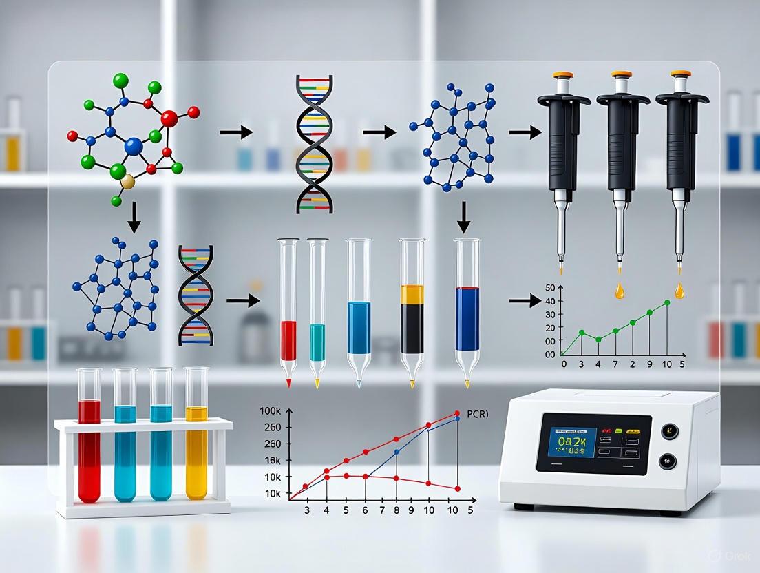
The following diagram illustrates the core ddPCR workflow, from sample partitioning to absolute quantification.
Quantitative Performance: dPCR vs. qPCR
The technical differences between qPCR and dPCR translate into significant performance advantages for dPCR in specific applications. The following table summarizes the key characteristics of each technology.
Table 1: Comparative Analysis of qPCR and dPCR/ddPCR
| Feature | Quantitative Real-Time PCR (qPCR) | Digital/Droplet Digital PCR (dPCR/ddPCR) |
|---|---|---|
| Quantification Method | Relative (requires standard curve) | Absolute (no standard curve) [4] [1] |
| Sensitivity | Distinguishes twofold changes | Detects differences <30%; identifies alleles <0.1% [4] |
| Robustness to Inhibitors | Sensitive to PCR inhibitors | Less sensitive due to partitioning [4] [1] |
| Precision | Lower precision for small copy number variations | High precision; measures single copy number variations [3] [4] |
| Data Output | Cycle threshold (Ct) value | Copies per microliter (copies/μL) [4] |
| Dynamic Range | Wider dynamic range | Comparable, but requires dilution for high-concentration targets [1] |
| Throughput | High | Evolving, but can be lower than qPCR [1] |
| Cost | Lower consumable costs | Higher consumable costs [1] |
Recent studies directly compare the performance of dPCR and qPCR in clinical scenarios. A 2025 study on respiratory virus diagnostics during the 2023–2024 tripledemic found that dPCR demonstrated superior accuracy, particularly for high viral loads of influenza A, influenza B, and SARS-CoV-2 [6]. dPCR also showed greater consistency and precision than Real-Time RT-PCR, especially in quantifying intermediate viral levels in complex sample matrices like nasopharyngeal swabs [6].
Furthermore, comparative studies of dPCR platforms themselves are essential for robust data interpretation. A 2025 study in Scientific Reports compared the QX200 ddPCR system (Bio-Rad) with the QIAcuity One ndPCR system (QIAGEN). It found that while both platforms had similar limits of detection (LOD) and quantification (LOQ), their precision could be influenced by factors such as the choice of restriction enzyme, highlighting the importance of assay optimization [3].
Table 2: Performance Metrics from Recent Comparative Studies
| Performance Metric | qPCR / Real-Time RT-PCR | dPCR (Nanoplate-based) | ddPCR (Droplet-based) |
|---|---|---|---|
| Limit of Detection (LOD) | Varies with assay and standard curve | ~0.39 copies/μL input [3] | ~0.17 copies/μL input [3] |
| Limit of Quantification (LOQ) | Varies with assay and standard curve | ~1.35 copies/μL input [3] | ~4.26 copies/μL input [3] |
| Accuracy in Viral Load Quantification | High, but challenged by co-infections and inhibitors [6] | Superior for medium loads of RSV and high loads of other viruses [6] | N/A in cited sources |
| Precision (Coefficient of Variation) | Higher variability, especially with inhibitors [4] | High precision; CVs can be <5% with optimized assays [3] | High precision; CVs can be <5% with optimized assays [3] |
Experimental Protocol: Droplet Digital PCR for Rare Mutation Detection
The following detailed protocol, adapted from a visualized experimental method, outlines the key steps for using ddPCR to detect rare tumor mutations, a common application that leverages the technology's high sensitivity [2].
Research Reagent Solutions and Materials
Table 3: Essential Materials for ddPCR Experiments
| Item | Function |
|---|---|
| ddPCR Master Mix | Contains DNA polymerase, dNTPs, buffer, and other core PCR reagents. |
| Fluorogenic Probe Assays | Target-specific primers and fluorescently labeled probes (e.g., FAM, HEX) for detection. |
| Droplet Stabilizing Oil | Creates an immiscible phase for generating stable, uniform droplets. |
| Restriction Enzymes (e.g., HaeIII) | Can be added to increase amplicon accessibility and improve precision [3] [5]. |
| DNA Sample | The template nucleic acid of interest, which must be of high quality and accurately quantified. |
| Droplet Generator Cartridge | A specialized microfluidic chip used to partition the sample into nanodroplets. |
| Thermal Cycler | Instrument for performing end-point PCR amplification under precise temperature control. |
| Droplet Reader | Instrument that flows droplets single-file past a laser to detect fluorescence in each droplet. |
Step-by-Step Methodology
Preparation and Setup:
- Decontaminate the workspace and equipment with 10% bleach followed by 70% ethanol.
- Gently vortex and briefly centrifuge all reagents, except the droplet-stabilizing oil.
- Prepare the ddPCR reaction mixture by combining the master mix, fluorescently labeled oligonucleotide probes, and other required reagents.
- Add the prepared DNA sample to the reaction mixture. Pipette the entire volume multiple times (approximately 10x) to ensure thorough mixing. It is critical to avoid air bubbles.
Droplet Generation:
- Transfer the entire reaction mixture into the sample wells of a specialized droplet generator cartridge.
- Load a fresh PCR strip tube into the droplet generator instrument and place the cartridge.
- Initiate the droplet generation run. The instrument will combine the sample mixture with oil to form an emulsion of tens of thousands of nanoliter-sized droplets, each acting as an independent microreactor. This process partitions the DNA molecules randomly into the droplets.
End-Point PCR Amplification:
- Once droplet generation is complete, transfer the emulsion to a standard PCR tube strip and seal it.
- Place the tube strip in a thermal cycler and run a standard PCR protocol optimized for the specific target. The cycling conditions (denaturation, annealing, extension) are performed to endpoint, amplifying the target DNA within each droplet.
Droplet Reading and Data Analysis:
- After thermal cycling, transfer the PCR tube strip to the droplet reader.
- The droplet reader aspirates the emulsion and streams the droplets single-file past a fluorescence detector. The detector measures the fluorescence in each droplet, classifying it as positive (contains the target mutation) or negative (does not contain the target).
- The instrument software uses Poisson statistics to analyze the fraction of positive droplets and calculate the absolute concentration of the target DNA (in copies per microliter) in the original sample.
Applications and Future Directions in Research and Drug Development
The unique advantages of ddPCR have made it indispensable in several advanced research and clinical applications.
Precision Oncology and Rare Mutation Detection: ddPCR's ability to detect mutant allele frequencies below 0.1% makes it a powerful tool for cancer biomarker discovery, monitoring minimal residual disease (MRD), and detecting resistance mutations in liquid biopsies [4] [2]. Its high precision allows for tracking minute changes in tumor DNA concentration over time.
Cell and Gene Therapy Development: ddPCR is widely used to quantify the titer of lentiviral vectors and to determine the vector copy number (VCN) in transduced cells, a critical quality control step in therapeutic development [5]. Its absolute quantification eliminates the need for variable standard curves, improving reproducibility across runs and operators [5].
Infectious Disease Diagnostics and Viral Load Monitoring: dPCR demonstrates superior accuracy in quantifying viral loads, as seen in respiratory virus studies [6]. This is crucial for understanding disease progression, transmissibility, and treatment efficacy. Its robustness to inhibitors also makes it valuable for direct testing in complex clinical samples.
Environmental Monitoring and Water Microbiology: dPCR is increasingly applied in environmental surveillance, such as wastewater-based epidemiology and monitoring waterborne pathogens, due to its sensitivity and ability to detect low-abundance targets in complex sample matrices [1].
The future of dPCR technology points toward increased multiplexing capabilities, allowing for the simultaneous quantification of multiple targets from a single sample [5]. This is particularly relevant for complex characterizations, such as analyzing multiple components of a lentiviral construct in gene therapy. Further integration with automation and next-generation sequencing (NGS) for validation will also expand its utility in both research and clinical diagnostics [1].
Quantitative PCR (qPCR) has long been the gold standard for nucleic acid detection and quantification. However, this technique relies on measuring amplification cycles relative to standard curves, making it susceptible to artifacts from sample contaminants and amplification inefficiencies that can compromise data accuracy and reproducibility [7]. Droplet Digital PCR (ddPCR) represents a fundamental shift in this paradigm by harnessing the power of microfluidic partitioning to create thousands of nanoliter-sized reactors that enable absolute nucleic acid quantification without standard curves [8] [9].
This revolutionary approach transforms biological quantification from analog measurements vulnerable to environmental interference to robust digital assays that generate binary, counting-based data. The core innovation lies in microfluidic partitioning, where samples are divided into thousands to millions of discrete droplets, each functioning as an independent PCR reactor [8]. This partitioning enables a fundamental shift from relative quantification based on amplification kinetics to absolute quantification based on Poisson statistics of positive versus negative reactions [9].
The implications of this technology are particularly profound for applications requiring exceptional sensitivity and precision, including liquid biopsy testing, pathogen detection, rare mutation identification, and gene expression analysis of low-abundance targets [9]. By distributing target molecules across numerous partitions, ddPCR achieves a significantly improved signal-to-noise ratio, enabling detection of rare mutations and minor fold changes that would be indistinguishable from technical artifacts using traditional qPCR [7].
Core Principles: The Science of Partitioning
Microfluidic Foundations of Partitioning
Microfluidics, the science of manipulating fluids at the microscale, enables the creation of these nano-reactors through sophisticated channel designs that exploit the unique physical properties of fluids at microscopic dimensions [10]. At these scales, fluid behavior is governed primarily by viscous forces rather than inertia, resulting in strictly laminar flow regimes where mixing occurs predominantly through molecular diffusion rather than turbulence [11] [12]. This predictable fluid behavior allows for precise control over reaction conditions and enables the generation of highly monodisperse droplets.
The creation of these nano-reactors employs two primary microfluidic approaches. In continuous-flow microfluidics, fluids are precisely directed through microchannels where hydrodynamic focusing techniques compress a central sample stream with outer sheath flows, dramatically reducing diffusion distances and mixing times to as little as 50 microseconds [12]. Alternatively, droplet-based microfluidics utilizes flow-focusing geometries where immiscible fluids intersect, breaking the aqueous sample stream into discrete picoliter-volume droplets suspended in oil [8]. This latter approach forms the basis for most commercial ddPCR systems, generating approximately 20,000 individual reaction droplets from a single sample [13].
The Digital PCR Principle: From Analog to Digital Detection
The fundamental principle of digital PCR involves partitioning a PCR reaction into thousands of individual reactions such that each partition contains zero, one, or several target nucleic acid molecules [9]. Following PCR amplification, each partition is analyzed for the presence (positive) or absence (negative) of fluorescence signal, creating a binary digital readout [9]. The absolute concentration of the target nucleic acid in the original sample is then calculated using Poisson statistics based on the ratio of positive to negative partitions [8] [9].
The mathematical foundation for this quantification relies on the Poisson distribution, which describes the probability of a given number of events occurring in a fixed interval of space or time when these events occur with a known constant rate and independently of the time since the last event. In ddPCR, the Poisson model determines the probability of a partition receiving k copies of the target molecule according to the formula:
[P(k) = \frac{\lambda^k e^{-\lambda}}{k!}]
Where λ represents the average number of target molecules per partition, and k is the actual number of target molecules in a given partition [9]. For optimal quantification, the partitioning is designed such that λ is low enough (typically <1) to ensure that most positive partitions contain only a single target molecule, enabling precise absolute quantification without competition effects [9].
Table 1: Key Advantages of Partitioning in Digital PCR
| Advantage | Technical Basis | Application Benefit |
|---|---|---|
| Absolute Quantification | Eliminates need for standard curves through direct counting | Improved reproducibility across laboratories [9] |
| Enhanced Sensitivity | Partitions increase effective concentration of rare targets | Detection of rare mutations and low-abundance targets [9] |
| Superior Precision | Thousands of data points from a single sample | Identification of small fold changes (<2x) with statistical significance [9] [7] |
| High Tolerance to Inhibitors | Dilution of inhibitors across partitions, endpoint measurement | Reliable results from complex samples (e.g., stool, blood, wastewater) [14] [9] |
| Rare Allele Detection | Statistical enrichment through partitioning | Identification of mutations present at <0.1% frequency [9] |
The ddPCR Workflow: From Sample to Answer
Microfluidic Droplet Generation
The ddPCR process begins with the preparation of a conventional PCR reaction mixture containing template DNA, primers, probes, nucleotides, and DNA polymerase. This mixture is then loaded into a droplet generator cartridge along with droplet generation oil [13]. Within the microfluidic cartridge, a flow-focusing nozzle creates a water-in-oil emulsion by vacuum-driven manipulation of the aqueous and oil phases [13]. This process generates approximately 20,000 uniform droplets per sample, with each droplet measuring about 1 nL in volume and serving as an independent PCR reactor [13]. The stability of this emulsion is critical to maintaining partition integrity throughout the amplification process, preventing droplet coalescence or disintegration.
Endpoint PCR Amplification
Following droplet generation, the emulsion is transferred to a 96-well PCR plate, sealed, and placed in a conventional thermal cycler [13]. The PCR amplification follows standard temperature cycling protocols (e.g., initial denaturation at 95°C for 10 minutes, followed by 40-45 cycles of denaturation at 95°C for 15 seconds and annealing/extension at 60°C for 60 seconds) [13]. Unlike qPCR, which monitors amplification in real-time, ddPCR uses endpoint detection, where the final fluorescence of each droplet is measured after amplification completion [7]. This approach eliminates dependence on amplification efficiency, as the critical factor becomes whether a droplet crossed the fluorescence threshold during amplification, not how efficiently it did so [7].
Droplet Reading and Data Analysis
After thermal cycling, the plate is transferred to a droplet reader that sequentially aspirates each sample [13]. The reader flows the droplets in a single file past a dual-color optical detection system that measures the fluorescence of each droplet [7]. The raw fluorescence data is processed by sophisticated algorithms that classify droplets as positive or negative based on their fluorescence amplitude [13]. Specialized software, such as the open-access tool "definetherain," applies clustering algorithms (k-nearest neighbor) to improve accuracy in distinguishing positive from negative droplets, particularly important when analyzing samples with low target concentrations [13]. The absolute quantification of target molecules is then automatically calculated using Poisson statistics, providing concentration measurements in copies per microliter [9].
Diagram 1: Complete ddPCR workflow from sample preparation to absolute quantification
Performance Comparison: ddPCR vs. qPCR
Quantitative Performance Metrics
Direct comparisons between ddPCR and qPCR reveal distinct performance advantages depending on application requirements. For samples with low levels of contaminants and moderate-to-high target concentrations, both technologies demonstrate comparable performance [7]. However, in challenging applications involving low abundant targets (Cq ≥ 29) and/or variable amounts of chemical and protein contaminants, ddPCR generates more precise, reproducible, and statistically significant results [7]. This enhanced performance is particularly evident in applications requiring detection of small expression differences (2-fold or lower), where ddPCR's partitioning approach provides superior precision [7].
The tolerance to PCR inhibitors represents another significant advantage of ddPCR. Studies have demonstrated that while the reverse transcription (RT) mix components can inhibit Taq polymerase and alter qPCR reaction efficiency (from 89.6% to 67.1% with increasing RT contamination), ddPCR maintains quantification accuracy despite the presence of these inhibitors [7]. This resilience stems from the endpoint detection methodology and the dilution of inhibitors across thousands of partitions, reducing their local concentration and mitigating interference with amplification [9].
Application-Specific Performance
The application of ddPCR for SARS-CoV-2 detection in complex sample matrices demonstrates its practical advantages. In a comprehensive analysis of stool and urine samples from COVID-19 patients, ddPCR demonstrated 100% detection rate in stool samples and 27.1% in urine samples, outperforming traditional qRT-PCR approaches, particularly for samples with low viral target concentrations [14]. Similarly, in food microbiology, ddPCR enabled specific detection of Lacticaseibacillus casei with a sensitivity of 100 CFU/mL, superior to real-time PCR in detecting low bacterial concentrations in spiked milk samples [15].
Table 2: Technical Comparison of ddPCR and qPCR Performance Characteristics
| Parameter | Droplet Digital PCR (ddPCR) | Quantitative PCR (qPCR) |
|---|---|---|
| Quantification Method | Absolute counting via Poisson statistics | Relative quantification based on standard curves [9] |
| Detection Principle | Endpoint measurement [7] | Real-time monitoring of amplification [7] |
| Precision at Low Targets | Superior precision for low copy numbers and small fold changes [7] | Variable results for low targets, highly dependent on reaction efficiency [7] |
| Tolerance to Inhibitors | High tolerance due to partitioning and endpoint detection [9] [7] | Sensitive to inhibitors that affect amplification efficiency [7] |
| Dynamic Range | Limited by number of partitions [9] | Broader dynamic range [9] |
| Data Reproducibility | High inter-laboratory reproducibility [9] | More variable between laboratories and instruments [7] |
| Optimal Application | Rare mutation detection, copy number variation, viral load in complex samples [14] [9] | High-throughput screening, expression analysis of abundant targets [7] |
Essential Reagents and Materials
Successful implementation of ddPCR requires specific reagents and materials optimized for microfluidic partitioning and droplet stability. The core components include:
Table 3: Essential Research Reagent Solutions for ddPCR Experiments
| Reagent/Material | Function | Specification Notes |
|---|---|---|
| Droplet Generator Cartridge | Microfluidic chamber for droplet formation | Compatible with specific ddPCR instrumentation [13] |
| Droplet Generation Oil | Creates water-in-oil emulsion for partitioning | Formulated for stable droplet formation and thermal stability [13] |
| ddPCR Supermix | Optimized PCR reagents for droplet reactions | Contains DNA polymerase, dNTPs, buffers with enhanced droplet compatibility [13] |
| Target-Specific Primers/Probes | Sequence-specific amplification | Similar design requirements as qPCR but require validation for droplet partitioning [13] |
| Detection Reagents | Fluorescent probes for target detection | FAM, HEX/VIC probes most common; compatible with droplet environment [13] |
| Positive/Negative Controls | Assay validation and quality control | Essential for establishing fluorescence thresholds [13] |
Implementation Protocols: Methodologies for Robust ddPCR
Sample Preparation and Assay Optimization
Proper sample preparation is critical for successful ddPCR applications. Nucleic acid extraction should be performed using methods that maximize yield while minimizing inhibitors. For difficult sample matrices such as stool, the recommended protocol involves creating 30% fecal suspensions in phosphate-buffered saline (PBS) followed by centrifugation at 4000 rpm for 10 minutes to remove debris [14]. For urine samples, processing includes centrifugation at 500 g for 20 minutes at 4°C to pellet cellular material before nucleic acid extraction [14]. RNA extraction is typically performed using spin column-based kits specifically validated for the sample type [14].
Assay optimization requires careful validation of primer and probe concentrations. A standard 20 μL ddPCR reaction typically contains 8 μL of 2× ddPCR Supermix, 400 nM of forward and reverse primers, and 125 nM of probe [13]. However, these concentrations may require optimization for specific targets. The development of specific genetic markers through pangenome analysis has been shown to improve specificity, particularly for closely related species where conventional markers like 16S rRNA may fail to provide sufficient discrimination [15].
Data Analysis and Interpretation
Robust data analysis is essential for accurate ddPCR quantification. The initial step involves defining the threshold between positive and negative droplets, which can be challenging with low target concentrations or in the presence of inhibitors [13]. Commercial software typically provides automated thresholding, but manual adjustment or advanced algorithms may be necessary for optimal performance. The open-source tool "definetherain" implements k-nearest neighbor clustering to improve droplet classification accuracy, particularly important for samples with low target numbers where traditional thresholding may fail [13].
The application of Poisson statistics accounts for the possibility that some positive partitions may contain more than one target molecule. The concentration calculation uses the formula:
[c = \frac{-\ln{(N{\text{neg}}/N)}}{V{\text{droplet}}}]
Where (N{\text{neg}}) is the number of negative droplets, (N) is the total number of valid droplets, and (V{\text{droplet}}) is the volume of each droplet [13]. This calculation provides the absolute concentration in copies per microliter, which can be extrapolated to determine the total target molecules in the original sample.
Droplet Digital PCR represents a transformative approach to nucleic acid quantification that leverages microfluidic partitioning to create thousands of nano-reactors. This technology enables absolute quantification with exceptional precision, sensitivity, and robustness to inhibitors, addressing fundamental limitations of traditional qPCR. The partitioning principle distributes target molecules across discrete reaction compartments, effectively enriching rare targets and diluting inhibitors, while the digital counting approach provides direct absolute quantification without reference standards.
As microfluidic technologies continue to advance, further miniaturization of partitions will enable even greater sensitivity and precision while reducing reagent consumption and costs. The integration of ddPCR with emerging applications in liquid biopsy, infectious disease monitoring, and single-cell analysis underscores its growing importance in both research and clinical diagnostics. By harnessing the power of partitioning, ddPCR has established a new paradigm in molecular quantification that will continue to drive innovations across biological research and medical diagnostics.
Droplet Digital PCR (ddPCR) represents a paradigm shift in nucleic acid quantification, enabling absolute target measurement without reliance on external standards. This precision is powered not by novel biochemistry, but by the application of Poisson statistics to a massively partitioned reaction system. This technical guide explores the mathematical engine of absolute quantification, detailing how the random distribution of target molecules into thousands of nanodroplets, combined with Poisson probability law, allows researchers to achieve unparalleled accuracy, sensitivity, and precision in molecular quantification. Framed within broader ddPCR research, this whitepaper provides researchers, scientists, and drug development professionals with the statistical foundations, practical protocols, and analytical frameworks essential for leveraging this powerful technology.
The evolution of polymerase chain reaction (PCR) technology from conventional to digital formats has transformed nucleic acid quantification from a relative measurement to an absolute count. While quantitative real-time PCR (qPCR) quantifies nucleic acids relative to a standard curve, introducing potential variability due to differences in amplification efficiency between samples and standards, digital PCR (dPCR) and its droplet-based variant (ddPCR) achieve absolute quantification by dividing a single sample into thousands to millions of individual partitions [16]. This partitioning creates a digital assay where each compartment functions as an independent PCR microreactor. After amplification, the simple binary counting of positive versus negative partitions, analyzed through Poisson statistics, yields an absolute count of target molecules in the original sample, eliminating the need for standard curves and providing inherently calibrated results [17] [9].
The Principle of Poisson Distribution in ddPCR
Foundations of the Poisson Model
In ddPCR, the sample is partitioned into a large number (n) of droplets, typically 20,000 in systems like the Bio-Rad QX100 [17] [18]. The target molecules, numbering m in the original sample, are randomly distributed throughout these partitions. The average number of target molecules per droplet (λ) is given by λ = m/n. The Poisson distribution describes the probability of finding k target molecules in any given droplet when the molecules are distributed randomly and independently [16]. The fundamental equation is:
P(k) = (λ^k * e^(-λ)) / k!
Where:
- P(k) is the probability that a droplet contains exactly k target molecules
- λ is the average number of target molecules per droplet
- e is the base of the natural logarithm (approximately 2.71828)
- k! is the factorial of k
For quantification, the most critical probability is P(0), the fraction of droplets expected to contain zero target molecules: P(0) = e^(-λ) [16]. After the amplification reaction, the droplet reader counts the total number of droplets (N) and the number of positive droplets (Npos). The fraction of negative droplets (Nneg/N) provides an experimental estimate of P(0). Rearranging the equation gives: λ = -ln(Nneg/N) = -ln(1 - Npos/N), where ln is the natural logarithm [18] [16]. This calculated λ represents the average number of target molecules per droplet, from which the absolute concentration in the original sample is directly derived.
Conceptual Workflow of ddPCR
The following diagram illustrates the complete ddPCR workflow, from sample partitioning to final quantification, highlighting where Poisson statistics are applied:
Diagram 1: The ddPCR workflow, highlighting the statistical analysis phase. The process transforms a sample into thousands of individual reactions, whose binary results (positive/negative) are analyzed via Poisson statistics to achieve absolute quantification.
Statistical Analysis and Quantification Accuracy
Precision and Confidence Intervals
The precision of ddPCR quantification is statistically defined and depends primarily on the total number of partitions analyzed and the value of λ [16]. The fundamental limit of precision is governed by Poisson statistics, where the best possible relative standard deviation (RSD) is 1/√m, with m being the total number of template molecules measured [19]. This relationship means precision improves as the number of target molecules increases.
The confidence in the estimated target concentration is maximized when the dynamic range of the assay is appropriately utilized. Intuitively, confidence is lowest when most partitions are either empty or full, as this pattern provides less statistical information. With 10,000 or more partitions, optimal precision is achieved at a λ value of approximately 1.6, which corresponds to about 20% of partitions being negative [16]. At this λ value, the Poisson distribution provides the most statistically robust quantification.
Dynamic Range and Optimization
The following table summarizes the relationship between λ values, partition status, and quantification confidence:
Table 1: Poisson Distribution Characteristics at Different λ Values
| λ value | Droplets with 0 targets | Droplets with 1 target | Droplets with ≥2 targets | Confidence in Quantification |
|---|---|---|---|---|
| λ = 0.1 | ~90% | ~9% | ~1% | Low (Too many empty droplets) |
| λ = 0.5 | ~61% | ~30% | ~9% | Medium |
| λ = 1.6 | ~20% | ~32% | ~48% | Optimal [16] |
| λ = 5.0 | <1% | ~3% | ~96% | Low (Too many saturated droplets) |
Statistical methods for calculating confidence intervals include the Wald method (which approximates the binomial distribution with a normal distribution) and the preferred Wilson method, which provides more accurate results when most partitions are empty or filled [16]. The Wilson confidence interval is calculated as:
CI = [p + α²/2n ± α√(p(1-p)/n + α²/4n²)] / (1 + α²/n)
Where p is the fraction of positive partitions, n is the total number of partitions, and α is 1.96 for a 95% confidence interval [16].
Experimental Protocol for ddPCR
Detailed Workflow for Absolute Quantification
The ddPCR workflow follows a standardized process that can be adapted for various applications including copy number variation (CNV) analysis, rare mutation detection, and transcript quantification [18].
1. Reaction Setup Prepare a 20-25μL reaction mixture containing:
- 2× ddPCR Supermix: Contains buffer, DNA polymerase, and dNTPs optimized for droplet generation [18] [20]
- 20× Primer/Probe Mix: Target-specific primers and fluorescent probes (typically FAM and VIC/HEX for multiplexing) [18]
- Template DNA: 1-100 ng of high-quality DNA, potentially digested with restriction enzymes (e.g., AluI) to reduce viscosity and separate linked duplicates [18]
2. Droplet Generation
- Load the reaction mixture into a droplet generator cartridge with 70μL of droplet generation oil [20]
- The microfluidic chip creates ~20,000 nanoliter-sized droplets using a water-oil emulsion system [17]
- Transfer the generated droplets to a 96-well PCR plate and seal with pierceable foil [18]
3. PCR Amplification
- Perform endpoint PCR amplification on a thermal cycler (typically 40 cycles) [17]
- Optimize annealing/extension temperature for specific primer/probe sets [18]
4. Droplet Reading and Analysis
- Load the plate into a droplet reader that counts each droplet serially [17]
- The reader identifies positive (fluorescent) and negative (non-fluorescent) droplets for each channel [18]
- Software applies Poisson statistics to calculate the absolute concentration of the target sequence in copies/μL [17] [9]
Essential Research Reagent Solutions
Table 2: Key Reagents and Materials for ddPCR Experiments
| Reagent/Equipment | Function | Application Notes |
|---|---|---|
| ddPCR Supermix | Provides optimized buffer, enzymes, and nucleotides for droplet-based PCR | Essential for proper droplet formation; contains dUTP for contamination control [18] [20] |
| TaqMan Probes | Sequence-specific fluorescent detection with 5' fluorophore and 3' quencher | Design for 60-150 bp amplicons; Tm ~8-10°C higher than primers [18] |
| Droplet Generation Oil | Creates water-oil emulsion for partitioning | Forms stable, uniform droplets; specific to ddPCR system [20] |
| Restriction Enzymes (e.g., AluI) | Digests genomic DNA to reduce viscosity and separate linked duplicates | Improves partitioning efficiency; avoid enzymes that cut within amplicon [18] |
| Droplet Generator | Microfluidic device creating ~20,000 droplets from sample | Creates uniform nanoliter-sized droplets [17] [20] |
| Droplet Reader | Analyzes fluorescence in each droplet post-amplification | Distinguishes positive/negative droplets; typically two-color detection [18] |
Comparison with qPCR and Applications
Fundamental Differences Between dPCR and qPCR
The statistical framework of ddPCR creates distinct advantages and disadvantages compared to traditional qPCR:
Table 3: dPCR vs. qPCR Comparison
| Parameter | Digital PCR (dPCR) | Quantitative PCR (qPCR) |
|---|---|---|
| Quantification Basis | Absolute count via Poisson statistics | Relative to standard curve |
| Signal Measurement | End-point (binary) | Real-time (continuous) |
| Precision Source | Number of partitions | Amplification efficiency |
| Dynamic Range | Limited by partition count [9] | Typically broader [9] |
| Inhibitor Tolerance | Higher due to partitioning [8] [16] | Lower |
| Sensitivity | Superior for rare variants [21] [9] | Limited for low-abundance targets |
| Standard Curve | Not required [17] [9] | Required |
Key Applications Leveraging Poisson Statistics
The unique capabilities of ddPCR make it particularly valuable for:
- Rare Mutation Detection: Partitioning effectively enriches rare alleles by separating them from wild-type sequences, enabling detection of mutations at frequencies as low as 0.001% [21] [9].
- Copy Number Variation (CNV) Analysis: Absolute quantification allows precise determination of gene copy numbers without reference standards, using ratio of target to reference genes [18].
- Liquid Biopsy Applications: High sensitivity enables detection of circulating tumor DNA in oncology and non-invasive prenatal testing [22] [8].
- Pathogen Detection and Quantification: Precise absolute quantification benefits viral load monitoring and detection of low-abundance pathogens like Mycobacterium tuberculosis [23].
- Gene Expression Analysis: Absolute quantification of transcripts without reference genes, particularly valuable for low-abundance mRNA targets [18] [9].
Limitations and Practical Considerations
Despite its powerful statistical foundation, ddPCR has limitations that researchers must consider:
- Dynamic Range Constraints: The finite number of partitions (typically 20,000 in many systems) limits the maximum number of molecules that can be accurately quantified without sample dilution [9].
- Sample Volume Limitations: At very low concentrations, precision is limited by the total sample volume processed, potentially giving qPCR an advantage in these specific scenarios [19].
- Technical Artifacts: DNA denaturation during partitioning can cause strand separation into different droplets, potentially leading to overestimation [9]. Template linkage, partition volume variance, and "molecular dropout" can cause underestimation [9].
- Throughput and Cost: While improving, ddPCR throughput may be lower than qPCR, and consumable costs can be higher for some applications [8].
The following diagram illustrates the core relationship between sample partitioning and statistical analysis:
Diagram 2: The core principle of Poisson analysis in ddPCR. The random distribution of target molecules into partitions enables back-calculation of the original concentration through Poisson statistics.
Poisson statistics provides the mathematical foundation that enables ddPCR to achieve absolute quantification of nucleic acids, transforming how researchers measure DNA, RNA, and rare genetic variants. By understanding and properly applying these statistical principles, researchers can design experiments that maximize precision, accurately interpret results with appropriate confidence intervals, and leverage the full potential of digital PCR technology across diverse applications from basic research to clinical diagnostics. As ddPCR continues to evolve with increasing partition numbers and improved workflows, its statistical engine will remain central to its unique capability to deliver absolute molecular counts that are redefining measurement standards in molecular biology.
The advent of the Polymerase Chain Reaction (PCR) revolutionized molecular biology by enabling exponential amplification of specific DNA sequences. However, accurate quantification of nucleic acids remained challenging with conventional PCR methods. The evolution from digital PCR (dPCR) to droplet digital PCR (ddPCR) represents a paradigm shift in nucleic acid quantification, moving from relative to absolute measurement without requiring standard curves. This transformation began with fundamental limiting dilution concepts in the late 1980s and early 1990s [24]. These early approaches recognized that by partitioning nucleic acids to single-molecule levels, researchers could apply Poisson statistics to achieve precise quantification—a principle that would eventually form the foundation of modern ddPCR. The journey from these rudimentary beginnings to today's sophisticated microfluidic platforms reflects how technological innovation has dramatically expanded our capability to detect and quantify genetic targets with unprecedented precision across diverse fields including virology, oncology, and genetic disease research [25] [26].
This technical guide examines the historical development, methodological principles, and practical applications of ddPCR within the broader context of how this technology enables advanced molecular analysis. By tracing its evolution from early digital PCR concepts to current state-of-the-art implementations, we provide researchers with comprehensive insights into both the theoretical foundations and practical implementation of this transformative technology.
Historical Development and Technological Evolution
Early Foundations: Limiting Dilution PCR (1990-1999)
The conceptual foundation of digital PCR emerged not as a single discovery but through independent developments across multiple research fields. In 1990, Simmonds et al. pioneered what was then termed "limiting dilution PCR" to quantify HIV provirus molecules in blood samples from infected individuals [24]. This approach involved performing PCR on replicate samples at limiting dilutions, then applying Poisson statistics to calculate original target concentrations based on the proportion of positive amplifications. Concurrently, other research groups were developing similar methodologies under different names including "single molecule PCR," with Jeffreys et al. applying the technique to minisatellite evolution studies and Ruano et al. utilizing it for haplotyping analysis [24].
These early implementations shared a common principle: sample partitioning to endpoint dilution where each reaction contained zero or one target molecule, followed by binary detection (positive/negative) of amplification products. While revolutionary in concept, these methods faced significant practical limitations as open-system approaches requiring manual sample partitioning, typically using multi-well plates with limited partition numbers [24]. The labor-intensive nature of these protocols, combined with contamination risks from post-amplification processing, restricted their widespread adoption despite demonstrated utility in sensitive applications such as monitoring minimal residual disease in leukemia patients [24].
Conceptual Formalization: Digital PCR (1999)
The field reached a pivotal moment in 1999 when Vogelstein and Kinzler formally introduced the term "digital PCR" in their landmark publication [24]. Their work demonstrated quantification of ras mutations by partitioning samples across 384-well plates, but more importantly, it established the digital quantification framework and terminology that would define the field. The term "digital PCR" effectively captured both the binary nature of the detection system (positive/negative reactions) and reflected the increasingly digital orientation of biological research at the time [24].
Despite this conceptual advance, practical limitations persisted. The method remained relatively laborious compared to emerging alternatives, particularly real-time quantitative PCR (qPCR) described by Heid et al. in 1996 [24]. The automation and closed-tube nature of qPCR systems addressed key limitations of early dPCR approaches, leading to a temporary decline in dPCR utilization between 2000-2002 as researchers embraced the more practical qPCR platform for routine quantification applications [24].
Technological Renaissance: Microfluidics and ddPCR (2007-Present)
The modern renaissance of digital PCR began around 2007, driven primarily by advances in microfluidics that addressed previous practical limitations [24]. The key innovation was the development of systems that could automatically partition samples into thousands of nanoliter-sized droplets, creating the water-in-oil emulsion reactors that define droplet digital PCR (ddPCR) [18]. This technological leap transformed dPCR from a specialized manual technique to a practical, high-throughput platform capable of precise absolute quantification.
The fundamental breakthrough was the creation of systems that could generate thousands to millions of uniform partitions without manual intervention, dramatically increasing partition numbers while reducing labor requirements [27]. Commercial platforms including Bio-Rad's QX series, RainDance Technologies' systems, and Fluidigm's BioMark HD enabled researchers to implement ddPCR workflows with practical turnaround times and minimal specialized training [28] [26]. This automation, combined with the inherent advantages of digital quantification, spurred exponential growth in ddPCR applications across diverse research areas from viral load monitoring to copy number variation analysis [24] [25].
Table 1: Historical Milestones in Digital PCR Development
| Year | Development | Key Innovators/Group | Significance |
|---|---|---|---|
| 1988 | Early single molecule amplification | Saiki et al. | Demonstrated PCR amplification of single β-globin molecules [24] |
| 1990 | Limiting dilution PCR for HIV quantification | Simmonds et al. | First application of Poisson statistics to quantify viral targets [24] |
| 1991 | Single molecule PCR for haplotyping | Ruano et al. | Applied partitioning for genetic analysis [24] |
| 1992 | Formalized limiting dilution PCR methodology | Research group | Established protocol for quantitative applications [24] |
| 1999 | "Digital PCR" term coined | Vogelstein & Kinzler | Introduced terminology and framework for digital quantification [24] |
| 2007+ | Microfluidic ddPCR systems | Multiple companies | Enabled automated partitioning, driving widespread adoption [24] |
Technical Principles and Methodological Advances
Fundamental Working Principles
Droplet digital PCR operates through a series of well-defined steps that transform sample analysis from analog to digital quantification. The process begins with sample partitioning, where each reaction mixture is divided into approximately 20,000 nanoliter-sized droplets using microfluidic technology [18]. This partitioning occurs through either passive methods like T-junction and flow-focusing geometries or active approaches using external forces, with polydimethylsiloxane (PDMS) being the most common material for droplet generation devices [26]. The stochastic distribution of nucleic acid molecules follows Poisson statistics, ensuring that each droplet contains zero, one, or a few target molecules based on their original concentration [18].
Following partitioning, PCR amplification proceeds within each droplet using standard thermal cycling conditions. The critical difference from conventional PCR lies in the detection method—rather than monitoring amplification in real-time, ddPCR uses endpoint detection with fluorescent TaqMan probes [18]. After amplification, droplets pass through a reader that measures fluorescence in two channels (typically FAM and VIC/VHEX), classifying each droplet as positive or negative for the target sequence [18]. The absolute concentration of the target nucleic acid is then calculated using Poisson statistics based on the ratio of positive to negative droplets, according to the formula: λ = -ln(1-p), where λ represents the average number of copies per droplet and p is the proportion of positive droplets [18]. This approach eliminates the need for standard curves and provides direct absolute quantification.
Comparative Analysis: ddPCR vs. qPCR
The fundamental differences between ddPCR and quantitative PCR (qPCR) translate to distinct performance characteristics that determine their appropriate applications. While qPCR measures amplification at the cycle threshold (Cq) during exponential phase and requires calibration curves for quantification, ddPCR's binary endpoint detection and statistical analysis provide direct absolute quantification without reference standards [7]. This distinction becomes particularly significant when analyzing samples with low target abundance or containing PCR inhibitors [7].
Research has demonstrated that for target sequences with low abundance (Cq ≥ 29) or in samples with variable amounts of chemical and protein contaminants, ddPCR generates more precise, reproducible, and statistically significant results [7]. The partitioning process in ddPCR effectively dilutes inhibitors across thousands of droplets, making the technology more tolerant to substances that would otherwise compromise qPCR reaction efficiency [7]. In copy number variation studies, ddPCR has shown 95% concordance with pulsed-field gel electrophoresis (considered a gold standard) compared to only 60% concordance for qPCR, with ddPCR results differing by just 5% on average from PFGE values while qPCR differed by 22% [25].
Table 2: Performance Comparison Between qPCR and ddPCR
| Parameter | qPCR | ddPCR |
|---|---|---|
| Quantification Method | Relative (requires standard curve) | Absolute (no standard curve) [18] |
| Detection Principle | Measurement during exponential phase (Cq) | Endpoint binary detection [18] [27] |
| Precision with Low Abundance Targets | Highly variable (Cq ≥ 29) [7] | Highly precise and reproducible [7] |
| Tolerance to Inhibitors | Low to moderate [7] | High (inhibitors diluted across partitions) [7] |
| Dynamic Range | ~5-6 logs | ~5 logs |
| Precision in CNV Analysis | 60% concordance with PFGE [25] | 95% concordance with PFGE [25] |
| Multiplexing Capability | Moderate | Available (4-12 targets with newer systems) [28] |
Experimental Protocols and Methodologies
Standard ddPCR Workflow for CNV Analysis
The application of ddPCR for copy number variation (CNV) analysis represents one of its most impactful uses, particularly for clinically relevant genes like DEFA1A3 where copy number ranges from 2-16 per diploid genome and correlates with disease susceptibility [25]. The following protocol, adapted from Bio-Rad's QX100 system, provides a robust framework for CNV determination [18]:
Step 1: DNA Preparation and Digestion Begin with 100 ng of genomic DNA, though the assay demonstrates a broad dynamic range from 10 pg to 350 ng. To reduce viscosity and separate closely linked duplications, perform enzymatic digestion with AluI (or another appropriate restriction enzyme that doesn't cut within the target amplicon). Combine 200 ng DNA diluted in nuclease-free water to 8.9 μL with 1 μL of 10× restriction enzyme buffer and 0.1 μL AluI enzyme (10,000 U/mL). Incubate at 37°C for at least 1 hour, then dilute the reaction 1:2 by adding 10 μL nuclease-free water to stop digestion and dilute buffer salts that might interfere with PCR [18].
Step 2: Reaction Assembly Assemble reactions in a 96-well plate with the following components: 12.5 μL of 2× ddPCR master mix (containing buffer, DNA polymerase, and dNTPs), 1.25 μL of 20× ROI target primer/TaqMan probe mix, 1.25 μL of 20× reference target (RPP30 recommended) primer/TaqMan probe mix, and 10 μL digested DNA diluted in nuclease-free water [18]. The total reaction volume of 25 μL includes excess to prevent air bubble formation during droplet generation. Centrifuge the plate briefly at 150 × g for 15 seconds to ensure contents settle at the bottom, then mix by pipetting 15-20 times to achieve a homogeneous mixture [18].
Step 3: Droplet Generation and PCR Amplification Transfer 20 μL of each reaction to DG8 droplet generator cartridges. Following droplet generation, carefully transfer the emulsified samples to a 96-well PCR plate. Seal the plate with heat-sealing foil and perform PCR amplification with the following thermal cycling conditions: 95°C for 10 minutes (enzyme activation), followed by 40 cycles of 94°C for 30 seconds (denaturation) and 60°C for 60 seconds (annealing/extension), with a final 98°C incubation for 10 minutes (enzyme deactivation) and infinite hold at 4°C [18].
Step 4: Droplet Reading and Data Analysis Load the PCR plate into the droplet reader, which processes each droplet individually, measuring fluorescence in both channels. The QuantaSoft software analyzes the data, applying Poisson statistics to calculate the absolute concentration of both target and reference genes. The copy number is determined from the ratio of target to reference concentrations, with the diploid reference gene expected to yield two copies per genome [18].
Critical Parameters for Assay Design
Successful ddPCR assays require careful attention to several technical parameters. TaqMan assays should amplify 60-150 bp fragments, with smaller products generally demonstrating superior amplification efficiency [18]. Primer melting temperature (Tm) typically targets 60°C, while the internal hybridization probe should have a Tm approximately 8-10°C higher than the primers [18]. Avoid designing probes with a 5' guanine (which can partially quench fluorescence) and homopolymer runs longer than 3 bases to minimize secondary structure formation [18].
For CNV analysis, ensure the region of interest (ROI) amplicon resides completely within the putative CNV, while the reference amplicon should target a stable diploid gene like RPP30 [18]. Utilize tools like RepeatMasker to avoid known repeats and perform in silico PCR verification to confirm single-product amplification [18]. When detecting rare variants or single nucleotide polymorphisms, consider adjusting the input DNA concentration to ensure appropriate numbers of positive droplets for statistical validity.
Research Reagent Solutions
Table 3: Essential Reagents and Materials for ddPCR Experiments
| Reagent/Material | Function | Application Notes |
|---|---|---|
| ddPCR Master Mix | Provides buffer, dNTPs, and hot-start DNA polymerase | Optimized for droplet generation; substitution may cause droplet failure [18] |
| TaqMan Probes | Sequence-specific fluorescent detection | FAM and VIC/HEX most common; avoid 5' guanine [18] |
| Restriction Enzymes (AluI) | DNA digestion to reduce viscosity | 4-cutter enzyme; verify absence of cut sites in amplicons [18] |
| Droplet Generation Oil | Creates water-in-oil emulsion for partitioning | Formulated for specific droplet generators [18] |
| DG8 Cartridges | Microfluidic chambers for droplet generation | Single-use components [18] |
| Detection Primers | Target amplification | Design for 60°C Tm; 60-150 bp products preferred [18] |
| Nuclease-Free Water | Reaction assembly | Ensures no enzymatic degradation of components [18] |
Current Applications and Future Perspectives
Diverse Research and Clinical Applications
The unique capabilities of ddPCR have enabled its application across diverse fields. In virology, ddPCR has proven invaluable for detecting low-level viral persistence, with researchers demonstrating that SARS-CoV-2 RNA detection in plasma correlates with clinical deterioration and predicts patient outcomes [27] [26]. The technology's precision at low target levels makes it particularly suitable for monitoring viral load in patients undergoing antiviral therapy and for detecting residual virus in vaccine production [26].
In oncology, ddPCR enables sensitive detection of rare mutations and copy number variations, with applications in cancer diagnostics, minimal residual disease monitoring, and therapy selection [24] [25]. The technology's ability to provide absolute quantification of transgene copies has also made it indispensable in biopharmaceutical development, where it's used to characterize manufacturing cell lines and ensure consistent production of biologic therapeutics [29]. Additionally, ddPCR plays a crucial role in quality control for cell and gene therapies, particularly for vector copy number quantification and residual DNA detection [28].
Emerging Trends and Future Directions
The future evolution of ddPCR technology focuses on several key areas. Integration with automated systems and development of higher-throughput platforms address current limitations in workflow efficiency, particularly for quality control applications in regulated environments [28]. Chip-based dPCR systems offer streamlined "sample-to-result" workflows that reduce hands-on time and contamination risk, making them particularly suitable for quality control environments [28].
Multiplexing capabilities continue to expand, with newer systems capable of detecting up to 12 targets simultaneously, enabling more comprehensive genetic analysis in limited sample volumes [28]. The combination of ddPCR with emerging techniques like next-generation sequencing creates powerful synergistic approaches, using ddPCR for validation and precise quantification of NGS findings [29]. As the technology matures, standardization of protocols and analytical validation for clinical applications will be essential for broader adoption in diagnostic settings [28] [29].
The evolution from digital PCR to modern ddPCR represents more than just technical refinement—it embodies a fundamental shift in how researchers approach nucleic acid quantification. By providing absolute quantification without standard curves, exceptional sensitivity for rare targets, and superior tolerance to inhibitors, ddPCR has overcome limitations that constrained earlier PCR technologies [7]. The historical journey from limiting dilution concepts in the 1990s to today's automated microfluidic systems demonstrates how parallel advances in engineering, biochemistry, and data analysis have converged to create a powerful platform that continues to expand scientific possibilities [24].
As ddPCR technology evolves, its integration with automated workflows, expansion of multiplexing capabilities, and validation for clinical applications will further solidify its role as an essential tool for life science research, drug development, and molecular diagnostics [28] [29]. The technology's ability to generate publication-quality data from challenging samples ensures its continued adoption across diverse fields, enabling researchers to address biological questions that were previously beyond the reach of molecular quantification methods [7].
ddPCR in Action: Standard Workflow and Transformative Applications
Droplet Digital PCR (ddPCR) is a third-generation polymerase chain reaction technology that enables absolute quantification of nucleic acids without the need for a standard curve [30] [17]. This technique provides superior sensitivity, precision, and accuracy compared to conventional quantitative PCR (qPCR) by combining microfluidic partitioning with Poisson statistics [25] [31]. The core principle involves distributing a PCR reaction mixture into thousands of nanoliter-sized water-in-oil droplets, effectively creating numerous independent PCR reactions [18]. After amplification, each droplet is analyzed individually to determine the fraction of positive reactions, allowing calculation of the absolute target concentration in the original sample [30]. This guide provides a comprehensive technical workflow for ddPCR, framed within broader research on its mechanisms and applications in clinical and scientific settings.
The complete ddPCR process transforms a bulk sample into thousands of individual data points for digital analysis. Partitioning divides the PCR reaction into 20,000 nanoliter-sized droplets using microfluidic technology [17]. Following partitioning, amplification occurs through standard thermal cycling, with each droplet functioning as an independent PCR reaction [18]. Finally, detection and analysis involve reading each droplet's fluorescence and applying Poisson statistics to determine the initial target concentration [30] [18]. This workflow enables precise absolute quantification, making it particularly valuable for detecting rare mutations, copy number variations, and low-abundance pathogens [25] [32] [33].
Detailed Experimental Protocol
Sample Preparation and Reaction Assembly
Proper sample preparation is critical for successful ddPCR analysis. Begin with DNA extraction using appropriate kits for your sample type (e.g., DNeasy Plant Mini Kit for plant tissues or PowerSoil Kit for soil samples) [33]. Assess DNA quality and concentration using spectrophotometry [33]. For genomic DNA, perform enzymatic digestion to reduce viscosity: combine 200 ng DNA, 1 μL 10× restriction enzyme buffer, and 0.1 μL AluI enzyme (10,000 U/mL) in 8.9 μL nuclease-free water [18]. Incubate at 37°C for ≥1 hour, then dilute 1:2 with nuclease-free water to stop the reaction and dilute buffer salts [18].
Assemble the PCR reaction in a 96-well plate with the following components:
- 12.5 μL of 2× ddPCR master mix
- 1.25 μL of 20× ROI target primer/probe mix
- 1.25 μL of 20× reference gene primer/probe mix
- 10 μL digested DNA diluted in nuclease-free water [18]
Centrifuge the plate briefly (15 seconds at 150 × g) to ensure contents settle at the bottom [18]. Mix reactions by pipetting 15-20 times to achieve a homogeneous mixture [18].
Table 1: Essential Research Reagent Solutions for ddPCR
| Reagent/Material | Function | Specifications |
|---|---|---|
| ddPCR Master Mix | Provides buffer, DNA polymerase, dNTPs for amplification | Must be optimized for droplet generation; Bio-Rad "Supermix for Probes" recommended [31] |
| Primer/Probe Mix | Target-specific amplification and detection | 20× concentration; Tm ~60°C for primers, +8-10°C for probes [18] |
| Restriction Enzyme (AluI) | Reduces DNA viscosity for better partitioning | 4-cutter (cuts every 256 bp on average); avoid sites in amplicon [18] |
| Droplet Generation Oil | Creates water-in-oil emulsion for partitioning | Contains surfactants for droplet stability [18] |
| DG8 Cartridges | Microfluidic chamber for droplet generation | Single-use consumable [18] |
Droplet Generation and PCR Amplification
Load 20 μL of the prepared reaction mixture into individual wells of a DG8 droplet generator cartridge [18] [17]. The droplet generator uses microfluidics and specific reagents to partition each sample into 20,000 nanoliter-sized droplets [17]. This process creates a water-in-oil emulsion where target molecules are randomly distributed among droplets according to Poisson distribution principles [30]. Proper droplet formation requires using the recommended ddPCR master mix, as substitutions may lead to droplet generation failure [18].
Transfer the generated droplets to a 96-well PCR plate and seal with pierceable foil using a heat sealer [18]. Perform PCR amplification using standard thermal cycling conditions. A typical protocol includes:
- Initial denaturation: 95°C for 10 minutes
- 40-45 cycles of:
- Denaturation: 94°C for 30 seconds
- Annealing/extension: 58°C for 1 minute (temperature varies based on primer Tm)
- Final extension: 98°C for 10 minutes
- Hold at 4°C [33]
Droplets remain stable throughout thermal cycling due to appropriate surfactant composition in the droplet generation oil [30].
Droplet Reading and Data Analysis
Following amplification, load samples into the droplet reader [17]. The reader processes droplets in a single file, passing each through a two-color detection system that measures fluorescence [17]. The reader classifies each droplet as positive (containing target) or negative (no target) based on fluorescence thresholds [18]. Data acquisition occurs at the endpoint, with each droplet providing a discrete fluorescent signal indicating target presence or absence [34].
Apply Poisson statistics to calculate target concentration using the formula: λ = -ln(1-p) where λ represents the average number of copies per droplet and p is the ratio of positive droplets to the total number of droplets [18]. Convert copies per droplet to copies per microliter using the known droplet volume (~1 nL) [18]. This statistical approach accounts for the random distribution of targets and enables absolute quantification without standard curves [17].
Table 2: Analytical Performance Metrics for ddPCR Assay Validation
| Performance Parameter | Assessment Method | Acceptance Criteria |
|---|---|---|
| Limit of Blank (LoB) | 60 measurements on blank samples | Define fluorescence threshold for background [33] |
| Limit of Detection (LoD) | Probit regression on 70 low-concentration measurements | Lowest concentration detectable with 95% CI [33] |
| Limit of Quantification (LoQ) | 20 measurements across serial dilutions | Lowest concentration with CV <25% [33] |
| Dynamic Range | Linear fit with 9 replicates per concentration | R² value demonstrating linearity [33] |
| Specificity | Testing against related species/pathogens | No cross-reactivity with non-targets [33] |
Data Visualization and Interpretation
Visualize results using software such as QuantaSoft, which provides multiple data representation options [18] [17]. The software typically displays droplet clusters in two-dimensional plots, differentiating positive and negative populations for each fluorescence channel [18]. This visualization allows for clear discrimination of target-containing droplets from background.
For copy number variation analysis, calculate the target concentration relative to the reference gene using the formula: CNV = (ROI copies/μL) / (REF copies/μL) × 2 [18] This calculation enables precise determination of copy number variations, with studies showing 95% concordance with gold standard methods like PFGE [25].
Applications and Advantages in Research
ddPCR provides significant advantages for clinical research and diagnostic applications. Its exceptional sensitivity enables rare variant detection at frequencies below 0.1%, making it invaluable for liquid biopsy applications in oncology [35]. The technology demonstrates superior tolerance to PCR inhibitors compared to qPCR, particularly beneficial when analyzing complex samples like soil, plant tissues, or clinical specimens [32] [33]. ddPCR also excels in absolute quantification without requiring standard curves, providing more reliable results for copy number variation analysis [25] [17].
Recent technological advancements include platforms supporting six-color multiplexing, portable systems for point-of-care testing, and integration of AI for automated droplet classification [35]. These developments expand ddPCR's utility across diverse fields including oncology, infectious disease surveillance, genetic disorder research, and environmental testing [32] [35]. The technology's precision, sensitivity, and reproducibility continue to drive its adoption in both research and clinical diagnostics [31] [34].
Droplet Digital PCR (ddPCR) represents a significant advancement in nucleic acid quantification, enabling absolute target measurement without reliance on standard curves. This technique partitions a single PCR sample into thousands of nanoliter-sized droplets, creating discrete reaction chambers where endpoint amplification occurs independently [17] [8]. The fundamental principle involves massive sample partitioning followed by Poisson statistical analysis to determine the absolute concentration of the target nucleic acid in the original sample [9]. Each droplet functions as an individual PCR reactor, with fluorescence detection post-amplification categorizing droplets as positive (containing at least one target molecule) or negative (containing no target molecules) [17]. This binary readout simplifies quantification and enhances precision, making ddPCR particularly valuable for detecting rare mutations, copy number variations, and low-abundance targets in complex biological samples [8] [9].
The ddPCR workflow overcomes limitations of traditional quantitative PCR (qPCR) by separating the amplification from the quantification step. While qPCR requires continuous monitoring of amplification curves and extrapolation from reference standards, ddPCR utilizes endpoint detection after full amplification cycles are complete [17]. This partitioning and endpoint detection approach provides ddPCR with higher tolerance to PCR inhibitors, as these interfering substances are diluted across thousands of individual droplets, reducing their effective concentration in any single reaction chamber [36] [9]. The technology's capacity for absolute quantification has established it as a powerful tool in clinical diagnostics, oncology, infectious disease monitoring, and gene expression analysis where precise nucleic acid quantification is critical [17] [36].
Technical Workflow of ddPCR
The droplet digital PCR process follows a meticulously optimized sequence from sample preparation through data analysis, with each stage critically influencing the final results. The workflow can be systematically divided into five key stages, each requiring specific reagents, instruments, and quality control measures to ensure accurate quantification.
Sample Preparation and Assay Design
Every ddPCR analysis begins with comprehensive sample preparation. The nucleic acid template (DNA, RNA, or cDNA) must be properly extracted and purified, with recommended removal of PCR inhibitors which can be achieved through sample dilution if necessary [17]. The reaction mixture is typically prepared in a 20-25µL volume containing ddPCR supermix, primers, and fluorescent probes [17] [37]. For probe-based detection, hydrolysis probes with a 5' fluorescent label and 3' quencher are commonly employed, similar to qPCR assays [37]. Assay design requires special consideration for clear differentiation between positive and negative droplets during endpoint analysis [17]. Template quality is paramount, with high-quality purified gDNA free from inhibitors recommended. For genomic DNA targets, restriction enzyme digestion may be necessary to ensure proper template partitioning, followed by heat inactivation and dilution [37]. Template quantity must be optimized based on target abundance, with systems like the Bio-Rad QX100 recommending approximately 100ng gDNA for single-copy targets to avoid saturation effects [37].
Droplet Generation and Partitioning
The prepared sample undergoes partitioning into nanoliter-sized droplets using microfluidic technology and a water-oil emulsion system [17] [8]. A standard ddPCR system creates approximately 20,000 independent partitions per sample [17] [37]. The droplet generator combines the aqueous PCR reaction mix with droplet generation oil and surfactants to form monodisperse droplets [37] [2]. This process requires specialized equipment such as the QX100 Droplet Generator or automated droplet generation systems [37]. The partitioning occurs randomly, with some droplets containing no template molecules, some containing a single molecule, and others containing multiple template molecules [17]. The uniformity of droplet size and volume is critical for quantification accuracy, as volume variance can introduce statistical errors in concentration calculations [9]. After generation, the emulsified samples are typically transferred to a 96-well PCR plate for the amplification process [37].
Endpoint PCR Amplification
The partitioned droplets undergo thermal cycling in a standard thermal cycler to amplify the target nucleic acids. The amplification proceeds to the full 40 cycles to reach the reaction endpoint, unlike qPCR which monitors amplification in real-time [17]. Standard two-step qPCR thermal cycling conditions with a controlled ramp rate (e.g., 3°C/sec) are typically employed [37]. For new primer/probe sets, temperature gradient optimization is recommended to identify the ideal anneal/extend temperature [37]. During amplification, droplets containing at least one target molecule will accumulate fluorescent product, while negative droplets remain non-fluorescent. The endpoint approach eliminates dependence on amplification efficiency variations that can affect qPCR quantification, as the final fluorescence signal depends only on the presence or absence of the target sequence, not the rate at which it amplified [9]. This makes ddPCR less susceptible to inhibition and amplification biases compared to other PCR methods [9].
Droplet Reading and Fluorescence Detection
Following amplification, droplets are analyzed serially using a droplet reader equipped with a two-color detection system [17]. The reader arranges droplets in a single file, passing them through a fluorescence detector droplet by droplet [17] [2]. The detection system measures fluorescence in specific channels (typically FAM and HEX for multiplexing), classifying each droplet as positive or negative based on predefined fluorescence thresholds [37]. Modern droplet readers can process thousands of droplets per sample, counting the number of positive and negative droplets for subsequent statistical analysis [2]. The binary nature of the detection (on/off) simplifies instrument requirements, as the system must only distinguish between two states rather than a full range of fluorescence intensities [9]. This digital detection approach enhances reproducibility across laboratories and platforms compared to analog quantification methods [9].
Data Analysis and Poisson Statistics
The final stage involves calculating the absolute concentration of the target nucleic acid using Poisson statistics. The ratio of positive to negative droplets determines the initial copy number concentration in units of copies per µL [17]. Poisson statistics accounts for the random distribution of template molecules across droplets, accounting for the probability that some positive droplets may have contained more than one target molecule [9]. The formula for calculating the target concentration is:
[ \lambda = -\ln(1 - p) ]
Where λ represents the average number of target molecules per partition (copies/partition), and p is the ratio of positive partitions to total partitions [9]. The absolute concentration in copies/µL is then calculated based on the known partition volume and the input sample volume [9]. Specialized software (e.g., Quantasoft for Bio-Rad systems) facilitates this calculation and provides visualization tools such as 1D and 2D scatterplots to distinguish different target populations and assess assay quality [37].
ddPCR Workflow Diagram
Quantitative Data and Performance Metrics
ddPCR provides distinct advantages in quantification accuracy, sensitivity, and precision compared to traditional PCR methods. The technology's performance can be evaluated through several key metrics that highlight its capabilities for absolute quantification and detection of rare targets.
Table 1: Quantitative Comparison of ddPCR Performance Characteristics
| Performance Metric | ddPCR Capability | Comparative Advantage |
|---|---|---|
| Quantification Method | Absolute quantification | No standard curves required; direct calculation of target concentration [17] [9] |
| Sensitivity | High (detection of rare sequences) | Can identify minute amounts of mutant DNA against large wild-type background [8] [2] |
| Precision | Superior | Thousands of data points per sample; highly reproducible across laboratories [9] |
| Tolerance to Inhibitors | High | Sample partitioning dilutes inhibitors; endpoint detection unaffected by efficiency variations [36] [9] |
| Dynamic Range | Limited by partition number | Narrower than qPCR but sufficient for most applications [9] |
Recent applications in clinical diagnostics demonstrate ddPCR's practical performance. In bovine tuberculosis detection, a ddPCR assay targeting IS6110 demonstrated 90.76% sensitivity and 100% specificity when testing microbiological culture samples, improving to 94.80% sensitivity with 100% specificity when directly applied to fresh lymph node tissues [36]. The assay's limit of detection was established at 10 copies per 20μL reaction, highlighting the technique's capacity for precise quantification at low target concentrations [36]. The false negative rate was reduced from 9.23% to 5.20% when moving from culture samples to direct tissue analysis, underscoring ddPCR's utility for direct clinical specimen testing without culture amplification [36].
The partitioning nature of ddPCR provides statistical advantages that enhance quantification reliability. With 20,000 partitions per sample, the technology achieves a theoretical dynamic range of approximately 0.0001 to 100,000 copies per reaction, though practical considerations typically narrow this range [9]. The massive partitioning also enables detection of rare mutations present at frequencies as low as 0.001%, making ddPCR invaluable for liquid biopsy applications in oncology where rare circulating tumor DNA must be identified against a background of wild-type DNA [8] [9].
Experimental Protocol: Bio-Rad QX100 System
The following detailed protocol outlines the specific procedures for conducting ddPCR analysis using the Bio-Rad QX100 system, a widely adopted platform for droplet digital PCR applications. This protocol can be adapted for various targets including rare mutation detection, copy number variation analysis, and gene expression quantification.
Reagent Preparation and Sample Setup
Begin by preparing the reaction master mix on ice. For each 20μL reaction, combine the following components in the specified order:
- 10μL of 2× ddPCR Supermix [37]
- 1-2μL of 20× primer/probe mix (final concentration 500-900nM primers, 250nM probe) [37]
- 1-5μL of template DNA (recommended: 100ng gDNA for single-copy targets) [37]
- Nuclease-free water to a final volume of 20μL
Gently mix the reaction by pipetting, avoiding vortexing which could introduce air bubbles. Centrifuge briefly to collect all liquid at the bottom of the tube. For template DNA preparation, high-quality purified gDNA free from inhibitors is essential. When working with genomic DNA, digestion with appropriate restriction endonucleases (1μg DNA/40μL reactions) followed by heat inactivation at 65°C for 20 minutes is recommended [37]. After heat inactivation, dilute the template DNA at least 7.5× to ensure proper template partitioning during droplet generation [37].
Droplet Generation Procedure
Load 20μL of the prepared PCR sample into individual microwells of the DG8 droplet generator cartridge [37]. Add 70μL of droplet generation oil into the adjacent oil wells [37]. Carefully place a rubber gasket over the cartridge to create a vacuum seal [37]. Insert the cartridge into the QX100 Droplet Generator and close the lid. Activate the instrument to generate droplets—this process typically takes 30-60 seconds per sample. The generator will produce approximately 20,000 droplets per sample with uniform size and volume [37]. Using a 50μL multichannel pipette, transfer 40μL of the generated droplets to a semi-skirted 96-well PCR plate [37]. Seal the plate with pierceable foil heat seal using a plate sealer at 180°C for 5 seconds. Ensure the seal is complete and free from leaks which could compromise amplification.
Thermal Cycling Conditions
Transfer the sealed 96-well plate to a thermal cycler (e.g., C1000 Touch Thermal Cycler with 96-well Fast Reaction Module). Use the following standard two-step thermal cycling protocol [37]:
- Enzyme activation: 95°C for 10 minutes
- 40 cycles of:
- Denaturation: 94°C for 30 seconds
- Annealing/Extension: 55-60°C for 60 seconds (optimize temperature for specific assay)
- Enzyme deactivation: 98°C for 10 minutes
- Hold: 4°C ∞
Use a ramp rate of 3°C/second to ensure efficient thermal transfer [37]. For new primer/probe sets, it is recommended to test a temperature gradient (e.g., 55-65°C) during assay development to optimize the anneal/extend temperature [37]. The amplification proceeds to the full 40 cycles to reach the endpoint, unlike qPCR which typically uses fewer cycles.
Droplet Reading and Data Analysis
Following thermal cycling, remove the plate from the thermal cycler and carefully remove the foil seal. Load the plate into the QX100 Droplet Reader [37]. The reader will serially analyze each well, counting the number of positive and negative droplets based on fluorescence thresholds. Analyze the results using Quantasoft software or similar platform-specific analysis tools. The software will apply Poisson statistics to calculate the absolute concentration of the target nucleic acid in copies/μL based on the fraction of positive droplets [37]. Set appropriate fluorescence thresholds to distinguish positive and negative populations clearly. The software provides visualization options including 1D and 2D scatterplots to assess assay quality and identify potential issues with droplet classification.
Research Reagent Solutions and Materials
Successful ddPCR experiments require specific reagents and equipment optimized for partitioning, amplification, and detection. The following essential materials represent the core components of a functional ddPCR workflow.
Table 2: Essential Research Reagents and Materials for ddPCR
| Category | Specific Items | Function and Application Notes |
|---|---|---|
| Core Reagents | ddPCR supermix [37], Primer/Probe sets [37], Droplet generation oil [37], Template DNA/RNA | Supermix contains DNA polymerase, dNTPs, and optimized buffers; Hydrolysis probes (FAM/HEX) enable multiplex detection [37] |
| Consumables | DG8 cartridges [37], Rubber gaskets [37], Semi-skirted 96-well PCR plates [37], Pierceable foil heat seals [37] | Cartridges enable microfluidic droplet generation; Gaskets create vacuum seal for partitioning; Foil seals prevent evaporation during cycling [37] |
| Specialized Equipment | Droplet generator [37], Droplet reader [37], Thermal cycler [37], Plate sealer | QX100 system generates 20,000 droplets/sample; Reader serially analyzes droplet fluorescence; Standard thermal cycler used for amplification [37] |
Additional specialized reagents may be required for specific applications. For RNA targets, reverse transcriptase must be incorporated into the supermix for cDNA synthesis. For multiplex assays, probes with distinct fluorescent labels (e.g., FAM, HEX, Cy5) and compatible quenchers are necessary. For enhanced specificity, locked nucleic acid (LNA) probes or other modified nucleic acids may be incorporated into the assay design. EvaGreen dye-based assays represent an alternative to probe-based detection for some applications, though with potentially reduced specificity [17]. Proper storage and handling of all reagents, particularly fluorescent probes susceptible to photodegradation, is essential for assay reproducibility.
Applications and Future Perspectives
ddPCR has established itself as a powerful technology across diverse research and diagnostic fields due to its precision in absolute quantification and sensitivity for rare target detection. In clinical diagnostics, ddPCR has proven particularly valuable for detecting low-abundance nucleic acids that traditional methods might miss. In bovine tuberculosis detection, ddPCR demonstrated 94.80% sensitivity and 100% specificity when directly applied to fresh lymph node tissues, outperforming culture-based methods that require weeks to produce results [36]. This application highlights ddPCR's capacity for rapid, accurate pathogen detection directly from clinical samples, significantly reducing diagnostic turnaround time.
In oncology, ddPCR enables liquid biopsy approaches by detecting rare circulating tumor DNA (ctDNA) in blood samples. The technology can identify mutant alleles present at frequencies as low as 0.001% against a background of wild-type DNA, facilitating early cancer detection, treatment monitoring, and minimal residual disease assessment [8] [2]. This exceptional sensitivity stems from the massive partitioning that effectively enriches rare targets by separating them from abundant background DNA. Additionally, ddPCR provides precise copy number variation (CNV) analysis, offering advantages over microarray-based methods for detecting small copy number changes or mosaicism [9].
Beyond clinical applications, ddPCR serves important roles in gene expression analysis, miRNA quantification, viral load monitoring, and quality control for next-generation sequencing libraries [8] [9]. In wastewater surveillance, ddPCR has enabled absolute quantification of SARS-CoV-2 RNA, supporting public health monitoring efforts [9]. The technology's tolerance to inhibitors makes it particularly suitable for complex sample matrices like wastewater, soil, and food samples where traditional PCR methods might fail [9].
Future developments in ddPCR technology will likely focus on increasing throughput, reducing costs, and enhancing multiplexing capabilities. Current systems typically support 2-5 color multiplexing, but advances in fluorescent chemistry and detection systems may expand this capacity. Integration with automated sample preparation systems and microfluidic platforms will further streamline ddPCR workflows, making the technology more accessible for clinical diagnostics and high-throughput screening applications. As the field progresses, ddPCR is poised to become an increasingly indispensable tool for precise nucleic acid quantification across life sciences research, clinical diagnostics, and environmental monitoring.
Droplet Digital PCR (ddPCR) represents a transformative third-generation PCR technology that enables absolute quantification of nucleic acids with unparalleled sensitivity and precision. [38] [30] In oncology, this technology has revolutionized minimal residual disease (MRD) monitoring and rare mutation detection through liquid biopsy—the analysis of circulating tumor DNA (ctDNA) and other biomarkers in blood. [39] [40] Unlike traditional tissue biopsies, liquid biopsies offer a non-invasive means to track tumor dynamics, assess treatment response, and detect emerging resistance mutations long before clinical symptoms or radiographic changes appear. [40] The exceptional sensitivity of ddPCR, capable of detecting mutant alleles at frequencies as low as 0.001%-0.1% against a background of wild-type DNA, makes it particularly suited for monitoring the molecular residual disease that often precedes clinical relapse in hematological malignancies and solid tumors. [41] [38] [42] This technical guide explores the principles, applications, and methodologies of ddPCR in the context of liquid biopsy for modern oncology research and drug development.
Fundamental Principles of Droplet Digital PCR
Core Technological Framework
Droplet Digital PCR operates through a partitioning process that divides a conventional PCR reaction into thousands of nanoliter-sized water-in-oil droplets, effectively creating individual microreactors. [17] [39] Each droplet functions as an independent PCR amplification chamber, with the random distribution of nucleic acid molecules following Poisson statistics. [17] Following endpoint PCR amplification, each droplet is analyzed sequentially through a two-color detection system to determine its fluorescence status (positive or negative for the target sequence). [17] The fundamental formula for absolute quantification relies on Poisson distribution: the concentration of target molecules is calculated based on the proportion of positive droplets, providing direct quantification without standard curves. [17] [38] This digital approach achieves remarkable sensitivity because it dilutes the background wild-type sequences while concentrating the target molecules within individual partitions, significantly enhancing the signal-to-noise ratio for rare mutation detection. [4]
Comparative Advantages Over Other PCR Technologies
dPCR provides significant advantages for detecting low-abundance nucleic acids, especially compared to quantitative PCR (qPCR) and next-generation sequencing (NGS).
Table 1: Comparison of Digital PCR with Other Nucleic Acid Quantification Methods
| Method | Principle | Application | Advantages/Disadvantages |
|---|---|---|---|
| Digital PCR | Partitions sample into small volumes; counts positive/negative reactions after endpoint amplification | Copy number variation; absolute viral load; rare mutations; gene expression; NGS library QC | Advantages: Absolute quantification, no standard curve, high sensitivity, better resistance to PCR inhibitorsDisadvantages: Narrow dynamic range, high cost |
| Real-time PCR (qPCR) | Fluorescence signal measured per cycle; quantification via CT values and standard curves | Pathogen detection/quantification; relative gene expression; SNP analysis; tumor markers | Advantages: Wide dynamic range, broad applications, low costDisadvantages: Amplification efficiency affected by PCR inhibitors |
| Next Generation Sequencing (NGS) | Massively parallel sequencing of nucleic acid fragments | Mutation discovery, tumor heterogeneity, unknown pathogen identification | Advantages: Comprehensive, detects unknown variantsDisadvantages: Less sensitive for rare mutations, more expensive, complex data analysis |
ddPCR demonstrates particular superiority in scenarios requiring precise quantification of rare events, where its partitioning system provides a significantly improved signal-to-noise ratio compared to qPCR. [4] While qPCR remains adequate for higher-abundance targets, ddPCR can distinguish differences in gene expression of less than 30%, identify alleles at frequencies below 0.1%, and detect single-copy number variations. [4] Furthermore, ddPCR is less susceptible to PCR inhibitors and amplification efficiency variations, as it utilizes endpoint detection rather than relying on cycle threshold (CT) values that can be influenced by reaction kinetics. [38] [4] Compared to NGS, ddPCR offers substantially lower limits of detection for known mutations at a fraction of the cost and complexity, making it ideal for monitoring specific mutations during treatment and disease surveillance. [41]
ddPCR Workflow and Visualization
The complete ddPCR process involves sample preparation, droplet generation, amplification, and analysis, with careful optimization required at each stage to ensure accurate rare mutation detection.
Diagram 1: Comprehensive ddPCR Workflow for Rare Mutation Detection
Critical Optimization Steps
Successful rare mutation detection requires meticulous assay optimization to maximize sensitivity and specificity. [41] Thermal cycling conditions must be empirically determined for each assay, with annealing/extension temperatures typically ranging from 58-64°C and cycle numbers between 40-45. [41] For instance, in detecting CEBPA mutations, increasing denaturation temperature from 94°C to 96°C significantly improved PCR efficiency, reducing "raindrop" patterns and increasing total copies per well from 2,000-3,000 to 7,000-10,000. [41] Probe and primer concentrations also require optimization, with hydrolysis probes typically used at 250 nM and primers at 500 nM final concentration. [43] For challenging targets like CBFB::MYH11 Type I with larger amplicons, additional extension steps or separate wells for different probes may be necessary to resolve clustering issues. [41] These optimization procedures ensure clear separation between positive and negative droplets, which is fundamental for accurate binary classification and subsequent quantification.
Clinical Applications in MRD Monitoring
Hematological Malignancies
ddPCR has demonstrated exceptional utility in monitoring minimal residual disease in various leukemias, where detection of residual malignant cells predicts relapse and guides treatment decisions. [41] In juvenile myelomonocytic leukemia (JMML), ddPCR detected relapse manifestations earlier than conventional methods, identifying a critical 1- to 3-month window post-hematopoietic stem cell transplantation (HSCT) when 66.7% (8/12) of relapses occurred. [44] Bone marrow ddPCR-MRD levels at 1-month post-HSCT proved highly prognostically significant, with a cut-off of 0.465% effectively stratifying overall survival (p=0.007), event-free survival (p=0.035), and cumulative incidence of relapse (p=0.035). [44] The technology has been successfully applied to monitor various fusion transcripts and mutations, including atypical BCR::ABL1 variants (e19a2, e23a2ins52, e13a2ins74), CBFB::MYH11 (types G and I), PCM1::JAK2, KMT2A::ELL2, PICALM::MLLT10 fusions, and CEBPA frame-shift/insertion mutations. [41]
Solid Tumors and Circulating Tumor DNA
In solid tumors, ddPCR enables non-invasive monitoring through detection of circulating tumor DNA (ctDNA). [40] The COMBI-AD trial validated ddPCR assays for detecting BRAF V600-mutant ctDNA as a prognostic biomarker in stage III melanoma. [40] Baseline ctDNA was detectable in 13% (79/597) of patients and strongly predicted worse recurrence-free survival (HR 2.91-2.98, p<0.0001) and overall survival (HR 3.35-4.27, p<0.0001) in both placebo and combination therapy groups. [40] Patients with adverse longitudinal ctDNA kinetics (molecular relapse or persistently positive) had markedly shorter median recurrence-free survival (5.32-8.31 months) compared to those with favorable kinetics (not reached, p<0.0001). [40] This demonstrates ddPCR's capacity to identify high-risk patients who might benefit from treatment escalation or alternative therapeutic strategies.
Table 2: Clinical Performance of ddPCR in Oncology Applications
| Cancer Type | Molecular Target | Clinical Utility | Performance Metrics |
|---|---|---|---|
| Juvenile Myelomonocytic Leukemia (JMML) | Various mutations | MRD monitoring post-HSCT | 0.465% cut-off predicted survival (p=0.007); 66.7% relapses detected within 1-3 months post-HSCT |
| Melanoma (Stage III) | BRAF V600E/V600K ctDNA | Prognostic biomarker for adjuvant therapy | HR 2.91-4.27 for survival; 13% baseline positivity; predicted recurrence with 95% confidence |
| Various Leukemias | Atypical fusion transcripts (BCR::ABL1, CBFB::MYH11, etc.) | MRD monitoring and treatment guidance | Detection sensitivity of 0.001%-0.1%; enables "digitalized" serial monitoring |
| Lung Cancer | EGFR T790M | Detection of resistance mutations | Detection down to 0.1% mutation abundance in ctDNA |
Advanced Methodologies and Protocols
Rare Mutation Detection Protocol
The detection of rare mutations such as the EGFR T790M resistance mutation in non-small cell lung cancer exemplifies a standardized ddPCR approach. [43] The PCR mixture is prepared with a 1X mastermix concentration, 500 nM primers, 250 nM each of FAM-labeled wild-type and HEX-labeled mutant probes, and template DNA. [43] Critical to sensitivity is the DNA input calculation: for human genomic DNA, the number of copies = mass of DNA (in ng)/0.003 (since 3 pg = 1 haploid genome). [43] With a 10ng DNA input, this provides approximately 3,333 genome copies, enabling detection sensitivity down to 0.15% with 95% confidence when using a system with 0.2 copies/μL theoretical limit of detection. [43] Thermal cycling employs a two-step protocol: initial enzyme activation at 95°C for 10 minutes, followed by 45 cycles of 95°C for 30 seconds and 62°C for 15 seconds. [43] Proper controls including non-template controls and monocolor controls for fluorescence compensation are essential for assay validation. [43]
Enhanced Mutation Scanning with COLD-ddPCR
For mutation scanning across genomic regions rather than single known positions, COLD-PCR (Co-amplification at Lower Denaturation temperature) combined with ddPCR enables detection of multiple mutations within a target amplicon. [42] This method uses two FAM/HEX-labeled hydrolysis probes both matching the wild-type sequence. [42] The ratio of FAM/HEX-positive droplets remains constant with wild-type templates but deviates when mutations occur under either probe's binding site. [42] COLD-PCR cycling conditions enrich mutation-containing sequences, enhancing the ratio change to achieve detection sensitivities of 0.2% to 1.2% mutation abundance. [42] This approach has been validated for TP53 exon 8 mutations and EGFR T790M in cell-free DNA, providing a streamlined method for scanning multiple potential mutation sites without requiring prior knowledge of specific variants. [42]
Research Reagent Solutions
Table 3: Essential Research Reagents for ddPCR Experiments
| Reagent/Category | Specific Examples | Function/Purpose |
|---|---|---|
| ddPCR Systems | QX200 (Bio-Rad), Naica (Stilla Technologies), QIAcuity (Qiagen) | Instrument platforms for droplet generation, amplification, and reading |
| Mastermix | ddPCR Supermix, PerfeCTa Multiplex qPCR Mastermix | Provides DNA polymerase, dNTPs, buffer, MgCl₂ optimized for ddPCR |
| Probe Chemistry | Hydrolysis probes (TaqMan), EvaGreen dye | Target-specific detection with fluorophore-quencher systems |
| Reference Genes | ABL1, GUSB, B2M | Endogenous controls for sample quality and quantification normalization |
| Partitioning Oil/Reagents | Droplet Generation Oil for Probes, Surfactants | Creates stable water-in-oil emulsion for compartmentalization |
| Enzymes for Specialized Applications | EcoRI-HF Restriction Enzyme | Additional digestion step for mutation detection assays |
Droplet Digital PCR has firmly established itself as an indispensable technology for liquid biopsy applications in oncology, particularly for minimal residual disease monitoring and rare mutation detection. [41] [44] [40] Current developments focus on increasing multiplexing capabilities, streamlining workflows through integrated microfluidic systems, and enhancing portability for point-of-care testing. [39] The integration of ddPCR with other technologies like COLD-PCR and NGS validation creates powerful synergistic approaches for comprehensive mutation profiling. [38] [42] As the field advances toward automation and miniaturization, ddPCR platforms are becoming more accessible for clinical implementation. [39] [30] The exceptional sensitivity and absolute quantification capabilities of ddPCR position it as a cornerstone technology for personalized oncology, enabling earlier intervention, dynamic treatment adaptation, and improved patient outcomes through meticulous molecular monitoring. Future applications will likely expand to include complex biomarker signatures, treatment response prediction, and real-time therapeutic monitoring across diverse cancer types.
Absolute viral load quantification is critical for accurate diagnosis, patient management, and treatment monitoring in infectious diseases. Droplet Digital PCR (ddPCR) represents a transformative approach that enables precise, absolute quantification of viral nucleic acids without reliance on standard curves. This technical guide explores the principles of ddPCR technology, detailing its superior sensitivity and robustness compared to traditional quantitative PCR (qPCR), particularly for direct detection from crude lysates. We provide comprehensive experimental protocols, data analysis frameworks, and applications demonstrating ddPCR's pivotal role in advancing infectious disease diagnostics and research.
Accurate viral load measurement is essential for diagnosing infections, determining disease progression, selecting appropriate antiviral therapies, and monitoring treatment efficacy [45]. Traditional quantitative PCR (qPCR) has been the gold standard for nucleic acid detection but provides only relative quantification, requiring calibration curves that introduce variability and potential inaccuracies [17] [26]. The COVID-19 pandemic highlighted critical limitations in conventional testing, including false-negative results in early infection stages and significant variability in viral load measurements across platforms [45]. These challenges underscored the need for more precise quantification methods that could reliably detect low viral loads and function robustly across diverse sample matrices.
Droplet Digital PCR (ddPCR) has emerged as a powerful solution to these challenges by providing absolute quantification of viral DNA and RNA without standard curves [8]. This technology partitions samples into thousands of nanoliter-sized droplets, allowing precise target molecule counting through end-point amplification and Poisson statistical analysis [17] [18]. The partitioning process naturally dilutes PCR inhibitors present in clinical samples, conferring greater resistance to inhibition compared to qPCR [45] [26]. This technical advance is particularly valuable for infectious disease diagnostics, where sample quality can vary considerably and low viral loads must be detected with high confidence.
The Principle of Droplet Digital PCR Technology
Fundamental Workflow and Partitioning
The ddPCR process transforms a bulk nucleic acid sample into thousands of individual reaction chambers through a water-oil emulsion droplet system [17] [26]. Each sample is partitioned into approximately 20,000 nanoliter-sized droplets, with target molecules distributed randomly according to Poisson statistics [17] [46]. This massive partitioning enables a digital readout where each droplet functions as an independent PCR reactor, containing either zero, one, or a few template molecules [26]. Following PCR amplification to endpoint, each droplet is analyzed sequentially in a flow-based droplet reader that detects fluorescence signals [18]. Droplets containing at least one target molecule (positive) exhibit significantly higher fluorescence than those without target molecules (negative), typically showing an approximately two-fold increase in fluorescence intensity [45].
Absolute Quantification Through Poisson Statistics
The core innovation of ddPCR lies in its application of Poisson statistics to determine target concentration without reference standards [18]. The random distribution of nucleic acid molecules across droplets follows Poisson's law of small numbers, allowing precise calculation of initial template concentration based on the proportion of positive to total droplets [17] [18]. The fundamental equation used is λ = -ln(1-p), where λ represents the average number of target molecules per droplet and p is the fraction of positive droplets [18]. This calculation provides absolute quantification in copies per microliter, as the droplet volume is precisely known (approximately 1 nL for the QX200 system) [18]. The massive partitioning into thousands of droplets provides significantly more data points than conventional qPCR, leading to superior precision and accuracy, particularly at low target concentrations [17] [26].
Figure 1: ddPCR Workflow for Absolute Quantification. The sample undergoes massive partitioning into approximately 20,000 droplets, followed by endpoint PCR amplification and fluorescence detection to distinguish positive from negative droplets for absolute quantification using Poisson statistics.
Advantages of ddPCR Over qPCR for Viral Load Quantification
Droplet Digital PCR offers several critical advantages for viral load quantification that address specific limitations of traditional quantitative PCR methods.
Absolute Quantification Without Standard Curves
Unlike qPCR, which requires standard curves with known concentrations of reference materials, ddPCR provides direct absolute quantification of target nucleic acids [17] [26]. This eliminates variability introduced by external calibrators, which often demonstrate instability and day-to-day variability in qPCR assays [26]. Studies have shown that ddPCR values can differ significantly from qPCR measurements, with one report noting that ddPCR values averaged only 60% of qPCR values when using the 8E5 calibration standard for HIV DNA quantification, likely due to DNA loss from the cell calibrant [26]. By providing direct counting of target molecules, ddPCR eliminates this source of measurement error and variability between laboratories.
Enhanced Sensitivity and Robustness
ddPCR demonstrates superior sensitivity and robustness, particularly at low viral copy numbers and in challenging sample matrices. Research has confirmed that ddPCR maintains accurate quantification directly from crude viral lysates without nucleic acid purification, while qPCR performance significantly declines under these conditions [45]. This resistance to inhibitors stems from the partitioning process that effectively separates inhibitory substances from target molecules, diluting their effect across thousands of individual reactions [45] [26]. This capability is particularly valuable for diagnostic workflows where RNA extraction represents a bottleneck or where sample quality may be compromised.
Table 1: Performance Comparison Between ddPCR and qPCR for Viral Load Quantification
| Parameter | ddPCR | Traditional qPCR |
|---|---|---|
| Quantification Method | Absolute quantification by direct counting [17] | Relative quantification requiring standard curve [26] |
| Sensitivity | Higher sensitivity, particularly at low copy numbers [45] | Reduced sensitivity at low target concentrations [45] |
| Tolerance to Inhibitors | High tolerance to PCR inhibitors [45] [26] | Sensitive to inhibitors in clinical samples [45] |
| Precision | High precision due to thousands of data points [17] | Lower precision, especially for low-abundance targets [45] |
| Sample Input | Works with crude lysate without RNA extraction [45] | Typically requires purified nucleic acids [45] |
| Reproducibility | High reproducibility between laboratories and operators [47] | Variable reproducibility dependent on standard curve quality [26] |
Applications in Infectious Disease Research
The unique advantages of ddPCR make it particularly suitable for multiple applications in viral research and diagnostics. These include sensitive detection of low-abundance pathogens, accurate monitoring of viral load changes during treatment, precise characterization of persistent viral reservoirs, and robust quantification directly from clinical samples without extensive processing [26] [8]. In SARS-CoV-2 research, ddPCR has demonstrated utility in detecting viral RNA in clinically suspicious cases that tested negative by conventional qPCR, suggesting potential for reducing false-negative results in diagnostic settings [45]. The technology has also been applied to HIV reservoir quantification, hepatitis viral load monitoring, and detection of emerging viral pathogens where established reference materials may not yet be available [26].
Experimental Protocol for SARS-CoV-2 Viral Load Quantification
Sample Preparation and Crude Lysate Processing
For direct detection from clinical samples without RNA extraction, nasopharyngeal swab specimens stored in universal transport medium (UTM) can be processed using crude lysis methods. In published protocols, samples are mixed with QuickExtract lysis buffer (Lucigen) or simply heated at 95°C for 5 minutes to inactivate the virus and release nucleic acids [45]. This straightforward processing obviates the need for commercial RNA extraction kits, addressing supply chain limitations while maintaining detection sensitivity. All sample processing should be performed following appropriate biosafety protocols for handling infectious materials, with heat inactivation serving dual purposes of sample preparation and safety [45].
ddPCR Reaction Setup
The ddPCR reaction utilizes the same primer-probe systems established for qPCR assays, facilitating method comparison and transition. For SARS-CoV-2 detection, the CDC N1 and N2 nucleocapsid primers have been successfully adapted to ddPCR [45]. The reaction mixture is prepared using a 20 μL volume containing:
- 2 μL reverse transcriptase (for RNA targets)
- 1 μL DTT
- 1.5 μL primer master mix
- 5.5 μL nuclease-free water
- 5-10 μL input template (purified RNA or crude lysate) [45]
The Bio-Rad 2× ddPCR supermix is specifically optimized for proper droplet formation and should not be substituted with other master mixes to ensure successful partitioning [18] [48]. For DNA viruses, the reverse transcription step can be omitted, and DNA polymerase included directly in the reaction mix.
Droplet Generation, Amplification, and Reading
The reaction mixture is loaded into designated wells of a DG8 cartridge, with 70 μL of droplet generation oil added to adjacent wells. A rubber gasket is placed over the cartridge to create a vacuum seal before loading into the QX200 Droplet Generator [18] [48]. This instrument partitions each sample into approximately 20,000 nanoliter-sized droplets through microfluidics technology [17]. The generated droplets are then transferred to a 96-well PCR plate, sealed with pierceable foil, and amplified on a thermal cycler using standard conditions. For SARS-CoV-2 detection, protocols typically follow the CDC-recommended cycling parameters with reverse transcription at 50°C for 60 minutes, enzyme activation at 95°C for 10 minutes, followed by 40-45 cycles of denaturation (95°C for 20 seconds) and annealing/extension (55°C for 30 seconds) [45]. Following amplification, plates are transferred to the droplet reader, which counts positive and negative droplets in each sample using a two-color detection system [18].
Table 2: Essential Research Reagent Solutions for ddPCR Viral Detection
| Reagent/Component | Function | Example/Specification |
|---|---|---|
| ddPCR Supermix | Provides optimized buffer, polymerase, dNTPs for droplet formation and amplification [48] | Bio-Rad 2× ddPCR supermix [18] |
| Primer/Probe Sets | Target-specific amplification and detection | CDC N1/N2 assays for SARS-CoV-2 [45] |
| Droplet Generation Oil | Creates water-oil emulsion for partitioning | Bio-Rad DG oil [48] |
| Reverse Transcriptase | Converts RNA to cDNA for RNA virus detection | Included in 1-step RT-ddPCR kits [45] |
| Restriction Enzymes | Digests genomic DNA to reduce viscosity and separate linked targets | AluI (4-cutter) for DNA viruses [18] |
| Crude Lysis Buffer | Releases nucleic acids directly from clinical samples without purification | QuickExtract buffer (Lucigen) [45] |
Data Analysis and Interpretation
Droplet reader data is analyzed using instrument-specific software such as QuantaSoft (Bio-Rad), which applies Poisson statistics to calculate the absolute concentration of target molecules in copies per microliter [18]. Thresholds between positive and negative droplets are established based on fluorescence amplitude, typically with manual inspection to verify automated calling [45]. The software provides direct readout of target concentration, which can be normalized to sample input volume or reference genes as needed. For viral load quantification, results are typically reported as copies/μL of original sample, enabling direct comparison across samples and time points without additional normalization curves [45] [18].
Applications in Infectious Disease Research and Diagnostics
Direct Detection from Crude Lysates
A significant application of ddPCR in infectious disease diagnostics is direct detection from crude lysates without nucleic acid purification. Research has demonstrated that ddPCR accurately quantifies SARS-CoV-2 viral load directly from nasopharyngeal swab specimens stored in universal transport medium, showing high concordance with measurements from purified RNA [45]. This capability simplifies testing workflows, reduces processing time, and alleviates dependence on RNA extraction kits during supply shortages. The robustness to inhibitors present in crude samples makes ddPCR particularly suitable for point-of-care testing and resource-limited settings where extensive sample processing may not be feasible [45] [26].
Quality Control in Gene Therapy and Viral Vector Production
Beyond direct pathogen detection, ddPCR has emerged as a valuable quality control tool in gene therapy development, particularly for quantifying adeno-associated virus (AAV) vector titers. Studies have shown that ddPCR provides reproducible quantification of AAV vectors without being affected by primer and probe selection, unlike qPCR methods [47]. This application extends to detecting replication-competent lentiviruses in CAR T-cell production, where ddPCR offers sensitive, specific, and reproducible detection that expedites quality-control assessments compared to traditional cell-based assays or qPCR [47]. The absolute quantification capability ensures reliable standardization of viral vector preparations critical for therapeutic applications.
Pathogen Detection and Monitoring
ddPCR has been successfully applied to detect and monitor various viral pathogens, including HIV, cytomegalovirus (CMV), human herpes virus 6 (HHV-6), and hepatitis viruses [45] [26]. The technology's sensitivity makes it particularly valuable for detecting low-level persistence and monitoring treatment response, where small changes in viral load carry clinical significance [26]. In HIV research, ddPCR has been used to precisely characterize the persistent viral reservoir, which remains a major barrier to cure [26]. The ability to detect rare variants and subtle changes in viral load provides researchers and clinicians with powerful tools for understanding disease progression and treatment efficacy.
Droplet Digital PCR represents a significant advancement in viral load quantification, offering absolute quantification without standard curves, superior sensitivity at low copy numbers, and exceptional robustness to inhibitors in complex sample matrices. The technology enables simplified workflows, including direct detection from crude lysates, while providing precise, reproducible results across laboratories. As infectious disease diagnostics continue to evolve, ddPCR stands as a powerful tool for researchers and clinicians seeking to improve diagnostic accuracy, monitor treatment response, and advance our understanding of viral pathogenesis. Its applications span from basic research to clinical diagnostics and therapeutic quality control, underscoring its versatility and transformative potential in the field of infectious diseases.
Gene Expression and Copy Number Variation (CNV) Analysis with High Precision
Copy Number Variations (CNVs) are genomic alterations involving duplications or deletions of DNA segments that can significantly influence gene expression and contribute to diverse disease phenotypes, including cancer, neurodevelopmental disorders, and cardiovascular conditions [49] [50]. Achieving high precision in CNV analysis is therefore critical for both basic research and clinical diagnostics. This whitepaper provides an in-depth technical guide on droplet digital PCR (ddPCR), a powerful method for absolute quantification of nucleic acids that enables precise CNV analysis and gene expression studies. We detail the underlying principles of ddPCR technology, present structured comparisons of current CNV detection platforms, and provide exhaustive experimental protocols validated for CNV detection. Furthermore, we situate ddPCR within the broader context of a research thesis by contrasting its capabilities with next-generation sequencing (NGS) and microarray-based approaches, highlighting its unique advantages for applications requiring exceptional sensitivity and accuracy, such as rare allele detection and validation of findings from high-throughput discovery platforms [17] [18] [50].
Copy number variations are imbalances that alter the diploid status of a genomic locus, resulting in either an increase (duplication) or decrease (deletion) in copy number [49]. Typically ranging from 50 base pairs to 1 megabase in length, CNVs affect a substantially greater portion of the human genome than single nucleotide polymorphisms (SNPs) and contribute substantially to population diversity and disease susceptibility [49]. On average, an individual carries approximately 12 CNVs compared to the reference genome, with about 15% of the human genome being affected by CNVs [49]. The functional consequences of CNVs can be profound, including changes in gene expression levels through gene dosage effects, positional effects, and potentially gene disruption or fusion events [49].
Multiple technological platforms have been developed for CNV detection, each with distinct strengths and limitations. Table 1 provides a comprehensive comparison of the primary methodologies currently employed.
Table 1: Comparison of Major CNV Detection Platforms
| Platform | Key Principle | Resolution | Throughput | Key Advantages | Key Limitations |
|---|---|---|---|---|---|
| Digital PCR (dPCR/ddPCR) | Partitioning of PCR reactions and Poisson statistics for absolute quantification | High (single copy detection) | Medium | Absolute quantification without standards; high sensitivity and precision; resistant to PCR inhibitors [17] [9] [50] | Limited dynamic range; not suitable for discovery of novel variants [9] |
| Next-Generation Sequencing (NGS) | Read-depth, split-read, read-pair, or assembly-based detection from sequencing data | Variable (depends on coverage and method) | High | Genome-wide detection; simultaneous SNV and CNV detection; precise breakpoint mapping [51] | Computational complexity; higher cost; requires bioinformatics expertise [51] |
| Microarrays (CGH & SNP) | Comparative hybridization to arrayed probes | Medium to High | High | Established technology; robust analysis algorithms; high throughput [49] | Requires prior knowledge of genome; lower resolution than NGS for breakpoints [49] |
| Quantitative PCR (qPCR) | Relative quantification using standard curves | Medium | Low | Widely accessible; cost-effective for targeted analysis [49] | Requires standard curves; less precise than dPCR; susceptible to amplification efficiency variations [17] |
Each platform occupies a specific niche in the CNV analysis ecosystem. While NGS methods excel at genome-wide discovery and microarrays offer cost-effective genotyping, ddPCR provides the gold standard for precise validation and quantification of known CNVs, particularly in complex samples where high background or inhibitors may challenge other methods [51] [50].
The Principle of Droplet Digital PCR
Droplet digital PCR represents a fundamental advancement in nucleic acid quantification by combining microfluidics with Poisson statistics to achieve absolute quantification without requiring standard curves [17] [18] [9]. The core innovation of ddPCR lies in the partitioning of a conventional PCR reaction into thousands of nanoliter-sized water-in-oil droplets, effectively creating numerous independent PCR reactions [17]. Each droplet functions as an individual microreactor that contains all necessary PCR components—including primers, probes, and enzymes—with a random distribution of target DNA molecules according to Poisson statistics [18].
Following partitioning, the droplets undergo standard thermal cycling to amplify target sequences. In probe-based assays, amplification leads to fluorescent signal generation in droplets containing the target sequence [18]. After amplification is complete, each droplet is analyzed in a flow-based droplet reader that counts the fluorescent-positive and negative droplets [17] [18]. The fundamental digital aspect of this technology stems from this binary readout—each droplet is scored as either positive (1, containing target) or negative (0, lacking target) [9].
The absolute quantification of target molecules is achieved through Poisson statistical analysis, which accounts for the random distribution of molecules across droplets [18] [9]. The formula for calculating the initial target concentration is:
λ = -ln(1 - p)
Where λ represents the average number of target molecules per droplet, and p is the ratio of positive droplets to the total number of droplets analyzed [18]. This calculation enables direct determination of target concentration without reference to standard curves, a significant advantage over quantitative PCR methods [17] [50].
Diagram: Droplet Digital PCR Workflow
Droplet Digital PCR Workflow: This diagram illustrates the five fundamental steps in the ddPCR process, from sample preparation through partitioning, amplification, reading, and final data analysis.
For CNV analysis, ddPCR typically employs a duplex reaction where one primer-probe set targets the region of interest (ROI) and a second set targets a reference gene (REF) with known copy number (usually two copies per diploid genome) [18]. The reference gene serves as an internal control for normalization. The copy number of the ROI is then calculated using the formula:
CNV = (λₜₐᵣ₉ₑₜ / λᵣₑƒ) × 2
Where λₜₐᵣ₉ₑₜ and λᵣₑƒ represent the concentration of target and reference molecules, respectively, and the multiplier of 2 accounts for the expected two copies of the reference gene in a diploid genome [18] [50].
Comparative Performance of CNV Detection Methods
The selection of an appropriate CNV detection method depends on multiple factors, including the required sensitivity, specificity, throughput, and whether the analysis is targeted or genome-wide. Recent comparative studies have systematically evaluated the performance of various computational tools for CNV detection from NGS data, examining factors such as variant size, sequencing depth, and tumor purity [52]. These comprehensive comparisons reveal that no single method excels across all scenarios, and performance is significantly influenced by experimental parameters.
Table 2 presents a quantitative comparison of CNV detection methodologies based on recent benchmarking studies, highlighting how different technologies perform across key performance metrics.
Table 2: Quantitative Performance Comparison of CNV Detection Methods
| Method Category | Detection Sensitivity for Rare Variants (<1%) | Precision (Boundary Bias) | Impact of Sequencing Depth | Optimal Variant Size Range | Performance with Low Purity Samples (<40%) |
|---|---|---|---|---|---|
| ddPCR | Very High (0.001%-0.01%) [17] [9] | Very High (exact copy number) [50] | Minimal effect [9] | 60-150 bp (amplicon size) [18] | High tolerance [50] |
| NGS Read-Depth | Medium (1-5%) [52] | Low-Medium (depends on bin size) [51] | High impact (improves with depth) [52] | 100 bp - Whole chromosome [51] | Low (requires high purity) [52] |
| NGS Split-Read | Medium (1-5%) [51] | High (single bp for breakpoints) [51] | Moderate impact [52] | 10 bp - 1 Mb [51] | Medium [52] |
| NGS Read-Pair | Low-Medium (5-10%) [51] | Medium (100 bp - 1 kb) [51] | Moderate impact [52] | 100 bp - 1 Mb [51] | Low-Medium [52] |
| Microarrays | Low (>10%) [49] | Medium (5-10 kb) [49] | N/A | 5 kb - Whole chromosome [49] | Low [49] |
The exceptional sensitivity and precision of ddPCR positions it as the ideal technology for validating CNVs identified through initial screening with NGS or microarrays, particularly for clinical applications where accuracy is paramount [50]. Furthermore, ddPCR's resilience to PCR inhibitors and ability to provide precise quantification without external standards makes it exceptionally valuable for analyzing challenging samples, including formalin-fixed paraffin-embedded (FFPE) tissues and liquid biopsies [9] [50].
Detailed Experimental Protocol for CNV Analysis Using ddPCR
This section provides a comprehensive protocol for CNV analysis using the Bio-Rad QX100/QX200 droplet digital PCR system, though the fundamental principles are applicable to other ddPCR platforms [18]. The protocol has been optimized for robust CNV detection and can be adapted for various genomic targets.
Strategic Planning and Assay Design
Proper assay design is critical for successful ddPCR experiments. TaqMan PCR assays should amplify fragments between 60-150 base pairs, with smaller products generally amplifying more efficiently [18]. Primers should be designed with a melting temperature (Tm) of approximately 60°C, while the internal hybridization probe should have a Tm 8-10°C higher than the primers [18]. Avoid designing probes with a 5' guanine base, as this can partially quench fluorescence, and avoid homopolymer runs longer than 3 bases to minimize secondary structure [18].
For CNV analysis, a duplex PCR approach is employed with one assay targeting the region of interest (ROI) and a second targeting a reference gene (REF). The reference gene RPP30 is recommended as it demonstrates stable copy number across populations [18]. The ROI amplicon should be designed to fall completely within the putative CNV region. It is essential to verify that PCR primers amplify a single product using tools such as "in silico PCR" available on the UCSC genome browser, and to design assays in genomic regions masked for repeats using RepeatMasker [18].
DNA Preparation and Restriction Digestion
While ddPCR can tolerate various DNA qualities, proper preparation enhances droplet generation and assay performance. The protocol typically uses 100 ng of genomic DNA as input, though the dynamic range extends from 10 pg to 350 ng per reaction depending on the application [18].
- Check Amplicon Sequences: Verify that neither the ROI nor REF amplicon sequences contain recognition sites for your selected restriction enzyme (e.g., AluI) [18].
- Prepare Digest Reaction: Combine the following components in a microcentrifuge tube:
- 200 ng genomic DNA in 8.9 μL nuclease-free water
- 1.0 μL 10× restriction enzyme buffer
- 0.1 μL AluI enzyme (10,000 U/μL)
- Incubate: Digest for at least 1 hour at 37°C in a thermal cycler or water bath [18].
- Dilute: After incubation, stop the reaction and dilute salts by adding 10 μL nuclease-free water, creating a 1:2 dilution. Mix thoroughly by pipetting [18].
While restriction digestion is recommended, excellent results can be obtained with undigested DNA. However, digestion is particularly important when detecting duplication events to separate closely linked copies [18].
Reaction Setup and Droplet Generation
- Assemble PCR Reactions: In a 96-well plate, combine the following components for each reaction:
- 12.5 μL 2× ddPCR Supermix (contains buffer, DNA polymerase, dNTPs)
- 1.25 μL 20× ROI primer/TaqMan probe mix
- 1.25 μL 20× RPP30 (REF) primer/TaqMan probe mix
- 10 μL digested DNA (from previous section) Total reaction volume: 25 μL [18]
- Briefly Centrifuge: Spin the plate at 150 × g for 15 seconds to collect contents at the bottom of wells [18].
- Mix Reactions: Homogenize by pipetting 15-20 μL up and down approximately 15 times [18].
- Generate Droplets: Transfer 20 μL of each reaction to individual wells of a DG8 cartridge. Place a DG8 gasket over the cartridge and insert into the QX100/QX200 Droplet Generator. Following the manufacturer's protocol, this will create approximately 20,000 droplets per sample [18].
PCR Amplification and Droplet Reading
- Transfer Droplets: Carefully transfer 40 μL of the generated droplets to a twin.tec 96-well PCR plate [18].
- Seal Plate: Use a heat sealer with foil seals to securely seal the plate [18].
- Amplify: Perform PCR amplification on a thermal cycler using the following conditions:
- 95°C for 10 minutes (enzyme activation)
- 40 cycles of:
- 94°C for 30 seconds (denaturation)
- 60°C for 60 seconds (annealing/extension)
- 98°C for 10 minutes (enzyme deactivation)
- 4°C hold [18] Note: The annealing temperature may need optimization based on primer Tm.
- Read Droplets: After amplification, place the plate in the droplet reader. The instrument will sequentially read each well, counting the number of positive and negative droplets for each fluorescence channel [18].
Data Analysis and Interpretation
Data analysis is performed using the instrument's accompanying software (e.g., QuantaSoft for Bio-Rad systems):
- Threshold Setting: Set fluorescence thresholds to clearly distinguish positive and negative droplet populations [18].
- Concentration Calculation: The software automatically applies Poisson statistics to calculate the concentration (copies/μL) of both ROI and REF targets [18].
- Copy Number Calculation: Calculate copy number using the formula: CNV = (λₜₐᵣ₉ₑₜ / λᵣₑƒ) × 2 Where λₜₐᵣ₉ₑₜ and λᵣₑƒ represent the concentration of target and reference molecules, respectively [18] [50].
- Quality Assessment: Ensure sufficient numbers of droplets (typically >10,000 valid droplets) and clear separation between positive and negative populations for reliable results [18].
Diagram: Data Analysis Workflow for CNV Detection
CNV Data Analysis Workflow: This diagram outlines the sequential steps for analyzing ddPCR data to determine copy number variation, from initial fluorescence data through threshold setting, Poisson correction, ratio calculation, and final CNV determination.
Essential Reagents and Materials for ddPCR-Based CNV Analysis
Successful implementation of ddPCR for CNV analysis requires specific reagents and instrumentation. The following table details the essential components of a ddPCR workflow and their functions.
Table 3: Research Reagent Solutions for ddPCR CNV Analysis
| Reagent/Material | Function | Specifications/Notes |
|---|---|---|
| Droplet Generator | Partitions reaction into nanoliter droplets | Bio-Rad QX100/QX200 or equivalent; creates ~20,000 droplets/sample [18] |
| Droplet Reader | Measures fluorescence in each droplet | Flow cytometer-based detection; counts positive/negative droplets [18] |
| ddPCR Supermix | Provides optimized buffer for droplet generation | Contains DNA polymerase, dNTPs, and stabilizers; specifically formulated for droplet formation [18] |
| TaqMan Probes | Sequence-specific detection with fluorescent reporters | FAM and VIC/HEX labels for multiplexing; should avoid 5' guanine [18] |
| Restriction Enzyme (AluI) | Digests genomic DNA to reduce viscosity | 4-cutter (cuts every 256 bp on average); alternative enzymes possible if recognition site not in amplicon [18] |
| Primer/Probe Sets | Target-specific amplification | ROI and REF sets; 60-150 bp amplicons; designed against RepeatMasked genome [18] |
| Droplet Generation Oil | Creates water-in-oil emulsion | Specific surfactant formulation for stable droplet formation [18] |
| Thermal Sealer and Foil | Seals plates before amplification | Prevents evaporation and cross-contamination during thermal cycling [18] |
Proper storage and handling of reagents is essential for maintaining assay performance. TaqMan probes are light-sensitive and should be protected from prolonged exposure to light. All reagents should be stored according to manufacturer specifications, and master mixes should be prepared on ice to maintain enzyme activity [18].
Advanced Applications and Integration in Research
The exceptional precision of ddPCR has enabled its application across diverse research domains, particularly where quantitative accuracy is paramount. In oncology, ddPCR is extensively used for detecting HER2 gene amplification in breast cancer, where it can accurately determine copy number even in samples with significant normal cell contamination [50]. The technology's ability to detect small copy number differences (as low as 10%) makes it invaluable for determining gene amplification status for therapeutic guidance [50].
In the study of neurodevelopmental disorders, ddPCR provides precise quantification of CNVs associated with conditions such as autism and schizophrenia, where specific copy number changes can significantly impact disease risk and progression [49]. Similarly, in pharmacogenomics, ddPCR is employed to analyze CNVs in genes involved in drug metabolism, enabling personalized dosing regimens based on gene copy number [49].
When situated within the context of a broader research thesis, ddPCR serves as a crucial bridge between high-throughput discovery platforms and clinical validation. A typical research workflow might involve:
- Discovery Phase: Genome-wide CNV screening using NGS or microarrays to identify candidate variants [49] [51] [52]
- Validation Phase: Precise quantification of candidate CNVs in larger sample cohorts using ddPCR [18] [50]
- Functional Analysis: Correlation of CNV status with gene expression and clinical phenotypes
This integrated approach leverages the complementary strengths of each technology—the breadth of NGS for discovery and the precision of ddPCR for validation—enabling robust conclusions about the functional and clinical significance of CNVs.
Furthermore, ddPCR is increasingly employed for quality control in NGS-based CNV detection, serving as an orthogonal validation method to verify uncertain calls, particularly in regions with complex architecture or low coverage [53]. The technology's ability to provide absolute quantification also makes it ideal for establishing threshold values for clinical decision-making and for analyzing samples with limited quantity or quality where NGS may be challenging [50].
Droplet digital PCR represents a transformative technology for precise CNV analysis and gene expression studies, offering unparalleled accuracy, sensitivity, and reproducibility for targeted nucleic acid quantification. Its ability to provide absolute quantification without standard curves, combined with remarkable resilience to PCR inhibitors, positions ddPCR as the gold standard for validating CNVs identified through discovery-based platforms such as NGS and microarrays. As research continues to elucidate the functional consequences of CNVs in human health and disease, the role of ddPCR in providing definitive quantitative data will remain indispensable. By following the detailed protocols and guidelines presented in this technical guide, researchers can effectively implement ddPCR in their investigative workflows, advancing our understanding of how copy number variations influence gene expression and contribute to disease pathogenesis.
Droplet Digital PCR (ddPCR) represents a transformative evolution in polymerase chain reaction technology, enabling absolute quantification of nucleic acids with unparalleled precision. This technique has emerged as a powerful tool in molecular biology, particularly for applications requiring high sensitivity and accuracy, such as gene therapy and environmental monitoring. Unlike conventional quantitative PCR (qPCR), which relies on relative quantification against standard curves, ddPCR provides direct, absolute quantification without the need for external references [17] [1]. The core innovation of ddPCR lies in its partitioning approach, where a single sample is divided into thousands of nanoliter-sized droplets, each functioning as an individual PCR reactor [17]. This digital partitioning allows for precise measurement of target sequences based on Poisson distribution statistics, making it exceptionally valuable for detecting rare mutations, validating gene edits, and monitoring minimal residual disease [54] [4]. The technology's ability to provide reproducible results independent of amplification efficiency has positioned it as a critical methodology for advancing precision medicine and environmental surveillance.
Core Principles and Workflow of ddPCR
The Partitioning Principle and Poisson Statistics
The fundamental operating principle of ddPCR involves massive sample partitioning followed by end-point detection and Poisson statistical analysis. A typical ddPCR reaction partitions a 20μL sample mixture into approximately 20,000 nanoliter-sized droplets [17] [4]. This partitioning process occurs through a water-oil emulsion system utilizing microfluidic technology and surfactants to create uniform compartments [17]. The random distribution of target nucleic acid molecules across these droplets follows Poisson statistics, where some droplets contain zero target molecules (negative droplets), some contain one molecule, and others may contain multiple molecules [17]. After PCR amplification to endpoint (typically 40 cycles), each droplet is analyzed individually using a flow-based droplet reader equipped with a two-color detection system [17]. The ratio of positive to negative droplets, when applied to Poisson distribution algorithms, enables absolute quantification of the target nucleic acid concentration in the original sample, expressed as copies per microliter [17] [54].
Comparative Advantage Over Traditional PCR Methods
ddPCR's digital nature provides significant advantages over analog quantification methods. While quantitative real-time PCR (qPCR) measures fluorescence intensity at each amplification cycle and requires standard curves for relative quantification, ddPCR utilizes end-point detection with binary readouts (positive or negative) for absolute quantification [7] [4]. This approach makes ddPCR less susceptible to factors that typically affect PCR efficiency, such as the presence of inhibitors or suboptimal primer annealing [7] [4]. Notably, ddPCR can distinguish differences in gene expression smaller than 30% and identify rare alleles with frequencies below 0.1%, surpassing qPCR's capability which is generally limited to detecting twofold or greater differences [54] [4]. The technology's precision stems from its massive partitioning, which generates tens of thousands of data points compared to the single measurement obtained from conventional PCR [17].
Technical Specifications and Performance Metrics
Quantitative Comparison of ddPCR Performance
Table 1: Comparative Analysis of ddPCR Versus qPCR Technical Capabilities
| Parameter | ddPCR | qPCR |
|---|---|---|
| Quantification Method | Absolute, without standard curves [17] [4] | Relative, requires standard curve and reference genes [17] [7] |
| Detection Sensitivity | Can detect differences <30% in gene expression; identifies alleles <0.1% frequency [54] [4] | Limited to detecting >2-fold differences [54] [4] |
| Tolerance to Inhibitors | High tolerance to PCR inhibitors [7] [4] | Highly sensitive to inhibitors affecting reaction efficiency [7] |
| Precision | Exceptional precision due to massive partitioning (20,000 data points) [17] [4] | Lower precision with single data point per sample [17] |
| Dynamic Range | Linear range demonstrated from ~5 to >44,000 copies/μL [55] | Broader dynamic range but dependent on reference materials [4] |
| Data Acquisition | End-point analysis [17] [7] | Real-time monitoring during amplification cycles [17] [54] |
Workflow Visualization
Figure 1: ddPCR Workflow. The process begins with sample preparation, followed by droplet generation, PCR amplification, droplet reading, and data analysis using Poisson statistics for absolute quantification [17].
Essential Reagents and Research Solutions
Core Reagent System Components
Table 2: Essential Research Reagents for ddPCR Experiments
| Reagent/Material | Function | Application Notes |
|---|---|---|
| ddPCR Supermix | Provides optimized buffer, dNTPs, and DNA polymerase for droplet-based reactions [17] | Formulated specifically for water-oil emulsion stability; contains reference dyes for droplet detection [17] |
| Hydrolysis Probes (TaqMan) | Sequence-specific detection with fluorescent reporter and quencher [17] | Increased specificity and signal-to-noise ratio; enables multiplexing [17] |
| EvaGreen Dye | Intercalating dye for non-specific detection [17] | Cost-effective for single-plex assays; requires careful optimization to minimize non-specific signals [17] |
| Droplet Generation Oil | Creates stable water-oil emulsion for partitioning [17] | Formulated with specific surfactants to maintain droplet integrity during thermal cycling [17] |
| Primer Sets | Target-specific amplification [17] | Require rigorous validation; design considerations similar to qPCR but with emphasis on clear positive/negative separation [17] [7] |
Applications in Gene Therapy
Genome Edit Validation and Quality Control
ddPCR has become an indispensable tool in gene therapy development, particularly for validating and quantifying gene edits. The technology's ability to precisely measure copy number variations (CNVs) that differ by just a single copy makes it ideal for verifying successful integration of therapeutic genes [54] [4]. In CRISPR-based therapies, ddPCR can detect and quantify edit efficiencies at specific genomic loci, even in complex backgrounds where wild-type sequences predominate [54]. This application is critical for quality control in gene therapy product development, as it enables researchers to precisely measure the ratio of edited to unedited sequences, ensuring consistency and potency of therapeutic preparations [4]. The digital nature of the quantification provides absolute measurements of vector copy numbers per cell, a critical safety parameter in gene therapy dosing [54].
Rare Mutation Detection and Minimal Residual Disease Monitoring
The exceptional sensitivity of ddPCR enables detection of rare mutant sequences in a background of wild-type DNA at frequencies as low as 0.001% [54]. This capability is particularly valuable for monitoring minimal residual disease (MRD) in gene therapy applications for hematological disorders, where early detection of residual malignant cells can inform therapeutic decisions [54]. In liquid biopsy applications, ddPCR can identify circulating tumor DNA with mutant alleles present at frequencies less than 0.1%, providing a non-invasive method for tracking treatment response and disease recurrence [54] [4]. The technology's precision in measuring fractional abundance (mutant to wild-type ratio) makes it superior to other methods for tracking clonal evolution and emerging resistance mutations during treatment [4].
Vector Titer Determination and Quality Assessment
Accurate determination of viral vector titer is essential for dose standardization in gene therapy products. ddPCR provides absolute quantification of vector genomes without the need for standard curves, eliminating a significant source of variability in titer assessment [54]. This application is particularly valuable for lentiviral and adeno-associated virus (AAV) vectors, where precise dosing correlates with both efficacy and safety [54]. Additionally, ddPCR can distinguish between full-length and truncated vector genomes, providing crucial information about product quality that is difficult to obtain with other methods [4]. The technology's tolerance to inhibitors makes it particularly suitable for analyzing crude vector preparations during purification process development [4].
Applications in Environmental Monitoring
Wastewater Surveillance and Pathogen Detection
ddPCR has emerged as a powerful tool for environmental surveillance, particularly in wastewater-based epidemiology. The technology's superior sensitivity enables detection of pathogenic targets in complex environmental matrices that often contain PCR inhibitors [39] [55]. During the COVID-19 pandemic, ddPCR demonstrated enhanced detection of SARS-CoV-2 in wastewater compared to RT-qPCR, with studies showing it could identify viral RNA in samples that tested negative by conventional methods [55]. This application provides early warning of disease outbreaks in communities and enables tracking of pathogen variants without the need for individual clinical testing [39] [55]. The absolute quantification capability of ddPCR allows for more accurate estimation of viral load trends in wastewater, supporting public health decision-making [55].
Water Safety and Fecal Contamination Monitoring
The detection of fecal contamination in drinking water represents another critical application of ddPCR in environmental monitoring. Traditional culture-based methods for detecting indicator bacteria require days to obtain results, while ddPCR can provide quantitative results within hours [54]. This rapid turnaround enables quicker responses to water contamination events, potentially preventing widespread exposure to waterborne pathogens. ddPCR's ability to precisely quantify specific bacterial markers, such as Bacteroides spp., allows for more accurate source tracking of fecal pollution in watersheds [54]. The technology's high tolerance to environmental inhibitors present in water samples makes it particularly suitable for this application, as it reduces the need for extensive sample purification that can lead to target loss [39].
Genetically Modified Organism (GMO) Detection
In environmental monitoring, ddPCR provides highly sensitive detection and quantification of genetically modified organisms in complex samples [39] [54]. The technology can identify specific transgenic sequences even when present at very low abundance relative to wild-type background, making it invaluable for monitoring unauthorized GMO releases and assessing compliance with regulatory thresholds [54]. ddPCR's absolute quantification capability eliminates uncertainties associated with standard curve-based methods, providing more reliable data for regulatory decisions [39]. This application extends to monitoring agricultural products and imported goods for the presence of approved and unapproved genetically modified materials [39].
Experimental Protocol: Detection of Rare Mutations via ddPCR
Sample Preparation and Assay Design
Begin with DNA extraction using methods that minimize fragmentation, such as column-based kits designed for long fragments. Quantify DNA using fluorometric methods to ensure accuracy. For rare mutation detection, typically 10-100ng of input DNA is required, depending on the expected mutation frequency [17]. Design hydrolysis probes with the mutation site positioned in the middle third of the probe sequence to maximize discrimination between wild-type and mutant templates [17]. For multiplex detection, use probes with distinct fluorophores (e.g., FAM and HEX/VIC) that have minimal spectral overlap. Validate assay specificity using control templates with known mutation status before proceeding with test samples [7]. Prepare the reaction mixture containing ddPCR supermix, primers (900nM final concentration), probes (250nM final concentration), and DNA template in a total volume of 20μL [17].
Droplet Generation and PCR Amplification
Transfer the 20μL reaction mixture to the designated well of a droplet generator cartridge. Following manufacturer instructions, place the cartridge in the droplet generator, which will automatically partition the sample into approximately 20,000 nanoliter-sized droplets via microfluidics [17]. Carefully transfer the generated droplets to a 96-well PCR plate, seal the plate with a foil heat seal, and place it in a thermal cycler. Perform PCR amplification with the following cycling conditions: initial denaturation at 95°C for 10 minutes; 40 cycles of denaturation at 94°C for 30 seconds and annealing/extension at 60°C for 60 seconds (optimize temperature based on primer Tm); and a final enzyme deactivation step at 98°C for 10 minutes [17]. After amplification, store the plate at 4°C until droplet reading (preferably within 4 hours).
Droplet Reading and Data Analysis
Place the PCR plate in the droplet reader, which will automatically aspirate each sample and stream the droplets in a single file past a two-color detection system [17]. The reader will count the number of positive and negative droplets for each fluorescence channel. Analyze the data using manufacturer-provided software, which applies Poisson statistics to calculate the absolute concentration of target molecules in the original sample [17]. For rare mutation detection, the fractional abundance is calculated as [mutant concentration / (mutant + wild-type concentration)] × 100%. Ensure that the number of negative droplets is sufficient for precise Poisson adjustment (typically >10,000 droplets per sample) [17]. Results with >95% confidence interval should be considered reliable.
Future Perspectives and Development Trends
The future development of ddPCR technology is moving toward greater automation, integration, and miniaturization [39]. Emerging systems aim to combine nucleic acid extraction, purification, and ddPCR analysis on integrated microfluidic platforms, creating lab-on-a-chip devices suitable for point-of-care testing [39]. The ongoing refinement of multiplexing capabilities will enable simultaneous detection of multiple targets in limited sample volumes, expanding applications in complex gene expression analysis and pathogen detection panels [39]. Portable, field-deployable ddPCR systems are under development to enable real-time environmental monitoring in resource-limited settings [39]. Additionally, the integration of ddPCR with next-generation sequencing workflows provides orthogonal validation for rare variants detected by NGS, creating powerful complementary approaches for comprehensive genetic analysis [54]. As these technological advances continue, ddPCR is poised to become increasingly accessible and applicable across diverse fields from clinical diagnostics to environmental science.
Maximizing ddPCR Performance: Critical Optimization and Troubleshooting Strategies
Droplet Digital PCR (ddPCR) represents a third-generation PCR technology that enables absolute quantification of nucleic acids without the need for a standard curve. This technique operates by partitioning a PCR reaction into thousands of nanoliter-sized droplets, effectively creating individual microreactors where amplification occurs. The fundamental principle relies on Poisson statistics to determine target concentration based on the ratio of positive to negative droplets after end-point fluorescence detection [56]. Unlike quantitative PCR (qPCR), which measures amplification in real-time and requires external calibration, ddPCR provides direct nucleic acid quantification, offering enhanced sensitivity, precision, and superior tolerance to PCR inhibitors [57] [33].
The performance of ddPCR is profoundly influenced by assay design parameters, particularly primer and probe selection and optimization. Proper design is crucial for achieving clear signal separation between positive and negative droplets, maximizing amplification efficiency, and ensuring accurate absolute quantification. This technical guide provides researchers with evidence-based strategies for optimizing these critical components within the broader context of understanding how droplet digital PCR works, specifically focusing on the molecular interactions that determine assay success.
Core Principles of Primer and Probe Design
The foundation of a robust ddPCR assay lies in the careful design of primers and probes. While many conventional qPCR design principles apply, ddPCR's unique partitioning and end-point detection mechanics impose specific considerations.
Primer Design Specifications
Primers for ddPCR should be designed to generate short amplicons, typically between 60-150 base pairs. This is particularly critical when analyzing fragmented DNA, such as that extracted from Formalin-Fixed Paraffin-Embedded (FFPE) tissue samples, as it ensures high amplification efficiency and reduces quantification bias [58]. The design process should target regions with balanced GC content (ideally 40-60%) to promote stable hybridization while avoiding secondary structures that could interfere with amplification [59].
Specificity validation through in silico analysis using tools like BLAST is essential to prevent non-specific amplification. Furthermore, primers should be highly purified, typically using HPLC or equivalent methods, to minimize truncated products that could consume reagents without contributing to signal generation [59]. This combination of length optimization, sequence stability, and purity ensures efficient amplification within the nanoliter-scale reaction environments of individual droplets.
Probe Design and Selection
Hydrolysis probes (TaqMan) represent the most common detection chemistry for ddPCR assays due to their specific signal generation mechanism. These probes should be designed to have a higher melting temperature (Tm) than the associated primers, typically by 5-10°C, to ensure probe hybridization occurs prior to primer annealing during each thermal cycle [59].
Fluorophore selection must align with the detection channels available on the ddPCR instrument being used. Common combinations include FAM coupled with quenchers like BHQ-1 or TAMRA [33]. For multiplex assays, careful selection of non-overlapping fluorophores with minimal spectral bleed-through is paramount. The probe sequence itself must be thoroughly checked for secondary structures and potential primer-dimer interactions using specialized software tools. This comprehensive approach to probe design ensures specific target detection and clear fluorescence signal differentiation.
Optimization of Primer and Probe Concentrations
Optimal concentration of primers and probes is a critical determinant of ddPCR performance, directly impacting amplification efficiency, signal intensity, and the clear discrimination between positive and negative droplets.
Empirical Concentration Guidelines
Extensive research across various applications has established practical concentration ranges for ddPCR reagents. The table below summarizes recommended concentrations based on experimental data from peer-reviewed studies:
Table 1: Optimized Primer and Probe Concentrations for ddPCR Assays
| Application Area | Primer Concentration (nM) | Probe Concentration (nM) | Key Findings | Source |
|---|---|---|---|---|
| Plant Pathogen Detection (Phytophthora nicotianae) | 500 | 250 | Achieved higher sensitivity (96.4% vs 83.9%) and better quantification at low concentrations compared to qPCR. | [33] |
| SARS-CoV-2 Detection (N1/N2 genes in stool/urine) | 500 | 250 | Enabled detection of low viral targets (avg. 327.10 copies/μL); 100% positivity in stool, 27.1% in urine. | [14] |
| Multiplexed Cancer Biomarkers (CNAs in FFPE samples) | 900 (total) | 250 (per probe) | Required careful balancing for clean droplet cluster segregation in multiplex reactions. | [58] |
These concentrations typically fall within a range of 400-900 nM for primers and 100-300 nM for probes in a standard 20-25 μL reaction volume. However, these values are not universal; they represent a starting point for assay-specific optimization. This is especially true for multiplex assays, where each primer-probe set may require individual titration to balance amplification efficiency and prevent competitive inhibition [58].
Protocol for Concentration Titration
A systematic empirical approach is necessary to determine the optimal concentrations for a specific assay. The following protocol outlines a standard titration procedure:
- Prepare Reaction Master Mixes: Create a series of reactions where the concentration of one component (e.g., forward primer) is varied while keeping all other components, including the template (a well-characterized positive control), constant.
- Establish Concentration Ranges:
- Test primers across a range of 100-900 nM.
- Test probes across a range of 50-400 nM.
- Execute ddPCR Run: Process all reactions using a standardized thermocycling protocol.
- Analyze Results:
- Assess the amplification efficiency by reviewing the calculated copies/μL.
- Evaluate the separation between positive and negative droplet clusters using the 2D amplitude plot in the analysis software. Optimal concentrations yield the clearest separation and minimal "rain" (droplets with intermediate fluorescence).
- For multiplex assays, ensure that all target clusters are well-defined and distinct from one another.
This titration process identifies the concentration that provides the highest signal-to-noise ratio, which is directly observable in the droplet cluster plot, leading to the most precise and accurate quantification [58] [59].
Advanced Strategies for Multiplex Assay Design
Multiplexing in ddPCR allows for the simultaneous quantification of multiple targets in a single reaction, maximizing information output from precious samples. This requires advanced design strategies to ensure each target is accurately measured.
Color-Based Multiplexing Approaches
Two primary multiplexing strategies are employed in ddPCR:
- One-Target-Per-Channel Approach: This conventional method assigns a single target to each available fluorescence detection channel. While straightforward, it limits multiplexing capacity to the number of optical channels on the instrument (typically 4-6) [60].
- Color-Combination Approach: This advanced strategy uses a combinatorial code of fluorophores to increase multiplexing capacity. A single target is identified by a unique combination of two or more fluorophores. For instance, using four fluorophores in pairwise combinations can theoretically encode for up to six distinct targets. This method requires probes with different fluorophores but the same quencher to be combined at distinct concentrations ("lo" and "hi") to create unique fluorescence amplitude signatures for each target [60].
The implementation of color-combination strategies demands rigorous optimization of probe concentrations to ensure that the fluorescence intensity bands for each target are distinct and reliably quantifiable.
Experimental Workflow for Multiplex Assay Development and Validation
The following diagram illustrates the comprehensive workflow for developing and validating a multiplex ddPCR assay, from initial design to data analysis.
Diagram: Multiplex ddPCR Assay Development Workflow.
This workflow underscores the iterative nature of multiplex assay development. A critical final step is the validation of specificity and sensitivity against singleplex assays and external standards to ensure that quantification accuracy is maintained in the multiplexed format [60] [58].
Validation and Quality Control
Adhering to established validation guidelines is paramount for generating reliable, publication-quality ddPCR data that can be compared across different laboratories and instrument platforms.
The dMIQE Guidelines
The Minimum Information for Publication of Quantitative Digital PCR Experiments (dMIQE) guidelines provide a foundational framework for assay validation and reporting [59]. Key essential information related to assay design includes:
- Sample Description and Nucleic Acid Quality: Detailed information on sample processing, storage, and DNA/RNA quality assessment.
- Sequence Information: Accession numbers of target sequences and the specific locations of primer and probe binding sites.
- Reaction Conditions: Detailed reagent concentrations (primers, probes), reaction volumes, and thermocycling parameters.
- Partitioning Characteristics: The number of partitions analyzed and their volume.
- Data Analysis Details: The software and analysis method used, including the threshold settings for defining positive droplets.
- Experimental Validation: Data demonstrating assay specificity, and results from negative template controls (NTCs).
Following dMIQE ensures that the optimized assay is thoroughly documented, enabling other researchers to critically evaluate and replicate the experimental work [59].
Determining Analytical Performance
A robust ddPCR assay must be characterized by its key analytical performance metrics:
- Limit of Blank (LoB): Determined by running at least 60 replicates of a no-template control, the LoB is the highest apparent concentration expected when no target is present.
- Limit of Detection (LoD): The lowest target concentration that can be reliably detected with a 95% confidence interval. This is typically established using probit regression analysis on a dilution series of the target.
- Limit of Quantification (LoQ): The lowest target concentration that can be quantified with acceptable precision, often defined as a coefficient of variation (CV) below 25% [33].
For example, a ddPCR assay for Phytophthora nicotianae demonstrated superior diagnostic performance with an Area Under the Curve (AUC) of 0.913 in ROC analysis, compared to 0.885 for qPCR, highlighting its enhanced detection capability in complex environmental samples [33].
The Scientist's Toolkit: Essential Research Reagents
Successful ddPCR assay development relies on a set of core reagents and instruments, each playing a critical role in the workflow.
Table 2: Key Research Reagent Solutions for ddPCR Assay Development
| Tool Category | Specific Examples | Function in Assay Development & Optimization |
|---|---|---|
| ddPCR Systems | QX200 Droplet Digital PCR System (Bio-Rad), QIAcuity (Qiagen) | Instruments for droplet generation, thermocycling, and droplet reading. Platform choice influences channel availability and partition count. |
| PCR Master Mix | ddPCR Supermix for Probes (Bio-Rad) | Optimized buffer containing DNA polymerase, dNTPs, and stabilizers tailored for the ddPCR environment. |
| Nucleic Acid Kits | DNeasy Plant Mini Kit, DNeasy PowerSoil Kit (Qiagen), Viral RNA Kit | Reagents for high-quality DNA/RNA extraction from various sample matrices (tissue, soil, clinical swabs). |
| Oligo Synthesis & Purification | HPLC-purified primers and probes | Ensures high-purity oligonucleotides to maximize assay efficiency and minimize non-specific amplification. |
| Digital Analysis Software | QuantaSoft (Bio-Rad) | Software for absolute quantification, threshold setting, and visualization of droplet clusters for performance assessment. |
Optimizing primer and probe selection and concentration is a decisive factor in harnessing the full potential of droplet digital PCR. By adhering to design principles that favor short amplicons and specific probes, followed by systematic empirical titration of reagent concentrations, researchers can develop assays with exceptional sensitivity and precision. The implementation of advanced color-combination strategies further extends this capability, enabling highly multiplexed detection. Throughout this process, rigorous validation in accordance with dMIQE guidelines ensures the generation of robust, reproducible, and scientifically defensible data. As ddPCR continues to evolve as a cornerstone technology in molecular diagnostics and life science research, meticulous assay design remains the foundation upon which all reliable results are built.
The Impact of Restriction Enzymes on Accessing Complex Genomic Regions
Restriction enzymes are indispensable tools in molecular biology, serving as precise molecular scissors that cleave DNA at specific recognition sequences. Their application extends from fundamental cloning to sophisticated genomic analyses. However, when tasked with probing complex genomic regions—areas characterized by dense chromatin packaging, specific epigenetic marks, or repetitive sequences—the efficiency and reliability of these enzymes can be significantly compromised. This technical guide explores the intrinsic and experimental factors that limit restriction enzyme access to such regions and examines how advanced methodologies, particularly droplet digital PCR (ddPCR), are being leveraged to overcome these challenges. Framed within broader research on ddPCR mechanics, this review highlights how the absolute quantification and partitioning principles of ddPCR provide a robust framework for validating and utilizing restriction enzyme digests in the analysis of intricate genomic landscapes, thereby enabling advancements in genomics, diagnostics, and therapeutic development [61] [62].
The Fundamental Challenge of Chromatin and Structural Accessibility
The primary barrier restricting enzyme access to genomic DNA is not the sequence itself, but the higher-order packaging of DNA into chromatin. In vivo studies using transgenic Drosophila models have provided direct evidence of this phenomenon. When embryonic nuclei were treated with restriction enzymes, the cutting efficiency at specific integrated target sites was found to be biphasic. A fraction of the chromosomal DNA was cut rapidly, while the remainder remained refractory to digestion. This pattern was consistent across different chromosomal locations and embryonic stages, suggesting that a subset of the target sites was rendered inaccessible due to their incorporation into higher-order chromatin structures that physically block the enzyme's access [63].
This inherent inaccessibility is a critical consideration for in vitro experiments as well. While purified genomic DNA used in laboratory settings is largely free of histones and nucleosomes, other structural features can still impede uniform digestion. The persistence of protein complexes, secondary DNA structures, or simply the physical coiling of long DNA molecules can shield restriction sites from complete and simultaneous enzymatic cleavage [63]. This foundational challenge underscores the necessity for optimized experimental protocols to achieve reliable results.
Quantitative Assessment of Restriction Enzyme Efficacy
The performance of restriction enzymes in genomic applications is quantifiable through several key parameters, including genomic coverage and CpG island representation. The choice of restriction enzyme, particularly its recognition sequence length and specificity, directly determines the extent and nature of genomic regions that can be effectively accessed and analyzed.
Table 1: Genomic Coverage of Selected Four-Nucleotide Restriction Enzymes in the Human Genome
| Restriction Enzyme | Recognition Sequence | Theoretical CpG Coverage | CpG/Fragment Ratio | Key Genomic Regions Targeted |
|---|---|---|---|---|
| MspI | C▼CGG | High | Highest | CpG Islands (CGIs) |
| HaeIII | GG▼CC | ~4.8% | High | CGIs, Shore Regions |
| AluI | AG▼CT | ~2.2% | Medium | Broad Coverage |
| MseI | T▼TAA | ~1.1% | Low | Low CG-density Regions |
| MspI & TaqαI | C▼CGG & T▼CGA | ~6.6% (Combined) | Combined High | Expanded CGI Coverage |
| MspI & ApeKI | C▼CGG & G▼CWGC | ~13.2% (Combined) | Combined High | CGIs, Shores, Coding Sequences |
Data derived from in silico digestions reveal that commonly used enzymes like MspI provide high coverage of CpG-rich regions but leave large portions of the genome unexamined [64]. Combining enzymes with complementary sequence preferences, such as MspI and ApeKI, can dramatically increase theoretical epigenome coverage to nearly half of the human genome, thereby facilitating a more comprehensive analysis of complex regions, including CpG island "shores" and coding sequences [64].
Advanced Workflow: Methylation-Sensitive Restriction Enzyme-ddPCR (MSRE-ddPCR)
The integration of methylation-sensitive restriction enzymes (MSREs) with droplet digital PCR (ddPCR) represents a powerful modern workflow that overcomes traditional limitations for analyzing epigenetic marks in complex regions, particularly with low-quantity or low-quality DNA samples such as cell-free DNA from liquid biopsies [62].
The following diagram illustrates the MSRE-ddPCR workflow, which provides absolute quantification of DNA methylation status:
Detailed Protocol and Reagent Solutions
The MSRE-ddPCR protocol is a one-tube method that enhances sensitivity and simplifies the analysis of DNA methylation [62].
- DNA Sample Preparation: Extract genomic DNA from sources such as cell lines, formalin-fixed paraffin-embedded (FFPE) tissues, or serum. For low-concentration cell-free DNA (cfDNA), a fixed input volume (e.g., 5 µL) is used.
- MSRE Digestion in ddPCR Mix: Combine the DNA sample (up to 0.625 ng) directly with the ddPCR supermix, primers, fluorescent probes (e.g., FAM/HEX), and the MSRE (e.g., HpaII). This one-tube setup reduces handling and loss. A spike-in control with known methylation status is included to monitor digestion efficiency.
- Droplet Generation: Load the reaction mix (typically 20-40 µL) into a droplet generator cartridge. Using microfluidics, the sample is partitioned into approximately 20,000 nanoliter-sized water-in-oil droplets.
- Endpoint PCR Amplification: Transfer the droplets to a 96-well plate, seal, and perform standard PCR amplification to the endpoint (40 cycles). In droplets containing intact, methylated target DNA (protected from MSRE digestion), amplification will occur, generating a fluorescent signal.
- Droplet Reading and Analysis: Load the plate into a droplet reader, which counts the fluorescent (positive) and non-fluorescent (negative) droplets. The ratio of positive to total droplets is analyzed using Poisson statistics to provide an absolute count of the target methylated molecules in the original sample, without the need for standard curves [17] [9] [62].
Table 2: Research Reagent Solutions for MSRE-ddPCR
| Reagent / Tool | Function / Description | Example Uses & Notes |
|---|---|---|
| Methylation-Sensitive Restriction Enzymes (MSREs) | Cleave DNA only at unmethylated recognition sites. | HpaII (cuts unmethylated C▼CGG). Isoschizomer MspI (cuts regardless of methylation) can be used for control digests. |
| ddPCR Supermix | A master mix optimized for digital PCR, containing DNA polymerase, dNTPs, and buffer. | Formulated for efficient amplification after partitioning. Compatible with MSRE buffers. |
| Fluorescent Probes | Sequence-specific oligonucleotides labeled with a fluorophore and quencher for detection. | Hydrolysis (TaqMan) probes offer high specificity. Assays can be multiplexed (e.g., FAM for target, HEX for control). |
| Droplet Generator | Microfluidic device that partitions the sample into thousands of uniform nanoliter droplets. | Creates the "digital" component, enabling absolute quantification by Poisson statistics. |
| Methylation Control (Spike-in) | Synthetic DNA fragment with known methylation status. | Added to the reaction to monitor and normalize for variations in MSRE digestion efficiency. |
Implications for Genomic Analysis and Drug Development
The ability to reliably access and characterize complex genomic regions using restriction enzyme-based methods has profound implications for basic research and applied drug development.
- Biomarker Discovery and Validation: The MSRE-ddPCR workflow enables the precise quantification of methylation hotspots in genes associated with cancer, such as the Solute Carrier Family 22 Member 17 (SLC22A17) in melanoma. These epigenetic signatures can serve as valuable diagnostic or prognostic biomarkers [62].
- Analysis of Liquid Biopsies: The high sensitivity and tolerance to fragmented DNA make MSRE-ddPCR ideal for analyzing cell-free DNA (cfDNA) from patient serum. This facilitates non-invasive "liquid biopsy" approaches for cancer detection, monitoring treatment response, and tracking tumor heterogeneity [62].
- Pharmacoepigenetics: Understanding the methylation status of genes involved in drug metabolism or disease pathways can inform personalized treatment strategies. Restriction enzyme assays can help identify patient subgroups based on their epigenetic profiles, guiding the choice of therapeutics [61] [62].
- Pathogen Genotyping and Molecular Taxonomy: Restriction Fragment Length Polymorphism (RFLP) analysis of conserved genes, like the 16S rRNA in bacteria, remains a robust method for pathogen identification and strain typing, which is critical for infectious disease diagnostics and epidemiology [61].
Restriction enzymes remain powerful tools for dissecting the genome, yet their effectiveness is intrinsically limited by the structural and epigenetic complexity of genomic DNA. The advent of highly sensitive and quantitative technologies like droplet digital PCR has revitalized their application. By coupling the sequence specificity of methylation-sensitive restriction enzymes with the absolute quantification power of ddPCR, researchers can now navigate and interrogate complex genomic regions with unprecedented accuracy and sensitivity. This synergy is paving the way for breakthroughs in the discovery of epigenetic biomarkers and the development of targeted therapies, solidifying the role of restriction enzyme-based methodologies in the next generation of genomic science and precision medicine.
An In-depth Technical Guide
Droplet Digital PCR (ddPCR) represents a third-generation PCR technology that enables absolute quantification of nucleic acids without the need for a standard curve. The core principle involves partitioning a PCR reaction into thousands of nanoliter-sized water-in-oil droplets, effectively creating individual reaction chambers. Following end-point PCR amplification, the system counts positive and negative droplets to provide absolute target quantification using Poisson statistics [26] [30].
Within this sophisticated system, the master mix serves as the fundamental biochemical environment that supports amplification across all partitions. Its composition directly determines the efficiency, sensitivity, and reliability of the entire assay. As research into how droplet digital PCR work expands, selecting the appropriate master mix has emerged as a critical methodological variable influencing data quality, particularly when analyzing complex samples or low-abundance targets [33] [65].
Core Components of a ddPCR Master Mix
A ddPCR master mix is a precisely formulated solution containing all essential components required for nucleic acid amplification except for the primers, probe, and template. Its composition must be optimized for the unique physics and chemistry of micro-droplet reactions.
Table 1: Core Components of a Typical ddPCR Master Mix
| Component | Function | Considerations for ddPCR |
|---|---|---|
| DNA Polymerase | Enzyme that catalyzes DNA synthesis. | Must be highly processive and efficient for robust endpoint amplification. |
| dNTPs | Nucleotide building blocks for new DNA strands. | Require high purity and balanced concentration for faithful amplification. |
| Buffer Salts | Maintain optimal pH and ionic strength for enzyme activity. | Formulated for compatibility with droplet generation and stability. |
| MgCl₂ | Cofactor essential for DNA polymerase activity. | Concentration requires optimization as it affects primer hybridization and enzyme efficiency. |
| Stabilizers | Protect reaction components and enhance droplet integrity. | Crucial for maintaining droplet stability during thermal cycling. |
| Passive Reference Dye | Normalizes fluorescence signals between droplets. | Not always included; may require separate addition. |
Beyond these core components, master mix formulations can be broadly categorized by their fluorescence detection chemistry:
Probe-Based Master Mix (e.g., ddPCR Supermix for Probes): Contains additives optimized for 5' nuclease assay (TaqMan) chemistry. It typically includes dUTP and Uracil-DNA Glycosylase (UDG) for carryover contamination prevention [33] [66]. This is exemplified by the Bio-Rad ddPCR Supermix for Probes, successfully used for sensitive detection of the plant pathogen Phytophthora nicotianae [33].
Dye-Based Master Mix (e.g., ddPCR Supermix for SYBR Green): Optimized for intercalating DNA dyes. While potentially less specific than probe-based assays, it offers a lower-cost alternative and has been validated for detecting variable targets like the 'Candidatus Phytoplasma solani' in grapevines [65].
The Master Mix's Impact on Experimental Outcomes: Quantitative Evidence
The choice of master mix directly influences key performance metrics. Research comparing ddPCR to qPCR consistently highlights how the master mix's tolerance to inhibitors and reaction efficiency underpins ddPCR's advantages.
Table 2: Performance Comparison of ddPCR vs. qPCR in Recent Studies
| Study / Pathogen | Sample Type | Key Finding | Implied Master Mix Performance |
|---|---|---|---|
| Phytophthora nicotianae [33] | Tobacco root & soil | ddPCR positive rate: 96.4% vs. qPCR: 83.9%; better quantification in soil. | Superior tolerance to PCR inhibitors present in complex soil matrices. |
| 'Candidatus Phytoplasma solani' [65] | Grapevine roots & leaves | ddPCR sensitivity 10x higher than qPCR; detected 75% vs. 41.6% in symptomatic roots. | Effective amplification in inhibitor-rich root tissues where qPCR failed. |
| Human Gene DEFA1A3 (CNV) [25] | Human genomic DNA | ddPCR showed 95% concordance with gold standard (PFGE) vs. qPCR's 60%. | Excellent accuracy and precision for absolute copy number quantification. |
The data demonstrates that master mixes formulated for ddPCR confer significant robustness, allowing accurate quantification in samples where traditional qPCR is inhibited or insufficiently sensitive. This is largely attributed to the compartmentalization of the reaction, which effectively dilutes inhibitors present in the sample across thousands of droplets, thereby reducing their interference in any single positive reaction [26] [65].
Detailed Experimental Protocol for ddPCR Assay Setup
The following methodology, adapted from validated protocols, ensures reliable assay performance [33] [66].
Reagent Preparation and Reaction Setup
- Thaw all reagents (master mix, primers, probe, template) completely and mix by gentle vortexing. Centrifuge briefly to collect contents at the bottom of the tube.
- Prepare the reaction mix on ice in a nuclease-free microcentrifuge tube according to the table below. A slight excess (e.g., for 1-2 extra reactions) is recommended to account for pipetting loss.
Table 3: Example 20 µL Reaction Setup for Probe-Based ddPCR
| Component | Final Concentration | Volume per 20 µL Reaction |
|---|---|---|
| ddPCR Supermix for Probes (2X) | 1X | 10 µL |
| Forward Primer (20 µM) | 500 nM | 0.5 µL |
| Reverse Primer (20 µM) | 500 nM | 0.5 µL |
| Probe (10 µM) | 250 nM | 0.5 µL |
| Template DNA | Variable | X µL (typically 1-4 µL) |
| Nuclease-Free Water | - | To a final volume of 20 µL |
- Gently mix the reaction cocktail by inverting the tube 5-10 times. Do not vortex after adding the template, as high shear forces can fragment DNA.
- Load the reaction mix into a DG8 cartridge followed by droplet generation oil. Use the QX200 Droplet Generator to create nanoliter-sized water-in-oil droplets [33] [66].
Thermal Cycling and Droplet Reading
- Transfer the emulsified samples to a 96-well PCR plate and seal it with a pierceable foil heat seal using a plate sealer.
- Perform PCR amplification on a thermal cycler using an optimized protocol. An example profile is:
- Initial Denaturation: 95°C for 10 minutes (enzyme activation)
- 45 Cycles of:
- Denaturation: 94°C for 30 seconds
- Annealing/Extension: 58°C for 1 minute
- Final Enzyme Deactivation: 98°C for 10 minutes
- Hold: 4°C ∞
- Read the plate using a QX200 Droplet Reader. The reader measures the fluorescence of each droplet (positive or negative) in each channel.
- Analyze the data using vendor-specific software (e.g., QuantaSoft). The software applies Poisson statistics to the count of positive and negative droplets to calculate the absolute concentration of the target in copies per microliter of input reaction mix [33] [30].
The Scientist's Toolkit: Essential Research Reagent Solutions
Table 4: Key Reagents and Equipment for ddPCR Workflows
| Item | Specific Example | Function in Workflow |
|---|---|---|
| ddPCR Master Mix | ddPCR Supermix for Probes (No dUTP) [66] | Provides optimized core biochemistry for probe-based amplification in droplets. |
| Nucleic Acid Extraction Kit | DNeasy PowerSoil Kit [33] | Isolates high-quality, inhibitor-free DNA from complex samples like soil. |
| Droplet Generation Oil | Droplet Generation Oil for Probes [66] | Immiscible oil phase for generating stable, monodisperse droplets. |
| Digital PCR System | QX200 Droplet Digital PCR System [33] [66] | Integrated system for droplet generation, thermal cycling, and droplet reading. |
| Assay Plates & Cartridges | DG8 Cartridges & 96-Well Plates [66] | Consumables specifically designed for the droplet generation and reading process. |
Workflow and Master Mix Function Visualization
ddPCR Workflow from Reaction Setup to Result
Master Mix Functions and Impact on Results
The selection of an appropriate master mix is a foundational decision that directly controls the accuracy, sensitivity, and robustness of any ddPCR assay. It transcends being a mere reagent and functions as an integral component of the measurement system itself. As ddPCR technology continues to be adopted in diverse fields—from clinical virology [26] and oncology [30] to plant pathology [33] [65]—understanding and optimizing this key factor will remain paramount for researchers demanding the highest standard of data quality in nucleic acid quantification.
Core Principle: Reaction Partitioning as a Robustness Engine
At the heart of Droplet Digital PCR (ddPCR) lies a simple yet powerful concept: reaction partitioning. Unlike conventional real-time quantitative PCR (qPCR), where the entire reaction occurs in a single, unified volume, ddPCR divides the PCR mixture into thousands to millions of nanoliter-sized, water-in-oil droplets [67] [68]. Each droplet functions as an individual, isolated micro-reactor. This fundamental architectural difference is the primary source of ddPCR's renowned resilience to PCR inhibitors.
The mechanism of increased tolerance can be understood through the Poisson distribution. In a partitioned reaction, template DNA molecules and potential inhibitory substances are randomly distributed across the vast number of droplets [69]. Theoretically, each droplet ideally contains either one or zero copies of the template, and similarly, inhibitory molecules will be isolated in only a subset of the droplets [69]. Consequently, in droplets that contain a template molecule but no inhibitor molecules, amplification proceeds with optimal efficiency. Even in droplets where partial inhibition occurs—leading to delayed amplification or reduced efficiency—the endpoint fluorescence signal is often still sufficient to be confidently classified as a "positive" event through careful threshold setting during analysis [69] [7]. This binary (positive/negative) readout at the endpoint makes the quantification less dependent on the precise kinetics of the amplification curve, which is highly susceptible to inhibition in qPCR [7]. In essence, partitioning localizes and contains the effect of inhibitors, preventing them from degrading the performance of the entire reaction.
Experimental Evidence: Quantitative Tolerance Data
The superior tolerance of ddPCR has been demonstrated across diverse sample types and inhibitors. The following table summarizes key experimental findings from published studies.
Table 1: Experimental Evidence of ddPCR's Inhibition Tolerance Across Sample Types
| Inhibitor/Matrix | Effect on qPCR | Effect on ddPCR | Experimental Context & Key Finding |
|---|---|---|---|
| SDS & Heparin [69] | Significant suppression; >50% reduction in apparent concentration at low IC~50~. | High resilience; IC~50~ values >0.5 log higher than qPCR. | Clinical samples: ddPCR's partitioning mitigates impact by retaining discernible positive signal in partially inhibited droplets. |
| Reverse Transcriptase (RT) Components [7] | Artifactual Cq values & perceived 4-fold reduction in quantity; efficiency drops to ~67%. | Minimal impact on absolute concentration; maintains precise quantification. | Gene Expression (synthetic DNA): Contaminants variably inhibit Taq polymerase in qPCR, ddPCR gives more precise, reproducible data. |
| Humic Acids, Tannic Acid, Plant/Soil Matrices [70] | Underestimation of target concentration; high susceptibility. | Higher resilience; successful detection and quantification where qPCR fails. | Plant/Environmental RNA virus (PMMoV): ddPCR showed improved detection in seeds, plants, soil, and wastewater. |
| Complex Wastewater Matrix [71] | False negatives and underestimation of viral load common. | 100% detection frequency; higher reported viral concentrations. | SARS-CoV-2 detection: Optimized qPCR with additives required; ddPCR performed robustly without special enhancements. |
Visualization of the ddPCR Workflow and Inhibition Mechanism
The following diagram illustrates the core ddPCR workflow and how the partitioning process inherently mitigates the effect of inhibitors.
Diagram Title: ddPCR Workflow and Inhibition Localization
Detailed Experimental Protocol for Inhibition Testing
To systematically evaluate the tolerance of a ddPCR assay to inhibitors, the following protocol, adapted from a study on Cytomegalovirus (CMV) detection, can be employed [69].
Materials and Reagent Preparation
- PCR Mix: Prepare a master mix containing ddPCR Supermix, primers, and probes specific to the target (e.g., CMV UL123 and UL55 genes). Use the same primer/probe sets for both ddPCR and qPCR comparisons [69].
- Target Template: Use a standardized template source, such as whole virus or synthetic nucleic acids at a known concentration (e.g., ~9 copies/μL in the final reaction) [69].
- Inhibitor Stock Solutions: Prepare a panel of clinically or environmentally relevant inhibitors:
- Sodium Dodecyl Sulfate (SDS)
- Ethylenediaminetetraacetic Acid (EDTA)
- Heparin [69]
- Equipment: Droplet generator, thermal cycler, droplet reader, and real-time PCR instrument.
Methodological Procedure
- Inhibitor Spiking: Serially dilute each inhibitor stock solution and spike them directly into the separate, identical PCR master mixes [69].
- Reaction Partitioning: For ddPCR, load the inhibited mixtures into a droplet generator to create nanoliter-sized droplets. For qPCR, keep the reaction as a bulk mixture.
- PCR Amplification:
- ddPCR: Perform endpoint PCR amplification on a thermal cycler. A lowered ramp rate (e.g., 2°C/second) is recommended to ensure uniform thermal transfer to all droplets [67].
- qPCR: Run standard real-time PCR with fluorescence acquisition over 40-50 cycles.
- Data Acquisition and Analysis:
- ddPCR: Read the droplets on a droplet reader. In the analysis software, apply a broad threshold near the negative droplet cluster to incorporate partially inhibited droplets that show intermediate fluorescence [69].
- qPCR: Record the quantification cycle (Cq) for each reaction.
- Quantifying Inhibition: For both technologies, plot the measured target concentration (or Cq value for qPCR) against the inhibitor concentration. Calculate the half-maximal inhibitory concentration (IC~50~) for each inhibitor and assay. A higher IC~50~ value indicates greater tolerance to the inhibitor [69].
The Scientist's Toolkit: Essential Reagent Solutions
The following table lists key reagents and their specific roles in developing robust, inhibition-tolerant ddPCR assays.
Table 2: Key Research Reagent Solutions for ddPCR
| Reagent / Solution | Function / Purpose | Application Note |
|---|---|---|
| ddPCR Supermix | Provides core components (polymerase, dNTPs, buffer) optimized for partitioning and droplet stability. | Choice of mastermix is a critical factor for system accuracy [31]. |
| T4 Gene 32 Protein (gp32) | Binds to single-stranded DNA, preventing the action of inhibitors like humic acids; a potent PCR enhancer. | Found to be the most significant additive for removing inhibition in wastewater samples at 0.2 μg/μL [71]. |
| Bovine Serum Albumin (BSA) | Binds to inhibitors, neutralizing their interfering effects on the DNA polymerase. | Effective in eliminating false negative results in complex samples like wastewater [71]. |
| Restriction Enzymes | Digests complex genomic DNA, reducing viscosity and improving access to supercoiled DNA for accurate quantification. | Use high-fidelity enzymes that do not cut within the amplicon [67]. |
| Inhibitor-Resistant Polymerase | Specific enzyme blends engineered to be less susceptible to common sample contaminants. | An adaptation in buffer/polymerase choice can facilitate PCR amplification [71]. |
The inherent tolerance of ddPCR makes it the technology of choice for challenging sample matrices known to harbor high levels of PCR inhibitors. Its application is particularly impactful in:
- Clinical Diagnostics: Analysis of inhibition-prone samples like stool, sputum, and blood for pathogen detection (e.g., SARS-CoV-2, CMV), where it can reduce false negatives [69] [55].
- Environmental Microbiology: Accurate quantification of microbial targets and indicators in wastewater, soil, and fecal-contaminated water, overcoming interference from humic substances, tannins, and heavy metals [71] [70].
- Food Safety and Quality Control: Detection and absolute quantification of starter cultures or pathogens in complex food matrices like milk and dairy products without the need for standard curves [15].
In conclusion, the partitioning of reactions in ddPCR is not merely a different method of measurement; it is a fundamental engineering solution to the pervasive problem of PCR inhibition. By confining interference to a subset of nano-reactions, ddPCR ensures that a sufficient number of amplification events proceed unimpeded, thereby transforming the challenge of quantifying nucleic acids in complex, "dirty" samples from a technical hurdle into a routine analysis.
Droplet Digital PCR (ddPCR) represents a significant advancement in molecular quantification technology, operating on the principle of sample partitioning into thousands to millions of nanoliter-sized droplets, each serving as an individual PCR microreactor. This partitioning enables absolute nucleic acid quantification without requiring standard curves, as calculations are based on the fraction of positive versus negative droplets according to Poisson statistics [30]. However, a critical limitation of this system is signal saturation, which occurs when the initial template concentration is too high, leading to multiple target molecules being compartmentalized within individual droplets. This saturation violates the fundamental Poisson distribution assumption of single-molecule partitioning, resulting in underestimated target concentrations and compromised data accuracy [30] [25].
The precision of ddPCR hinges on optimal sample loading to ensure most droplets contain either zero or one target molecule. When droplets contain multiple molecules, the digital binary readout (positive/negative) fails to reflect the true template concentration, creating a non-linear response curve that plateaus at high concentrations. This technical guide provides evidence-based protocols and dilution strategies to prevent signal saturation across various application scenarios, enabling researchers to maintain the exceptional sensitivity, accuracy, and precision that make ddPCR invaluable for detecting rare genetic mutations, quantifying pathogen load, analyzing copy number variations, and validating gene expression biomarkers in clinical, agricultural, and biopharmaceutical research [30] [72] [25].
Fundamental Principles and Consequences of Oversaturation
The Partitioning Principle and Poisson Distribution
The theoretical foundation of ddPCR rests on Poisson distribution statistics, which describe the probability of target molecules distributing randomly across partitions when the system is properly optimized [30]. The absolute quantification is calculated using the formula:
- Concentration (copies/μL) = −ln(1 − p) / V, where p is the fraction of positive droplets and V is the droplet volume [30] [72]. This calculation assumes that each droplet receives either zero or one target molecule. When excessive template DNA is loaded, the probability of droplets containing two, three, or more molecules increases significantly, leading to an underestimation of the true concentration because multiple molecules are counted as a single positive event [30].
Practical Consequences of Signal Saturation
The practical impact of signal saturation manifests in several critical ways:
- Concentration Underestimation: Saturation causes a non-linear relationship between expected and measured concentrations, particularly noticeable above approximately 100,000 copies per 20 μL reaction [25] [73]. The measured values plateau while the actual concentration continues to increase.
- Reduced Dynamic Range: The effective dynamic range of the assay becomes compressed at the upper end, limiting the technology's utility for high-concentration samples without appropriate dilution [73].
- Compromised Data Quality: Oversaturation increases measurement variability and reduces precision, potentially leading to false conclusions in applications requiring high accuracy, such as copy number variation analysis [25] or viral load quantification [72].
Figure 1: The Impact of Template Input on ddPCR Quantification Accuracy. Optimal loading ensures single-molecule partitioning for precise quantification, while oversaturation leads to significant measurement errors.
Evidence-Based Guidelines for Sample Input
General DNA Input Recommendations
Extensive validation studies have established fundamental guidelines for DNA input in ddPCR reactions. For genomic DNA targets present at 1-8 copies per diploid genome, approximately 100 ng of DNA per 20 μL reaction is generally recommended [25] [73]. However, this requirement must be adjusted based on the specific characteristics of the target and application. When working with high-copy-number targets (exceeding 8 copies per diploid genome), it is essential to reduce the input DNA amount proportionally to prevent oversaturation of the partitions [73]. For example, when quantifying a target with 16 copies per genome (double the typical variation), input DNA should be reduced to approximately 50 ng to maintain the same effective concentration per droplet.
Sample-Specific Input Optimization
Different sample types and applications require tailored input strategies, as demonstrated across multiple studies:
Table 1: Optimal Sample Input Recommendations for Different ddPCR Applications
| Application / Sample Type | Recommended Input | Key Considerations | Supporting Evidence |
|---|---|---|---|
| Genomic DNA (CNV Analysis) | 100 ng/reaction (for 1-8 copy targets); reduce for high CNV | Template may require restriction digest for better partitioning | [25] [73] |
| Pathogen Detection in Plants | Varies; ddPCR showed 10x higher sensitivity than qPCR in complex matrices | Superior tolerance to inhibitors in roots and leaves | [65] |
| Viral RNA Quantification | Dilute to target ~1,000 copies/reaction for optimal dynamic range | Use conversion factor (0.77 for HDV) to convert copies/mL to IU/mL | [72] |
| Food Allergen Detection | LOD: 0.08 pg/μL; LOQ: 0.31 pg/μL for fish 18S rRNA gene | Validate with serially diluted samples in complex food matrices | [74] |
| Gene Expression (cDNA) | Varies by transcript abundance; ideal for low-abundance targets | ddPCR detects subtle fold changes with higher precision than qPCR | [75] |
Quantitative Dilution Framework
Establishing a systematic dilution protocol is essential for preventing saturation. The following framework provides a practical approach:
- Initial Test Run: Perform an initial ddPCR run with undiluted sample and a series of dilutions (e.g., 1:10, 1:100, 1:1000) to identify the optimal concentration range.
- Saturation Threshold: Monitor for the saturation threshold, typically indicated when the fraction of positive droplets exceeds ~70% or when repeated dilutions fail to produce a linear decrease in calculated concentration.
- Optimal Range Targeting: Aim for a final concentration in the reaction mixture that yields 10,000-100,000 copies per 20 μL reaction (approximately 500-5,000 copies/μL) for most systems [73]. For the QX200 system that generates ~20,000 droplets, this typically corresponds to a positive droplet fraction of 10%-60%, well below the saturation point.
- Reaction Volume Considerations: Standard ddPCR reactions typically use 20 μL volumes [33] [73], which are then partitioned into nanoliter-sized droplets. The total number of partitions varies by system but generally ranges from 20,000 to millions of partitions.
Detailed Experimental Protocols
Core ddPCR Workflow Protocol
The following protocol outlines the standard procedure for ddPCR analysis using droplet-based systems, with incorporated steps to prevent signal saturation:
Figure 2: Comprehensive ddPCR Workflow with Integrated Saturation Check. The critical quality control step after data analysis ensures results fall within the optimal quantification range.
Materials Required:
- QX200 Droplet Generator or equivalent system [73]
- 2× ddPCR Supermix (probe-based for target-specific detection) [33] [73]
- Droplet generation oil and appropriate cartridges/gaskets [73]
- Primers and probes (100 μM stocks) [33] [73]
- Thermal cycler with precise temperature control
- Droplet reader capable of detecting FAM/HEX fluorescence channels [73]
Step-by-Step Procedure:
Template Preparation:
- For genomic DNA, digest 1 μg of DNA with appropriate restriction enzymes in a 40 μL reaction to improve partitioning efficiency. Heat-inactivate enzymes at 65°C for 20 minutes, then dilute the template at least 7.5-fold to ensure proper partitioning [73].
- For RNA templates, use high-quality extracted RNA. In hepatitis D virus (HDV) detection, the QIAamp Viral RNA Mini Kit and similar kits have been successfully used, with RNA eluted in 60 μL of RNase-free water [72].
Reaction Mixture Assembly:
- Prepare a 20 μL reaction mixture containing [33] [73]:
- 10 μL of 2× ddPCR Supermix for Probes
- 1 μL of each primer (final concentration 500 nM)
- 0.5 μL of probe (final concentration 250 nM)
- 2 μL of template DNA (adjusted based on dilution factor)
- Nuclease-free water to 20 μL
- For one-step RT-ddPCR, use: 5 μL Supermix, 2 μL primer-probe mix, 2 μL reverse transcriptase, 1 μL 300 mM DTT, 8 μL nuclease-free water, and 2 μL RNA template [76].
- Prepare a 20 μL reaction mixture containing [33] [73]:
Droplet Generation:
- Load 20 μL of the reaction mixture into the middle wells of an 8-chamber cartridge.
- Add 70 μL of droplet generation oil to the bottom wells of each chamber.
- Place a rubber gasket over the cartridge and generate droplets using the droplet generator [73].
PCR Amplification:
- Transfer 40 μL of generated droplets to a 96-well PCR plate and seal with pierceable foil.
- Amplify using the following standard conditions [33]:
- Initial denaturation: 95°C for 10 minutes
- 40-45 cycles of:
- Denaturation: 94°C for 30 seconds
- Annealing/extension: 58-60°C for 1 minute
- Enzyme deactivation: 98°C for 10 minutes
- Hold at 4°C
- For SYBR Green-based ddPCR (as used in phytoplasma detection [65]), adjust annealing temperature based on primer optimization.
Droplet Reading and Analysis:
Validation Protocol for Optimal Input Determination
Purpose: To empirically determine the optimal sample input and dilution factor for a specific target.
Procedure:
- Prepare a dilution series of the template (e.g., undiluted, 1:10, 1:100, 1:1000).
- Run all dilutions in the same ddPCR experiment following the core protocol.
- Analyze results and plot the measured concentration against the dilution factor.
- Identify the linear range where measured concentration decreases proportionally with dilution.
- Select the dilution factor that falls within the optimal range of 10,000-100,000 copies per 20 μL reaction while maintaining a positive droplet fraction below 70%.
- Use this empirically determined dilution factor for future experiments with similar sample types.
The Scientist's Toolkit: Essential Research Reagents
Table 2: Essential Reagents and Equipment for ddPCR Experiments
| Item | Function/Application | Examples/Specifications |
|---|---|---|
| ddPCR System | Partitions samples, performs PCR, and reads droplets | QX200 (Bio-Rad), Naica System (Stilla) [72] [73] |
| ddPCR Supermix | Optimized reaction buffer for droplet generation | 2× ddPCR Supermix for Probes [33] [73] |
| Droplet Generation Oil | Creates water-in-oil emulsion for partitioning | Bio-Rad Droplet Generation Oil [73] |
| Primer/Probe Sets | Target-specific amplification | 100 μM stocks; FAM/HEX labels with BHQ quenchers [33] [72] |
| RNA Extraction Kits | Nucleic acid purification from various samples | QIAamp Viral RNA Mini Kit, INSTANT Virus RNA/DNA Kit [72] |
| Restriction Enzymes | Digest genomic DNA for improved partitioning | Used for gDNA templates (1 μg in 40 μL reaction) [73] |
| Thermal Cycler | Precise temperature cycling for amplification | Veriti 96-Well Thermal Cycler [33] |
| Digital PCR Plates/Seals | Specialized consumables for droplet handling | Semi-skirted 96-well PCR plates, pierceable foil heat seals [73] |
Troubleshooting Common Saturation Issues
Recognizing Saturation in Data Output
Key indicators of signal saturation in ddPCR data include:
- High Positive Droplet Fraction: Consistently exceeding 70% positive droplets across samples [73].
- Non-Linear Dilution Series: Failure of measured concentration to decrease proportionally with increasing dilution factors.
- Reduced Precision: Increased coefficient of variation (CV) between technical replicates at higher concentrations.
- Plateau Effect: Measured concentrations cluster at an upper limit despite expected differences in template amount.
Corrective Actions for Oversaturated Samples
When saturation is detected, implement these corrective measures:
- Increase Dilution Factor: Based on the initial results, apply a higher dilution factor to bring the template concentration into the optimal range.
- Reduce Input Volume: Decrease the volume of template added to the reaction while maintaining the total reaction volume with additional nuclease-free water.
- Optimize Template Preparation: For genomic DNA, ensure complete restriction digestion and adequate dilution post-digestion as originally recommended [73].
- Validate with Reference Material: Use standardized reference materials (e.g., WHO international standards for viral loads [72]) to establish proper dilution schemes for specific applications.
Optimal sample input and appropriate dilution practices are fundamental to harnessing the full potential of ddPCR technology. By adhering to the evidence-based guidelines presented in this technical guide—including the systematic dilution framework, application-specific recommendations, and integrated saturation checks—researchers can prevent signal saturation and ensure accurate absolute quantification across diverse experimental contexts. The rigorous application of these protocols maintains the superior sensitivity and precision that make ddPCR indispensable for modern molecular diagnostics, biomarker validation, and genetic analysis in both research and clinical settings.
Droplet Digital PCR (ddPCR) is a powerful third-generation PCR technology that enables absolute quantification of nucleic acids without the need for standard curves [39] [30]. Its core principle involves partitioning a PCR reaction into thousands to millions of nanoliter-sized water-in-oil droplets, effectively creating individual microreactions [17]. The random distribution of target DNA molecules across these partitions follows Poisson statistics, allowing for precise calculation of target concentration based on the ratio of positive to negative droplets after endpoint amplification [9]. This partitioning process is fundamental to ddPCR's advantages, including exceptional sensitivity, precision, and high tolerance to PCR inhibitors compared to quantitative PCR (qPCR) [72] [25].
The validity and accuracy of ddPCR results are fundamentally dependent on the quality and quantity of partitions analyzed [31]. A higher count of valid partitions directly translates to improved statistical power, enhanced measurement precision, and greater confidence in absolute quantification [25] [77]. Partitions can be deemed invalid for various reasons, including droplet coalescence, inadequate emulsion stability, improper droplet size, or the presence of debris interfering with fluorescence reading [39] [31]. Consequently, optimizing experimental conditions to maximize valid partition counts represents a critical methodological focus for researchers seeking to leverage the full potential of ddPCR technology across diverse applications, from rare mutation detection in liquid biopsies to copy number variation analysis and pathogen quantification [30] [78]. This technical guide synthesizes current evidence and methodologies to provide researchers with actionable strategies for improving ddPCR data quality through enhanced partition integrity.
Core Principles: Partitioning and Its Impact on Data Quality
The Fundamental Relationship Between Partition Count and Statistical Power
In ddPCR, the random distribution of target molecules across partitions follows Poisson statistics, where the probability of a partition containing k target molecules is given by P(k) = (λ^k * e^(-λ))/k!, with λ representing the average number of target molecules per partition [9]. The accuracy of this model is inherently dependent on having a sufficient number of partitions to ensure representative sampling. A higher number of valid partitions reduces the confidence interval around the concentration measurement, thereby increasing the statistical power of the experiment [31]. This is particularly crucial for applications requiring high sensitivity, such as detecting rare mutations in oncological research or quantifying minimal residual disease, where precision at low target concentrations is paramount [72] [78].
The dynamic range of ddPCR is directly determined by the number of partitions available for analysis [9]. Commercial ddPCR systems typically generate around 20,000 droplets per sample, providing a theoretical dynamic range that supports accurate quantification across varying target concentrations [17] [79]. However, when valid partition counts are substantially reduced due to technical issues, the effective dynamic range narrows, potentially leading to inaccurate quantification, especially at concentration extremes. This fundamental relationship underscores why maximizing valid partition counts is not merely an optimization step but a core requirement for generating publication-quality ddPCR data [77].
Common Causes of Invalid Partitions
Multiple technical factors can compromise partition integrity and reduce valid droplet counts. Understanding these factors is the first step toward developing effective mitigation strategies. Droplet coalescence, where individual partitions merge, represents a primary failure mode that reduces the total number of independent reactions and violates the statistical assumption of independent partitioning [30]. This phenomenon often occurs during thermal cycling if the emulsion stability is compromised, potentially due to inadequate surfactant concentration or improper oil formulation [30].
Inconsistent droplet size represents another significant challenge, as variations in partition volume introduce quantification errors by violating the assumption of uniform partition volume essential for accurate concentration calculations [72]. Microfluidic irregularities, viscosity variations in the reaction mixture, or improper droplet generation techniques can all contribute to droplet size heterogeneity [39]. Additionally, practical issues such as failed or incomplete droplet generation, droplet loss during transfer steps, and the presence of debris or air bubbles that interfere with fluorescence detection can further reduce the number of analyzable partitions [31] [79]. Recognizing these potential failure points enables researchers to implement targeted quality control measures throughout the ddPCR workflow.
Systematic Optimization Strategies
Reagent and Reaction Composition
The composition of the reaction mixture profoundly impacts emulsion stability and partition quality. The choice of ddPCR master mix has been identified as a critical factor affecting system accuracy across the working range [31]. Different commercial master mixes vary in their surfactant composition, buffer capacity, and polymerase characteristics, all influencing droplet integrity during thermal cycling. When troubleshooting partition quality issues, systematically comparing master mixes from different manufacturers can identify formulations that provide optimal stability for specific applications.
Restriction enzyme treatment of template DNA represents another valuable optimization strategy, particularly for complex genomic DNA targets. Digesting high-molecular-weight DNA reduces sample viscosity and minimizes the risk of template linkage, where multiple target molecules remain physically connected and co-partition into the same droplet, violating the Poisson distribution assumptions [79]. The standard protocol involves digesting 1μg of template DNA in a 40μL reaction volume, followed by heat inactivation at 65°C for 20 minutes and appropriate dilution to ensure proper template partitioning [79]. Additionally, managing input DNA quantity is essential, as exceeding recommended amounts (typically 100ng per 20μL reaction for single-copy targets in systems generating 20,000 droplets) can lead to saturation effects where too many partitions contain multiple targets, reducing quantification accuracy [79].
Instrumentation and Procedural Techniques
Microfluidic design and droplet generation technology significantly influence partition quality and consistency. Different ddPCR platforms employ various microfluidic approaches for droplet generation, each with specific advantages and technical considerations [39]. The Bio-Rad QX200 system uses manual droplet generation with discrete cartridges, while the Stilla Technologies Naica System employs chips with automatically produced droplets [72]. Understanding the specific operational requirements of each system is essential for maximizing partition quality.
Droplet stabilization techniques after generation can markedly improve partition integrity. Evidence indicates that overnight cooling of droplets before reading increases statistical power for analysis, potentially by allowing emulsion equilibration and stabilizing the water-oil interface [31]. This simple intervention can be implemented in most laboratory settings without additional equipment. Proper thermal cycling protocols also contribute to partition stability; using controlled ramp rates (e.g., 3°C/sec) rather than maximum speeds minimizes thermal stress on the emulsion, preserving droplet integrity throughout amplification [79]. Finally, meticulous technique during droplet transfer steps—using wide-bore tips, avoiding air bubble introduction, and ensuring complete loading—prevents preventable partition loss between generation and amplification [17].
Table 1: Optimization Strategies for Increasing Valid Partition Counts
| Optimization Area | Specific Technique | Mechanism of Action | Evidence/Effect |
|---|---|---|---|
| Reagent Composition | Master mix selection | Optimal surfactant composition and buffer capacity | Critical for system accuracy across working range [31] |
| Restriction enzyme digestion | Reduces sample viscosity and prevents template linkage | Improves partitioning efficiency for genomic DNA [79] | |
| Input DNA quantification | Prevents partition saturation | Maintains optimal target molecule-to-droplet ratio [79] | |
| Procedural Techniques | Post-generation cooling | Stabilizes water-oil interface | Increases statistical power for analysis [31] |
| Controlled thermal cycling | Minimizes thermal stress on emulsions | Preserves droplet integrity during amplification [79] | |
| Careful droplet transfer | Prevents physical loss of partitions | Maximizes number of droplets reaching detection [17] |
Experimental Protocols for Validation and Quality Control
Protocol: Systematic Validation of Partition Quality
Implementing rigorous quality control measures is essential for monitoring partition integrity and identifying potential issues. Begin by preparing the reaction mixture according to optimized parameters, including appropriate master mix, 100ng of restricted DNA template, and target-specific primers/probes in a final volume of 20μL [79]. Load the sample into the droplet generator cartridge alongside 70μL of droplet generation oil, ensuring precise pipetting to avoid air bubbles. Generate droplets following manufacturer specifications, noting any irregularities in the emulsion appearance.
After thermal cycling, carefully transfer 40μL of generated droplets to a PCR-compatible plate and quantify the total partition count using the droplet reader [79]. Calculate the valid partition percentage by dividing the number of accepted droplets by the theoretical maximum (typically 20,000 for systems like the QX200). Acceptable valid partition rates should exceed 85% under optimal conditions [31]. For comprehensive quality assessment, include droplet size measurements via optical microscopy to confirm volume uniformity, as significant variation (>10% coefficient of variation) can introduce quantification errors [72]. This protocol should be performed periodically as part of routine system maintenance and whenever partition quality issues are suspected.
Protocol: Determining Optimal Input DNA Quantity
Template quantity directly impacts partition quality and quantification accuracy. To establish the optimal input DNA range for a specific target, prepare a dilution series spanning 10-500ng of DNA in a constant reaction volume [79]. Fix the primer and probe concentrations while varying only the template amount. Process all samples through the complete ddPCR workflow using standardized partitioning and amplification conditions. Analyze the resulting data to identify the DNA input range that maximizes valid partition count while maintaining the calculated copies/μL within the system's dynamic range.
The ideal input DNA quantity provides a sufficient number of positive partitions for robust statistical analysis without approaching saturation, where most partitions contain at least one target molecule [9]. For rare targets (<1% abundance), higher input DNA may be necessary to ensure adequate positive partitions, though this must be balanced against potential increases in background signal [78]. Document the optimal range for each assay and template type to guide future experiments and ensure consistent partition quality across studies.
Table 2: Quality Control Parameters for ddPCR Partition Assessment
| Parameter | Assessment Method | Acceptance Criterion | Corrective Action if Failed |
|---|---|---|---|
| Valid Partition Percentage | Droplet reader count | >85% of theoretical maximum | Check reagent freshness, droplet generation technique |
| Droplet Size Uniformity | Optical microscopy | <10% coefficient of variation | Verify oil and sample viscosity; clean microfluidics |
| Template Saturation | Positive partition rate | λ < 5 for accurate Poisson correction | Reduce input DNA quantity |
| Emulsion Stability | Visual inspection pre/post cycling | No visible coalescence | Optimize surfactant concentration; adjust thermal cycling |
| Partition Loss | Transfer efficiency calculation | <5% loss during handling | Improve pipetting technique; use appropriate tips |
The Scientist's Toolkit: Essential Research Reagent Solutions
Table 3: Key Research Reagent Solutions for Optimal ddPCR Partitioning
| Reagent/Material | Function | Technical Considerations |
|---|---|---|
| ddPCR Master Mix | Provides core amplification components with optimized surfactants | Critical for emulsion stability; "Supermix for Probes (no dUTP)" validated for accuracy [31] |
| Droplet Generation Oil | Creates immiscible phase for water-in-oil emulsion | Formulation-specific; must match cartridge/microfluidic system [79] |
| Surface-Active Agents | Stabilize droplet interface to prevent coalescence | Concentration critical; included in master mix but may require optimization [30] |
| Restriction Enzymes | Fragment genomic DNA to reduce viscosity and linkage | Essential for complex templates; requires subsequent heat inactivation [79] |
| Nuclease-Free Water | Diluent for reaction setup | Must be pure; contaminants can disrupt emulsion formation [17] |
| Positive Control Template | System performance verification | Validates entire workflow; WHO international standards available for specific targets [72] |
Integrated Workflow and Visualization
The droplet digital PCR workflow for maximizing data quality involves interconnected optimization points across the entire experimental process. The diagram below visualizes these critical steps and their relationships:
This workflow visualization illustrates the sequential process of ddPCR with integrated quality control checkpoints. The diagram highlights how optimization at each stage contributes to the final goal of maximizing valid partition counts, with feedback loops enabling continuous methodological improvement. The color-coded nodes distinguish between major workflow stages (preparation, processing, and analysis), while the dashed connections represent critical quality assessment points that directly impact partition integrity. The dotted feedback lines emphasize the iterative nature of method optimization, where analytical results inform adjustments to upstream processes.
Maximizing valid partition counts in ddPCR requires a systematic approach addressing reagent quality, procedural technique, and rigorous quality control. The strategies outlined in this guide—from master mix selection and template preparation to droplet stabilization and transfer optimization—provide researchers with evidence-based methods to enhance partition integrity and data quality. Implementation of these techniques supports the generation of robust, reproducible results across diverse applications, reinforcing ddPCR's value as a precise quantification tool in biomedical research and clinical diagnostics. As ddPCR technology continues to evolve, maintaining focus on partition quality fundamentals will remain essential for harnessing its full analytical potential.
Validating ddPCR: Performance Metrics and Comparative Analysis with Other Platforms
Droplet Digital PCR (ddPCR) represents a transformative advancement in molecular diagnostics, enabling the absolute quantification of nucleic acids without the need for standard curves. This third-generation PCR technology partitions a sample into thousands of nanoliter-sized droplets, functioning as individual PCR microreactors. The core principle hinges on Poisson statistics applied to the ratio of positive to negative droplets after end-point amplification to calculate absolute target concentrations [56] [16]. Within this framework, the Limit of Detection (LOD) and Limit of Quantification (LOQ) emerge as fundamental performance parameters that define the operational boundaries of any ddPCR assay. The LOD specifies the lowest concentration at which a target can be detected with a defined confidence level, typically 95%, while the LOQ defines the lowest concentration that can be quantitatively measured with acceptable precision, often expressed as a coefficient of variation (CV) below 25% [33] [80]. Establishing these limits is not merely a procedural formality but a critical validation step that determines the assay's suitability for detecting rare mutations in oncology, tracking minimal residual disease, identifying low-abundance pathogens in public health, and quantifying genetically modified organisms in food safety [81] [74] [82].
This guide provides an in-depth technical framework for determining LOD and LOQ within the broader context of ddPCR research, offering detailed protocols, statistical foundations, and practical considerations for researchers and drug development professionals.
Theoretical Foundations: From Binomial Statistics to Performance Limits
The exceptional sensitivity of ddPCR stems from its fundamental statistical architecture. Unlike quantitative PCR (qPCR), which relies on relative quantification based on amplification curves, ddPCR converts analog concentration measurements into digital binary counts (positive or negative droplets) [16]. This partitioning of the sample into thousands of discrete units allows for the application of binomial statistics and Poisson distribution principles to determine the initial target concentration with a high degree of precision [16].
The Poisson distribution models the random distribution of target molecules across droplets. The probability of a droplet containing at least one target molecule is given by ( p = 1 - e^{-\lambda} ), where ( \lambda ) is the average number of target molecules per droplet (the concentration in copies per droplet) [16]. After amplification and reading, the fraction of positive droplets (( p = k/n ), where ( k ) is the number of positive droplets and ( n ) is the total number of analyzed droplets) is used to back-calculate the concentration: ( \lambda = -\ln(1 - k/n) ) [16].
The Limit of Blank (LoB) is a prerequisite metric for determining the LOD. The LoB is defined as the highest apparent target concentration expected to be found in 95% of replicate measurements of a blank sample containing no target [33] [80]. Experimentally, it is determined by analyzing a large number (e.g., N ≥ 30) of blank samples, which should be representative of the sample matrix (e.g., wild-type genomic DNA if testing for mutations in a background of wild-type sequences) [80].
The LOD is then defined as the lowest target concentration that can be distinguished from the LoB with a 95% confidence level. In practical terms, it is the concentration at which the probability of a false-negative is 5% (β = 0.05) [80]. The LOD is always greater than the LoB.
Finally, the LOQ is the lowest target concentration that can be quantitatively measured with acceptable precision and accuracy. A common criterion for the LOQ is a CV of less than 25% across replicate measurements [33]. The relationship between these metrics and their position on a concentration curve is illustrated below.
Experimental Protocol for Determining LoB, LOD, and LOQ
A rigorous, step-by-step experimental approach is essential for robust determination of LoB, LOD, and LOQ. The following protocol, visualized in the workflow below, adapts guidelines from the Clinical and Laboratory Standards Institute (CLSI) EP17-A2 standard for ddPCR [80].
Step-by-Step Workflow
Detailed Experimental Procedures
Step 1: Define Blank and Low-Level (LL) Samples
- Blank Sample: This should be a matrix-matched negative control. For example, when detecting a cancer mutation, the blank should be wild-type genomic DNA. When detecting a pathogen in soil, the blank should be pathogen-free soil extract [80]. A No Template Control (NTC) with nuclease-free water is insufficient as it does not account for background interference.
- Low-Level (LL) Samples: Prepare positive samples with concentrations between one and five times the expected LoB. This can be done by spiking a known, low quantity of the target (e.g., synthetic oligonucleotide, plasmid DNA, or cultured pathogen genomic DNA) into the blank matrix [80]. Using the sample matrix is crucial for assessing the impact of inhibitors and background noise.
Step 2: Determine the Limit of Blank (LoB)
The LoB is best calculated using a non-parametric method, which does not assume a normal distribution of the blank measurements [80].
- Experiment: Perform ddPCR analysis on at least N=30 replicate blank samples. These should be run across multiple days and reagent batches if the final assay is intended for use over time.
- Calculation:
- Order the measured concentrations from the 30 blanks in ascending order (Rank 1 to Rank N).
- For a 95% confidence level (α=0.05), calculate the rank position: ( X = 0.5 + (N \times 0.95) ). For N=30, ( X = 0.5 + (30 \times 0.95) = 29 ).
- The LoB is the concentration value at the 29th rank [80].
- Note: If X is not an integer, interpolate between the concentrations at the ranks below and above X.
Step 3: Determine the Limit of Detection (LOD)
The LOD is calculated using a parametric approach, which assumes the measurements of the low-level samples follow a normal distribution [80].
- Experiment: Prepare at least five different low-level (LL) samples. For each LL sample, perform a minimum of six replicate ddPCR measurements.
- Calculation:
- Calculate the standard deviation (SD) for the replicates of each LL sample (SD₁, SD₂, ..., SD₅).
- Calculate a pooled, global standard deviation (SDL) using the formula: ( SDL = \sqrt{\frac{\sum (ni - 1) SDi^2}{\sum (ni - J)}} ) where ( ni ) is the number of replicates for the i-th LL sample, and J is the number of LL samples (J=5). If all LL samples have the same number of replicates (n), this simplifies to ( SDL = \sqrt{\frac{\sum SD_i^2}{J}} ) [80].
- Calculate the LOD: ( LOD = LoB + Cp \times SDL ) The multiplier ( Cp ) is derived from the 95th percentiles of the normal distribution and depends on the total number of measurements. For a total of 30 measurements (5 LL samples × 6 replicates), ( Cp ) is approximately 2.05 [80].
Step 4: Determine the Limit of Quantification (LOQ)
The LOQ is the lowest concentration that can be measured with defined precision.
- Experiment: Create a dilution series of the target, with concentrations around and above the estimated LOD. Analyze a minimum of 20 replicates for each concentration level [33].
- Calculation:
- For each concentration level, calculate the Coefficient of Variation (CV = Standard Deviation / Mean × 100%).
- The LOQ is the lowest concentration level where the CV is less than or equal to 25% [33]. Some applications may require a more stringent CV, such as 20% or 10%.
Performance Metrics and Data Analysis in Practice
The theoretical and experimental framework for LOD/LOQ is applied across diverse fields. The table below summarizes performance data from recent peer-reviewed studies, illustrating how these metrics are reported and the superior sensitivity often achieved by ddPCR.
Table 1: Experimental LOD and LOQ Values from Recent ddPCR Applications
| Application Target | Sample Matrix | Reported LOD | Reported LOQ | Comparative qPCR Performance | Citation |
|---|---|---|---|---|---|
| Hepatitis D Virus (HDV) | Plasma | 0.56 IU/mL | 8 IU/mL | 31% of samples negative by RT-qPCR were positive by RT-dPCR | [72] |
| Phytophthora nicotianae | Tobacco root & soil | Determined by Probit Regression (95% CI) | CV < 25% | Higher positive detection rate (96.4% vs 83.9%) | [33] |
| Fish Allergen (DNA) | Processed Food | 0.08 pg/μL | 0.31 pg/μL | Not directly compared, but method deemed highly sensitive and specific | [74] |
| Lacticaseibacillus casei | Spiked Milk | 100 CFU/mL | Not specified | Lower detection limit than real-time PCR in spiked samples | [15] |
| Salmonella spp. | Bacterial Culture | 0.5 copies/μL | Within dynamic range | Method validated for high precision (CV 5-10%) | [82] |
Advanced Statistical Analysis: Probit Regression for LOD
While the parametric method is common, a more advanced statistical approach for determining LOD is probit regression. This method is explicitly mentioned in the Phytophthora nicotianae study and is outlined in CLSI guideline EP17-A2 [33] [80].
- Experiment: Prepare a dilution series with at least 5-7 concentration levels around the expected LOD. Run a large number of replicates (e.g., 70 measurements distributed across the dilution series) [33].
- Analysis: Use statistical software to perform probit analysis. This models the relationship between the concentration (independent variable) and the probability of detection (a binary outcome, dependent variable).
- Result: The LOD is derived from the regression as the concentration that yields a 95% probability of detection [33]. This method is particularly robust as it directly models the dose-response relationship at the critical limit of detection.
The Scientist's Toolkit: Essential Reagents and Materials
Successful development and validation of a ddPCR assay require specific, high-quality reagents and materials. The following table details the core components of the ddPCR workflow.
Table 2: Essential Research Reagent Solutions for ddPCR Assay Development
| Reagent / Material | Function / Description | Key Considerations |
|---|---|---|
| ddPCR Supermix for Probes | Provides DNA polymerase, dNTPs, buffer, and MgCl₂ optimized for probe-based ddPCR. | Essential for robust droplet formation and stability during thermal cycling. Must be compatible with the droplet generator oil. |
| Sequence-Specific Primers & Probes | Oligonucleotides that define the target amplicon. Hydrolysis probes (e.g., TaqMan) are most common. | Probes are typically labeled with 5' fluorophores (FAM, HEX, VIC) and 3' quenchers (BHQ1, BHQ2). Specificity is paramount [33] [82]. |
| Droplet Generation Oil | Immiscible oil used to partition the aqueous PCR reaction into nanoliter droplets. | Formulated with surfactants to stabilize droplets against coalescence during thermal cycling. Platform-specific [82]. |
| DNA Extraction Kits | To isolate high-quality, inhibitor-free genomic DNA from the sample matrix. | Choice of kit (e.g., DNeasy Plant Mini Kit, DNeasy PowerSoil Kit) depends on the sample source (plant, soil, blood, food) [33] [72]. |
| Nuclease-Free Water | The solvent for preparing reaction mixes and sample dilutions. | Must be free of nucleases to prevent degradation of primers, probes, and template. |
| Positive & Negative Controls | Certified template DNA for the target (positive) and matrix without the target (negative). | Critical for assay validation, routine quality control, and determining the LoB [80]. |
The accurate determination of LOD and LOQ is a cornerstone of a rigorously validated ddPCR assay. By adhering to the structured experimental protocols and statistical principles outlined in this guide—defining appropriate blank samples, running sufficient replicates, and applying the correct calculations for LoB, LOD, and LOQ—researchers can confidently establish the sensitivity and quantitative boundaries of their methods. The growing body of literature demonstrates that ddPCR consistently offers exceptional sensitivity and robustness, particularly for challenging applications involving low-abundance targets, complex matrices, and rare allele detection. As ddPCR continues to be adopted in clinical diagnostics, drug development, and food safety, a deep and practical understanding of how to establish these critical limits will remain an indispensable skill for scientists worldwide.
Within the broader thesis of how droplet digital PCR (ddPCR) works and its role in research, the technology's value is ultimately determined by its performance in real-world applications. ddPCR operates by partitioning a PCR reaction into thousands of nanoliter-sized droplets, effectively creating a massive array of parallel PCR reactions [7]. Following amplification, droplets are analyzed one-by-one to count the number of positive (containing the target sequence) and negative reactions, allowing for the absolute quantification of nucleic acids without the need for a standard curve [15] [25]. This fundamental principle offers potential advantages in precision, sensitivity, and tolerance to inhibitors compared to quantitative PCR (qPCR) [83] [7]. However, these claims require rigorous validation through structured experimental frameworks that benchmark its precision and accuracy against established reference methods. This guide explores the core components of such validation studies, providing a technical roadmap for researchers and drug development professionals to critically assess and demonstrate the performance of ddPCR assays in their specific fields.
Core Validation Parameters for Analytical Performance
A robust validation framework for a ddPCR assay must systematically evaluate key analytical performance parameters. These parameters collectively define the operational boundaries and reliability of the method.
Specificity: This refers to the assay's ability to exclusively detect the intended target. High specificity is often achieved through careful in silico analysis and empirical testing against a panel of non-target, but closely related, organisms or genetic variants [33] [15]. For instance, a ddPCR assay for Lacticaseibacillus casei was validated against 102 non-target bacterial strains, including phylogenetically close relatives, to confirm the absence of cross-reactivity [15].
Sensitivity and Limits of Detection: Sensitivity is frequently defined by three key metrics, determined through serial dilution studies of the target analyte [33]:
- Limit of Blank (LoB): The highest apparent analyte concentration expected from replicates of a blank sample containing no target.
- Limit of Detection (LoD): The lowest target concentration that can be reliably detected with a 95% confidence interval.
- Limit of Quantification (LoQ): The lowest target concentration that can be reliably quantified with acceptable precision, typically defined by a coefficient of variation (CV) of less than 25%.
Dynamic Range and Linearity: The dynamic range is the interval of target concentrations over which the ddPCR assay provides quantitative results with acceptable accuracy and precision. It is established by testing a series of samples with known, varying concentrations and assessing the linearity of the response [83] [33].
Precision and Accuracy: Precision (repeatability and reproducibility) measures the agreement between independent test results under specified conditions, often reported as the CV across technical replicates [25]. Accuracy reflects the closeness of agreement between the ddPCR result and an accepted reference value or method, such as pulsed-field gel electrophoresis (PFGE) for copy number variation [25].
Table 1: Key Analytical Performance Parameters and Their Definitions
| Parameter | Definition | Typical Validation Approach |
|---|---|---|
| Specificity | Ability to detect only the intended target. | Testing against a panel of non-target samples (e.g., 102 non-target bacteria) [15]. |
| Limit of Detection (LoD) | Lowest target concentration detectable with 95% confidence. | Probit regression analysis on serial dilution data [33]. |
| Limit of Quantification (LoQ) | Lowest target concentration quantifiable with CV <25%. | Multiple replicate measurements (e.g., 20) across low-concentration dilutions [33]. |
| Dynamic Range | Concentration range with accurate quantification. | Linear fit analysis of data from a dilution series [33]. |
| Precision | Closeness of agreement between independent results. | Calculation of Coefficient of Variation (CV) across technical replicates [25]. |
| Accuracy | Closeness of agreement to a reference value. | Comparison to a gold-standard method (e.g., ddPCR vs. PFGE) [25]. |
Frameworks for Comparative Validation: ddPCR vs. qPCR
A common validation framework involves a head-to-head comparison of ddPCR with the more established qPCR technology. The design of such studies must ensure a direct comparison by using identical reaction components, such as primers, probes, and template DNA, which are split for analysis on both platforms [7].
Experimental Protocol for Side-by-Side Comparison
A typical methodology for comparing ddPCR and qPCR performance is outlined below [33]:
- Sample Preparation: A uniform reaction mix is prepared for each sample, containing the template DNA, primers, probes, and master mix.
- Platform Splitting: The reaction mix is split into two aliquots; one is analyzed by ddPCR and the other by qPCR.
- ddPCR Workflow:
- Droplet Generation: The aliquot is loaded into a droplet generator, which partitions the sample into ~20,000 nanoliter-sized oil-emulsion droplets [25].
- PCR Amplification: The droplets undergo endpoint PCR amplification in a thermal cycler.
- Droplet Reading: A droplet reader counts the number of positive and negative droplets based on fluorescence.
- Data Analysis: The target concentration is calculated absolutely using Poisson statistics, without a standard curve [15].
- qPCR Workflow:
- Amplification: The aliquot undergoes real-time PCR in a thermocycler, with fluorescence measured each cycle.
- Quantification Cycle (Cq): The cycle at which the fluorescence crosses a predetermined threshold is recorded.
- Data Analysis: Concentration is determined by comparing the Cq value to a standard curve run on the same plate.
- Data Comparison: Results from both platforms are compared for sensitivity, precision, and agreement with expected values.
Key Findings from Comparative Studies
Comparative studies consistently highlight specific scenarios where ddPCR exhibits superior performance:
- Detection of Low-Abundance Targets: ddPCR demonstrates a significantly lower limit of detection, often 10 to 100 times lower than qPCR, making it ideal for targets with minimal nucleic acid copies [83] [15]. For example, in detecting the fish pathogen VNNV, the ddPCR LoD was 10- to 100-fold lower than that of RT-qPCR [83].
- Tolerance to PCR Inhibitors: The partitioning process in ddPCR dilutes out the effect of PCR inhibitors present in complex sample matrices like soil, plant tissue, or processed food. This allows for more accurate quantification in samples where qPCR results might be skewed or yield false negatives [7] [33].
- Precision in Copy Number Variation (CNV) Analysis: For absolute quantification, such as in CNV studies, ddPCR outperforms qPCR in accuracy and precision, especially at higher copy numbers. One study comparing ddPCR to the gold-standard PFGE showed 95% concordance, whereas qPCR was only 60% concordant and consistently underestimated copy number at higher values [25].
Table 2: Representative Performance Data from ddPCR Validation Studies
| Application Field | Reported LoD / Sensitivity | Performance vs. qPCR / Reference Method |
|---|---|---|
| Fish Allergen Detection [74] | LOD: 0.08 pg/μL | Detected fish DNA in 88.9% of labeled fish-containing food samples. |
| Viral Pathogen (VNNV) [83] | 10-100x lower than RT-qPCR | Extreme sensitivity for early disease diagnosis in fish. |
| Plant Pathogen (P. nicotianae) [33] | Positive Rate: 96.4% (vs. 83.9% for qPCR) | Better tolerance to PCR inhibitors in soil; higher diagnostic accuracy (AUC: 0.913 vs. 0.885). |
| Probiotic Detection (L. casei) [15] | 100 CFU/ml (10x better than qPCR in milk) | High specificity against 102 non-target bacteria; superior performance in complex food matrix. |
| Human CNV (DEFA1A3) [25] | 95% concordance with PFGE (vs. 60% for qPCR) | Accurate and precise absolute quantification across a wide range of copy numbers. |
Advanced and Clinical Validation Frameworks
Beyond direct comparison with qPCR, validation frameworks are evolving to assess the role of ddPCR in multi-technology diagnostic workflows and its direct impact on clinical outcomes.
Joint Application with Other Molecular Technologies
In clinical settings, particularly for critically ill patients, a single diagnostic technology may be insufficient. Validation frameworks can assess the synergistic value of using ddPCR alongside other advanced methods like metagenomic Next-Generation Sequencing (mNGS) and multiplex drop-off dPCR (MDO-dPCR). One study evaluated this joint approach on 97 patients with suspected infections [84]. The framework measured the sensitivity, specificity, and predictive values of each technology alone and in combination. The study found that while mNGS had the highest sensitivity (96.6%), ddPCR and MDO-dPCR offered greater specificity (73.3% and 72.5%, respectively). Using the tests in combination provided a comprehensive diagnostic picture, which led to adjustments in therapeutic regimens for over half of the patients (51.5%), demonstrating a direct clinical utility [84].
Validation Against Clinical Outcomes
The most impactful validation frameworks correlate ddPCR findings with patient clinical data. This involves:
- Correlation with Biomarkers: Measuring the association between pathogen load quantified by ddPCR and established clinical biomarkers like procalcitonin (PCT) or severity scores (APACHE II, SOFA) [84].
- Therapeutic Monitoring: Tracking changes in pathogen DNA copy number over time (e.g., Day 1 vs. Day 5 of hospitalization) to monitor response to anti-infective treatments [84].
- Predictive Value: Assessing the negative and positive predictive values of the ddPCR assay for specific clinical outcomes, such as susceptibility to infection [25].
The Scientist's Toolkit: Essential Research Reagents and Materials
The development and execution of a validated ddPCR assay require a specific set of reagents and instruments. The following table details the core components and their functions.
Table 3: Essential Research Reagent Solutions for ddPCR Assay Development
| Item | Function | Example from Literature |
|---|---|---|
| Primers & Probe | Specifically hybridize and amplify the target DNA sequence. A hydrolysis (TaqMan) probe is standard. | Designed to target the ribosomal 18S rRNA gene for fish allergen detection [74]; or the RNA2 segment for VNNV detection [83]. |
| ddPCR Supermix | A specialized PCR master mix containing DNA polymerase, dNTPs, and buffers optimized for droplet stability. | 2x ddPCR Supermix for Probes (Bio-Rad) was used for detecting P. nicotianae [33]. |
| Droplet Generation Oil | Used in the droplet generator to create the water-in-oil emulsion partitions. | Specific oil for the QX200 Droplet Generator (Bio-Rad) [33]. |
| Template DNA | The nucleic acid extract from the sample of interest (e.g., gDNA, cfDNA, RNA). | DNA extracted from food samples [74], fish tissues [83], or patient plasma [84]. |
| Nuclease-Free Water | Serves as a diluent and ensures the reaction mixture is free of contaminating nucleases. | Used to achieve the final reaction volume [33]. |
| DNA Extraction Kit | For purifying high-quality nucleic acids from complex sample matrices. | DNeasy Plant Mini Kit for tobacco roots [33]; TIANamp Micro DNA Kit for BAL samples [84]. |
| Droplet Reader Oil | A specific oil used to facilitate the flow of droplets through the droplet reader. | Reader oil for the QX200 Droplet Reader (Bio-Rad). |
| Positive Control Template | A known quantity of the target sequence, used to validate assay performance. | Titrated viral strains [83] or reference bacterial strains [15]. |
Experimental Workflow and Signaling Pathways
The following diagram illustrates the core workflow of a ddPCR assay, from sample preparation to data analysis, highlighting the steps that are critical for its precision and accuracy.
Diagram 1: ddPCR Workflow and Key Precision Factors. The workflow involves sample preparation, droplet partitioning, amplification, and analysis. Critical steps that underpin the technology's high precision and accuracy include the dilution of PCR inhibitors across thousands of partitions, independent amplification in each droplet, and absolute quantification via Poisson statistics [7] [33] [25].
Validation studies for droplet digital PCR are built on rigorous, structured frameworks that assess specificity, sensitivity, dynamic range, and, crucially, performance against reference methods like qPCR or PFGE. The evidence consistently shows that ddPCR offers distinct advantages in scenarios requiring the detection of rare targets, quantification in inhibitor-rich complex samples, and absolute measurement of copy number variations. By employing comprehensive validation protocols that include side-by-side platform comparisons, joint application with other molecular technologies, and correlation with clinical outcomes, researchers can fully benchmark the precision and accuracy of ddPCR. This ensures the technology is correctly deployed to enhance diagnostic confidence, guide therapeutic decisions, and advance scientific discovery.
Droplet Digital PCR (ddPCR) and quantitative PCR (qPCR) represent two generations of polymerase chain reaction technology with distinct methodological approaches and performance characteristics. This technical analysis provides a comprehensive comparison of these platforms, evaluating quantification methods, sensitivity, precision, dynamic range, and robustness across diverse applications from clinical diagnostics to environmental monitoring. Current research demonstrates that while qPCR remains a robust tool for quantifying moderate-to-high abundance targets, ddPCR offers superior performance for low-abundance targets, subtle expression changes, and inhibitor-rich samples. Within the broader thesis of how droplet digital PCR works, this review synthesizes evidence from recent studies that validate ddPCR's enhanced precision and reduced variability, particularly in applications requiring absolute quantification of rare targets or operation in complex matrices.
The evolution of polymerase chain reaction technology has progressed through three distinct generations, each offering unique capabilities for nucleic acid detection and quantification. First-generation conventional PCR provided qualitative analysis through gel electrophoresis, while second-generation quantitative real-time PCR (qPCR) introduced fluorescence-based monitoring during amplification cycles, enabling relative quantification against standard curves. Droplet Digital PCR (ddPCR), a third-generation technology, revolutionized nucleic acid quantification by implementing a partitioned, end-point detection system that enables absolute quantification without standard curves [38].
The fundamental distinction between these platforms lies in their quantification methodologies. qPCR relies on relative quantification using the ΔΔCq method, where target concentration is determined by comparing amplification cycle thresholds (Cq) to standard curves of known concentrations. This approach depends on highly efficient and consistent amplification efficiency across all reactions. In contrast, ddPCR employs absolute quantification through Poisson statistics, where the reaction mixture is partitioned into thousands of nanoliter-sized droplets, each functioning as an individual PCR reactor. After endpoint amplification, droplets are counted as positive or negative based on fluorescence, enabling direct calculation of target concentration without reference to standard curves [75] [38].
This methodological difference underpins the distinct performance characteristics observed across applications ranging from gene expression analysis and liquid biopsy to pathogen detection and copy number variation studies. Understanding these core technological principles provides context for evaluating the comparative performance data presented in subsequent sections.
Methodological Comparison
Fundamental Workflows and Quantification Approaches
The operational workflows for qPCR and ddPCR share initial steps but diverge significantly in preparation, amplification, and data analysis phases. Both methods begin with nucleic acid extraction and reaction mixture preparation containing template DNA, primers, probes, and PCR master mix. At this point, the workflows separate based on each technology's underlying architecture and detection philosophy.
Figure 1: Comparative workflows of qPCR and ddPCR technologies highlighting fundamental methodological differences
In qPCR, the complete reaction mixture undergoes bulk amplification in a single tube or well with real-time fluorescence monitoring. The quantification cycle (Cq), representing the cycle number at which fluorescence crosses a threshold, is recorded for each reaction. These Cq values are compared against a standard curve of known concentrations to determine the quantity of the target nucleic acid in the sample, resulting in relative quantification [75]. This approach necessitates careful optimization of reaction efficiency, which typically must remain between 90-110% for valid results, and requires reference genes for normalization in gene expression studies [7].
Conversely, ddPCR partitions each sample into thousands of nanoliter-sized droplets, creating discrete reaction chambers. After endpoint PCR amplification, each droplet is analyzed for fluorescence to determine if it contains the target sequence (positive) or not (negative). The ratio of positive to total droplets allows absolute quantification of the target concentration using Poisson distribution statistics to account for multiple targets per droplet [38]. This approach eliminates the need for standard curves and reduces dependence on amplification efficiency, making it less susceptible to factors that typically impair qPCR performance [7].
Key Technical Distinctions
The quantification methodology represents just one of several fundamental technical distinctions between these platforms:
- Amplification Efficiency Dependence: qPCR quantification is highly dependent on consistent, near-perfect amplification efficiency between samples and standards. Variations in efficiency due to inhibitors or suboptimal conditions directly impact Cq values and result accuracy. ddPCR's endpoint detection and binary counting system make it largely independent of amplification efficiency variations [38].
- Tolerance to Inhibitors: The partitioned nature of ddPCR dilutes PCR inhibitors across thousands of droplets, with many droplets remaining unaffected. This provides superior tolerance to common inhibitors such as SDS and heparin, with studies showing ddPCR tolerates up to a half-log higher concentration of these substances than qPCR [69].
- Multiplexing Capability: While both platforms support multiplexing, ddPCR typically requires less optimization because amplification efficiency matching between targets is less critical than in qPCR [75].
Performance Comparison: Quantitative Data
Sensitivity, Precision, and Dynamic Range
Direct comparative studies reveal distinct performance profiles for qPCR and ddPCR across key parameters. The following table synthesizes quantitative data from multiple recent studies evaluating both technologies across diverse applications.
Table 1: Comprehensive performance comparison between qPCR and ddPCR across key metrics
| Performance Parameter | qPCR | ddPCR | Experimental Context |
|---|---|---|---|
| Limit of Detection (LOD) | ~32 copies (RCR assay) [85] | ~10 copies (RCR assay) [85] | CAR-T manufacturing validation |
| 100 CFU/mL [15] | 100 CFU/mL [15] | Lacticaseibacillus casei detection in milk | |
| Not detected (Cq ≥35) [75] | 0.5 copies/μL [75] | Gene expression analysis | |
| Precision (Coefficient of Variation) | Median CV%: >4.5% [86] | Median CV%: 4.5% [86] | Periodontal pathobiont quantification |
| Up to 20% variation [85] | Significantly less variation [85] | CAR-T manufacturing (copy number ratio) | |
| 7-13% CV (varying by concentration) [3] | 6-11% CV (varying by concentration) [3] | Gene copy number analysis in protists | |
| Dynamic Range | 8 logs [85] | 6 logs [85] | Using gBlocks |
| Effectively quantifies Cq <30 [7] | Maintains precision for low targets (Cq ≥29) [7] | Gene expression with low-abundance targets | |
| Inhibitor Tolerance | Susceptible to SDS, heparin, sample contaminants [69] | More tolerant to SDS, heparin (0.5-0.8 log higher IC50) [69] | Spiked inhibitor experiments |
| Requires adequate dilution of contaminants [7] | Robust to variable contamination levels [7] | Reverse transcription mix contamination | |
| Multiplexing Efficiency | Requires validation/optimization for efficiency matching [75] | Simplified multiplex development [75] | Gene expression analysis |
Application-Specific Performance Analysis
Beyond these fundamental metrics, technology performance varies significantly across application domains:
- Microbial Detection: In periodontal pathobiont detection, dPCR demonstrated superior sensitivity, identifying low bacterial loads that resulted in qPCR false negatives. Specifically, dPCR detected a 5-fold higher prevalence of Aggregatibacter actinomycetemcomitans in periodontitis patients compared to qPCR, with improved linearity (R² > 0.99) and lower intra-assay variability [86].
- Gene Expression Analysis: For low-abundance targets, ddPCR provided higher precision and statistical significance for subtle expression changes. In one study, ddPCR detected a statistically significant 2.07-fold change in BCL2 expression where qPCR results were non-significant, despite using identical reaction conditions [75].
- Clinical Diagnostics: In CAR-T manufacturing validation, dPCR showed superior correlation (R² = 0.99 vs. 0.78 for qPCR) for genes linked in one construct and demonstrated a "less variable and significantly more compact array of regulatory tests" [85].
- Probiotic Detection: Comparative analysis of human clinical trial samples found ddPCR offered a 10-100 fold lower limit of detection for multi-strain probiotic detection in fecal samples, though properly optimized qPCR still performed comparably for many targets [87].
Experimental Protocols from Key Studies
Periodontal Pathobiont Detection Protocol
A 2025 study comparing multiplex dPCR and qPCR for detecting periodontal pathogens established optimized protocols for both technologies [86]:
Sample Collection and DNA Extraction:
- Subgingival plaque samples collected using absorbent paper points from 20 periodontitis patients and 20 healthy controls
- Samples pooled in reduced transport fluid with 10% glycerol and stored at -20°C
- DNA extraction performed using QIAamp DNA Mini kit (Qiagen) following manufacturer's instructions
dPCR Assay Conditions:
- Instrument: QIAcuity Four (Qiagen) with Nanoplate 26k 24-well plates
- Reaction volume: 40 μL containing 10 μL sample DNA, 10 μL 4× Probe PCR Master Mix, 0.4 μM of each specific primer, 0.2 μM of each specific probe, 0.025 U/μL restriction enzyme Anza 52 PvuII
- Thermocycling conditions: 2 min at 95°C; 45 cycles of 15 s at 95°C and 1 min at 58°C
- Imaging: Three channels (green for A. actinomycetemcomitans, yellow for P. gingivalis, crimson for F. nucleatum)
- Data analysis: QIAcuity Software Suite v2.5.0.1 with Volume Precision Factor v9.0 applied
qPCR Assay Conditions:
- Same primer/probe sequences and concentrations as dPCR assay
- Standard curves generated using serial dilutions of DNA standards
- Reactions performed on standard real-time PCR instrumentation
This study demonstrated dPCR's superior performance for detecting low bacterial loads, particularly for P. gingivalis and A. actinomycetemcomitans, while both methods showed high concordance for F. nucleatum detection across study groups [86].
Probiotic Detection in Human Clinical Trial
A 2025 study compared qRT-PCR and ddPCR for detecting multi-strain probiotics in human clinical trial samples [87]:
Sample Processing:
- 200 mg fecal samples stored at -80°C immediately after collection
- DNA extraction using AM1840 MagMax Total Nucleic Acid Isolation kit on MagMax Express 96 Magnetic Particle Processor
- Prior to isolation, samples lysed using lysis/binding buffer with bead beating (Precellys VK01 bead tubes, 2 cycles of 3 pulses for 30 s at 6800 RPM)
- DNA quantified using Qubit HS kit on Qubit 3.0 Fluorimeter
qRT-PCR Conditions:
- Instruments: 7500FAST Real-Time PCR Systems
- Master mixes: SYBR Fast or Taqman Fast Advanced
- Reaction volume: Not specified, but 10 ng of isolated fecal DNA used per reaction
- Assays run individually (not multiplexed) with previously published primers and probes
ddPCR Conditions:
- Instruments: Bio-Rad QX200 Droplet Reader and Automated Droplet Generator
- Master mixes: ddPCR Supermixes either EvaGreen or for Probes (No dUTP)
- Reaction volume: Not specified, but 10 ng of isolated fecal DNA used per reaction
- Minimum of 10⁵ droplets analyzed per sample
- Fluorescence intensity thresholds set to minimize "rain"
This study demonstrated that while ddPCR offered 10-100 fold lower detection limits, both methods could effectively discriminate treated and untreated groups when properly optimized and validated [87].
Research Reagent Solutions
Successful implementation of either qPCR or ddPCR requires appropriate selection of reagents and tools optimized for each platform. The following table outlines essential materials and their functions based on the cited studies.
Table 2: Essential research reagents and tools for qPCR and ddPCR applications
| Reagent/Tool | Function | Example Products/References |
|---|---|---|
| DNA Extraction Kits | Nucleic acid purification from complex samples | QIAamp DNA Mini Kit [86], AM1840 MagMax Total Nucleic Acid Isolation Kit [87] |
| dPCR Instruments | Partitioning, amplification, and droplet reading | QIAcuity Four [86], QX200 Droplet Reader [87], QX600 [75] |
| qPCR Instruments | Real-time amplification and fluorescence monitoring | CFX Opus Real-Time PCR System [75], 7500FAST Real-Time PCR Systems [87] |
| Restriction Enzymes | Improve DNA accessibility, especially for high GC targets | Anza 52 PvuII [86], HaeIII, EcoRI [3] |
| Probe-Based Master Mixes | Fluorogenic probe-based detection with optimized reagents | QIAcuity Probe PCR Kit [86], Taqman Fast Advanced [87] |
| Digital PCR Plates/Cartridges | Microfluidic partitioning of reactions | QIAcuity Nanoplate 26k [86] |
| Reference Materials | Standards for quantification and quality control | gBlocks [85], synthetic oligonucleotides [3] |
| Inhibitor-Resistant Master Mixes | Enhanced performance with inhibitor-rich samples | Specialized supermixes for complex matrices [75] |
Technology Selection Guidelines
Application-Specific Recommendations
Choosing between ddPCR and qPCR requires careful consideration of experimental goals, sample characteristics, and performance requirements. The following decision framework visualizes the technology selection process based on application needs:
Figure 2: Decision framework for selecting between qPCR and ddPCR based on experimental requirements
Implementation Considerations
Beyond application-specific factors, practical considerations significantly impact technology selection:
- Throughput Requirements: qPCR typically offers higher throughput for routine analysis of moderate-to-high abundance targets, with faster run times and higher well density on standard instruments [75].
- Cost Considerations: qPCR generally has lower per-reaction costs and benefits from more established infrastructure in most laboratories. ddPCR costs are decreasing but remain higher for routine applications.
- Data Reproducibility: For regulated environments requiring high reproducibility, ddPCR's absolute quantification and reduced variability offer advantages, particularly in clinical diagnostics and quality control applications [85].
- Workflow Integration: Laboratories with established qPCR workflows can transition validated assays to ddPCR with minimal reoptimization, particularly when using pre-validated assay systems [88].
The direct comparison between ddPCR and qPCR reveals a complex performance landscape where each technology excels in distinct application spaces. qPCR remains the workhorse for high-throughput analysis of moderate-to-high abundance targets, benefiting from established protocols, lower costs, and broad dynamic range. Conversely, ddPCR offers compelling advantages for low-abundance targets, subtle expression changes, and challenging sample matrices, with absolute quantification capabilities that eliminate standard curve dependencies.
The evolving methodology of how droplet digital PCR works continues to expand its applications across diverse fields from clinical diagnostics to environmental monitoring. As both technologies develop, their complementary strengths suggest a continuing role for each in the molecular biology toolkit, with selection dependent on specific experimental requirements rather than universal superiority of either platform. Future developments will likely focus on increasing ddPCR throughput and reducing costs while enhancing qPCR's sensitivity and robustness, further blurring the boundaries between these powerful quantification technologies.
Digital PCR (dPCR) represents a transformative third-generation PCR technology that enables absolute quantification of nucleic acids without the need for standard curves, fundamentally advancing molecular diagnostics and research [30] [86]. The core principle of dPCR involves partitioning a PCR reaction into thousands to millions of individual reactions, performing endpoint amplification, and applying Poisson statistics to calculate absolute target concentration based on the ratio of positive to negative partitions [89] [30]. This partitioning approach confers significant advantages over quantitative PCR (qPCR), including enhanced precision, greater resistance to inhibitors, and superior sensitivity for detecting rare targets [89] [86]. Two dominant partitioning methodologies have emerged in the dPCR landscape: droplet-based dPCR (ddPCR) systems, such as the Bio-Rad QX200, which utilize water-in-oil emulsions to create partitions [89], and nanoplate-based dPCR systems, such as the QIAGEN QIAcuity, which employ microfluidic chips with fixed wells [68] [90]. This technical evaluation provides a comprehensive cross-platform comparison of these two technologies, examining their performance characteristics, operational workflows, and optimal applications within life sciences research and drug development.
The evolution of dPCR began with foundational work in the 1990s that combined limiting dilution PCR with Poisson statistics [30]. The technology gained significant momentum with the development of the BEAMing (beads, emulsion, amplification, and magnetics) technology in 2003, which simplified compartmentalization using water-in-oil droplets [30]. Commercial platforms subsequently diversified into various partitioning approaches, including droplet-based systems, microchamber-based arrays, and more recently, integrated nanoplate systems [68] [30]. This technological diversification has created a critical need for systematic performance comparisons to guide platform selection for specific research applications, particularly in fields requiring precise nucleic acid quantification such as oncology, infectious disease monitoring, and cell and gene therapy [28] [30] [86].
Fundamental Principles and System Architectures
Droplet-Based dPCR (ddPCR) Systems
Droplet digital PCR (ddPCR) systems employ microfluidic technology to partition PCR reactions into thousands of uniform water-in-oil emulsion droplets, typically generating 20,000 or more nanoliter-sized partitions per reaction [89] [68]. The workflow begins with preparing a conventional PCR mixture containing template DNA/RNA, primers, probes, and master mix. This mixture is then loaded into a specialized droplet generation cartridge that uses immiscible fluids to create monodisperse droplets through passive or active microfluidic forces [89] [30]. Each droplet functions as an independent micro-reactor that randomly encapsulates zero, one, or a few nucleic acid molecules. Following droplet generation, the emulsion is transferred to a standard PCR plate for endpoint thermal cycling. After amplification, droplets are streamed sequentially through a flow-based detector that measures fluorescence from each droplet [89] [68]. The ratio of positive to negative droplets is then analyzed using Poisson statistics to determine the absolute concentration of the target nucleic acid in copies per microliter [89].
A key advantage of the droplet-based system is its high degree of partitioning, which enhances sensitivity for rare allele detection [89]. The random distribution of template molecules also confers resistance to PCR inhibitors, as inhibitors are similarly distributed and diluted across thousands of partitions, minimizing their impact in individual droplets [89]. However, the ddPCR workflow involves multiple instruments (droplet generator, thermocycler, droplet reader) and requires manual transfer steps between them, increasing hands-on time and potential contamination risks [28] [68]. Additionally, maintaining droplet stability throughout thermal cycling is critical, as temperature variations can cause droplet coalescence or shearing, potentially affecting data quality [68].
Nanoplate-Based dPCR Systems
Nanoplate-based dPCR systems utilize microfluidic chips containing fixed arrays of nanoscale wells to achieve partitionment [68] [90]. Systems such as the QIAGEN QIAcuity employ dedicated digital PCR plates with either 8,500 or 26,000 partitions per well, depending on the plate configuration [68] [90]. The nanoplate workflow begins with preparing a PCR mixture similar to conventional qPCR, which is then loaded directly into the nanoplate wells. The entire plate is subsequently placed into an integrated instrument that performs partitioning, thermocycling, and imaging in a fully automated process without requiring manual transfer steps [68] [90]. Partitioning occurs through a microfluidic network that distributes the reaction mixture into the nanowells. Following thermal cycling, the instrument captures fluorescence images of the entire plate using multi-channel detection optics, and dedicated software analyzes the images to identify positive and negative partitions [68].
The primary advantage of the nanoplate system is its streamlined workflow, which significantly reduces hands-on time and minimizes contamination risk by eliminating transfer steps [28] [68]. The fixed partition volumes also enhance reproducibility by eliminating variability in droplet size [68]. Additionally, nanoplate systems typically offer faster time-to-results, with complete run times of approximately 2 hours compared to several hours for ddPCR workflows [68]. However, the fixed number of partitions limits flexibility, and the cost per sample may be higher for some applications [68]. The integrated nature of the system also means that sample throughput is determined by plate configuration, with systems offering 24-well to 96-well formats [68].
Table 1: Fundamental Characteristics of Droplet-Based and Nanoplate-Based dPCR Systems
| Parameter | Droplet-Based dPCR (ddPCR) | Nanoplate-Based dPCR |
|---|---|---|
| Partitioning Method | Water-in-oil emulsion droplets | Microfluidic nanoplates with fixed wells |
| Typical Number of Partitions | 20,000 (QX200) [89] | 8,500-26,000 (QIAcuity) [68] [90] |
| Partition Volume | Nanoliter range (~10-100 pL) [68] | Nanoliter range (~10 nL) [68] |
| Instrument Configuration | Multiple instruments: droplet generator, thermocycler, droplet reader [68] | Integrated system: single instrument for partitioning, cycling, and imaging [68] [90] |
| Detection Method | Flow-based fluorescence detection of individual droplets [89] [68] | Planar imaging of entire nanoplate [68] |
| Multiplexing Capability | Limited (typically 2-plex, newer models up to 12-plex) [28] | Higher (up to 5-plex available) [28] [68] |
Experimental Comparison of Platform Performance
Limits of Detection and Quantification
Comprehensive evaluation of sensitivity metrics reveals nuanced performance differences between droplet-based and nanoplate-based dPCR systems. A 2025 study comparing the Bio-Rad QX200 ddPCR system and QIAGEN QIAcuity ndPCR system using synthetic oligonucleotides demonstrated that both platforms exhibit excellent sensitivity, though with distinct limits of detection (LOD) and quantification (LOQ) [3]. The LOD for ndPCR was approximately 0.39 copies/μL input (15.60 copies/reaction for a 40μL reaction), while ddPCR showed a slightly lower LOD of approximately 0.17 copies/μL input (3.31 copies/reaction for a 20μL reaction) [3]. Conversely, the LOQ analysis using a third-degree polynomial model determined an LOQ of 1.35 copies/μL input (54 copies/reaction) for ndPCR and 4.26 copies/μL input (85.2 copies/reaction) for ddPCR [3]. These findings suggest that while ddPCR may have a marginal advantage in detection sensitivity, ndPCR demonstrates superior quantification accuracy at low target concentrations.
The precision of measurements at concentrations near the LOQ also showed platform-specific characteristics. Coefficient of variation (CV) values for both platforms indicated precise results for all dilution groups above the LOQ thresholds, with CVs ranging between 7% and 11% for ndPCR and 6% to 13% for ddPCR [3]. The highest precision for ddPCR was achieved at concentrations of approximately 270 copies/μL input, while ndPCR showed highest precision at approximately 3000 copies/μL input but maintained similar precision (CV ~8%) across a broader concentration range of 31-534 copies/μL input [3]. This consistent performance across intermediate concentrations makes ndPCR particularly suitable for applications requiring quantification across varying sample types.
Accuracy and Precision Across Dynamic Range
Both dPCR platforms demonstrate high accuracy across their dynamic ranges, though with distinctive patterns in quantification performance. Comparative analysis of expected versus measured gene copy numbers of synthetic oligonucleotides showed excellent model fit for both platforms (ndPCR: R²adj = 0.98; ddPCR: R²adj = 0.99) [3]. However, both systems consistently measured gene copy numbers lower than expected, with this effect particularly pronounced for ddPCR at both ends of the dynamic range and for ndPCR with increasing concentrations [3]. The highest accuracy was achieved for mid-concentration dilution levels for ddPCR and for the two lowest dilution levels for ndPCR [3]. This pattern suggests that optimal accuracy for ddPCR occurs within a defined middle range of its dynamic range, while ndPCR maintains accuracy better at lower concentrations.
The precision of copy number quantification using biological samples demonstrates important practical considerations for platform selection. A study utilizing DNA extracted from varying cell numbers of the ciliate Paramecium tetraurelia revealed notable differences in precision depending on restriction enzyme selection, particularly for the ddPCR system [3]. Coefficient of variation values were generally higher for ddPCR compared to ndPCR when using EcoRI restriction enzyme, with ddPCR CV-values ranging between 2.5% and 62.1% depending on cell numbers [3]. When using HaeIII restriction enzyme, the overall precision improved substantially for ddPCR, with all CVs lower than 5% [3]. For ndPCR, restriction enzyme choice had less impact on overall precision, with CV-values ranging between 0.6% and 27.7% for EcoRI and 1.6% to 14.6% for HaeIII [3]. This finding highlights the importance of assay optimization for ddPCR applications, while ndPCR demonstrates greater robustness to variations in sample preparation.
Table 2: Performance Metrics for Droplet-Based and Nanoplate-Based dPCR Systems
| Performance Metric | Droplet-Based dPCR (QX200) | Nanoplate-Based dPCR (QIAcuity) |
|---|---|---|
| Limit of Detection (LOD) | 0.17 copies/μL input [3] | 0.39 copies/μL input [3] |
| Limit of Quantification (LOQ) | 4.26 copies/μL input [3] | 1.35 copies/μL input [3] |
| Dynamic Range | <0.5 to >3000 copies/μL input [3] | <0.5 to >3000 copies/μL input [3] |
| Precision (CV Range) | 6-13% (synthetic DNA) [3] | 7-11% (synthetic DNA) [3] |
| Accuracy (R²) | R²adj = 0.99 [3] | R²adj = 0.98 [3] |
| Impact of Restriction Enzyme | Significant (CV: 2.5-62.1% with EcoRI; <5% with HaeIII) [3] | Moderate (CV: 0.6-27.7% with EcoRI; 1.6-14.6% with HaeIII) [3] |
| Inhibition Resistance | High (inhibitors partitioned and diluted) [89] | High [86] |
Practical Workflow Considerations
Operational workflow characteristics represent a significant differentiator between droplet-based and nanoplate-based dPCR systems. The ddPCR workflow typically requires multiple instruments and manual transfer steps, including preparation of the reaction mix, droplet generation using a specialized cartridge, transfer of the emulsion to a PCR plate, thermal cycling, and finally droplet reading [28] [68]. This multi-step process typically requires 6-8 hours from sample preparation to results [28]. In contrast, the nanoplate-based system utilizes a fully integrated workflow where the reaction mix is loaded into a nanoplate, which is then placed into a single instrument that performs partitioning, thermal cycling, and imaging automatically [68] [90]. This streamlined process reduces hands-on time and can provide results in approximately 2 hours for a complete run [68].
The difference in workflow complexity has practical implications for laboratory operations and data quality. The multiple transfer steps in ddPCR workflows increase the risk of cross-contamination and require more extensive technical training to ensure reproducible results [68]. Additionally, droplet-based systems are susceptible to "rain" - droplets with intermediate fluorescence that complicate threshold setting and data analysis [68]. Nanoplate-based systems eliminate these issues through fixed partition volumes and integrated processing, providing more consistent results across operators and runs [68]. However, the fixed configuration of nanoplates limits flexibility in reaction volume compared to droplet-based systems, which can be adapted to different reaction volumes through adjustments in droplet generation parameters.
Application-Specific Protocol Recommendations
Gene Copy Number Variation Analysis in Protist Models
Experimental Objective: Absolute quantification of gene copy numbers in the ciliate Paramecium tetraurelia to understand gene amplification dynamics in unicellular eukaryotes [3].
Sample Preparation Protocol:
- Cultivate Paramecium tetraurelia under controlled conditions and enumerate cells using microscopy and counting chambers
- Extract genomic DNA using a standardized kit-based method, with DNA quantification via fluorometry
- Digest DNA with restriction enzymes (HaeIII demonstrated superior performance over EcoRI, particularly for ddPCR) [3]
- Dilute DNA to appropriate concentrations in nuclease-free water based on preliminary quantification
dPCR Setup:
- For ddPCR (Bio-Rad QX200): Prepare 20μL reactions containing 10μL of 2× ddPCR Supermix for Probes, final concentration of 250nmol/L for each probe, 450nmol/L for forward and reverse primers, and 50ng DNA [3]
- For ndPCR (QIAGEN QIAcuity): Prepare 40μL reactions containing 1× QIAcuity Probe PCR Master Mix, equivalent primer and probe concentrations, and 50ng DNA [3]
- Include no-template controls and positive controls with known copy numbers in each run
Partitioning and Amplification:
- ddPCR: Generate droplets using DG8 cartridges and the QX200 Droplet Generator. Transfer emulsions to a 96-well PCR plate, seal, and amplify with the following cycling conditions: 10min at 95°C; 40 cycles of 30s at 94°C, 60s at 60°C; 10min at 98°C; infinite hold at 4°C [3]
- ndPCR: Load reactions into 26k nanaoplate, seal, and run on QIAcuity One with integrated partitioning and cycling: 2min at 95°C; 45 cycles of 15s at 95°C, 1min at 58°C [3]
Data Analysis:
- ddPCR: Read droplets using QX200 Droplet Reader and analyze with QuantaSoft software applying Poisson correction
- ndPCR: Analyze images automatically with QIAcuity Software Suite using volume precision factor for improved accuracy
- Normalize gene copy numbers to cell counts and compare linearity across cell dilution series
Key Considerations: Restriction enzyme choice significantly impacts precision, especially for ddPCR. HaeIII is recommended over EcoRI for more reproducible results [3]. For cross-platform comparisons, ensure identical sample input amounts and normalization methods.
GMO Quantification in Food Samples
Experimental Objective: Accurate quantification of genetically modified organism (GMO) content in soybean samples to comply with EU labeling regulations (threshold of 0.9%) [90].
Sample Preparation Protocol:
- Extract DNA from certified reference materials (ERM-BF410 series) using validated CTAB-based method or commercial kits (e.g., RSC PureFood GMO kit) [90]
- Determine DNA purity and concentration using spectrophotometry; verify absence of inhibition through dilution series testing
- Prepare samples at multiple GMO percentages (0.05%, 0.5%, 2%) by mixing GM and non-GM materials based on absolute copy number of reference gene (lectin) [90]
Duplex dPCR Setup:
- For ddPCR: Prepare 20μL reactions with 1× ddPCR Supermix for Probes, 900nM primers, 250nM probes for both target (MON-04032-6 or MON89788) and reference (lectin) genes, and approximately 50ng DNA [90]
- For ndPCR: Prepare 40μL reactions with 1× QIAcuity Probe PCR Master Mix, identical primer and probe concentrations, and equivalent DNA input [90]
- Include calibration standards and negative controls in each run
Partitioning and Amplification:
- ddPCR: Generate droplets, amplify with standard cycling conditions, and read with droplet reader
- ndPCR: Use 26k nanoplate for partitioning, amplify with optimized cycling conditions, and image with integrated camera
Data Analysis and Validation:
- Calculate GMO percentage using ratio of target to reference gene copy numbers
- Evaluate method performance based on specificity, dynamic range, linearity, limit of quantification, and accuracy (trueness and precision) [90]
- Determine measurement uncertainty following established guidelines for compliance testing
Key Considerations: Both platforms demonstrate equivalent performance for GMO quantification when properly validated [90]. The streamlined workflow of ndPCR provides advantage for high-throughput testing environments, while ddPCR offers established regulatory precedence.
Table 3: Research Reagent Solutions for dPCR Experiments
| Reagent Category | Specific Examples | Function and Application Notes |
|---|---|---|
| Master Mixes | ddPCR Supermix for Probes (Bio-Rad), QIAcuity Probe PCR Kit (QIAGEN) | Provides optimized buffer, enzymes, and dNTPs for probe-based digital PCR; formulation specific to each platform [3] [90] |
| Restriction Enzymes | HaeIII, EcoRI, PvuII | Digest genomic DNA to improve access to target sequences; HaeIII demonstrated superior precision in protist gene copy number analysis [3] [86] |
| DNA Standards | Synthetic oligonucleotides, Certified plasmid DNA (pNIM-001) | Enable absolute quantification and method validation; essential for determining limits of detection and quantification [3] [91] |
| Probes and Primers | Hydrolysis probes (FAM, HEX/VIC, Cy5) | Target-specific detection systems; must be optimized for each dPCR platform with empirical concentration testing [3] [86] |
| Partitioning Consumables | DG8 Cartridges (Bio-Rad), Nanoplates (QIAGEN) | Platform-specific materials for creating partitions; critical for reproducible partition generation [3] [90] |
The comprehensive evaluation of droplet-based and nanoplate-based dPCR systems reveals a nuanced landscape where each technology demonstrates distinct advantages depending on application requirements. Both platforms provide excellent accuracy, precision, and sensitivity for absolute nucleic acid quantification, surpassing the capabilities of traditional qPCR, particularly for complex samples and low-abundance targets [3] [86] [90]. The fundamental differentiators between these technologies extend beyond basic performance metrics to encompass workflow efficiency, operational considerations, and application-specific optimization requirements.
Droplet-based dPCR systems offer established methodology with extensive literature support and demonstrated sensitivity for rare allele detection [89]. The partitioning power of droplet systems provides advantages for applications requiring maximum sensitivity, while the flexible reaction volumes accommodate diverse experimental needs. However, these systems require more complex workflows with multiple instruments and transfer steps, increasing hands-on time and technical expertise requirements [28] [68]. Additionally, droplet-based systems show greater susceptibility to variations in sample preparation, such as restriction enzyme selection, necessitating more extensive assay optimization [3].
Nanoplate-based dPCR systems excel in operational efficiency with integrated instrumentation that significantly reduces hands-on time and contamination risk [68]. The fixed partition volumes enhance reproducibility across runs and operators, making these systems particularly suitable for regulated environments and quality control applications [28]. While demonstrating slightly higher limits of detection in some studies, nanoplate systems show superior performance in quantification accuracy at low concentrations and greater robustness to sample preparation variables [3]. The streamlined workflow and faster time-to-results make these systems advantageous for high-throughput applications and clinical diagnostics [86] [6].
Platform selection should be guided by specific application requirements, with droplet-based systems preferred for maximum sensitivity and established regulatory applications, while nanoplate-based systems offer advantages for routine testing, high-throughput environments, and applications requiring minimal manual intervention. As both technologies continue to evolve, performance convergence is likely, with ongoing innovations addressing current limitations and expanding the capabilities of digital PCR for life sciences research and molecular diagnostics.
In the field of molecular genetics, the accurate quantification of copy number variations (CNVs) is critical for both research and clinical diagnostics. CNVs—deletions or duplications of DNA segments ranging from 50 base pairs to several megabases—comprise at least 12% of the human genome and have been linked to a wide spectrum of physiological states and diseases, from inherited neuropathies like Charcot-Marie-Tooth disease to susceptibility to infectious diseases such as urinary tract infection [25] [92]. A significant obstacle in the field has been the standardization of a methodology that is both highly accurate and amenable to clinical application.
Pulsed Field Gel Electrophoresis (PFGE) has long been considered a gold standard for CNV enumeration due to its direct measurement of physical DNA fragment sizes, which provides high accuracy and reproducibility [25]. However, its utility is limited by being low-throughput, labor-intensive, technically demanding, and requiring specialized equipment [25] [93]. This creates a pressing need for alternative methods that can match PFGE's accuracy while overcoming its practical limitations.
Droplet Digital PCR (ddPCR) has emerged as a promising candidate, offering a high-throughput, partition-based approach to absolute nucleic acid quantification. This technical guide examines the evidence for validating ddPCR against the PFGE gold standard for CNV analysis, providing detailed experimental data, protocols, and analytical frameworks for researchers and drug development professionals working within the broader context of ddPCR research.
Experimental Validation: Concordance Between ddPCR and PFGE
Key Comparative Study of DEFA1A3 Copy Number Analysis
A pivotal 2025 study by Zhang et al. provides the most direct evidence of concordance between ddPCR and PFGE [25] [92] [94]. The researchers conducted a head-to-head comparison using the human alpha defensin 1-3 (DEFA1A3) locus as a model system. This multiallelic gene exhibits natural copy number variation from 2 to 16 copies per diploid genome, making it an ideal candidate for evaluating CNV enumeration techniques across a wide dynamic range [25].
The study analyzed 40 genomic DNA samples from a clinical cohort using three different methods: PFGE, ddPCR, and TaqMan quantitative PCR (qPCR). PFGE results served as the reference for evaluating the two PCR-based methods. Concordance was defined as a measured copy number within ±1 copy of the PFGE-determined value [25] [92].
Table 1: Comparison of CNV Enumeration Performance Between ddPCR and qPCR Using PFGE as Reference
| Method | Concordance with PFGE | Spearman Correlation (r) | Median of Differences | Average Difference from PFGE |
|---|---|---|---|---|
| ddPCR | 95% (38/40 samples) | 0.90 (p < 0.0001) | 0 (IQR [0,0]) | 5% |
| qPCR | 60% (24/40 samples) | 0.57 (p < 0.0001) | -1.0 (IQR [-2,1]) | 22% |
The data demonstrates that ddPCR achieved significantly higher concordance with PFGE than qPCR. The statistical analysis revealed an almost perfect correlation between ddPCR and PFGE, with a slope of 0.9953 (95% CI [0.9607, 1.030]) in constrained linear regression, indicating nearly 1:1 agreement between the two methods [25] [92]. In contrast, qPCR showed only moderate correlation and systematically underestimated copy number at higher values (regression slope = 0.8889, 95% CI [0.8114, 0.9664]), revealing a critical flaw in its quantitative accuracy for CNV enumeration [25].
Complementary Evidence from Pathogen Detection
Further supporting evidence for ddPCR's robustness comes from its application in agricultural diagnostics. A 2025 study developed a ddPCR assay for detecting Phytophthora nicotianae, a destructive plant pathogen [33]. When compared directly with qPCR using 213 field-collected samples (68 infected roots and 145 soil samples), ddPCR demonstrated superior clinical sensitivity, with a positive detection rate of 96.4% versus 83.9% for qPCR [33].
Receiver operating characteristic (ROC) analysis further confirmed ddPCR's enhanced diagnostic performance, showing an area under the curve (AUC) of 0.913 compared to 0.885 for qPCR [33]. The study also highlighted ddPCR's better tolerance to PCR inhibitors present in complex soil samples and its more accurate quantification at low pathogen concentrations [33]. While this study did not involve PFGE, it reinforces ddPCR's advantages over qPCR in challenging diagnostic scenarios where precision and sensitivity are paramount.
Detailed Methodological Protocols
Digital Droplet PCR (ddPCR) Protocol for CNV Analysis
The following protocol for ddPCR-based CNV analysis is adapted from the DEFA1A3 validation study and the Phytophthora nicotianae detection assay [25] [33].
Table 2: Research Reagent Solutions for ddPCR-Based CNV Analysis
| Reagent/Equipment | Specification | Function |
|---|---|---|
| ddPCR System | QX200 Droplet Digital PCR System (Bio-Rad) | Instrument platform for droplet generation, thermal cycling, and droplet reading |
| Supermix | 2× ddPCR Supermix for Probes (No dUTP) | Optimized PCR reaction mix containing DNA polymerase, dNTPs, and buffer |
| Primers | Target-specific forward and reverse primers (500 nM final concentration) | Amplification of the target CNV region |
| Probe | Fluorescent hydrolysis probe (250 nM final concentration), FAM-labeled with BHQ-1 quencher | Sequence-specific detection of amplified target |
| Reference Assay | Copy number stable reference gene assay (HEX-labeled) | Internal control for normalization of copy number |
| Template DNA | 2-100 ng genomic DNA in nuclease-free water | Target nucleic acid for analysis |
| Droplet Generation Oil | DG Oil for Probes | Generation of water-in-oil emulsion droplets |
Procedure:
Reaction Mixture Preparation: Prepare a 20 μL reaction mixture containing:
- 10 μL of 2× ddPCR Supermix for Probes
- 1 μL of each primer (500 nM final concentration)
- 0.5 μL of each probe (250 nM final concentration for both target and reference)
- 2-100 ng of template genomic DNA
- Nuclease-free water to 20 μL
Droplet Generation: Transfer the 20 μL reaction mixture to the DG8 Cartridge. Add 70 μL of Droplet Generation Oil to the appropriate well. Place the cartridge in the QX200 Droplet Generator to create approximately 20,000 nanodroplets.
PCR Amplification: Carefully transfer the generated droplets to a 96-well PCR plate. Seal the plate with a pierceable foil heat seal. Perform PCR amplification on a thermal cycler using the following protocol:
- Enzyme activation: 95°C for 10 minutes
- 40-45 cycles of:
- Denaturation: 94°C for 30 seconds
- Annealing/Extension: 55-60°C for 1 minute (assay-specific)
- Enzyme deactivation: 98°C for 10 minutes
- Hold at 4°C
Droplet Reading: Place the PCR plate in the QX200 Droplet Reader. The reader aspirates each sample and flows the droplets single-file past a two-color (FAM and HEX) optical detection system.
Data Analysis: Analyze the data using QuantaSoft software. The software applies Poisson statistics to the count of positive and negative droplets for both the target and reference assays to provide an absolute copy number per diploid genome [25] [56] [33].
Pulsed-Field Gel Electrophoresis (PFGE) Protocol for CNV Analysis
PFGE remains a benchmark for CNV validation due to its ability to separate large DNA fragments and provide a direct physical measurement of genomic structure [25].
Procedure:
DNA Preparation: Embed high-quality, intact genomic DNA in agarose plugs to prevent shearing. This critical step preserves the integrity of large DNA fragments.
Restriction Digestion: In-gel digestion of DNA using a rare-cutting restriction enzyme (e.g., SmaI [95]). The enzyme choice depends on the specific CNV locus and must flank the variable region to produce fragments of differing sizes corresponding to copy number.
Electrophoresis: Load the plugs into an agarose gel and perform electrophoresis using a CHEF (Contour-Clamped Homogeneous Electric Field) or similar PFGE system. The pulsed-field parameters are optimized to separate large DNA fragments ranging from 10 kb to over 10 Mb.
Analysis: After electrophoresis, stain the gel with ethidium bromide, visualize under UV light, and compare the restriction fragment sizes to a molecular weight marker. The copy number is inferred from the size of the restriction fragments [25] [95].
Discussion
Advantages and Limitations of ddPCR for CNV Analysis
The high concordance (95%) between ddPCR and PFGE demonstrated in the DEFA1A3 study establishes ddPCR as a viable, high-throughput alternative for precise CNV enumeration in both research and clinical settings [25] [92]. The key advantages of ddPCR include:
- Absolute Quantification: Unlike qPCR, which relies on standard curves, ddPCR uses Poisson statistics to provide absolute quantification of target DNA, making it a calibration-free method [56] [96].
- High Precision at Extreme Copy Numbers: ddPCR maintains accuracy across a wide range of copy numbers, while qPCR's performance deteriorates at higher copy numbers due to error compounding [25].
- Resilience to Amplification Efficiency: The binary end-point readout (positive vs. negative droplets) makes ddPCR less susceptible to factors that typically inhibit or reduce PCR efficiency, a significant advantage over qPCR [25] [33].
- High-Throughput and Cost-Effectiveness: Compared to the labor-intensive and time-consuming PFGE protocol, ddPCR offers a significantly faster and more scalable workflow, making it more suitable for clinical applications [25].
Despite these strengths, ddPCR is not without limitations. The technology requires specialized instrumentation and can be affected by the presence of PCR inhibitors, though to a lesser degree than qPCR [33] [96]. Multiplexing capabilities, while improving, are still more limited compared to some next-generation sequencing approaches [96].
Comparison with Other CNV Analysis Methods
Table 3: Comparison of Key Techniques for CNV Analysis
| Method | Principle | Key Advantages | Key Limitations |
|---|---|---|---|
| PFGE | Separation of large DNA fragments by pulsed-field electrophoresis | Direct measurement; High accuracy and reproducibility (Gold Standard) | Low-throughput; Labor-intensive; Requires high-quality DNA [25] |
| ddPCR | Partitioning-based absolute quantification using Poisson statistics | Absolute quantification without standard curves; High precision; High-throughput; Resistant to PCR efficiency variations [25] [56] | Limited multiplexing; Specialized equipment required [96] |
| qPCR | Relative quantification based on amplification kinetics | Low cost; High-throughput; Widely available | Relies on standard curves; Accuracy decreases at high copy numbers; Sensitive to PCR inhibitors [25] [92] |
| Next-Generation Sequencing (NGS) | High-throughput base-by-base sequencing | Base-pair resolution; Genome-wide CNV discovery | High cost; Complex data analysis; Lower throughput for targeted CNVs [25] |
| Array CGH | Competitive hybridization of test and reference DNA to arrays | Genome-wide view; High resolution for pre-designed targets | Only provides relative quantification; Cannot detect balanced rearrangements [25] |
Future Directions
The field of digital nucleic acid testing is rapidly evolving. The integration of artificial intelligence (AI) for fluorescence image analysis and data interpretation is poised to enhance the precision, scalability, and automation of ddPCR [97]. Furthermore, ongoing development of integrated, portable systems is a critical step toward making robust ddPCR-based CNV analysis available for point-of-care testing (POCT) [97].
The compelling concordance data between ddPCR and PFGE for the DEFA1A3 locus validates droplet digital PCR as a highly accurate and precise method for CNV analysis. Its performance surpasses that of qPCR, particularly at mid-to-high copy numbers, and it successfully overcomes the major throughput limitations of the PFGE gold standard. For researchers and drug development professionals, ddPCR represents a robust, reliable, and clinically feasible technology for CNV enumeration, offering an optimal balance of accuracy, precision, and practical efficiency in genetic research and molecular diagnostics.
Within the framework of research investigating how droplet digital PCR (ddPCR) works, the analysis of its key performance parameters is fundamental. ddPCR represents a significant advancement in nucleic acid quantification by partitioning a PCR reaction into thousands of nanoliter-sized droplets, each functioning as an individual microreactor [39]. Following end-point amplification, the droplets are analyzed based on fluorescence, and the absolute quantity of the target nucleic acid is calculated using Poisson statistics, without the need for a standard curve [9]. This principle underpins the technology's exceptional performance. This technical guide provides an in-depth examination of two critical parameters that define the quality and reliability of ddPCR data: the Coefficient of Variation (CV), which measures precision and reproducibility, and the Dynamic Range, which defines the span of concentrations over which the assay provides accurate and linear quantification. Understanding these parameters is crucial for researchers, scientists, and drug development professionals seeking to leverage ddPCR for applications requiring high precision, such as copy number variation analysis, rare mutation detection, and viral load monitoring [98].
Core Principles of ddPCR Performance
The partitioning principle of ddPCR is the foundation for its superior performance characteristics. By dividing the sample into thousands of discrete partitions, the method transforms the quantification challenge from an analog measurement of fluorescence intensity over time (as in qPCR) into a simple digital count of positive and negative events [9]. This fundamental difference confers specific advantages that are reflected in the CV and dynamic range.
A key consequence of partitioning is enhanced tolerance to PCR inhibitors. Because the amplification efficiency in one droplet does not affect neighboring droplets, and because data collection occurs at the end-point rather than during the exponential phase, ddPCR is less impacted by sample contaminants that would otherwise skew quantitative results in qPCR [7] [98]. This robustness directly contributes to better precision and a more reliable dynamic range, especially in complex sample matrices like FFPE tissue [99] or wastewater [76].
Furthermore, the application of Poisson statistics is essential for accurate absolute quantification. This statistical correction accounts for the probability that any positive partition may have contained more than one target molecule at the time of partitioning, ensuring reported copy numbers are accurate across the assay's dynamic range [9].
Coefficient of Variation (CV) in ddPCR
The Coefficient of Variation (CV) is a critical metric of precision, expressing the standard deviation of repeated measurements as a percentage of the mean. A low CV indicates high reproducibility and reliability of the quantification data.
Experimental Data on Precision
ddPCR consistently demonstrates low CV values, underscoring its high precision. The following table summarizes CV data from recent studies:
Table 1: Reported Coefficient of Variation (CV) in ddPCR Assays
| Application / Study | Sample Type | Intra-Assay CV | Inter-Assay CV | Citation |
|---|---|---|---|---|
| FRS2 Copy Number Analysis | Genomic DNA (20 ng input) | 2.58% | 2.68% | [99] |
| FRS2 Copy Number Analysis | Genomic DNA (2 ng input) | 3.75% | 3.79% | [99] |
| SARS-CoV-2 Variant Detection | RNA from Viral Strains | < 10% | < 10% | [76] |
| Feline Herpesvirus Type-1 | Recombinant Plasmid | - | < 1.35% | [100] |
The data from the FRS2 copy number assay highlights a key aspect of precision: the CV can be slightly higher at lower template inputs (2 ng vs. 20 ng), but remains excellent and well below the typical thresholds for acceptance in molecular assays [99]. The exceptional inter-assay CV of less than 1.35% reported for the FHV-1 assay further confirms the high reproducibility of ddPCR across different experimental runs [100].
Protocol for Determining Assay Precision
To establish the precision of a ddPCR assay, the following methodology can be employed, as exemplified by the FRS2 study [99]:
- Sample Preparation: Prepare a sample with a known concentration of the target nucleic acid. For genomic DNA assays, this can be DNA extracted from cell lines or healthy donor samples.
- Replication: For intra-assay precision, the same sample is measured in multiple technical replicates (e.g., n=3 or n=5) within the same ddPCR run.
- Repeatability: For inter-assay precision, the same sample is measured in multiple technical replicates across different days (e.g., for five consecutive days) to capture run-to-run variability.
- Data Analysis: Calculate the mean copy number concentration and standard deviation for the replicates at each level. The CV is then calculated as: CV (%) = (Standard Deviation / Mean) × 100.
Dynamic Range in ddPCR
The dynamic range of an assay refers to the interval of template concentrations over which it provides accurate and linear quantification. A wide dynamic range is essential for applications where target concentrations can vary vastly between samples.
Experimental Data on Linearity and Range
ddPCR assays consistently demonstrate a wide linear dynamic range, typically exceeding five orders of magnitude. The accuracy across this range is evidenced by high coefficients of determination (R²) when comparing expected versus measured concentrations.
Table 2: Reported Dynamic Range and Sensitivity of ddPCR Assays
| Application | Target | Linear Dynamic Range (R²) | Limit of Detection (LOD) | Citation |
|---|---|---|---|---|
| FRS2 CNV | Human FRS2 gene | > 0.99 | Reliable input: 2 ng DNA | [99] |
| FHV-1 Detection | Feline herpesvirus gD gene | ≥ 0.99 | 0.18 copies/μL | [100] |
| SARS-CoV-2 Detection | Viral N and S genes | - | ~4 copies/reaction | [76] |
| Porcine Detection | Recombinant plasmid | 0.9998 | < 5 copy number | [101] |
The strong linear relationship (R² >0.99) confirms that ddPCR provides accurate absolute quantification across a wide range of concentrations [99] [100]. Furthermore, the exceptional sensitivity, with LODs often down to a single copy per reaction, highlights the power of ddPCR for detecting rare targets or quantifying minimal residual disease [100] [101].
Protocol for Determining Dynamic Range
The following protocol outlines the steps to establish the dynamic range and limit of detection for a ddPCR assay [100] [101]:
- Standard Preparation: Create a standard curve using a serial dilution of a target of known concentration. This is often a recombinant plasmid containing the target sequence.
- Absolute Quantification: The copy number concentration of each dilution is calculated based on the measured DNA concentration and the molecular weight of the plasmid [100].
- ddPCR Run: Each dilution in the series is run in replicate on the ddPCR platform.
- Data Analysis: Plot the measured concentration (from ddPCR) against the expected concentration for each dilution. Perform linear regression analysis to determine the R² value, which quantifies the linearity of the assay. The LOD is determined as the lowest concentration at which the target is consistently detected with a CV below an acceptable threshold (e.g., <5-10%) [99] [76].
The Scientist's Toolkit: Essential Research Reagents
A successful ddPCR assay relies on a set of key reagents and components. The following table details these essential items and their functions.
Table 3: Key Research Reagent Solutions for ddPCR
| Reagent / Component | Function | Example from Literature |
|---|---|---|
| Primers & Probes | Sequence-specific amplification and detection. Typically use dual-labeled hydrolysis probes (e.g., FAM, HEX/VIC). | FRS2 (FAM) and reference gene RPP30 (ROX) probes [99]. |
| ddPCR Master Mix | Provides the core components for PCR, including DNA polymerase, dNTPs, and buffer. Specific "ddPCR" mixes are optimized for droplet stability. | 2× Aplµs Digital PCR Mix [99]; One-Step RT-ddPCR Advanced Kit for Probes [76]. |
| Reference Gene Assay | Used for copy number variation studies to normalize for DNA input quantity. | RPP30, a single-copy gene, used in the FRS2 CNV assay [99]. |
| Standard Plasmid | A cloned fragment of the target sequence used for generating standard curves, determining LOD, and assessing dynamic range. | Recombinant pCE-FHV-gD plasmid for FHV-1 assay [100]; pUC57-porcine plasmid for porcine detection [101]. |
| Droplet Generation Oil | Used to partition the aqueous PCR reaction into nanoliter-sized droplets in a water-in-oil emulsion. | Droplet generation oil used in the DropXpert S6 system [99]. |
Experimental Workflow and Signaling Pathway
The journey from sample to result in ddPCR involves a series of structured steps, from nucleic acid preparation to final data analysis. The following diagram visualizes this core workflow, highlighting the critical stages that influence the coefficient of variation and dynamic range.
Figure 1: The ddPCR workflow, from sample partitioning to final analysis.
The workflow begins with the preparation of a standard PCR reaction mix containing the sample nucleic acids, primers, probes, and master mix. This aqueous mix is then partitioned into up to 20,000 nanoliter-sized droplets in an oil emulsion [99] [39]. Each droplet acts as an independent PCR microreactor. The partitioned sample undergoes end-point PCR amplification. Droplets containing at least one copy of the target sequence will generate a strong fluorescent signal, while those without the target will remain dark [9]. After amplification, each droplet is streamed past a detector that reads its fluorescence, classifying it as positive or negative. Finally, the proportion of positive droplets is fed into a Poisson distribution algorithm to calculate the absolute concentration of the target in the original sample, expressed in copies per microliter [9]. The precision of the partitioning (affecting CV) and the number of partitions (affecting dynamic range) are fundamental to the accuracy of this final result.
The analysis of key performance parameters unequivocally demonstrates that ddPCR offers exceptional precision, characterized by low intra- and inter-assay coefficients of variation, and a broad dynamic range with high linearity. These attributes are direct consequences of the core digital principle: sample partitioning and binary end-point detection. For researchers in drug development and clinical diagnostics, this translates into a powerful tool for applications where detecting small fold-changes or quantifying rare events with high confidence is paramount. The robust protocols for determining CV and dynamic range, as detailed in this guide, provide a framework for the in-house validation of ddPCR assays, ensuring the generation of publication-quality data that meets the stringent demands of modern molecular research.
Conclusion
Droplet Digital PCR represents a paradigm shift in nucleic acid quantification, offering unparalleled precision, absolute quantification without standard curves, and exceptional sensitivity for rare targets. Its proven robustness against inhibitors and high reproducibility make it an indispensable tool for critical applications in clinical diagnostics, oncology, and biomedical research. As evidenced by recent studies, ddPCR consistently demonstrates superior performance and high concordance with gold-standard methods. Future directions point toward greater automation, miniaturization for point-of-care testing, and expanded multiplexing capabilities. The integration of ddPCR into routine clinical practice is set to deepen, particularly in liquid biopsy and personalized medicine, driving more accurate diagnosis, treatment monitoring, and ultimately, improved patient outcomes.
References
- 1. The Role of Digital PCR in Enhancing Diagnostic Power ... [https://pmc.ncbi.nlm.nih.gov/articles/PMC11311727/]
- 2. Droplet Digital Polymerase Chain Reaction: A Method to ... [https://www.jove.com/v/30167/droplet-digital-polymerase-chain-reaction-method-to-generate]
- 3. Comparing the precision of two digital PCR applications for ... [https://www.nature.com/articles/s41598-025-13143-8]
- 4. What Are the Advantages of ddPCR Over Other PCR ... [https://mogene.com/what-are-the-advantages-of-ddpcr-over-other-pcr-variants/]
- 5. The advantages of using droplet digital PCR technology [https://www.regmednet.com/the-advantages-of-using-droplet-digital-pcr-technology-an-interview-with-mark-white/]
- 6. Comparative Performance of Digital PCR and Real-Time RT ... [https://pmc.ncbi.nlm.nih.gov/articles/PMC12474457/]
- 7. Droplet Digital PCR versus qPCR for gene expression ... [https://www.nature.com/articles/s41598-017-02217-x]
- 8. Droplet Digital Polymerase Chain Reaction - an overview [https://www.sciencedirect.com/topics/biochemistry-genetics-and-molecular-biology/droplet-digital-polymerase-chain-reaction]
- 9. Fundamentals of digital PCR [https://www.qiagen.com/us/knowledge-and-support/knowledge-hub/bench-guide/pcr/digital-pcr/what-is-digital-pcr]
- 10. Microfluidics: A general overview of microfluidics [https://elveflow.com/microfluidic-reviews/a-general-overview-of-microfluidics/]
- 11. How to Perform a Microfluidic Cultivation Experiment—A ... [https://pmc.ncbi.nlm.nih.gov/articles/PMC8699335/]
- 12. Microfluidic Hydrodynamic Focusing for Synthesis of ... [https://pmc.ncbi.nlm.nih.gov/articles/PMC6191180/]
- 13. Low copy target detection by Droplet Digital PCR through ... [https://pmc.ncbi.nlm.nih.gov/articles/PMC4003534/]
- 14. Evaluation of droplet digital qRT-PCR (dd qRT ... - PMC - NIH [https://pmc.ncbi.nlm.nih.gov/articles/PMC10352106/]
- 15. Droplet digital PCR method for the absolute quantitative ... [https://www.sciencedirect.com/science/article/pii/S0740002023000527]
- 16. dPCR: A Technology Review - PMC - PubMed Central [https://pmc.ncbi.nlm.nih.gov/articles/PMC5948698/]
- 17. How does ddPCR work? [https://mogene.com/how-does-ddpcr-work/]
- 18. Digital Droplet PCR: CNV Analysis and Other Applications [https://pmc.ncbi.nlm.nih.gov/articles/PMC4355013/]
- 19. dPCR vs. qPCR: The role of Poisson statistics at low ... [https://www.sciencedirect.com/science/article/abs/pii/S0003269720304784]
- 20. dPCR Protocol [https://www.sigmaaldrich.com/US/en/technical-documents/protocol/genomics/qpcr/dpcr?srsltid=AfmBOorVxfbNv8LmRjlCrNRWyYDxx2kDTEUO34hi7jWU1MKdBfYlV3wv]
- 21. dPCR for Beginners [https://www.qiagen.com/us/applications/digital-pcr/beginners]
- 22. Current and Emerging Applications of Droplet in Oncology Digital PCR [https://pubmed.ncbi.nlm.nih.gov/28477149/]
- 23. of Application in Mycobacterium infectio | IDR droplet digital PCR [https://www.dovepress.com/application-of-droplet-digital-pcr-to-detection-of-mycobacterium-tuber-peer-reviewed-fulltext-article-IDR]
- 24. Digital PCR: A brief history - PMC [https://pmc.ncbi.nlm.nih.gov/articles/PMC5129430/]
- 25. Digital droplet PCR is an accurate and precise method to ... [https://www.nature.com/articles/s41598-025-20944-4]
- 26. Droplet digital PCR of viral DNA/RNA, current progress ... [https://pmc.ncbi.nlm.nih.gov/articles/PMC8013307/]
- 27. ddPCR: Exploring the Progress of PCR Technology and ... [https://oxfordglobal.com/precision-medicine/resources/ddpcr-exploring-the-progress-of-pcr-technology-and-novel-applications]
- 28. dPCR vs. ddPCR: Why Precision Matters in Cell and Gene ... [https://www.roslinct.com/insights/dpcr-vs-ddpcr/]
- 29. Droplet digital PCR: A comprehensive tool for genetic ... [https://www.sciencedirect.com/science/article/pii/S1871678423000523]
- 30. Digital PCR: from early developments to its future application ... [https://pubs.rsc.org/en/content/articlehtml/2025/lc/d5lc00055f]
- 31. In-house validation of a droplet digital PCR system using a ... [https://www.sciencedirect.com/science/article/pii/S0003267025005665]
- 32. Global trends in the application of droplet digital PCR ... [https://www.sciencedirect.com/science/article/pii/S0732889324004474]
- 33. Development and validation of a droplet digital PCR assay ... [https://www.frontiersin.org/journals/plant-science/articles/10.3389/fpls.2025.1573949/full]
- 34. Applications of Digital PCR in Clinical Research [https://cellcarta.com/science-hub/dpcrwebinar/]
- 35. Digital PCR-dPCR Market Insights & Growth Outlook 2025 ... [https://www.congruencemarketinsights.com/report/digital-pcr-dpcr-market]
- 36. Droplet digital PCR as alternative to microbiological culture ... [https://pmc.ncbi.nlm.nih.gov/articles/PMC10925636/]
- 37. dPCR Protocol [https://www.sigmaaldrich.com/US/en/technical-documents/protocol/genomics/qpcr/dpcr?srsltid=AfmBOooD80YAr_CxyCrLdfriJndGG6_cR2LU6NRY6SFZH3J6maeT3_DU]
- 38. Principles of digital PCR and its applications in current ... [https://pmc.ncbi.nlm.nih.gov/articles/PMC6943456/]
- 39. Droplet-based digital PCR (ddPCR) and its applications [https://www.sciencedirect.com/science/article/abs/pii/S0165993622003806]
- 40. Clinical validation of droplet digital PCR assays in ... [https://pubmed.ncbi.nlm.nih.gov/40250457/]
- 41. Application of droplet digital PCR in minimal residual ... [https://www.nature.com/articles/s41598-024-57016-y]
- 42. Enhanced Ratio of Signals Enables Digital Mutation ... [https://pmc.ncbi.nlm.nih.gov/articles/PMC4411249/]
- 43. Rare Mutation Detection Digital PCR Tutorial [https://www.stillatechnologies.com/digital-pcr/primer-design/rare-mutation-detection/]
- 44. Droplet digital PCR: An effective method for monitoring and ... [https://pubmed.ncbi.nlm.nih.gov/38622924/]
- 45. Digital droplet PCR accurately quantifies SARS-CoV-2 viral ... [https://pmc.ncbi.nlm.nih.gov/articles/PMC7804156/]
- 46. Expert Droplet Digital PCR (ddPCR) - Reliable Services [https://sampled.com/services/multiomics/pcr/droplet-digital-pcr/]
- 47. A quality control revolution for droplet digital PCR [https://www.nature.com/articles/d42473-020-00123-x]
- 48. dPCR Protocol [https://www.sigmaaldrich.com/US/en/technical-documents/protocol/genomics/qpcr/dpcr?srsltid=AfmBOoo3ywaJTXt4vNDZBrsthXq6jn8cO2Qdn8mCSByBW54yRb3S0-Em]
- 49. Current analysis platforms and methods for detecting copy ... [https://pmc.ncbi.nlm.nih.gov/articles/PMC3544484/]
- 50. Detection of CNV with digital PCR (dPCR) [https://sequencing.roche.com/us/en/article-listing/digital-pcr-dpcr-for-cnv.html]
- 51. Copy Number Variation (CNV) Analysis: A Guide for ... [https://pages.bionano.com/copy-number-variant-cnv-analysis]
- 52. Comparative study of tools for copy number variation ... [https://www.nature.com/articles/s41598-025-06527-3]
- 53. SV Variant Table and Cards [https://docs.varsome.com/en/cnv-variant-table]
- 54. Digital polymerase chain reaction - Wikipedia [https://en.wikipedia.org/wiki/Digital_polymerase_chain_reaction]
- 55. Analytical and Clinical Performance of Droplet Digital PCR ... [https://pmc.ncbi.nlm.nih.gov/articles/PMC8316104/]
- 56. Digital PCR: from early developments to its future application ... [https://pmc.ncbi.nlm.nih.gov/articles/PMC12278255/]
- 57. Comparison of Droplet Digital PCR and Quantitative PCR ... [https://journals.plos.org/plosone/article?id=10.1371/journal.pone.0159004]
- 58. A Robust Protocol for Using Multiplexed Droplet Digital PCR ... [https://journals.plos.org/plosone/article?id=10.1371/journal.pone.0161274]
- 59. MIQE Guidelines for Digital PCR [https://bitesizebio.com/20356/miqe-guidelines-for-digital-pcr/]
- 60. Data analysis method for multiplex digital pcr using color- ... [https://patents.google.com/patent/EP4372100A1/en]
- 61. Restriction Endonucleases in Genome Mapping and Analysis [https://www.thermofisher.com/us/en/home/life-science/cloning/cloning-learning-center/invitrogen-school-of-molecular-biology/molecular-cloning/restriction-enzymes/restriction-enzymes-genome-mapping.html]
- 62. Methylation-sensitive restriction enzyme-droplet digital ... [https://pmc.ncbi.nlm.nih.gov/articles/PMC10998716/]
- 63. Restriction enzymes have limited access to DNA ... [https://pmc.ncbi.nlm.nih.gov/articles/PMC552293/]
- 64. In silico analysis identifies novel restriction enzyme ... [https://pmc.ncbi.nlm.nih.gov/articles/PMC4141122/]
- 65. Droplet Digital PCR Assay for Detection and Quantification ... [https://pmc.ncbi.nlm.nih.gov/articles/PMC12467802/]
- 66. Polymerase Chain Reaction (PCR) and Droplet Digital ... [https://www.protocols.io/view/polymerase-chain-reaction-pcr-and-droplet-digital-dbb22iqe.html]
- 67. Helpful Tips When Using Droplet Partitioning Digital PCR ... [https://www.bioradiations.com/digital-pcr-helpful-tips-when-using-droplet-partitioning-technology/]
- 68. Comparison of digital PCR methods [https://www.qiagen.com/us/knowledge-and-support/knowledge-hub/bench-guide/pcr/digital-pcr/comparison-of-digital-pcr-methods]
- 69. Tolerance of droplet-digital PCR versus real-time ... [https://pmc.ncbi.nlm.nih.gov/articles/PMC4247175/]
- 70. Reverse transcriptase droplet digital PCR shows high ... [https://plantmethods.biomedcentral.com/articles/10.1186/s13007-014-0042-6]
- 71. Evaluation of PCR-enhancing approaches to reduce ... [https://www.sciencedirect.com/science/article/abs/pii/S0048969724069250]
- 72. Droplet Digital PCR: A Powerful Tool for Accurate ... [https://pmc.ncbi.nlm.nih.gov/articles/PMC12080313/]
- 73. dPCR Protocol [https://www.sigmaaldrich.com/US/en/technical-documents/protocol/genomics/qpcr/dpcr?srsltid=AfmBOorHMMnOl2Tu49WspsZiB_oboYEsj9qzgt5xHzLJW9a9D2oT2VrE]
- 74. Application of droplet digital PCR for the detection of fish ... [https://www.sciencedirect.com/science/article/pii/S0963996925014012]
- 75. qPCR vs. ddPCR: Which Is Best for You? [https://www.bioradiations.com/qpcr-vs-ddpcr-assays-select-the-best-gene-expression-analysis-method-for-your-application1125/]
- 76. Droplet digital RT-PCR method for SARS-CoV-2 variants ... [https://www.frontiersin.org/journals/microbiology/articles/10.3389/fmicb.2025.1635733/full]
- 77. Maximizing Droplet Digital PCR data quality for ... [https://www.selectscience.net/webinar/maximizing-droplet-digital-pcr-data-quality-for-biopharmaceutical-excellence-2037]
- 78. Development and validation of a methylation-specific ... [https://www.nature.com/articles/s41598-025-15228-w]
- 79. dPCR Protocol [https://www.sigmaaldrich.com/US/en/technical-documents/protocol/genomics/qpcr/dpcr?srsltid=AfmBOoqQXICIfLD_1EQMrTBJa-FZlEbVYyArWmPhSKac9TDbzJ_LT6eV]
- 80. How to characterize the Limit of Blank and ... [https://www.stillatechnologies.com/digital-pcr/naica-system-analysis/how-to-characterize-the-limit-of-blank-and-the-limit-of-detection-in-crystal-digital-pcr/]
- 81. Determining lower limits of detection of digital PCR assays ... [https://pmc.ncbi.nlm.nih.gov/articles/PMC5129438/]
- 82. Validation of Droplet Digital Polymerase Chain Reaction ... [https://www.frontiersin.org/journals/microbiology/articles/10.3389/fmicb.2020.01512/full]
- 83. Designing and Validation of a Droplet Digital PCR ... [https://pmc.ncbi.nlm.nih.gov/articles/PMC10536565/]
- 84. Joint application of multiplex drop-off digital PCR, droplet ... [https://pmc.ncbi.nlm.nih.gov/articles/PMC12572856/]
- 85. A Comparative Study of qPCR and dPCR [https://www.sciencedirect.com/science/article/abs/pii/S1465324925004992]
- 86. Digital PCR outperforms quantitative real-time PCR for the ... [https://pmc.ncbi.nlm.nih.gov/articles/PMC12288185/]
- 87. Comparison of qRT-PCR and ddPCR for multi-strain ... [https://www.frontiersin.org/journals/microbiology/articles/10.3389/fmicb.2025.1579797/full]
- 88. Multiplex ddPCR Offers Superior Sensitivity Over qPCR [https://www.technologynetworks.com/analysis/blog/multiplex-ddpcr-offers-new-precision-for-gene-expression-analysis-406631]
- 89. Droplet Digital PCR: Advantages and Applications in ... [https://www.labx.com/resources/droplet-digital-pcr-advantages-and-applications-in-molecular-diagnostics/5581]
- 90. Comparison of two digital PCR platforms for quantification ... [https://pmc.ncbi.nlm.nih.gov/articles/PMC12622311/]
- 91. Comparison of four digital PCR platforms for accurate ... [https://www.nature.com/articles/srep13174]
- 92. Digital droplet PCR is an accurate and precise method to ... [https://pmc.ncbi.nlm.nih.gov/articles/PMC12546813/]
- 93. Comparative Evaluation of MALDI-TOF MS and PFGE for ... [https://www.sciencedirect.com/science/article/abs/pii/S0732889325005000]
- 94. Digital droplet PCR is an accurate and precise method to ... [https://pubmed.ncbi.nlm.nih.gov/41125744/]
- 95. Comparison of Pulsed-Field Gel Electrophoresis and ... [https://pmc.ncbi.nlm.nih.gov/articles/PMC130728/]
- 96. Analytical techniques for nucleic acid and protein detection ... [https://www.nature.com/articles/s12276-025-01453-w]
- 97. From droplets to diagnosis: AI-driven imaging and system ... [https://www.sciencedirect.com/science/article/pii/S0956566325006177]
- 98. dPCR vs qPCR [https://www.qiagen.com/us/applications/digital-pcr/beginners/dpcr-vs-qpcr]
- 99. Droplet digital PCR assay for precise determination of ... [https://pmc.ncbi.nlm.nih.gov/articles/PMC12291395/]
- 100. Development and Validation of a Droplet Digital PCR ... [https://www.mdpi.com/2306-7381/12/11/1107]
- 101. Droplet digital PCR versus real-time PCR for in-house ... [https://journals.plos.org/plosone/article?id=10.1371/journal.pone.0287712]