Droplet Digital PCR in Pancreatic Ductal Adenocarcinoma: A Comprehensive Guide for Research and Translational Application
This article provides a comprehensive overview of the application of droplet digital PCR (ddPCR) in Pancreatic Ductal Adenocarcinoma (PDAC), tailored for researchers, scientists, and drug development professionals.
Droplet Digital PCR in Pancreatic Ductal Adenocarcinoma: A Comprehensive Guide for Research and Translational Application
Abstract
This article provides a comprehensive overview of the application of droplet digital PCR (ddPCR) in Pancreatic Ductal Adenocarcinoma (PDAC), tailored for researchers, scientists, and drug development professionals. It explores the foundational principles of ddPCR technology and its advantages over traditional qPCR. The piece delves into specific methodological workflows for detecting PDAC biomarkers, such as KRAS mutations in circulating tumor DNA (ctDNA), and addresses common troubleshooting and optimization strategies in the lab. Finally, it reviews clinical validation studies and comparative performance data against other molecular techniques, synthesizing the current evidence to highlight the transformative potential of ddPCR in advancing PDAC diagnostics, monitoring, and personalized therapy.
The Digital Revolution: Understanding ddPCR Principles and Its Niche in PDAC Research
Droplet Digital PCR (ddPCR) represents a third-generation PCR technology that enables absolute quantification of nucleic acids without the need for a standard curve. This core technology is pivotal for applications requiring high sensitivity and precision, such as detecting rare mutations in cancer research. In the context of pancreatic ductal adenocarcinoma (PDAC), ddPCR is used to analyze circulating biomarkers like mutant KRAS DNA and microRNAs, providing critical insights for diagnosis and treatment monitoring [1] [2]. The technology operates on three fundamental principles: sample partitioning, end-point fluorescence detection, and absolute quantification via Poisson statistics.
Core Technological Principles
Partitioning
The ddPCR process begins by partitioning a PCR reaction mixture into thousands to millions of nanoliter-sized water-in-oil droplets. This step creates a massive number of parallel, individual reactions. The partitioning is a random process, and according to Poisson distribution, each droplet will ideally contain zero, one, or a few nucleic acid target molecules [1]. This physical separation of DNA templates is the foundational step that enables digital detection and quantification.
End-point Analysis
Following partition creation, standard PCR amplification is performed to endpoint. Unlike quantitative real-time PCR (qPCR), which monitors amplification in real-time, ddPCR uses an end-point fluorescence measurement. After amplification is complete, each partition is analyzed to determine if it contains amplified target (positive) or not (negative). For this analysis, fluorescent probes, such as hydrolysis TaqMan probes or molecular beacons, are used to generate a detectable signal [3] [1].
Absolute Quantification via Poisson Statistics
The concentration of the target nucleic acid in the original sample is absolutely quantified using Poisson statistics. The fraction of negative partitions (p(0)) is determined, and the average number of target molecules per partition (λ) is calculated as λ = -ln(p(0)). The target concentration in the original sample is then computed based on the known partition volume and the degree of sample dilution, providing a direct, absolute count of target molecules without reference to standards [1] [4].
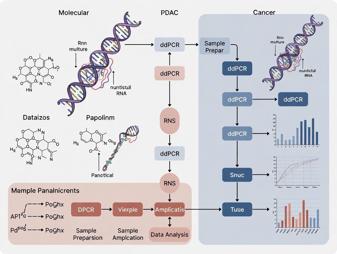
The following diagram illustrates the complete ddPCR workflow and its core principles:
Application in Pancreatic Ductal Adenocarcinoma Research
The absolute quantification capability of ddPCR is particularly valuable in PDAC research due to the need to detect low-abundance biomarkers in complex biological samples. Key applications include the analysis of circulating microRNAs and the detection of mutant KRAS genes in circulating tumor DNA (ctDNA), which are promising for liquid biopsy approaches [5] [2].
Table 1: Diagnostic Performance of ddPCR-Based Biomarkers in PDAC
| Biomarker | Sample Type | Study Groups | Key Quantitative Findings | Diagnostic Performance (AUC) |
|---|---|---|---|---|
| miR-1290 [5] | Plasma | 167 PC patients vs. 267 healthy subjects | Median level: 744 copies/μl (PC) vs. 360 copies/μl (Healthy) | 0.734 (miR-1290 alone); 0.956 (combined with CA 19-9) |
| KRAS Mutations [3] | Plasma ctDNA | Pancreatic cancer patients | Detection in 82.3% of patients with liver/lung metastasis; Limit of detection < 0.2% for target mutations | High concordance with tumor tissue; correlated with shorter survival |
Experimental Protocols
Protocol A: Absolute Quantification of Circulating miR-1290
This protocol is adapted from a study that used ddPCR to validate miR-1290 as a circulating biomarker for pancreatic cancer [5].
- Step 1: Sample Collection and Preparation. Collect peripheral blood into EDTA tubes. Process plasma by double-centrifugation to ensure complete cell removal. Aliquot and store plasma at -80°C until RNA extraction.
- Step 2: RNA Isolation. Purify total RNA from 500 μL to 1 mL of plasma using a phenol-guanidine-based isolation method (e.g., TRIzol LS) combined with silica membrane columns. Include spike-in synthetic RNAs as controls for extraction efficiency if desired.
- Step 3: Reverse Transcription. Convert RNA to cDNA using a stem-loop reverse transcription primer specific to miR-1290 and a reverse transcriptase enzyme. This step enhances specificity and efficiency for mature microRNAs.
- Step 4: ddPCR Reaction Setup. Prepare the PCR reaction mixture containing cDNA template, ddPCR Supermix for Probes, and TaqMan-based assay primers and probe for miR-1290.
- Step 5: Droplet Generation. Load the reaction mixture into a droplet generator to create ~20,000 nanoliter-sized droplets per sample.
- Step 6: PCR Amplification. Transfer the droplets to a 96-well plate and run the PCR protocol on a thermal cycler. Use the following cycling conditions: 95°C for 10 min (enzyme activation), followed by 40 cycles of 94°C for 30 sec (denaturation) and 60°C for 1 min (annealing/extension), with a final 98°C hold for 10 min (enzyme deactivation).
- Step 7: Droplet Reading and Analysis. Place the plate in a droplet reader to measure the fluorescence (FAM) in each droplet. Use the instrument's software to count the positive and negative droplets and apply Poisson statistics to calculate the absolute concentration of miR-1290 in copies/μL of the original plasma sample.
Protocol B: Multiplex KRAS Genotyping in ctDNA
This protocol is adapted from a study that utilized ddPCR combined with melting curve analysis for highly multiplexed KRAS genotyping, optimized for the short fragments typical of ctDNA [3].
- Step 1: ctDNA Extraction. Isolate cell-free DNA (cfDNA) from 3-5 mL of patient plasma using a commercially available cfDNA extraction kit. Quantify the yield using a fluorescence-based assay sensitive to low DNA concentrations.
- Step 2: Primer and Probe Design. Design primers to generate a short amplicon (~66 bp) to maximize the detection efficiency of fragmented ctDNA. Use molecular beacon probes for each KRAS mutation (e.g., G12D, G12V, G12R, G12C, G13D). Each probe should have a different fluorescent dye (color) and/or a distinct melting temperature (Tm).
- Step 3: ddPCR Reaction Setup. Prepare the reaction mix with the extracted ctDNA, supermix, and the panel of mutation-specific molecular beacon probes.
- Step 4: Droplet Generation and PCR. Generate droplets as in Protocol A. Perform asymmetric PCR to generate single-stranded amplicons for efficient probe hybridization.
- Step 5: Melting Curve Analysis. After PCR, place the droplet plate on a temperature-controlled stage. Gradually increase the temperature while continuously monitoring the fluorescence in each droplet channel. Generate melting curves for each positive droplet.
- Step 6: Genotyping and Quantification. The genotype in each positive droplet is determined by a combination of its fluorescent color and the Tm of its melting peak. The software then calculates the absolute concentration and mutant allele frequency for each KRAS mutation.
The workflow for this advanced multiplexing application is detailed below:
The Scientist's Toolkit: Research Reagent Solutions
Table 2: Essential Reagents and Materials for ddPCR in PDAC Research
| Item | Function/Description | Example Use Case |
|---|---|---|
| ddPCR Supermix for Probes | A ready-to-use reaction mix containing DNA polymerase, dNTPs, and buffer, optimized for droplet stability and PCR efficiency. | Core component of all ddPCR reactions, such as for miR-1290 or KRAS detection [5] [3]. |
| Mutation-Specific Probes | Hydrolysis probes (TaqMan) or Molecular Beacons labeled with fluorescent dyes (e.g., FAM, HEX). | Discriminates wild-type from mutant DNA sequences (e.g., KRAS G12D vs. G12V) [3]. |
| Droplet Generation Oil | Specially formulated oil to create stable, monodisperse water-in-oil emulsions. | Essential for the initial partitioning step in droplet-based systems [1]. |
| Cell-Free DNA Blood Collection Tubes | Tubes with preservatives that stabilize nucleated blood cells and prevent genomic DNA contamination of plasma. | Ensures high-quality, reproducible ctDNA samples for KRAS mutation analysis [3] [2]. |
| cfDNA/RNA Extraction Kits | Silica-membrane or bead-based kits designed to purify low-concentration, short-fragmented nucleic acids from body fluids. | Isolates ctDNA from plasma or microRNA from plasma for downstream ddPCR analysis [5] [3]. |
Digital droplet PCR (ddPCR) represents a transformative advancement in molecular diagnostics, providing unparalleled precision for pancreatic ductal adenocarcinoma (PDAC) research. As the third generation of PCR technology, ddPCR operates by partitioning a PCR reaction into thousands to millions of nanoliter-sized droplets, effectively creating a digital matrix where each droplet functions as an individual reaction vessel [1]. This partitioning enables absolute quantification of nucleic acids without requiring standard curves, a significant advantage over both conventional PCR and quantitative real-time PCR (qPCR) [6]. For PDAC—a malignancy characterized by late diagnosis, dense stroma, and limited tissue availability—ddPCR offers researchers a powerful tool to overcome fundamental challenges in biomarker detection and monitoring.
The clinical utility of ddPCR is particularly evident in liquid biopsy applications, where it detects circulating tumor DNA (ctDNA) harboring characteristic PDAC mutations such as KRAS, TP53, CDKN2A, and SMAD4 [7]. In the context of PDAC's genetic landscape, where over 90% of cases harbor KRAS mutations [7], ddPCR's ability to precisely quantify rare mutant alleles in complex biological samples makes it indispensable for early detection, minimal residual disease monitoring, and therapy response assessment. The technology's robust performance even with limited input material addresses critical bottlenecks in PDAC research and clinical translation, particularly when analyzing ctDNA from liquid biopsies where tumor-derived nucleic acids may represent only a small fraction of total circulating DNA [6] [8].
Key Technical Advantages of ddPCR
Superior Sensitivity and Precision for Rare Alleles
Droplet digital PCR achieves exceptional sensitivity through massive sample partitioning, which effectively enriches rare targets and reduces background noise. This partitioning enables the detection of mutant alleles at frequencies as low as 0.1% against a background of wild-type sequences, a level of sensitivity rarely achievable with conventional qPCR [9] [10]. This capability is critically important in PDAC research for detecting minimal residual disease following surgery and for identifying emerging resistance mutations during targeted therapy.
The precision of ddPCR for rare allele detection was demonstrated in a study analyzing urinary cfDNA, where researchers successfully detected NRAS and EGFR gene variants at allelic frequencies as low as 0.1% with excellent concordance between observed and expected values [9]. This precision remains robust even in challenging matrices such as urine, highlighting the technology's applicability to various liquid biopsy sources. For monitoring PDAC progression, studies have shown that KRAS mutant alleles can be reliably detected and quantified in plasma samples, with positive ctDNA status serving as a strong prognostic indicator for both progression-free and overall survival [8] [11].
Calibration-free Absolute Quantification
Unlike qPCR, which relies on standard curves and reference samples for relative quantification, ddPCR provides absolute quantification of target nucleic acids without external calibration [6] [12]. This capability stems from the binary nature of droplet reading (positive or negative) and the application of Poisson statistics to calculate target concentration based on the fraction of positive droplets [1]. The elimination of standard curves removes a significant source of variability and potential error, while also simplifying experimental workflow.
This absolute quantification capability was convincingly demonstrated in a CNV validation study, where ddPCR measurements of the DEFA1A3 gene copy number showed 95% concordance with pulsed-field gel electrophoresis (PFGE), considered a gold standard method [12]. In contrast, qPCR results showed only 60% concordance with PFGE, with a concerning average deviation of 22% from reference values [12]. For PDAC researchers, this calibration-free approach enables direct comparison of results across different experiments and laboratories, facilitating multi-center studies and accelerating biomarker validation.
Table 1: Comparison of ddPCR Performance Characteristics in PDAC Research
| Application | Performance Metric | Result | Context |
|---|---|---|---|
| Rare Mutation Detection | Limit of Detection | 0.1% mutant allele frequency [9] | Detection of NRAS/EGFR variants in urinary cfDNA |
| ctDNA Prognostication | Hazard Ratio for OS | HR = 2.3 (95% CI 1.9-2.8) [8] | High baseline ctDNA in non-resectable PDAC |
| Copy Number Quantification | Concordance with Gold Standard | 95% [12] | DEFA1A3 CNV vs. PFGE |
| Analysis from Limited Samples | Minimum Cell Input | 200 cells [6] | Crude lysate method for rare target quantification |
Enhanced Accuracy and Reproducibility
The partitioning approach underlying ddPCR not only enhances sensitivity but also improves overall assay accuracy and reproducibility. By distributing the reaction across thousands of individual partitions, the impact of PCR inhibitors is significantly reduced, as these compounds are similarly diluted across the droplet population [13]. This built-in tolerance to inhibitors is particularly valuable when analyzing challenging clinical samples from PDAC patients, which may contain various substances that interfere with PCR amplification.
The reproducibility of ddPCR was evidenced in a study monitoring Lacticaseibacillus casei, where the technology demonstrated high linearity and efficiency across a quantitative range of 100-105 CFU/mL, with a detection limit of 100 CFU/mL that surpassed the performance of real-time PCR [13]. This level of reproducibility is essential for PDAC biomarker studies that require longitudinal monitoring of ctDNA levels to assess treatment response and disease progression. Furthermore, the minimal equipment requirements and straightforward data interpretation lower technical barriers to implementation across different laboratory settings.
Application Notes: ddPCR Protocols for PDAC Research
Absolute Quantification of KRAS Mutations in Plasma ctDNA
Background: Detection of KRAS mutations in circulating tumor DNA provides valuable prognostic information in PDAC. Approximately 90-95% of PDAC cases harbor KRAS mutations, with G12D and G12V being the most common variants [7]. Establishing a reliable protocol for absolute quantification of these mutations enables non-invasive disease monitoring and treatment response assessment.
Methods:
- Sample Preparation: Collect peripheral blood in cell-stabilization tubes. Process within 2-6 hours with sequential centrifugations (1600×g followed by 16000×g) to obtain platelet-poor plasma [11]. Isect cfDNA using specialized kits (e.g., Mag-Bind cfDNA Kit) with elution in low-volume TE buffer.
- PCR Mix Preparation: Prepare reaction mixtures containing:
- ddPCR Supermix for Probes (No dUTP)
- FAM-labeled probe for KRAS mutant alleles
- HEX-labeled probe for wild-type KRAS sequence
- Primers amplifying the target KRAS codon
- Template cfDNA (typically 5-15 ng per reaction)
- Droplet Generation: Generate approximately 20,000 droplets per sample using a droplet generator [11].
- Thermal Cycling: Perform amplification with the following protocol:
- Initial denaturation: 95°C for 10 minutes
- 45 cycles of: 95°C for 30 seconds (denaturation) and 58-62°C for 60 seconds (annealing/extension)
- Enzyme deactivation: 98°C for 10 minutes
- Hold at 4°C
- Data Analysis: Read droplets using a droplet reader and analyze with companion software. Apply thresholding based on negative controls and calculate mutant allele concentration using Poisson statistics [10].
Key Considerations:
- Include negative controls (no-template and wild-type only) in each run
- For low-frequency mutations (<1%), analyze sufficient total DNA to ensure adequate detection sensitivity
- Use a minimum of 3 positive droplets for mutant calling to ensure statistical significance [11]
Table 2: Essential Research Reagent Solutions for ddPCR in PDAC Studies
| Reagent/Category | Specific Examples | Function/Application |
|---|---|---|
| Nucleic Acid Extraction | Mag-Bind cfDNA Kit [9] | Maximizes cfDNA yield from plasma/urine with minimal gDNA contamination |
| Sample Preservation | Colli-Pee UAS Preservative [9] | Stabilizes cfDNA in urine samples post-collection |
| ddPCR Master Mix | ddPCR Supermix for Probes [9] | Provides optimized reaction components for probe-based detection |
| Reference Standards | Mimix Multiplex I cfDNA Set [9] | Validates assay performance with predetermined allelic frequencies |
| Mutation Assays | Multiplex KRAS Screening Kit [11] | Simultaneously detects seven common KRAS mutations |
Crude Lysate ddPCR for Limited Input Samples
Background: Traditional nucleic acid extraction methods often lead to significant target loss when processing limited cell samples, creating a substantial barrier for PDAC research where sample material is often scarce. The crude lysate ddPCR approach eliminates the DNA extraction and purification steps, enabling accurate quantification of rare targets from as few as 200 cells [6].
Methods:
- Cell Lysis: Directly lyse 200-16,000 cells in 20μL lysis buffer (e.g., Buffer 2 from SuperScript IV CellsDirect cDNA Synthesis Kit) [6].
- Viscosity Breakdown: Incubate lysate with a viscosity breakdown solution to reduce sample viscosity that can interfere with droplet generation. This step is critical for achieving consistent droplet formation [6].
- PCR Setup: Prepare ddPCR reaction mix using crude lysate as template, reducing pipetting steps and potential sample loss.
- Droplet Generation and Amplification: Follow standard ddPCR workflow with optimized droplet generation and thermal cycling conditions.
- Data Interpretation: Calculate copies per cell using a reference assay (e.g., RPP30 for human cells) to normalize the target signal [6].
Validation: This method demonstrated excellent linearity (r² > 0.99) and accuracy compared to standard ddPCR with extracted DNA, while significantly reducing sample input requirements [6]. The approach is particularly valuable for analyzing rare cell populations or minimal tissue samples in PDAC research.
Experimental Design and Workflow
The typical ddPCR workflow for PDAC biomarker analysis involves several key stages, from sample collection through data interpretation, with specific considerations at each step to ensure reliable results:
ddPCR Workflow for PDAC Biomarker Analysis
Critical Experimental Considerations
Partition Quality and Number: The statistical power of ddPCR depends on both the number of partitions analyzed and the consistency of partition volume. Researchers should aim for a minimum of 15,000-20,000 high-quality droplets per sample to ensure accurate quantification, particularly for rare allele detection [9]. droplet volume should be verified experimentally, as deviations from manufacturer specifications can affect concentration calculations [6].
Assay Design and Optimization: Effective ddPCR assays require careful primer and probe design, following principles similar to qPCR. For rare mutation detection, use allele-specific probes or optimized primer sets to distinguish closely related sequences. Include appropriate controls:
- No-template controls to identify contamination
- Wild-type-only controls to assess assay specificity
- Reference assays for normalization in copy number studies [10]
DNA Input Optimization: The optimal DNA input amount depends on the application. For rare mutation detection, higher inputs increase the probability of detecting low-frequency mutations. However, excessive DNA can lead to partition overcrowding and violate Poisson distribution assumptions. As a general guideline, adjust input to maintain the majority of partitions as negative while ensuring sufficient positive partitions for statistical validity [10].
Droplet digital PCR technology provides PDAC researchers with a powerful analytical tool characterized by exceptional sensitivity, precision for rare alleles, and calibration-free absolute quantification. These advantages make it particularly suited for addressing the significant challenges in pancreatic cancer research, including late diagnosis, tissue heterogeneity, and limited biomarker availability. The technology's robust performance in liquid biopsy applications enables non-invasive assessment of tumor dynamics, treatment response, and disease evolution, offering new opportunities for improving PDAC management and patient outcomes.
As ddPCR platforms continue to evolve with increased automation, higher throughput, and expanded multiplexing capabilities, their integration into standardized PDAC research and clinical workflows promises to accelerate the development of personalized treatment approaches and enhance our understanding of this devastating disease. The protocols and applications outlined in this document provide a foundation for leveraging ddPCR's key advantages to advance pancreatic cancer research.
Pancreatic Ductal Adenocarcinoma (PDAC) remains one of the most formidable challenges in oncology, with a five-year survival rate below 12% and projections indicating it will become the second leading cause of cancer-related mortality by 2030 [14]. This dismal prognosis is primarily attributable to late-stage diagnosis, with over 50% of patients presenting with advanced or metastatic disease where curative surgical resection is no longer feasible [14]. The complex pathophysiology of PDAC—characterized by a dense desmoplastic stroma, early metastatic dissemination, and non-specific symptomatology—has rendered conventional diagnostic approaches insufficient for detecting the disease during its actionable stages [7].
In this challenging landscape, liquid biopsy has emerged as a promising non-invasive strategy for detecting tumor-derived components in bodily fluids. These analytes—including circulating tumor DNA (ctDNA), circulating tumor cells (CTCs), exosomes, and tumor-educated platelets—provide a dynamic window into tumor biology and evolution [7] [15]. However, the clinical utility of liquid biopsy is fundamentally constrained by the technological limitations of available detection platforms, particularly for identifying the low-abundance biomarkers characteristic of early-stage PDAC and minimal residual disease.
This application note establishes how Droplet Digital PCR (ddPCR) technology addresses the critical sensitivity and quantification challenges in PDAC biomarker analysis. We present validated experimental protocols and analytical frameworks that leverage ddPCR's capabilities to advance early detection, therapeutic monitoring, and resistance mutation tracking in pancreatic cancer research and drug development.
The Molecular Basis for ddPCR Application in PDAC
PDAC Genetics and Liquid Biopsy Targets
The genetic landscape of PDAC is characterized by four hallmark mutations that drive tumor initiation and progression. KRAS mutations occur in over 90-95% of cases, representing the earliest and most frequent alteration, primarily at codon 12 (e.g., p.G12D, p.G12V) [7]. These mutations lead to constitutive activation of MAPK and PI3K pathways, promoting uncontrolled proliferation and survival. TP53 mutations occur in approximately 70-75% of PDACs, disrupting apoptosis and genomic stability, while CDKN2A is inactivated in 35-40% of cases through mutation, deletion, or promoter methylation [7]. SMAD4 alterations appear in approximately 30% of PDACs and are associated with enhanced tumor progression and metastasis [7].
Beyond these canonical drivers, next-generation sequencing has identified mutations in DNA damage repair genes—including BRCA1, BRCA2, ATM, and PALB2—in 5-10% of PDACs, which confer sensitivity to platinum-based agents and PARP inhibitors [7]. The National Comprehensive Cancer Network (NCCN) guidelines (version 2.2025) now recommend comprehensive molecular profiling of tumor tissue or cell-free DNA to detect these and other actionable alterations when tissue is unavailable [7].
ctDNA as a Quantitative PDAC Biomarker
Circulating tumor DNA (ctDNA) has emerged as the most analytically tractable liquid biopsy analyte for PDAC management. ctDNA consists of fragmented tumor-derived DNA shed into the circulation through apoptosis, necrosis, or active secretion, with an average fragment length of 140 base pairs compared to 160 base pairs for healthy cell-free DNA [15]. With a short half-life of less than 2 hours, ctDNA provides a real-time snapshot of tumor burden and genetic heterogeneity [15].
Recent evidence demonstrates a significant correlation between ctDNA levels and disease burden in metastatic PDAC. A 2025 study measuring ctDNA via methylated markers (HOXD8 and POU4F1) found ctDNA detection in 66.2% of treatment-naïve metastatic PDAC patients (n=71), with strong correlations between ctDNA quantity and total tumor volume (Spearman's ρ=0.462, p<0.001) and liver metastasis volume (Spearman's ρ=0.692, p<0.001) [16]. Tumor volume thresholds for ctDNA detection were established at 90.1 mL for total tumor volume and 3.7 mL for liver metastases volume [16].
The prognostic value of ctDNA in advanced PDAC is well-established, with a recent meta-analysis of 64 studies (n=5,652) demonstrating that high baseline ctDNA levels predict shorter overall survival (HR=2.3, 95% CI 1.9-2.8) and progression-free survival (HR=2.1, 95% CI 1.8-2.4) [8]. Similarly, unfavorable ctDNA kinetics during treatment were associated with reduced overall survival (HR=3.1, 95% CI 2.3-4.3) and progression-free survival (HR=4.3, 95% CI 2.6-7.2) [8].
Table 1: Key Genetic Alterations in PDAC and Their Detection via Liquid Biopsy
| Gene | Mutation Prevalence | Clinical Significance | Detection Rate in Liquid Biopsy |
|---|---|---|---|
| KRAS | 90-95% | Early driver mutation; constitutive MAPK/PI3K pathway activation | 37-62% in newly diagnosed patients [17] |
| TP53 | 70-75% | Disrupted apoptosis and genomic stability | Varies by stage and detection method |
| CDKN2A | 35-40% | Cell cycle dysregulation | Often detected via promoter methylation |
| SMAD4 | ~30% | Associated with metastasis and poor prognosis | Lower detection rates in early disease |
| DNA Damage Repair Genes | 5-10% | Predicts sensitivity to platinum agents/PARP inhibitors | Concordant with tissue when detectable |
Why ddPCR? Technological Advantages for PDAC Biomarker Analysis
Fundamental Principles of ddPCR
Droplet Digital PCR represents the third generation of PCR technology, following conventional PCR and quantitative real-time PCR (qPCR) [1]. The fundamental innovation of ddPCR is sample partitioning—a PCR mixture containing the sample is divided into thousands to millions of nanoliter-sized water-in-oil droplets, effectively creating individual reaction chambers [1]. This partitioning enables a digital binary readout where each droplet is scored as positive or negative for the target sequence after amplification, allowing absolute quantification of target molecules without calibration curves through application of Poisson statistics [1].
The ddPCR workflow comprises four essential steps: (1) partitioning of the PCR mixture into droplets; (2) PCR amplification to endpoint; (3) fluorescence analysis of each droplet; and (4) calculation of target concentration based on the fraction of positive droplets [1]. This process provides unparalleled sensitivity for rare allele detection, precise quantification regardless of amplification efficiency, and exceptional reproducibility across a wide dynamic range [1].
Comparative Advantages Over Alternative Platforms
ddPCR offers distinct advantages for PDAC liquid biopsy applications compared to other molecular detection technologies:
Superior Sensitivity for Rare Mutations: ddPCR can detect mutant alleles at frequencies as low as 0.001% in a background of wild-type DNA [1]. This exceptional sensitivity is critical for PDAC applications where ctDNA fractions can be minimal in early-stage disease or minimal residual disease settings.
Absolute Quantification Without Standards: Unlike qPCR, which requires standard curves for relative quantification, ddPCR provides absolute quantification of target molecules, enabling precise measurement of ctDNA variant allele frequencies without reference materials [1].
Robust Performance in Inhibitory Samples: The partitioning process in ddPCR dilutes PCR inhibitors across thousands of droplets, making it more resilient to substances that commonly inhibit amplification in blood-derived samples [1].
Precision at Low Template Concentrations: ddPCR demonstrates superior accuracy and reproducibility for quantifying low-abundance targets compared to both qPCR and next-generation sequencing (NGS), with typical coefficient of variation <10% at single-digit copy numbers [1].
Table 2: Analytical Comparison of ddPCR with Alternative Detection Platforms
| Parameter | ddPCR | qPCR | NGS |
|---|---|---|---|
| Detection Sensitivity | 0.001%-0.01% mutant allele frequency | 1-5% mutant allele frequency | 0.1-5% mutant allele frequency |
| Quantification Method | Absolute (digital counting) | Relative (standard curve required) | Relative (with unique molecular identifiers) |
| Sample Throughput | Medium (1-96 samples/run) | High (96-384 samples/run) | Very high (hundreds to thousands) |
| Turnaround Time | 4-8 hours | 2-4 hours | 3-10 days |
| Cost per Sample | Medium | Low | High |
| Multiplexing Capacity | Limited (2-6 targets) | Limited (2-4 targets) | High (hundreds to thousands) |
| Ideal PDAC Application | MRD monitoring, therapy response assessment, resistance mutation tracking | High VAF mutation screening | Comprehensive genomic profiling, novel biomarker discovery |
Experimental Protocols for PDAC Biomarker Analysis
Protocol 1: KRAS Mutation Detection in Plasma ctDNA
Principle: This protocol enables sensitive detection and absolute quantification of KRAS hotspot mutations (G12D, G12V, G12C, G13D) in plasma-derived ctDNA from PDAC patients using allele-specific ddPCR assays.
Sample Collection and Processing:
- Collect whole blood in cell-stabilization tubes (e.g., Streck Cell-Free DNA BCT)
- Process within 6 hours of collection: centrifuge at 1,600 × g for 20 min at 4°C to separate plasma
- Transfer plasma to microcentrifuge tubes and centrifuge at 16,000 × g for 10 min to remove residual cells
- Store plasma at -80°C if not processing immediately
Cell-Free DNA Extraction:
- Extract cfDNA from 2-5 mL plasma using the QIAamp Circulating Nucleic Acid Kit (Qiagen)
- Elute in 20-50 μL AVE buffer
- Quantify using Qubit dsDNA HS Assay Kit
- Store extracted cfDNA at -20°C until ddPCR analysis
ddPCR Reaction Setup:
- Prepare ddPCR reaction mix (20 μL final volume):
- 10 μL ddPCR Supermix for Probes (No dUTP)
- 1.8 μL KRAS mutation-specific assay (FAM-labeled)
- 1.8 μL Reference assay (HEX-labeled, e.g., wild-type KRAS or reference gene)
- 2-10 μL template cfDNA (adjust volume based on concentration)
- Nuclease-free water to 20 μL
- Generate droplets using Automated Droplet Generator
- Transfer droplets to 96-well PCR plate and seal with foil heat seal
PCR Amplification:
- Run on conventional thermal cycler with the following protocol:
- Enzyme activation: 95°C for 10 min
- 40 cycles of:
- Denaturation: 94°C for 30 sec
- Annealing/Extension: 55-60°C (assay-specific) for 60 sec
- Enzyme deactivation: 98°C for 10 min
- Hold at 4°C
Droplet Reading and Analysis:
- Transfer plate to Droplet Reader
- Analyze using manufacturer's software (QuantaSoft)
- Set manual threshold based on negative controls and no-template controls
- Calculate mutant allele frequency: (mutant copies/μL) / (mutant copies/μL + wild-type copies/μL) × 100
Quality Control:
- Include no-template controls (water) in each run
- Include positive controls (synthetic mutant DNA) in each run
- Ensure >10,000 droplets per sample for reliable quantification
- Maintain contamination control practices including separate pre- and post-amplification areas
Protocol 2: Methylation-Based ctDNA Quantification
Principle: This protocol detects PDAC-specific DNA methylation patterns in plasma ctDNA using ddPCR assays targeting hypermethylated gene promoters (HOXD8, POU4F1), which have demonstrated prognostic value in metastatic PDAC [16].
Bisulfite Conversion:
- Treat 20-50 ng cfDNA with bisulfite using EZ DNA Methylation-Lightning Kit
- Follow manufacturer's protocol with modified conditions:
- Denaturation: 98°C for 5 min
- Incubation: 64°C for 2.5 hours
- Desulfonation: room temperature for 20 min
- Elute in 10-20 μL M-Elution Buffer
ddPCR Assay Design:
- Design primers and probes to recognize bisulfite-converted methylated sequences
- Target PDAC-specific methylated markers (HOXD8, POU4F1)
- Include reference assay for normalization (unmethylated reference gene or total cfDNA quantification)
ddPCR Reaction and Analysis:
- Prepare reaction mix as in Protocol 1 with methylation-specific assays
- Use 2-5 μL bisulfite-converted DNA per reaction
- Follow same droplet generation, amplification, and reading protocols
- Calculate methylated copies/μL and percentage of methylated molecules
Validation:
- Establish limit of detection using serial dilutions of methylated control DNA
- Determine linear range for quantitative applications
- Verify specificity against unmethylated genomic DNA
Research Reagent Solutions for PDAC ddPCR Applications
Table 3: Essential Research Reagents for PDAC ddPCR Studies
| Reagent Category | Specific Products | Application Notes |
|---|---|---|
| Blood Collection Tubes | Streck Cell-Free DNA BCT, PAXgene Blood cDNA Tubes | Preserves cell-free DNA integrity, prevents genomic DNA contamination |
| cfDNA Extraction Kits | QIAamp Circulating Nucleic Acid Kit, MagMAX Cell-Free DNA Isolation Kit | Optimized for low-abundance cfDNA recovery from plasma |
| ddPCR Master Mixes | ddPCR Supermix for Probes (No dUTP), ddPCR Mutation Detection Assay | No-dUTP formulation prevents carryover contamination |
| Assay Designs | Bio-Rad ddPCR Mutation Assays, Custom TaqMan SNP Genotyping Assays | KRAS G12D/V/C/R, G13D; TP53 hotspots; methylation-specific assays |
| Control Materials | Horizon Multiplex I cfDNA Reference Standards, Synthetic Oligonucleotides | Quality control, assay validation, quantification standards |
| Droplet Generation Oil | Droplet Generation Oil for Probes, DG8 Cartridges | Consistent droplet formation, stable emulsion during PCR |
| Consumables | DG8 Cartridges, Gaskets, 96-Well PCR Plates, Foil Seals | Compatible with Automated Droplet Generator systems |
Data Analysis and Interpretation Framework
Quantitative Analysis Methods
ddPCR data analysis requires specialized approaches to transform raw droplet counts into biologically meaningful metrics:
Absolute Quantification: Calculate target concentration using the fraction of positive droplets and Poisson statistics: [ \text{Concentration (copies/μL)} = \frac{-\ln(1 - p)}{V} ] Where ( p ) is the fraction of positive droplets and ( V ) is the droplet volume in μL.
Variant Allele Frequency (VAF) Calculation: [ \text{VAF} = \frac{\text{Mutant concentration (copies/μL)}}{\text{Mutant concentration + Wild-type concentration (copies/μL)}} \times 100\% ]
Limit of Blank (LOB) and Limit of Detection (LOD): Establish using negative controls and low-concentration standards:
- LOB = Mean{negative controls} + 1.645 × SD{negative controls}
- LOD = LOB + 1.645 × SD_{low concentration sample}
Statistical Significance Testing: Use Fisher's exact test or chi-square test to compare mutant droplet counts between samples and controls, with multiple testing correction for multiplex assays.
Clinical Correlation and Interpretation
Interpreting ddPCR results in the PDAC context requires integration with clinical parameters:
Tumor Burden Correlation: Relate ctDNA concentration to radiographic tumor volume, with published thresholds of 90.1 mL total tumor volume and 3.7 mL liver metastasis volume for ctDNA detection [16].
Therapeutic Monitoring: Define molecular response criteria:
- Complete molecular response: undetectable ctDNA
- Partial molecular response: >50% decrease in mutant copies/μL
- Molecular progression: >50% increase in mutant copies/μL or new mutation emergence
Prognostic Stratification: Apply validated ctDNA thresholds for risk stratification:
- High-risk: >5-10 mutant copies/μL plasma
- Low-risk: detectable but <5 mutant copies/μL
- Undetectable: favorable prognosis
Applications in PDAC Research and Drug Development
Minimal Residual Disease Detection
Following curative-intent resection, ddPCR enables ultrasensitive detection of minimal residual disease (MRD) that predicts clinical recurrence months before radiographic evidence. Research applications include:
Postoperative Monitoring Protocol:
- Collect plasma samples at defined intervals: preoperatively, 4-8 weeks postoperatively, then every 3 months
- Analyze using tumor-informed ddPCR assays targeting patient-specific mutations
- Define MRD positivity using validated thresholds (typically ≥1 mutant molecule/mL plasma)
Clinical Utility: Studies demonstrate that postoperative ctDNA detection predicts recurrence with 90-95% sensitivity and specificity, with median lead time of 6-9 months before radiographic recurrence [15].
Therapy Response Monitoring and Resistance Mechanisms
ddPCR facilitates real-time assessment of treatment efficacy and emergence of resistance:
Kinetic Monitoring:
- Establish baseline ctDNA level before treatment initiation
- Monitor at 2-4 week intervals during therapy
- Correlate ctDNA kinetics with radiographic response (RECIST criteria)
Resistance Mutation Tracking:
- Develop multiplex ddPCR assays for common resistance mechanisms
- Monitor for emergence of secondary KRAS mutations (e.g., G12R, Q61H) or parallel pathway alterations
- Guide therapy adaptation based on molecular evolution
Table 4: ddPCR Applications Across PDAC Disease Continuum
| Disease Stage | Primary Application | Key Analytes | Clinical Utility |
|---|---|---|---|
| Early Detection | Screening high-risk populations | KRAS mutations, methylated markers | Identification of actionable precursors, early intervention |
| Localized Disease | Surgical response assessment, MRD detection | Tumor-informed mutations | Recurrence risk stratification, adjuvant therapy guidance |
| Locally Advanced | Treatment response monitoring | KRAS, TP53 mutations | Response assessment, therapy modification |
| Metastatic Disease | Therapeutic resistance tracking | KRAS subclones, resistance mutations | Treatment selection, clinical trial stratification |
Droplet Digital PCR technology addresses fundamental limitations in PDAC biomarker analysis through its exceptional sensitivity, absolute quantification capabilities, and robust performance in challenging sample matrices. The protocols and frameworks presented herein provide researchers and drug development professionals with validated methodologies for leveraging ddPCR across the PDAC disease continuum—from early detection in high-risk cohorts to therapy monitoring and resistance mechanism elucidation in advanced disease.
As PDAC management evolves toward molecularly-guided approaches, ddPCR stands as an essential tool for translating liquid biopsy biomarkers into clinically actionable insights. Its implementation promises to accelerate therapeutic development and ultimately improve outcomes for this recalcitrant malignancy.
Pancreatic Ductal Adenocarcinoma (PDAC) is one of the most aggressive human malignancies, predicted to become the second leading cause of cancer-related death worldwide by 2030 [18] [17]. The overall 5-year survival rate remains a dismal 9%, the lowest among all cancer types [18]. This poor prognosis is largely attributed to late diagnosis, non-specific symptoms, and the limited therapeutic advancements [18]. The anatomical location of the pancreas makes obtaining adequate tumor tissue for molecular diagnosis challenging, creating a critical barrier to understanding tumor biology and implementing targeted therapies [18].
Liquid biopsy, particularly the analysis of circulating tumor DNA (ctDNA), has emerged as a promising non-invasive tool with enormous potential to overcome these limitations [18] [19]. ctDNA consists of short DNA fragments released into the bloodstream from apoptotic, necrotic cancer cells, and living tumor cells, carrying tumor-specific genetic and epigenetic alterations [18] [20]. As a minimally invasive "remote" biomarker, ctDNA provides a more comprehensive representation of the molecular composition of a malignant disease than a single tumor tissue specimen [18]. This application note details the cornerstone applications of ctDNA analysis using digital droplet PCR (ddPCR) technology for non-invasive PDAC management within research settings.
Current Applications and Quantitative Evidence
ctDNA analysis provides significant value across multiple clinical research domains in PDAC. The quantitative evidence supporting these applications, particularly from ddPCR-based studies, is summarized in the table below.
Table 1: Key Quantitative Evidence for ctDNA Applications in PDAC Management
| Application Domain | Key Marker/Method | Performance Evidence | Reference/Study Context |
|---|---|---|---|
| Diagnosis & Detection | KRAS mutation (ddPCR) | Sensitivity: 37%-62% in newly diagnosed patients [17] | Tumor-uninformed approach for screening |
| miR-1290 + CA19-9 (ddPCR) | AUC=0.956 for discriminating PC patients from healthy subjects [5] | Combined biomarker approach improves accuracy | |
| Prognostic Assessment | Baseline ctDNA level (Meta-analysis) | HR=2.3 for OS; HR=2.1 for PFS in non-resectable PDAC [8] | High baseline ctDNA predicts poorer survival |
| ctDNA Kinetics (Meta-analysis) | HR=3.1 for OS; HR=4.3 for PFS in non-resectable PDAC [8] | Unfavorable ctDNA changes predict poor outcomes | |
| Tumor Burden Correlation | Methylated markers (HOXD8, POU4F1) | Spearman's ρ=0.353 with total TV; ρ=0.500 with liver metastases TV [16] | ddPCR targeting methylation; stronger correlation with liver metastasis volume |
| Therapy Guidance | KRAS genotyping (ddPCR + Melting Curve) | Detected in 82.3% of patients with liver/lung metastasis [21] | Enables mutation-specific therapy selection |
Beyond the data in Table 1, research demonstrates that the presence of KRAS mutations in plasma is associated with poor survival, highlighting its prognostic utility [18]. Furthermore, ctDNA is undetectable in approximately one-third of metastatic PDAC patients, which may be related to smaller tumor volumes, particularly liver metastasis volumes below 3.7 mL [16].
Experimental Protocols for Key Applications
Protocol 1: KRAS Mutation Detection via ddPCR
Application: Diagnosis, Prognostication, and Therapy Selection [18] [21].
Workflow Overview: The following diagram illustrates the complete workflow for KRAS mutation detection and analysis using ddPCR.
Step-by-Step Methodology:
Sample Collection & Pre-processing: Collect 10-20 mL of peripheral blood into Streck Cell-Free DNA BCT tubes. Invert gently 8-10 times. Process within 6 hours with a double centrifugation protocol: first at 1,600 x g for 10 minutes at 4°C, then transfer plasma to a new tube and centrifuge at 16,000 x g for 10 minutes to remove residual cells [22].
cfDNA Extraction: Isolate cfDNA from the clarified plasma using commercially available silica-membrane column kits (e.g., QIAamp Circulating Nucleic Acid Kit). Elute in a low-EDTA TE buffer or nuclease-free water. Quantify using a fluorometer (e.g., Qubit dsDNA HS Assay) [22].
ddPCR Reaction Setup:
- Prepare a 20-22 μL reaction mixture containing:
- 11 μL of ddPCR Supermix for Probes (no dUTP)
- 1.1 μL of custom-designed KRAS primer/probe assay (e.g., for G12D, G12V, G12R)
- Up to 5.5 μL of template cfDNA (typically 5-20 ng)
- Nuclease-free water to the final volume.
- Vortex and spin down the mixture [21].
- Prepare a 20-22 μL reaction mixture containing:
Droplet Generation: Transfer the reaction mixture to a DG8 cartridge. Place the cartridge into the QX200 Droplet Generator along with DG8 Gaskets and 70 mL of Droplet Generation Oil for Probes. Generate approximately 20,000 droplets per sample [22].
PCR Amplification: Carefully transfer 40 μL of the generated droplets to a 96-well PCR plate. Seal the plate with a foil heat seal. Perform amplification in a thermal cycler using the following profile:
- 95°C for 10 minutes (enzyme activation)
- 40 cycles of:
- 94°C for 30 seconds (denaturation)
- 55-60°C for 1 minute (annealing/extension; temperature is assay-specific)
- 98°C for 10 minutes (enzyme deactivation)
- 4°C hold [21].
Droplet Reading and Analysis: Place the plate in the QX200 Droplet Reader. The reader measures the fluorescence amplitude (FAM and HEX) in each droplet. Analyze the data using the associated software (QuantaSoft). Set the threshold between positive and negative droplets manually based on the negative controls. The software calculates the mutant allele frequency (MAF) or concentration in copies/μL based on Poisson statistics [22] [21].
Protocol 2: Methylated ctDNA Marker Quantification
Application: Prognostic Assessment and Tumor Burden Monitoring [16].
Step-by-Step Methodology:
Sample Processing: Follow the same pre-analytical steps as in Protocol 1 for plasma collection, cfDNA extraction, and quantification.
Bisulfite Conversion: Treat 20-50 ng of extracted cfDNA using a bisulfite conversion kit (e.g., EZ DNA Methylation-Lightning Kit) according to the manufacturer's instructions. This process converts unmethylated cytosine residues to uracil, while methylated cytosines remain unchanged. Purify the converted DNA.
ddPCR Assay Setup: Design primers and probes specific for the bisulfite-converted sequence of methylated markers (e.g., HOXD8, POU4F1). Set up the ddPCR reaction similarly to Protocol 1, but using a supermix suitable for bisulfite-converted DNA.
Droplet Generation, PCR, and Analysis: Perform droplet generation, PCR amplification, and droplet reading as described in Protocol 1. The results will quantify the number of methylated DNA molecules present in the plasma sample, which can be correlated with total tumor volume, particularly liver metastasis volume [16].
The Scientist's Toolkit: Essential Research Reagents & Materials
Successful implementation of ctDNA analysis for PDAC requires specific, high-quality reagents and instruments. The table below details the essential components of the research toolkit.
Table 2: Key Research Reagent Solutions for ctDNA Analysis in PDAC
| Reagent/Material | Function/Application | Examples & Notes |
|---|---|---|
| Cell-Free DNA Blood Collection Tubes | Stabilizes nucleated blood cells to prevent genomic DNA contamination and preserve cfDNA profile. | Streck Cell-Free DNA BCT tubes are the current gold standard. |
| cfDNA Extraction Kits | Isolate short-fragment, low-concentration cfDNA from plasma with high efficiency and purity. | QIAamp Circulating Nucleic Acid Kit (Qiagen), MagMAX Cell-Free DNA Isolation Kit (Thermo Fisher). |
| ddPCR Supermix | Provides optimal buffer, nucleotides, and polymerase for PCR amplification in oil-emulsion droplets. | ddPCR Supermix for Probes (no dUTP) (Bio-Rad). |
| Mutation-Specific Assays | Primers and fluorescently labeled probes designed to detect specific point mutations (e.g., in KRAS). | Custom-designed TaqMan assays or Molecular Beacons for G12D, G12V, etc. [21]. |
| Methylation-Specific Assays | Primers and probes targeting bisulfite-converted, methylated DNA sequences. | Custom assays for PDAC-specific methylated markers (HOXD8, POU4F1) [16]. |
| Droplet Generation Oil & Consumables | Creates the water-in-oil emulsion necessary for partitioning the PCR reaction. | DG32 Cartridges, DG8 Gaskets, Droplet Generation Oil for Probes (Bio-Rad). |
Liquid biopsy analysis of ctDNA using ddPCR presents a transformative approach for the non-invasive management of PDAC in research settings. The protocols and data outlined in this application note provide a framework for employing this technology to advance our understanding of PDAC biology, prognosis, and response to therapy. The high sensitivity and absolute quantification capabilities of ddPCR make it ideally suited for detecting low-abundance ctDNA, tracking minimal residual disease, and performing longitudinal monitoring of tumor dynamics. As the field moves forward, standardizing assay protocols and validation thresholds will be critical for translating these research applications into validated clinical tools.
From Bench to Bedside: Implementing ddPCR Assays for PDAC Biomarker Analysis
Pancreatic ductal adenocarcinoma (PDAC) is one of the most aggressive human malignancies, with a predicted rise to become the second leading cause of cancer-related mortality by 2030 [23] [18]. The overall 5-year survival rate remains a dismal 9%, largely attributable to late diagnosis and limited therapeutic advancements [23]. Genomically, PDAC is characterized by a conserved set of driver mutations, with KRAS, TP53, and CDKN2A representing three of the four most frequently altered genes [24]. The anatomical location of the pancreas makes tissue biopsy challenging, often yielding insufficient material for comprehensive molecular profiling [23]. This limitation has accelerated the adoption of liquid biopsy approaches, particularly droplet digital PCR (ddPCR), which enables highly sensitive detection and quantification of circulating tumor DNA (ctDNA) harboring these characteristic mutations [23] [18]. This application note details the rationale and methodological framework for selecting KRAS, TP53, and CDKN2A as primary markers for PDAC research using ddPCR.
Molecular Rationale for Target Selection
Biological Functions and Mutational Significance
The selection of KRAS, TP53, and CDKN2A as core markers is grounded in their distinct and complementary roles in PDAC pathogenesis.
KRAS is a proto-oncogene located on chromosome 12p12.1 that functions as a critical molecular switch in cellular growth signaling pathways. In PDAC, KRAS mutations occur in 88-95% of cases, representing the earliest and most ubiquitous genetic event in pancreatic carcinogenesis [23] [25] [21]. The majority of mutations occur at codon 12 (e.g., G12D, G12V, G12R), leading to constitutive GTPase activity and continuous proliferation signaling through pathways including Raf/mitogen-activated protein kinase and Akt/protein kinase B [23] [24].
TP53, located on chromosome 17p13.1, is a tumor suppressor gene that functions as the "guardian of the genome" by regulating cell cycle arrest, apoptosis, and DNA repair. TP53 mutations occur in 60-77% of PDAC cases and represent a later event in tumor progression [26] [24]. These mutations predominantly include missense variants (approximately two-thirds) and truncating mutations (one-third), with gain-of-function variants altering the tumor microenvironment, promoting proliferation, and conferring chemotherapy resistance [24].
CDKN2A (p16) is a tumor suppressor gene on chromosome 9p21.3 that functions as a critical cell cycle regulator by inhibiting CDK4/6-mediated Rb phosphorylation. Inactivated in approximately 18% of PDAC cases, CDKN2A loss occurs at an intermediate stage of carcinogenesis and leads to uncontrolled cell cycle progression [24]. Alterations include homozygous deletions, mutations, and epigenetic silencing, all resulting in disrupted G1/S phase transition control.
Clinical and Prognostic Implications
The mutational status of these three genes carries significant clinical implications for prognosis and potential therapeutic targeting. KRAS mutations, particularly when detected in ctDNA, are significantly associated with advanced disease stage, metastatic burden, and poor survival outcomes [23] [27]. Recent evidence further indicates that KRAS mutant allele dosage gains, observed in 20% of KRAS-mutated diploid tumors, correlate with advanced disease and serve as prognostic indicators across all disease stages [25].
TP53 mutations independently predict for shorter overall survival, with overexpression correlating with aggressive tumor phenotypes and lymph node metastasis [24]. The complex interactions between TP53 mutations and co-occurring alterations in KRAS, CDKN2A, and SMAD4 significantly influence metastatic potential and survival outcomes [24].
CDKN2A loss is associated with more aggressive disease and therapeutic resistance, though its independent prognostic value is most significant when evaluated in the context of the broader mutational landscape [24]. Collectively, these three markers provide a comprehensive representation of the core molecular drivers of PDAC pathogenesis and progression.
Table 1: Prevalence and Clinical Significance of Primary PDAC Genetic Markers
| Gene | Function | Mutation Prevalence in PDAC | Common Mutation Types | Clinical Significance |
|---|---|---|---|---|
| KRAS | Oncogene | 88-95% [25] [21] | Codon 12 mutations (G12D, G12V, G12R) [23] | Early driver event; poor prognosis; associated with advanced stage [23] [25] |
| TP53 | Tumor suppressor | 60-77% [24] [25] | Missense (∼66%), truncating (∼33%) [24] | Late event; shorter overall survival; therapy resistance [24] |
| CDKN2A | Tumor suppressor | ∼18% [24] | Homozygous deletion, mutation, methylation [24] | Intermediate event; cell cycle dysregulation; aggressive disease [24] |
Quantitative Mutation Profiles in PDAC
Tissue-Based Mutation Frequencies
Comprehensive molecular profiling of PDAC tissues establishes baseline mutation frequencies essential for assay design. In a recent analysis of 50 patients with resectable PDAC, competitive allele-specific PCR (castPCR) identified KRAS p.G12D as the most frequent mutation, present in 48.0% of tumor DNA samples with a median mutation percentage of 7.0% (IQR 5.3-13.7%) [26]. Other selected KRAS and TP53 mutations occurred less frequently: KRAS p.G12V (2.0%), TP53 p.R273H (10.0%), and CDKN2A p.H83Y (4.0%) [26]. The majority of patients harbored only one primary mutation, though approximately 8% demonstrated multiple concomitant mutations [26].
Digital PCR analysis of the same cohort demonstrated higher sensitivity for mutation detection in tumor tissue. When employing a >0% mutation cutoff threshold, dPCR detected KRAS p.G12D in 95.9% of primary tumor samples with a median mutation percentage of 15.2% (IQR 0.2-26.2%) [26]. TP53 p.R273H was identified in 93.8% of tumors, though with a markedly lower median mutation percentage of 0.1% (IQR 0.1-0.1%), reflecting differences in tumor clonality and zygosity status between these genes [26].
Circulating Tumor DNA Detection Rates
In matched preoperative plasma samples, dPCR demonstrated variable detection efficiency for these mutations in cell-free DNA (cfDNA). KRAS p.G12D mutations were identified in 32.7% of plasma samples using a >0% cutoff threshold, with a median mutation percentage of 0.1% (IQR 0.0-0.2%) [26]. When applying a more stringent >0.1% cutoff to reduce false positives, the detection rate decreased to 10.2% with a median mutation percentage of 0.2% in positive samples [26]. TP53 p.R273H was detectable in only 8.2% (>0% cutoff) and 2.0% (>0.1% cutoff) of preoperative plasma samples, reflecting the lower abundance of ctDNA in resectable PDAC and technical challenges associated with low-frequency variant detection [26].
The fraction of ctDNA within total cfDNA is typically low in PDAC, particularly in early-stage and resectable disease, where it may represent less than 0.1% of total cfDNA [26]. This fundamental biological constraint necessitates highly sensitive detection methods like ddPCR for reliable mutation detection in liquid biopsies.
Table 2: Digital PCR Detection Efficiency in Matatched Tumor and Plasma Samples
| Mutation | Sample Type | Detection Rate (>0% cutoff) | Median Mutation % in Carriers | Detection Rate (>0.1% cutoff) | Median Mutation % in Carriers |
|---|---|---|---|---|---|
| KRAS p.G12D | Primary Tumor | 47/49 (95.9%) [26] | 15.2% (IQR 0.2-26.2%) [26] | 37/49 (75.5%) [26] | 16.7% (IQR 10.9-34.5%) [26] |
| KRAS p.G12D | Preoperative cfDNA | 16/49 (32.7%) [26] | 0.1% (IQR 0.0-0.2%) [26] | 5/49 (10.2%) [26] | 0.2% (IQR 0.2-0.2%) [26] |
| TP53 p.R273H | Primary Tumor | 93.8% [26] | 0.1% (IQR 0.1-0.1%) [26] | 47.9% [26] | 0.1% (IQR 0.1-0.2%) [26] |
| TP53 p.R273H | Preoperative cfDNA | 8.2% [26] | Not reported | 2.0% [26] | Not reported |
Experimental Protocols for ddPCR Detection
Sample Collection and Processing
Blood Collection and Plasma Separation:
- Collect peripheral blood using EDTA-containing tubes (cfDNA BCT tubes recommended).
- Process samples within 2 hours of collection to prevent genomic DNA contamination from leukocyte lysis.
- Centrifuge at 1,600-2,000 × g for 10 minutes at 4°C to separate plasma from cellular components.
- Transfer supernatant to microcentrifuge tubes and perform a second centrifugation at 16,000 × g for 10 minutes to remove residual cells.
- Store plasma at -80°C until cfDNA extraction [26] [28].
cfDNA Extraction:
- Use commercially available cfDNA extraction kits (QIAamp Circulating Nucleic Acid Kit, Maxwell RSC ccfDNA Plasma Kit).
- Elute cfDNA in low-EDTA TE buffer or nuclease-free water.
- Quantify cfDNA using fluorometric methods (Qubit dsDNA HS Assay); expected yield ranges from 5-50 ng/mL plasma depending on disease stage [26] [27].
- Assess DNA fragment size distribution using Bioanalyzer or TapeStation; expected peak at ~165 bp [21].
Direct Amplification Protocol for Limited Samples
For minute tissue samples (e.g., fine-needle aspirates) where conventional DNA extraction may result in significant loss, a "water-burst" direct amplification method has been developed [29]:
- Sample Preparation: Suspend tissue fragments or cell pellets in 20 μL nuclease-free water to cause osmotic burst and release genomic DNA.
- Incubation: Incubate for 10 minutes at room temperature with occasional vortexing.
- Centrifugation: Centrifuge at 3,000 × g for 1 minute to pellet debris.
- Supernatant Collection: Transfer 5-10 μL of supernatant directly to ddPCR reaction mix without DNA purification.
- ddPCR Setup: Proceed with emulsion generation and PCR amplification as described below [29].
This method enables detection of mutant KRAS with allele frequencies as low as 0.8% and completes sample processing within 30 minutes, compared to several hours for conventional extraction [29].
ddPCR Assay Setup and Optimization
Reaction Setup:
- Prepare 20-22 μL ddPCR reaction mix containing:
- 10-50 ng cfDNA or equivalent volume of direct lysate
- 1× ddPCR Supermix for Probes (no dUTP)
- 900 nM forward and reverse primers
- 250 nM mutant and wild-type probes (FAM/HEX labeled)
- Load sample into DG8 cartridge with 70 μL droplet generation oil for probes
- Generate droplets using QX200 Droplet Generator (approximately 20,000 droplets per sample) [26] [21]
Thermal Cycling Conditions:
- Enzyme activation: 95°C for 10 minutes
- 40-45 cycles of:
- Denaturation: 94°C for 30 seconds
- Annealing/Extension: 55-60°C for 60 seconds (optimize based on primer Tm)
- Enzyme deactivation: 98°C for 10 minutes
- 4°C hold [26] [21]
Droplet Reading and Analysis:
- Transfer droplets to QX200 Droplet Reader
- Analyze raw data using QuantaSoft software
- Set amplitude threshold based on negative controls and no-template controls
- Calculate mutant allele frequency using formula: (mutant droplets/total positive droplets) × 100 [26] [21]
Multiplex ddPCR with Melting Curve Analysis
For simultaneous genotyping of multiple KRAS mutations:
Probe Design:
- Use molecular beacon probes with stem-loop structure rather than hydrolysis probes
- Label with different fluorescent dyes (FAM, HEX, Cy5) and design for distinct melting temperatures (Tm differences >2°C)
- Target amplicon size of ~66 bp for efficient cfDNA amplification [21]
Melting Curve Analysis:
- After endpoint PCR, perform melting curve analysis from 45°C to 80°C with 0.5°C increments
- Hold for 5 seconds at each temperature step while measuring fluorescence
- Determine genotype based on combination of fluorescence color and Tm value [21]
This approach enables discrimination of 7 common KRAS mutations (G12D, G12R, G12V, G13D, G12A, G12C, G12S) with detection limits <0.2% for all targets and high concordance with conventional ddPCR (R² = 0.97) [21].
Signaling Pathways and Experimental Workflow
Research Reagent Solutions
Table 3: Essential Research Reagents for ddPCR-Based PDAC Mutation Detection
| Reagent Category | Specific Product Examples | Application Notes | Performance Characteristics |
|---|---|---|---|
| ddPCR Systems | Bio-Rad QX200, QX600; Thermo Fisher QuantStudio 3D | QX200 recommended for probe-based detection; QuantStudio 3D suitable for chip-based applications [21] | QX200: ~20,000 droplets/sample; detection sensitivity to 0.001% MAF [26] [21] |
| cfDNA Extraction Kits | QIAamp Circulating Nucleic Acid Kit; Maxwell RSC ccfDNA Plasma Kit | Optimized for low-abundance cfDNA; minimal contamination from genomic DNA [26] [28] | Yield: 5-50 ng/mL plasma; fragment size: ~165 bp [27] |
| ddPCR Supermixes | ddPCR Supermix for Probes (no dUTP); ddPCR Mutation Detection Assay | No dUTP recommended for direct amplification; validated mutation detection assays available for KRAS G12/G13 [26] [29] | Compatible with direct lysates; resistant to PCR inhibitors [29] |
| Reference Controls | Genomic DNA from cell lines (MIA PaCa-2, PANC-1); synthetic gBlocks | MIA PaCa-2: KRAS G12C, TP53 R248W; PANC-1: KRAS G12D, TP53 R273H [27] | Enable assay validation and quantification standardization [27] |
| Mutation-Specific Assays | Bio-Rad ddPCR Mutation Assays; Custom TaqMan Assays | KRAS: G12D, G12V, G12R, G12C, G13D; TP53: R175H, R248Q/W, R273H/C [26] [21] | Multiplexing possible with 2-3 colors; LOD: 0.01-0.1% MAF [21] |
The simultaneous detection of KRAS, TP53, and CDKN2A mutations using ddPCR technology provides a powerful approach for molecular profiling in PDAC research. These three markers collectively represent the core genetic drivers of pancreatic carcinogenesis, with complementary roles in disease initiation and progression. The exceptional sensitivity and absolute quantification capabilities of ddPCR make it particularly suited for analyzing low-abundance ctDNA in liquid biopsies and limited tissue samples. The protocols and reagents detailed in this application note establish a robust framework for implementing these markers in preclinical PDAC research, with potential applications in early detection, minimal residual disease monitoring, and therapeutic response assessment. As targeted therapies against specific KRAS variants and TP53-directed agents continue to develop, this tri-marker detection approach will increasingly inform both basic research and translational drug development efforts.
Pancreatic Ductal Adenocarcinoma (PDAC) is a lethal malignancy characterized by late diagnosis and a profoundly poor prognosis, with a 5-year survival rate of just 2–9% [7]. The complex tumor microenvironment and early metastasis of PDAC necessitate research tools capable of precise molecular analysis. Droplet Digital PCR (ddPCR) has emerged as a powerful technique for absolute nucleic acid quantification, offering the high sensitivity required to detect low-frequency mutations and copy number variations (CNVs) in PDAC driver genes such as KRAS and GNAS [30]. This application note provides a detailed workflow breakdown for implementing ddPCR in PDAC research, from sample preparation to data analysis.
Sample Preparation for PDAC Analysis
Proper sample preparation is foundational for reliable ddPCR data, especially when working with challenging PDAC-derived samples.
Sample Types and Input Considerations
The dynamic range of ddPCR is broad, accommodating various sample types relevant to pancreatic cancer research [31]. The required input DNA depends on the specific biological question, particularly for detecting rare mutations.
Table 1: Sample Input Guidelines for ddPCR in PDAC Research
| Sample Type | Recommended Input (Total Copies) | Mass Equivalent (Human gDNA) | Key Considerations for PDAC |
|---|---|---|---|
| High-Quality Genomic DNA | 1 – 100,000 [32] | 3.3 pg – 350 ng [32] | 100 ng is a standard starting point for CNV analysis [31]. |
| Formalin-Fixed Paraffin-Embedded (FFPE) Tissue | Varies | Varies; often requires concentration [32] | DNA is highly degraded; ~40% may be non-amplifiable. Concentrating samples is recommended [32]. |
| Cell-Free DNA (cfDNA) from Liquid Biopsy | Varies based on target abundance | N/A | Input volume is often maximized to enhance detection of rare KRAS mutants in plasma [7] [30]. |
| Bacterial/Viral DNA | 1 – 100,000 [32] | N/A | Serial dilutions are often necessary to fall within the optimal range [32]. |
For rare allele detection in PDAC, such as identifying a KRAS mutant among a vast excess of wild-type sequences, the total amount of DNA screened is critical. To detect one mutant in a background of one million wild-type molecules (0.0001% sensitivity), screening approximately 10 µg of DNA is necessary [32].
DNA Digestion and Quality Control
Digestion of DNA samples with restriction enzymes is a critical step for several applications [32] [31]:
- Reduce Structural Complexity: Undigested genomic DNA's viscosity can interfere with uniform droplet partitioning.
- Improve Quantification Accuracy: Linearizing supercoiled plasmid DNA ensures accurate copy number determination.
- Ensure Uniform Analysis: For CNV analysis, digestion ensures tandem repeats are separated. This is particularly important when analyzing FFPE samples to standardize the degree of digestion across samples [32].
A common protocol involves digesting 200 ng of DNA with a high-fidelity restriction enzyme (e.g., AluI) in a 10 µL reaction for at least one hour at 37°C, followed by a 1:2 dilution to stop the reaction and reduce buffer salts that might inhibit PCR [31].
Assay Design for PDAC Biomarkers
Robust assay design is paramount for targeting key PDAC mutations and reference genes.
Primer and Probe Design Specifications
TaqMan-based assays are standard for ddPCR. Key design principles include [31]:
- Amplicon Length: 60–150 base pairs. Smaller products are preferred due to higher amplification efficiency, making them ideal for fragmented DNA from FFPE or liquid biopsies.
- Primer Melting Temperature (Tm): Typically ~60°C.
- Probe Tm: Should be 8–10°C higher than the primer Tm.
- Probe Design: Avoid a guanine (G) at the 5' end, as it can quench the fluorophore. Also, avoid homopolymer runs of more than three bases to prevent secondary structures.
Targeting PDAC-Relevant Markers
A well-designed ddPCR experiment for PDAC often uses a duplex reaction to simultaneously target a region of interest (ROI) and a reference gene [31].
- Region of Interest (ROI): In PDAC, the most common ROIs are mutations in the KRAS oncogene (e.g., G12D, G12V, G12R) and GNAS (e.g., R201H, R201C), which are key drivers in pancreatic cancer precursors and invasive carcinoma [30].
- Reference Gene (REF): A reference gene with stable copy number, such as RPP30, is used for normalization in copy number variation analysis [31] [30]. Using multiple reference genes is advised in cancer research to ensure the reference itself is not amplified or deleted [32].
Recent advances enable highly multiplexed dPCR. One study developed a 14-plex assay that simultaneously detects eight KRAS mutations, two GNAS mutations, wild-type sequences, and the RPP30 reference gene, allowing for comprehensive analysis of PDAC precursors from limited sample material [30].
The Partitioning and Amplification Workflow
The core of ddPCR technology involves partitioning the sample into thousands of nanodroplets, followed by end-point PCR amplification.
Reaction Assembly and Droplet Generation
A standard 25 µL reaction is assembled for the Bio-Rad QX100 system as follows [31]:
- 12.5 µL of 2x ddPCR Supermix
- 1.25 µL of 20x ROI Primer/Probe mix (e.g., for KRAS G12D)
- 1.25 µL of 20x REF Primer/Probe mix (e.g., for RPP30)
- 10 µL of digested DNA sample
The reaction mix is loaded into a droplet generator cartridge along with droplet generation oil. Using microfluidics, the cartridge partitions the sample into ~20,000 nanoliter-sized droplets, following a Poisson distribution where droplets contain zero, one, or a few target molecules [1] [31].
Thermal Cycling Optimization
After droplet generation, the emulsion is transferred to a 96-well PCR plate, sealed, and placed in a thermal cycler. Standard cycling conditions can be modified to handle specific challenges in PDAC research [32]:
- Lower Ramp Rate: A slower ramp rate (e.g., 2°C per second) ensures uniform thermal transfer to all droplets, resulting in cleaner data [32].
- Difficult Templates:
- Long Amplicons (>400 bp): Switch from a two-step to a three-step protocol with a 72°C extension cycle.
- GC-Rich Templates: Increase denaturation temperature to 96°C for 10 seconds during cycling.
- Direct Lysis: For detecting pathogens or analyzing microbiota, an initial 10-minute step at 98°C can lyse cells or viruses within the droplets.
Data Analysis and Interpretation
Following amplification, droplets are read one-by-one in a droplet reader, which functions like a flow cytometer, measuring the fluorescence in each droplet [31].
Absolute Quantification and Statistical Analysis
The fundamental principle of ddPCR analysis is absolute quantification via Poisson statistics. The concentration of the target DNA is calculated without a standard curve using the formula [31]: λ = -ln(1-p) Where λ is the average number of target molecules per droplet, and p is the fraction of positive droplets [31].
Table 2: Key Data Analysis Metrics and Considerations in ddPCR
| Analysis Aspect | Description | Application in PDAC Research |
|---|---|---|
| Absolute Quantification | Calculated from the ratio of positive to total droplets using Poisson statistics [33]. | Determines absolute copies/µL of mutant KRAS or GNAS in a sample, enabling direct comparison between runs [30]. |
| Variant Allele Frequency (VAF) | The ratio of mutant allele concentration to total (mutant + wild-type) allele concentration. | Critical for monitoring tumor burden in liquid biopsies and assessing the clonal evolution of IPMNs [30]. |
| Copy Number Variation (CNV) | Determined by comparing the ratio of the target gene (e.g., KRAS) to the reference gene (e.g., RPP30). | Identifies gene amplifications, a valuable biomarker in PDAC progression [31] [30]. |
| Error Bars (95% CI) | Represent the 95% confidence interval based on Poisson statistics and the total number of droplets [32]. | Provides a measure of confidence in the quantification, important for assessing small changes in tumor DNA. |
| Limit of Detection (LOD) | The lowest mutant allele frequency detectable. Advanced multiplex assays report a LOD below 0.2% [30]. | Essential for early detection of recurrence or progression from precursor lesions using low VAF variants. |
Validation of Rare Events and Multiplexing
To confirm a true positive signal for a rare mutation, the "Rule of 3" is recommended. The false positive rate (FPR) is first determined from no-template controls (NTCs). A positive sample must contain at least three times the number of positive droplets than the FPR to be considered a true positive [32].
Modern analysis also leverages melting curve analysis after endpoint fluorescence reading. This allows for highly multiplexed assays by using probes with different melting temperatures (Tm) for different targets, enabling the simultaneous quantification of multiple PDAC-associated mutations and amplifications in a single well [30].
The Scientist's Toolkit: Essential Reagents and Materials
Table 3: Key Research Reagent Solutions for ddPCR in PDAC
| Item | Function/Application | Example |
|---|---|---|
| ddPCR Supermix | Optimized buffer containing DNA polymerase, dNTPs, and stabilizers for robust droplet formation and PCR. | Bio-Rad ddPCR Supermix for Probes [31] [34] |
| Restriction Enzymes | Digest genomic DNA to reduce viscosity, separate tandem repeats, and linearize plasmids for accurate quantification. | AluI (4-cutter) or other high-fidelity enzymes [31] |
| TaqMan Assays | Target-specific primers and fluorescently labeled probes (FAM, VIC/HEX) for detecting PDAC mutations and reference genes. | Custom assays for KRAS G12D/V/R, GNAS R201H/C, and RPP30 [31] [30] |
| Droplet Generation Oil & Surfactant | Creates a stable water-in-oil emulsion to form and maintain discrete droplets during thermal cycling. | Bio-Rad DG Oil; Poloxamer 188 [34] |
| Reference Gene Assays | Essential for normalizing data in CNV analysis; multiple references are recommended to ensure accuracy in cancer genomes. | RPP30 assay; pericentromeric assays [32] [31] |
| Positive Control Templates | Quantified synthetic DNA or cell line DNA with known mutations to validate assay performance and sensitivity. | KRAS mutant genomic DNA reference standards [30] |
The accurate molecular profiling of pancreatic ductal adenocarcinoma (PDAC) is critically limited by the scarce availability of tumor tissue, making liquid biopsy an indispensable tool for advancing precision oncology [35]. The detection of circulating tumor DNA (ctDNA) harboring key mutations and resistance markers provides a minimally invasive means for disease monitoring and treatment selection. However, traditional molecular methods often lack the sensitivity to detect low-abundance variants or the multiplexing capacity to interrogate multiple targets from a single, limited sample.
Droplet Digital PCR (ddPCR) technology overcomes these limitations by enabling the absolute quantification of nucleic acids without the need for a standard curve, offering superior sensitivity and precision for detecting rare genetic events [12] [1] [36]. This application note details robust multiplexing strategies that leverage the capabilities of ddPCR for the simultaneous detection of multiple PDAC-associated mutations and resistance genes, providing researchers with detailed protocols for enhancing molecular diagnostic assays in pancreatic cancer research.
Principles of Multiplex ddPCR
Digital PCR operates by partitioning a PCR reaction mixture into thousands of nanoscale reactions, effectively diluting the sample to a point where many partitions contain either zero or a single target molecule [1] [36]. Following end-point amplification, the fraction of positive partitions is counted, and the original target concentration is calculated using Poisson statistics, enabling absolute quantification [36].
Multiplex ddPCR expands this principle by allowing the simultaneous detection of multiple targets in a single reaction. This is achieved through several strategic approaches:
- Spectral Multiplexing: Modern ddPCR systems utilize multiple, distinct fluorescent dyes (e.g., FAM, HEX, VIC, Cy5, ROX) associated with target-specific probes. Each target is assigned a unique dye, and the fluorescence signature of each droplet is read in multiple channels, allowing for the discrimination of several targets within the same partition [37] [36].
- Amplitude-Based Multiplexing: When the number of targets exceeds the available fluorescent channels, targets can be differentiated within a single channel by using probes labeled with the same fluorophore but at different concentrations. This creates clusters of droplets with varying fluorescence intensities (high and low), each corresponding to a different target, effectively doubling the multiplexing capacity per channel [37].
- Ratio-Based Encoding: Combining different ratios of two fluorophores for a single probe can create a unique fluorescent signature, further expanding multiplexing potential, though this approach requires meticulous optimization.
The following diagram illustrates the core workflow of a multiplexed ddPCR assay, from sample partitioning to final target quantification.
Figure 1: Core workflow of a multiplex ddPCR assay. The sample is partitioned into thousands of nanodroplets, PCR amplification occurs within each droplet, fluorescence is measured via endpoint reading, and absolute target concentration is calculated using Poisson statistics [1] [36].
Application in PDAC Research: A Focus on KRAS and Beyond
In PDAC, the KRAS oncogene is mutated in over 90% of cases, making it a primary target for liquid biopsy assays [35]. Detecting KRAS mutant ctDNA in patient plasma provides critical prognostic information, with studies showing a significant association between KRAS mutation detection and inferior overall survival [35]. Furthermore, the ability to monitor KRAS mutant allele frequency during treatment offers a dynamic view of tumor response and emergence of resistance.
Multiplex ddPCR assays can be designed to simultaneously screen for the most common KRAS mutations (e.g., in codons 12, 13, and 61), maximizing the information obtained from a single aliquot of patient plasma. This is particularly valuable given the typically low concentration of ctDNA in PDAC, especially in early-stage or minimally residual disease.
Beyond KRAS, multiplexing strategies can incorporate probes for additional oncogenic drivers or resistance genes relevant to specific therapeutic contexts. This multi-analyte approach aligns with the broader trend in oncology biomarker discovery, where multiplex protein signatures are being developed to improve early detection of PDAC, significantly outperforming the single protein biomarker CA19-9 [38].
Experimental Protocol: A 6-Plex ddPCR Assay for PDAC-Associated Mutations
This protocol describes a method for the simultaneous detection of five different KRAS mutations and a reference control in a single ddPCR reaction, adapted for use with the Bio-Rad QX200 or QX600 systems.
Research Reagent Solutions
Table 1: Essential reagents and materials for the multiplex ddPCR assay.
| Item | Function / Description | Example / Source |
|---|---|---|
| ddPCR Supermix | Provides optimized buffer, enzymes, and dNTPs for partition PCR. Critical for assay performance and accuracy [39]. | Bio-Rad ddPCR Supermix for Probes (no dUTP) |
| Primer/Probe Assays | Target-specific oligonucleotides for amplification and detection. | Hydrolysis probes (e.g., TaqMan) with distinct fluorophores (FAM, HEX, Cy5) [37]. |
| Restriction Enzyme | May be used to digest long genomic DNA and improve access to target sequences. | Not a critical factor for quantification accuracy [39]. |
| DNAse/RNAse-Free Water | Solvent for adjusting reaction volume without introducing contaminants. | Various molecular biology suppliers |
| Reference Gene Assay | Probe for a wild-type genomic sequence to normalize DNA input and assess sample quality. | Labeled with a distinct fluorophore (e.g., HEX, Cy5). |
Step-by-Step Procedure
Reaction Mixture Preparation:
- Prepare the reaction mix on ice in a final volume of 20 µL:
- 10 µL of 2x ddPCR Supermix for Probes (no dUTP).
- 1 µL of each primer/probe set (final concentration 900 nM/300 nM for "high" targets, 400 nM/100 nM for "low" targets in amplitude multiplexing) [37].
- 4 µL of template DNA (recommended: 10-50 ng of cfDNA extracted from plasma).
- DNAse/RNAse-free water to 20 µL.
- Gently mix by pipetting. Do not vortex.
- Prepare the reaction mix on ice in a final volume of 20 µL:
Droplet Generation:
- Transfer the entire 20 µL reaction to the sample well of a DG8 cartridge.
- Add 70 µL of Droplet Generation Oil to the oil well.
- Place the cartridge into the Droplet Generator. The generator will create approximately 20,000 nanoliter-sized droplets per sample.
PCR Amplification:
- Carefully transfer 40 µL of the generated droplets to a 96-well PCR plate. Seal the plate with a foil heat seal.
- Place the plate in a thermal cycler and run the following protocol:
- Enzyme Activation: 95°C for 10 minutes.
- 40-45 Cycles of:
- Denaturation: 94°C for 30 seconds.
- Annealing/Extension: 55-61°C for 1 minute (optimize temperature for primer specificity).
- Enzyme Deactivation: 98°C for 10 minutes.
- Hold: 4°C (optional: post-amplification, the plate can be held at 4°C overnight, which may improve droplet stability for reading) [39].
- Use a ramp rate of 2°C/second.
Droplet Reading and Analysis:
- Place the PCR plate into the Droplet Reader.
- The reader will aspirate each sample and flow the droplets single-file past a two-color (QX200) or multi-color (QX600) detection system.
- Using the instrument's associated software (e.g., QuantaSoft), set the amplitude thresholds to distinguish positive and negative droplets for each fluorescent channel.
- The software will automatically apply Poisson statistics to calculate the absolute concentration (copies/µL) of each target in the original reaction.
Workflow Visualization
The complete experimental journey, from sample preparation to data analysis, is summarized in the following workflow.
Figure 2: Detailed workflow of the multiplex ddPCR protocol, highlighting key procedural steps and critical considerations for assay optimization.
Data Analysis and Interpretation
In a multiplexed ddPCR experiment, data analysis involves interpreting 1-dimensional or 2-dimensional fluorescence amplitude plots generated by the analysis software.
- 1D Plots: Display the fluorescence amplitude of a single channel, allowing for the discrimination of two targets (e.g., wild-type and mutant) based on pre-set thresholds.
- 2D Plots: Display fluorescence intensity from two channels simultaneously, where each cluster of droplets represents a distinct population (e.g., double-negative, single-positive for Channel 1, single-positive for Channel 2, and double-positive).
The following diagram illustrates how different droplet populations are distinguished in a 2-plex assay using two fluorescence channels, a principle that can be expanded for higher-plex assays.
Figure 3: Conceptual representation of a 2D droplet plot. Each dot represents a single droplet, and its position is determined by its fluorescence intensity in two channels. Distinct clusters correspond to droplets containing different target combinations, enabling multiplex quantification.
Performance Metrics and Validation
Robust validation is essential for implementing a reliable ddPCR assay. Key performance characteristics to evaluate include:
Table 2: Key performance metrics for validating a multiplex ddPCR assay.
| Metric | Description | Acceptance Criteria / Typical Performance |
|---|---|---|
| Accuracy/Trueness | Agreement between measured and known target concentration. | High concordance (e.g., 95%) with gold-standard methods like PFGE [12]. |
| Precision | Reproducibility of repeated measurements (within-run and between-run). | Demonstrated robustness; most experimental factors (operator, restriction enzymes) show no relevant effect [39]. |
| Limit of Detection (LoD) | Lowest concentration reliably distinguished from blank. | Detection limits can range from 1.4 to 2.9 copies/µL depending on the target [37]. |
| Linearity & Dynamic Range | Ability to provide accurate quantification across a range of concentrations. | Accurate over the entire working range; dependent on the ddPCR master mix used [39]. |
| Specificity | Ability to distinguish between different targets without cross-reactivity. | Clear separation of clusters in 1D or 2D amplitude plots. |
Multiplex ddPCR represents a powerful tool for advancing precision medicine in pancreatic ductal adenocarcinoma research. Its ability to perform absolute quantification of multiple low-abundance nucleic acid targets simultaneously, without reliance on standard curves, addresses critical challenges in PDAC biomarker analysis [12] [35] [36]. The protocols and strategies outlined here provide a foundation for researchers to develop robust assays for detecting key mutations like those in KRAS, facilitating studies on tumor dynamics, treatment response, and resistance mechanisms. As the technology continues to evolve with increased multiplexing capabilities and automation, its integration into comprehensive biomarker discovery pipelines—complementing proteomic approaches [38]—will be instrumental in improving outcomes for patients with this challenging disease.
Pancreatic Ductal Adenocarcinoma (PDAC) remains one of the most lethal malignancies, with a 5-year survival rate of only 9% [40]. The challenges in PDAC management include late diagnosis, early metastasis, and development of therapeutic resistance. Liquid biopsy, particularly through analysis of circulating tumor DNA (ctDNA), has emerged as a transformative approach for overcoming these challenges [7]. Among detection technologies, droplet digital PCR (ddPCR) provides exceptional sensitivity for quantifying rare mutations in blood samples, enabling non-invasive tumor genotyping and monitoring [35]. This application note details experimental protocols and key applications of ddPCR in PDAC research, focusing on early detection, minimal residual disease (MRD) assessment, therapy response monitoring, and tracking resistance mechanisms.
Key Research Applications and Clinical Evidence
The application of ddPCR for ctDNA analysis in PDAC spans the entire disease continuum, from initial diagnosis to advanced disease management. The table below summarizes the key applications and supporting clinical evidence.
Table 1: Key Applications of ddPCR in PDAC Research
| Application Area | Detected Alterations | Clinical/Rresearch Utility | Evidence |
|---|---|---|---|
| Early Detection & Diagnosis | KRAS mutations (G12D, G12V, G12R); TP53 mutations [41] | Aids in early diagnosis; identifies high-risk individuals; complements imaging [42] | KRAS mutations detected in >88% of PDAC tumors; ctDNA detection possible before clinical symptoms [41] |
| Minimal Residual Disease (MRD) & Prognosis | Post-resection KRAS mutations [35] [41] | Predicts recurrence risk after surgery; independent prognostic biomarker [35] | Patients with post-operative ctDNA have significantly worse RFS (HR: 3.26) and OS (HR: 5.46) [41] |
| Therapy Response Monitoring | Quantitative changes in mutant KRAS allele frequency [40] | Correlates with treatment efficacy; enables real-time response assessment [40] | ctDNA levels decrease with response to chemotherapy and increase upon progression; faster response indicator than imaging [40] |
| Tracking Resistance | Emergence of new mutations in KRAS or other genes [43] | Identifies molecular mechanisms of drug resistance; guides subsequent therapy [43] | ddPCR can track clonal evolution and emergence of resistance-associated variants during treatment [43] |
Experimental Protocols for ddPCR in PDAC Research
Sample Collection and cfDNA Extraction Protocol
Principle: High-quality cell-free DNA (cfDNA) extraction from blood plasma is critical for sensitive ctDNA detection. Plasma is preferred over serum as it minimizes background DNA from lysed blood cells [44].
Workflow Diagram: Sample Collection and Processing
Materials:
- Blood Collection Tubes: Streck Cell-Free DNA BCT or K2 EDTA tubes [43]
- Extraction Kits: QIAamp Circulating Nucleic Acid Kit (Qiagen) or equivalent [43]
- Equipment: Refrigerated centrifuge, vortex, thermal shaker, spectrophotometer or fluorometer for DNA quantification
Step-by-Step Procedure:
- Blood Collection and Processing: Draw venous blood into appropriate collection tubes. Invert gently 8-10 times. Process within 2-6 hours of collection.
- Plasma Separation: Centrifuge at 1,600-2,000 × g for 10-20 minutes at 4°C. Transfer supernatant (plasma) to a fresh tube without disturbing the buffy coat. Perform a second centrifugation at 16,000 × g for 10 minutes to remove residual cells [43].
- cfDNA Extraction: Follow manufacturer's instructions for the chosen extraction kit. Typically, this involves proteinase K digestion, binding to a silica membrane, washing, and elution in a low-EDTA buffer.
- Quality Control: Quantify cfDNA using a fluorometric method (e.g., Qubit). Assess fragment size distribution using a Bioanalyzer or TapeStation; expected peak ~166 bp [40].
ddPCR Assay Setup and Mutation Detection
Principle: ddPCR partitions a single PCR reaction into thousands of nanoliter-sized droplets, enabling absolute quantification of target DNA molecules. This allows for detection of mutant alleles at very low frequencies (<0.1%) in a background of wild-type DNA [43] [35].
Workflow Diagram: ddPCR Mutation Detection
Materials:
- ddPCR System: Bio-Rad QX200 Droplet Digital PCR System or equivalent [43]
- Reagents: ddPCR Supermix for Probes (no dUTP), droplet generation oil, primers, and fluorescent probes (FAM/HEX) [43]
- Assay Design: Custom assays with Locked Nucleic Acid (LNA) probes to enhance allele discrimination, especially for KRAS G12/13 mutations [43]
Step-by-Step Procedure:
- Reaction Setup: Prepare a 22 µL reaction mixture containing 11 µL of 2x ddPCR Supermix, target-specific primers and probes at optimized concentrations (e.g., 900 nM primers, 250 nM probes), and up to 8 µL of template cfDNA [43].
- Droplet Generation: Transfer the reaction mix to a DG8 cartridge. Add 70 µL of droplet generation oil and generate droplets using the QX200 Droplet Generator.
- PCR Amplification: Transfer the emulsified droplets to a 96-well plate, seal, and run PCR with a touchdown thermal cycling protocol optimized for the specific assay.
- Droplet Reading and Analysis: Read the plate on the QX200 Droplet Reader. Analyze data using QuantaSoft software. Set thresholds to distinguish positive (mutant and wild-type) and negative droplets based on fluorescence amplitude. The concentration (copies/µL) is calculated automatically via Poisson statistics [43].
The Scientist's Toolkit: Essential Research Reagents
Successful implementation of ddPCR assays for ctDNA analysis requires specific, high-quality reagents and controls.
Table 2: Essential Research Reagents for ddPCR-based ctDNA Analysis
| Reagent/Category | Specific Examples | Function & Importance |
|---|---|---|
| Nucleic Acid Extraction | QIAamp Circulating Nucleic Acid Kit (Qiagen), Maxwell RSC ccfDNA Plasma Kit (Promega) [43] | Isulates short-fragment cfDNA from plasma with high efficiency and minimal contamination. |
| ddPCR Master Mix | ddPCR Supermix for Probes (no dUTP) (Bio-Rad) [43] | Provides optimized reagents for PCR amplification in a droplet format. |
| Mutation-Specific Assays | Custom LNA-modified PrimeTime Probes (IDT) [43] | Enhances allele-specific discrimination, crucial for detecting single-base mutations (e.g., KRAS). |
| Reference Gene Assay | RPP30 Assay (Bio-Rad) [43] | Quantifies total human cfDNA, serving as a control for sample quality and input. |
| Positive Control Templates | gBlock Gene Fragments (IDT), HDx Reference Standards (Horizon Discovery) [43] | Validates assay performance; used for determining limit of detection (LOD) and false-positive rates. |
| Droplet Generation Oil | DG32 Droplet Generation Oil for Probes (Bio-Rad) [43] | Creates a stable water-in-oil emulsion for partitioning the PCR reaction. |
Data Analysis and Interpretation
Critical Validation Steps:
- False Positive Determination: Run multiple no-template controls (NTCs: water, elution buffer) to establish the background false positive rate. An assay is robust if the false positive rate is near zero [43].
- Extraction Efficiency: Spike a known quantity of non-human synthetic DNA (e.g., XenT gBlock) into plasma before extraction. Quantify recovery post-extraction using a dedicated ddPCR assay to correct for extraction losses and accurately calculate mutant copies per mL of plasma [43].
- Limit of Blank (LoB) & Detection (LOD): LoB is determined from the 95th percentile of false positives in NTCs. LOD is validated by spiking mutant DNA into wild-type background at low allele frequencies (e.g., 0.1%) [43].
Reporting Results: Report mutant allele frequency as a percentage and as absolute concentration (e.g., copies per mL of plasma). The latter is more robust for longitudinal monitoring. Results should be interpreted in the clinical context, integrating imaging and other biomarkers like CA19-9 [40] [41].
Maximizing Fidelity: Strategies for Optimizing ddPCR Assays and Mitigating Pre-analytical Variables
The reliability of any diagnostic test, including liquid biopsy for pancreatic ductal adenocarcinoma (PDAC) research, is heavily dependent on the integrity of the starting biological material. The pre-analytical phase encompasses all procedures from patient preparation and blood collection to sample processing and storage. Approximately 60-70% of laboratory errors originate in the pre-analytical phase [45]. For sensitive downstream applications like droplet digital PCR (ddPCR) to detect rare oncogenic mutations, pre-analytical variables can significantly impact the quantity, quality, and analytical validity of the final results. This document outlines evidence-based best practices for the pre-analytical pipeline, specifically optimized for cell-free DNA (cfDNA) analysis in PDAC research.
Quantifying Pre-analytical Errors and Their Impact
Understanding the sources and frequency of pre-analytical errors is the first step in mitigating them. The table below summarizes the distribution of laboratory errors and common pre-analytical integrity variables.
Table 1: Distribution and Sources of Laboratory Errors
| Category | Source of Error | Frequency/Impact |
|---|---|---|
| Phase of Error [45] | Pre-analytical Errors | 60-70% of total laboratory errors |
| Analytical Errors | Contribute to a smaller proportion of total errors | |
| Post-analytical Errors | Contribute to a smaller proportion of errors | |
| Common Pre-analytical Variables [46] | Collection Errors | Wrong tube type, under-filled tubes, poor technique, improper labeling |
| Transport & Storage Errors | Incorrect centrifugation, inappropriate temperature, prolonged storage | |
| Processing Errors | Delayed processing, misreading test requests | |
| Patient Variables | Non-fasting, recent heavy meal, medication schedule, exercise |
Poor blood sample quality is the essence of pre-analytical variability. Hemolyzed samples are the primary source of poor blood sample quality, accounting for 40-70% of such cases, followed by inappropriate sample volume (10-20%), use of the wrong container (5-15%), and clotted samples (5-10%) [45]. Hemolysis can cause spurious release of intracellular analytes and spectral interference, while lipemia from non-fasting patients can lead to pseudo-hyponatremia and volume displacement effects [45].
Standardized Protocols for Blood Collection and Processing
Patient Preparation and Identification
- Fasting: For tests involving metabolites like glucose and triglycerides, a 8-12 hour fast is required. Failure to fast can result in falsely elevated levels and lipemic samples [45].
- Medication and Lifestyle: Document patient intake of over-the-counter drugs, herbal preparations, and dietary supplements (e.g., biotin), which can cause drug-laboratory test interactions [45]. Advise patients to avoid chewing gum before blood collection, as it can stimulate gastric secretion and affect test results [45].
- Patient Identification: Implement a protocol using a minimum of two unique patient identifiers (e.g., full name and date of birth). 16% of phlebotomy errors are due to patient misidentification, and 56% are due to improper labeling [47]. Label tubes in the presence of the patient to ensure accuracy [47].
Phlebotomy and Sample Collection
- Order of Draw: Adhere strictly to the CLSI GP41 standard Order of Draw to prevent cross-contamination between tube additives [47].
- Tube Selection: Use EDTA tubes (e.g., BD K2 EDTA tubes) for cfDNA studies, as they are recommended for plasma separation for molecular analyses [48].
- Technique to Prevent Hemolysis:
- Preventing Under-filled Tubes: Push the tube forward so the stopper is fully penetrated and hold it in place until the vacuum is exhausted and blood flow ceases [47].
Plasma Separation for cfDNA Analysis
The method of plasma separation is critical for obtaining high-quality cfDNA. The following protocol, adapted from PDAC liquid biopsy studies, ensures the removal of cells that could contribute genomic DNA to the sample.
Table 2: Plasma Processing Parameters from PDAC Studies
| Parameter | Protocol Details | Rationale |
|---|---|---|
| Blood Collection Tube | BD K2 EDTA tube [48] | Prevents coagulation and preserves cfDNA. |
| Time to Initial Centrifugation | Process within 4 hours of draw [48] | Minimizes cell lysis and release of genomic DNA. |
| Initial Centrifugation | 2,500 x g for 10 minutes at room temperature [48] | Generates platelet-free plasma by removing cells and debris. |
| Second Centrifugation | (Optional) High-speed spin (e.g., 16,000 x g) of supernatant [49] | Further removes residual platelets and microparticles. |
| Plasma Storage | Aliquot and snap-freeze plasma at -80°C [48] | Preserves cfDNA integrity for long-term storage. |
Optimized cfDNA Extraction and Workflow Integration for PDAC
cfDNA Extraction Methodology
For cfDNA extraction from plasma, column-based purification methods are widely used and effective. One study on PDAC patients used a column purification method (Zymo Research Quick-cfDNA Serum & Plasma Kit) optimized to enrich for circulating cfDNA, followed by a concentration step using the DNA Clean & Concentrator (Zymo Research) [49]. This protocol successfully yielded a median of 37.3 ng of cfDNA from 4-6 mL of plasma (range: 6.8 - 836.8 ng), which was adequate for next-generation sequencing (NGS) [49].
Workflow Visualization: From Blood Draw to ddPCR
The following diagram illustrates the complete integrated workflow for processing blood samples for cfDNA analysis in PDAC research.
The Researcher's Toolkit: Essential Reagents and Kits
Table 3: Key Research Reagent Solutions for Pre-analytical Workflow
| Item | Function | Example Use Case |
|---|---|---|
| EDTA Blood Collection Tubes | Anticoagulant that prevents clotting and preserves cfDNA. | BD K2 EDTA tubes used in PDAC studies for plasma collection [48]. |
| Column-based cfDNA Kits | Isolation and purification of cfDNA from plasma. | Zymo Research Quick-cfDNA Serum & Plasma Kit used to extract cfDNA from PDAC patient plasma [49]. |
| DNA Clean & Concentrator Kits | Concentration of dilute cfDNA eluates to meet minimum input requirements. | Used in conjunction with extraction kits to increase cfDNA concentration for NGS [49]. |
| Size Exclusion Chromatography (SEC) | Isolation of extracellular vesicles (EVs) from plasma based on size. | qEVoriginal columns used to isolate EVs for miRNA analysis in PDAC research [48]. |
Analytical Considerations for ddPCR in PDAC Research
The ultimate goal of a robust pre-analytical protocol is to enable reliable detection of PDAC-associated mutations, such as those in the KRAS and TP53 genes, which are found in up to 95% and 75% of PDAC cases, respectively [49]. Studies have shown that the detection of these mutations in circulating tumor DNA (ctDNA) is correlated with worse survival outcomes, with one study reporting median overall survival of 10.5 months for ctDNA-positive patients versus 18 months for ctDNA-negative patients [49]. Furthermore, a 2025 meta-analysis confirmed that high baseline ctDNA levels are a strong prognostic indicator, associated with significantly shorter overall survival (HR=2.3) and progression-free survival (HR=2.1) in patients with non-resectable PDAC [8].
To ensure success in ddPCR:
- Input DNA: The pre-analytical steps must yield cfDNA of sufficient quantity and quality. The PDAC study using the Oncomine Lung cfDNA assay required a minimum of 20 ng of input cfDNA for NGS [49].
- Inhibition Control: Always include a positive control in ddPCR reactions to detect the presence of inhibitors that may have been co-purified during extraction.
- Assay Design: Design assays to detect common PDAC hotspots (e.g., KRAS G12D/V) with high specificity to distinguish low variant allele fractions (reported as low as 0.05% in PDAC studies) from wild-type signals [49].
Meticulous attention to the pre-analytical phase is non-negotiable for generating robust and reproducible data in PDAC ddPCR research. Standardizing procedures from blood draw to cfDNA extraction, as outlined in this document, minimizes artifacts and ensures that the resulting genetic data truly reflects the patient's disease state rather than pre-analytical variability. Implementing these best practices is foundational for advancing our understanding of pancreatic ductal adenocarcinoma through liquid biopsy.
Pancreatic ductal adenocarcinoma (PDAC) remains one of the most lethal malignancies, with a devastating 5-year survival rate of less than 9%, largely due to late-stage diagnosis and limited therapeutic options [50]. The complex molecular heterogeneity and rapid development of drug resistance in PDAC have underscored the critical need for highly sensitive and reproducible molecular detection methods. Digital droplet PCR (ddPCR) has emerged as a transformative technology in PDAC research, enabling absolute nucleic acid quantification without the need for standard curves and providing superior sensitivity for detecting rare mutations and low-abundance biomarkers in liquid biopsies [5] [51].
The performance of any PCR-based assay, including ddPCR, is fundamentally dependent on optimal primer and probe design, precise annealing temperature determination, and careful titration of reaction components. This technical note provides a comprehensive optimization framework specifically contextualized for PDAC biomarker research, encompassing circulating tumor DNA, extracellular vesicle-derived RNAs, and microRNAs. By implementing these standardized protocols, researchers can achieve robust, reproducible results that accelerate the development of non-invasive diagnostic and monitoring tools for this challenging disease [52] [5] [50].
Primer and Probe Design Fundamentals
Core Design Principles
Table 1: Primer Design Specifications for ddPCR Assays
| Parameter | Optimal Range | Importance |
|---|---|---|
| Length | 18-30 bases | Balances specificity and binding efficiency [53] [54] |
| Melting Temperature (Tm) | 60-64°C (ideal 62°C) | Ensures specific annealing; both primers should be within 2°C [53] |
| GC Content | 40-60% (ideal 50%) | Provides sequence complexity while maintaining specificity [53] [54] |
| GC Clamp | G or C at 3' end | Strengthens terminal binding due to stronger hydrogen bonding [54] |
| Self-complementarity | ΔG > -9.0 kcal/mol | Prevents primer-dimer formation and secondary structures [53] |
| Amplicon Length | 70-150 bp | Ideal for standard cycling conditions and efficient amplification [53] [55] |
For PDAC research, particularly when working with challenging samples such as circulating tumor DNA (ctDNA) or RNA from extracellular vesicles (EVs), additional considerations include designing assays to span exon-exon junctions when working with RNA to minimize genomic DNA amplification [53]. The sequence-independent binding of fluorescent dyes like SYBR Green I in quantitative PCR requires exceptional primer specificity to avoid detecting non-specific products such as primer-dimers [55].
Probe Design for Hydrolysis Probes
Table 2: Probe Design Guidelines for ddPCR
| Parameter | Recommendation | Rationale |
|---|---|---|
| Location | Close to but not overlapping primers | Ensures efficient hybridization and cleavage [53] |
| Melting Temperature (Tm) | 5-10°C higher than primers | Ensures probe is bound before primer extension [53] |
| Length | 20-30 bases (single-quenched); longer with double-quenching | Maintains appropriate Tm and dye-quencher proximity [53] |
| GC Content | 35-65% | Prevents secondary structures while maintaining specificity [53] |
| 5' End | Avoid G base | Prevents quenching of the 5' fluorophore [53] |
| Quenching | Double-quenched probes recommended | Lower background fluorescence and higher signal-to-noise ratio [53] |
For PDAC biomarker assays targeting low-abundance targets such as mutant alleles in liquid biopsies, double-quenched probes with internal ZEN or TAO quenchers provide significantly reduced background fluorescence, enhancing detection sensitivity for rare mutation detection [53].
Experimental Optimization Protocols
Annealing Temperature Optimization
Determining the optimal annealing temperature (Ta) is critical for assay specificity and efficiency. The Ta should be set no more than 5°C below the Tm of your primers [53].
Protocol: Annealing Temperature Gradient
- Reaction Setup: Prepare a master mix containing all reaction components (primers at 100-200 nM, probe if used, ddPCR supermix, and template DNA).
- Gradient Programming: Set a thermal gradient spanning at least 5°C above and below the calculated Tm of your primers (e.g., 55°C to 65°C for primers with Tm of 60°C).
- Amplification Parameters:
- Initial denaturation: 95°C for 10 minutes
- 40 cycles of:
- Denaturation: 95°C for 15 seconds
- Annealing/Extension: Gradient temperature for 60 seconds
- Signal acquisition: Read at the annealing/extension step for probe-based detection, or at the end of each cycle for SYBR Green I assays
- Analysis: Identify the temperature that provides the highest fluorescence amplitude with the cleanest separation between positive and negative droplets, minimal primer-dimer formation, and the highest target concentration.
For PDAC applications involving the detection of low-frequency mutations, such as PIK3CA mutations found in some pancreatic cancers, optimal annealing temperature is particularly crucial for discriminating true mutations from background amplification [51].
Primer and Probe Concentration Titration
Systematic titration of primer and probe concentrations maximizes assay efficiency while minimizing non-specific amplification.
Protocol: Primer/Probe Concentration Optimization
- Primer Titration Matrix: Test forward and reverse primer concentrations in a matrix format (50, 100, 200, 300, 400 nM) while maintaining probe concentration constant at 100-250 nM.
- Probe Titration: Once optimal primer concentrations are determined, titrate probe concentrations (50, 100, 200, 300 nM).
- Template: Use a positive control sample with known target concentration and a no-template control to assess background.
- Assessment Criteria:
- Maximum amplitude difference between positive and negative droplets
- Minimal signal in no-template controls
- Highest target concentration (copies/μL)
- Clear separation between positive and negative droplet populations
In PDAC research targeting plasma EV-derived RNAs, optimal primer concentration has been shown to significantly impact accurate quantification, with different primer sets requiring specific concentration adjustments to achieve a target-to-reference ratio of 1 in normal DNA samples [52] [55].
PDAC-Specific Applications and Validation
Biomarker Assays for Pancreatic Cancer
Table 3: Optimized Assays for PDAC Biomarker Detection
| Biomarker Type | Specific Targets | Application in PDAC | Reference |
|---|---|---|---|
| EV-derived mRNAs | FBXO7, MORF4L1, DDX17, TALDO1, AHNAK, TUBA1B, CD44, SETD3 | Diagnostic classifier with AUC 0.86-0.89 [52] | |
| Circulating miRNAs | miR-1290 | Diagnostic biomarker; 744 copies/μL in PDAC vs 360 in controls [5] | |
| Gene signature | LAMC2, TSPAN1, MYO1E, MYOF, SULF1 | 5-gene signature with AUC 0.83 in peripheral blood [50] | |
| Protein biomarker | CA19-9 | Current clinical standard; 299 U/mL in PDAC vs 4.3 in controls [5] |
The RNA ratio-based approach for EV-derived RNAs represents a particularly innovative method for PDAC detection, eliminating the need for internal references by using the ratio of any two candidate RNAs in the same sample as a new biomarker [52]. This normalizer-free circulating EVs RNA classifier has demonstrated excellent performance in distinguishing PDAC patients from noncancerous controls.
Assay Validation Guidelines
For PDAC assays to be clinically relevant, rigorous validation is essential:
- Linearity and Dynamic Range: Test serial dilutions of synthetic targets or positive control material across the expected concentration range (typically 1-10,000 copies/μL for ddPCR).
- Limit of Detection (LOD): Determine the lowest concentration detectable with 95% confidence using diluted samples.
- Precision: Assess inter-assay and intra-assay variability using replicates across multiple runs.
- Specificity: Evaluate against samples with known mutations and non-target sequences.
In the context of PDAC liquid biopsy applications, the ultrasensitive detection of PIK3CA mutations has shown high concordance (78.6-81%) between ddPCR and ultrasensitive real-time PCR methods, highlighting the importance of proper validation [51].
The Scientist's Toolkit: Essential Research Reagents
Table 4: Research Reagent Solutions for PDAC ddPCR Assays
| Reagent/Kit | Function | Application Note |
|---|---|---|
| QIAamp DSP cNA Kit | Plasma cell-free DNA extraction | Optimal recovery of fragmented DNA from liquid biopsies [51] |
| TRIzol LS Reagent | RNA isolation from liquid biopsies | Maintains RNA integrity from plasma samples [50] |
| SuperScript III First-Strand Synthesis System | cDNA synthesis | High-efficiency reverse transcription for RNA biomarkers [50] |
| ddPCR EvaGreen Supermix | DNA detection with intercalating dye | Alternative to probe-based detection; requires optimized primers [55] |
| ddPCR Mutation Assay | Detection of specific mutations | Optimized for PIK3CA and other PDAC-relevant mutations [51] |
| BCA Protein Assay Kit | EV protein quantification | Normalization of extracellular vesicle input [52] |
Workflow and Data Analysis
Figure 1: Comprehensive ddPCR assay optimization workflow for PDAC research
Data Analysis and Interpretation
Proper analysis of ddPCR data is essential for accurate biomarker quantification in PDAC research:
- Threshold Setting: Establish clear discrimination between positive and negative droplets based on fluorescence amplitude.
- Poisson Correction: Apply statistical analysis to account for the Poisson distribution of template molecules among droplets.
- Normalization: For EV-derived RNA biomarkers, consider ratio-based approaches to eliminate the need for internal references [52].
- Quality Metrics: Assess droplet count, amplitude separation, and negative control performance.
Figure 2: Data analysis pipeline for PDAC ddPCR assays
The quantification cycle (Cq) in real-time PCR or the absolute quantification in ddPCR is influenced not only by target concentration but also by PCR efficiency and quantification threshold setting [56]. For PDAC applications, particularly those involving combination biomarkers such as miR-1290 with CA19-9, the diagnostic performance improves significantly (AUC = 0.956 for combination vs. 0.734 for miR-1290 alone) [5].
By implementing these optimization protocols and analysis methods, researchers can develop robust ddPCR assays that advance PDAC detection and monitoring, potentially leading to earlier diagnosis and improved patient outcomes through more precise molecular profiling.
Droplet Digital PCR (ddPCR) is a powerful molecular technique that provides absolute quantification of nucleic acid molecules, offering higher sensitivity, precision, and accuracy compared to traditional PCR methods [57]. This technology partitions each sample into approximately 20,000 nanoliter-sized droplets, with each droplet functioning as an individual PCR reaction [57]. In pancreatic ductal adenocarcinoma (PDAC) research, ddPCR has emerged as a critical tool for detecting circulating tumor DNA (ctDNA), with particular focus on KRAS mutations which occur in up to 90% of PDAC cases and represent an important potential biomarker [23]. The exceptional sensitivity of ddPCR enables detection of very low amounts of pathogen DNA—as low as 1-2 copies per reaction—making effective contamination control paramount for generating reliable, reproducible results [58].
Contamination in ddPCR workflows can compromise research integrity through false positives or inaccurate quantification, particularly problematic when analyzing rare mutation sequences against a wild-type background [57]. This application note establishes detailed protocols for implementing dedicated pre- and post-PCR workstations specifically contextualized for PDAC research environments, ensuring the validity of ctDNA analysis for early diagnosis, prognosis estimation, and treatment monitoring [23].
Establishing Dedicated Workstations
Spatial Separation Requirements
Implement strict unidirectional workflow practices to prevent amplicon contamination. The table below outlines the essential characteristics for each dedicated zone:
Table 1: Workstation Zone Specifications for ddPCR Laboratories
| Workstation Zone | Primary Function | Physical Location | Equipment | Personnel Flow |
|---|---|---|---|---|
| Pre-PCR Area | Sample preparation, reagent setup, droplet generation | Separate room or dedicated enclosed space | Pipettes, centrifuges, droplet generator [57] | Entry before post-PCR work |
| Post-PCR Area | Droplet reading, data analysis [57] | Separate room distant from pre-PCR area | Droplet reader, analytical computer [57] | No re-entry to pre-PCR area |
Figure 1: Unidirectional Workflow. Movement between dedicated areas follows a strict one-way path to prevent contamination.
Procedural Controls and Decontamination
Establish rigorous cleaning protocols for each workstation. Pre-PCR areas require daily decontamination of all surfaces and equipment using DNA-degrading solutions (e.g., 10% fresh bleach, DNA-ExitusPlus, or DNA-Zap). Ultraviolet light irradiation (254 nm) for 15-30 minutes before and after procedures provides additional protection by cross-linking any contaminating DNA. Implement dedicated supplies for each zone, including laboratory coats, pipettes, tip boxes, and waste containers, with clear color-coding or labeling systems. Incorporate negative controls (no-template controls) in every ddPCR run to monitor for contamination, a critical quality measure given ddPCR's ability to detect approximately 1-2 target DNA copies per reaction [58].
Application in Pancreatic Ductal Adenocarcinoma Research
ddPCR Workflow for PDAC ctDNA Analysis
The application of ddPCR in PDAC research focuses particularly on detecting circulating tumor DNA (ctDNA) in liquid biopsies, which provides a non-invasive method for obtaining tumor-specific genetic and epigenetic information [23]. This approach is especially valuable for PDAC due to the anatomical location of the pancreas, which makes traditional tissue biopsies challenging [23]. The complete workflow, from blood collection to data analysis, requires meticulous contamination control at each stage.
Figure 2: PDAC ctDNA Analysis Workflow. The process transitions from pre-PCR to post-PCR areas after amplification, with no return pathway.
Key Genetic Targets in PDAC
ddPCR assays for PDAC primarily target specific genetic alterations. The most significant biomarker is KRAS mutation, particularly in codon 12, which appears at an early stage of pancreatic carcinogenesis and represents the most frequent genetic alteration in PDAC [23]. Other relevant targets include mutations in CDKN2A, TP53, and SMAD4 genes, as well as germline BRCA mutations which have therapeutic implications given the FDA approval of olaparib for BRCA-mutated metastatic pancreatic adenocarcinoma [23]. The following table outlines primary molecular targets and their research applications in PDAC:
Table 2: Key Molecular Targets for ddPCR in PDAC Research
| Target | Frequency in PDAC | Detection Method | Research Application |
|---|---|---|---|
| KRAS mutations | Up to 90% [23] | Mutation-specific probes [23] | Early detection, treatment monitoring, prognosis |
| BRCA1/2 mutations | 4-7% (germline) [23] | Mutation-specific probes | Predicting response to PARP inhibitors |
| TP53 mutations | Common | Mutation-specific probes | Tumor evolution studies |
| Methylation markers | Variable | Methylation-specific primers/probes | Early detection biomarkers |
Detailed Experimental Protocols
Pre-PCR Protocol: ctDNA Analysis Setup for PDAC
This protocol details the steps for preparing ddPCR reactions to detect KRAS mutations in plasma-derived ctDNA from PDAC patients, with emphasis on contamination control measures.
Step 1: Plasma Separation and DNA Extraction (Pre-PCR Area)
- Collect whole blood in EDTA or Streck tubes and process within 2 hours of collection.
- Centrifuge at 1600 × g for 10 minutes to separate plasma, then transfer supernatant to a clean tube.
- Centrifuge plasma at 16,000 × g for 10 minutes to remove remaining cells and debris.
- Extract ctDNA from 1-4 mL plasma using the QIAamp Circulating Nucleic Acid Kit or similar, eluting in 20-50 μL elution buffer.
- Store extracted DNA at -20°C if not used immediately.
Step 2: ddPCR Reaction Preparation (Pre-PCR Area)
- Prepare reaction mix on a clean, decontaminated bench using dedicated pre-PCR pipettes and filter tips.
- For each reaction, combine: 10 μL 2× ddPCR Supermix for Probes (no dUTP), 1 μL 20× primer-probe assay (KRAS G12D/V/C-specific FAM-labeled and wild-type HEX-labeled controls), 5 μL nuclease-free water, and 4 μL template DNA.
- Cap and gently mix reactions by inversion. Centrifuge briefly to collect contents at tube bottom.
Step 3: Droplet Generation (Pre-PCR Area)
- Transfer 20 μL of each reaction to individual wells of a DG8 cartridge.
- Add 70 μL of droplet generation oil to appropriate wells.
- Place gasket on cartridge and transfer to droplet generator.
- After droplet generation (approximately 1-2 minutes), carefully transfer droplets to a 96-well PCR plate.
- Seal plate with foil heat seal using a plate sealer at 180°C for 5 seconds.
Step 4: PCR Amplification (Separate Thermal Cycler Area)
- Place sealed plate in thermal cycler and run with standard cycling conditions: 95°C for 10 minutes; 40 cycles of 94°C for 30 seconds and 55-60°C (assay-specific) for 60 seconds; 98°C for 10 minutes; 4°C hold.
- After amplification, plates may be stored at 4°C for up to 24 hours before reading.
Post-PCR Protocol: Droplet Reading and Data Analysis
Step 1: Droplet Reading (Post-PCR Area)
- Transfer plate to droplet reader in the post-PCR laboratory.
- Initiate reading following manufacturer's protocols; the reader will count droplets and determine which are positive and negative [57].
- The software will apply Poisson statistics to determine the initial copy number of target nucleic acid in copies per μL [57].
Step 2: Data Analysis (Post-PCR Area)
- Analyze data using the manufacturer's software or the ddpcr R package, which provides an interactive graphical interface for exploring, visualizing, and analyzing two-channel ddPCR data [59].
- For KRAS mutation analysis, determine the mutant allele frequency (MAF) using the formula: MAF = (KRAS mutant copies / total KRAS copies) × 100.
- Export data for further statistical analysis and visualization.
Step 3: Post-Analysis Decontamination
- Decontaminate droplet reader with DNA-degrading solutions after each run.
- Dispose of all PCR plates and materials in dedicated post-PCR waste containers.
- Never open amplification plates in the post-PCR area or return them to pre-PCR areas.
Research Reagent Solutions
The following table details essential materials and reagents required for implementing contamination-controlled ddPCR in PDAC research:
Table 3: Essential Research Reagents for ddPCR in PDAC Studies
| Reagent/Material | Function | Example Products |
|---|---|---|
| ddPCR Supermix | Provides optimal reaction environment for amplification | ddPCR Supermix for Probes (Bio-Rad) |
| Primer-Probe Assays | Sequence-specific detection of targets | KRAS mutation assays, BRCA mutation assays |
| Droplet Generation Oil | Creates water-oil emulsion droplet system | Droplet Generation Oil for Probes (Bio-Rad) |
| DNA Decontamination Solutions | Eliminates contaminating DNA from surfaces | DNA-ExitusPlus, DNA-Zap, 10% fresh bleach |
| Nuclease-Free Water | PCR-grade water without nucleases | Molecular biology grade nuclease-free water |
| Plasma Collection Tubes | Stabilizes cell-free DNA in blood samples | Streck Cell-Free DNA BCT, EDTA tubes |
| Nucleic Acid Extraction Kits | Isolves ctDNA from plasma samples | QIAamp Circulating Nucleic Acid Kit |
| Filter Pipette Tips | Prevents aerosol contamination during pipetting | Sterile, nuclease-free filter tips |
Implementing dedicated pre- and post-PCR workstations with strict unidirectional workflow is essential for generating reliable ddPCR data in pancreatic ductal adenocarcinoma research. The exceptional sensitivity of ddPCR technology, capable of detecting single DNA molecules, necessitates rigorous contamination control measures to accurately identify low-frequency mutations such as KRAS in circulating tumor DNA. Following these application notes and protocols will enable researchers to maintain the integrity of their ddPCR experiments, ensuring valid results for PDAC early detection, monitoring, and therapeutic development.
Digital droplet PCR (ddPCR) represents a transformative technology in molecular diagnostics, enabling the absolute quantification of nucleic acids without the need for a standard curve. This technique operates by partitioning a PCR reaction into thousands of nanoliter-sized droplets, each functioning as an individual micro-reactor [1]. The statistical analysis of positive and negative droplets according to Poisson distribution allows for precise calculation of target DNA concentration, providing exceptional sensitivity and accuracy for detecting rare mutations and copy number variations [12] [1]. In the context of pancreatic ductal adenocarcinoma (PDAC)—a lethal malignancy with a 5-year survival rate of only 2-9%—ddPCR has emerged as a particularly promising tool for analyzing circulating tumor DNA (ctDNA) and other biomarkers obtained through liquid biopsy [7].
The clinical relevance of ctDNA analysis in PDAC is well-established. A recent systematic review and meta-analysis demonstrated that high baseline ctDNA levels are strongly prognostic, associated with shorter overall survival (HR = 2.3, 95% CI 1.9-2.8) and progression-free survival (HR = 2.1, 95% CI 1.8-2.4) in patients with non-resectable PDAC [8]. Furthermore, unfavorable ctDNA kinetics during treatment were correlated with even poorer outcomes for both overall survival (HR = 3.1, 95% CI 2.3-4.3) and progression-free survival (HR = 4.3, 95% CI 2.6-7.2) [8]. These findings underscore the critical importance of accurate ctDNA quantification in PDAC management. However, the full potential of ddPCR in PDAC research and clinical practice can only be realized through robust data interpretation protocols, particularly in setting analytical thresholds and resolving inconclusive partitions, which directly impact assay sensitivity, specificity, and reproducibility.
Critical Importance of Threshold Setting in ddPCR Data Analysis
Fundamental Principles of Partition Classification
In ddPCR, each droplet is categorized after amplification as positive, negative, or inconclusive based on its fluorescence intensity. Positive partitions contain at least one copy of the target sequence, negative partitions contain no target sequence, and inconclusive partitions exhibit fluorescence signals that cannot be confidently assigned to either category [1] [60]. The fundamental challenge in threshold setting stems from the need to distinguish true positive signals from background noise, experimental artifacts, and non-specific amplification, while accounting for technical variations that occur between runs, operators, and reagent batches [60] [39].
The accuracy of target concentration calculations depends entirely on correct partition classification. Misclassification errors can lead to substantial inaccuracies in absolute quantification, potentially affecting clinical interpretations and therapeutic decisions. For PDAC applications, where ctDNA often exists at very low frequencies amidst abundant wild-type DNA, optimal threshold setting becomes particularly crucial for reliable detection of minimal residual disease, early treatment response assessment, and emerging resistance mutation monitoring [8] [7].
Impact of Threshold Setting on Clinical Interpretation
In PDAC research, the clinical implications of improper threshold setting are substantial. A recent meta-analysis highlighted that methodological heterogeneity, particularly the use of study-specific, non-validated thresholds, currently limits the clinical translation of ctDNA analysis in PDAC [8]. The absence of standardized approaches contributes to inter-laboratory variability and complicates the comparison of results across studies. For instance, thresholds that are too permissive may increase false positives, potentially leading to premature discontinuation of effective therapies, while overly stringent thresholds may increase false negatives, delaying necessary treatment modifications [8] [7].
The problem is exacerbated in longitudinal monitoring scenarios, where consistent threshold application across multiple time points is essential for accurate assessment of ctDNA kinetics. Without standardized, externally validated thresholds for interpreting ctDNA changes, the clinical implementation of ddPCR in PDAC management remains challenging [8].
Methodological Approaches for Robust Threshold Determination
Experimental Design for Threshold Optimization
Establishing robust thresholds requires a systematic experimental approach that incorporates appropriate controls and replicates. A well-designed threshold optimization experiment should include multiple negative controls (samples without the target sequence) and positive controls (samples with known target concentrations) that span the expected dynamic range of the assay [39]. For PDAC applications, this might include cell-free DNA from healthy donors, synthetic reference materials with known KRAS mutation status, and patient-derived samples previously characterized by orthogonal methods.
A multifactorial validation approach, as demonstrated in a recent ddPCR system validation study, enhances the robustness of established thresholds by testing factors such as different operators, reagent lots, and instruments [39]. This study found that while most factors (including operator and primer/probe systems) had no relevant effect on DNA copy number quantification, the choice of ddPCR master mix was critical for accurate results [39]. Such comprehensive validation is essential for establishing thresholds that remain reliable across normal laboratory variations.
Statistical Methods for Threshold Setting
Advanced statistical methods improve threshold determination by providing objective, data-driven approaches that minimize user bias. The recently developed NonPVar and BinomVar methods offer flexible approaches for calculating variance in dPCR data, addressing limitations of traditional methods that assume strict binomial distribution of partitions [60].
Table 1: Comparison of Statistical Methods for Threshold Setting and Uncertainty Estimation in ddPCR
| Method | Principle | Advantages | Limitations | Best Applications |
|---|---|---|---|---|
| NonPVar | Generic, distribution-free variance estimation | Robust against pipetting errors and partition size variation; Handles multiple error sources | Less precise with few replicates; Wider confidence intervals | Complex quantification (CNV, fractional abundance) with additional error sources |
| BinomVar | Assumes binomial distribution of partitions | More precise variance estimates; Optimal for low concentrations | Fails with additional error sources (pipetting, partition variation) | Standard absolute quantification; Low target concentrations |
| Delta Method | Linear approximation of non-linear functions | Mathematical simplicity; Widely implemented | Poor performance for ratios (CNV); Underestimates variance with additional errors | Basic absolute quantification without complex errors |
| GLMM | Generalized linear mixed models | Accounts for multiple variance components | Complex implementation; Requires statistical expertise | Hierarchical experimental designs |
These methods are particularly valuable for complex ddPCR applications in PDAC research, including copy number variation (CNV) analysis, fractional abundance quantification of KRAS mutations, and DNA integrity assessment [60]. For CNV estimation in singleplex designs, where target and reference molecules are quantified separately, the NonPVar method demonstrates superior performance because it effectively handles additional sources of variability such as pipetting errors that affect target and reference molecules differently [60].
Strategies for Resolving Inconclusive Partitions
Inconclusive partitions—droplets exhibiting ambiguous fluorescence signals that fall between clearly positive and negative populations—arise from various technical and biological sources. Common causes include non-specific amplification, probe degradation, imperfect amplification efficiency, sample impurities, and suboptimal reaction conditions [60] [39]. In PDAC liquid biopsy applications, additional challenges may emerge from the low abundance of ctDNA in early-stage disease and the presence of PCR inhibitors in blood-derived samples [7].
Identifying inconclusive partitions requires careful examination of the ddPCR amplitude plot, typically visualizing fluorescence amplitude of one channel against another or against droplet count. Inconclusive clusters often appear as discrete populations between the main positive and negative clusters, or as increased scatter around the threshold regions [60]. Systematic approaches for characterizing these partitions include running dilution series to observe pattern consistency, comparing with template-free controls to identify non-specific amplification, and evaluating signal distribution across replicates to distinguish random from systematic errors.
Practical Approaches for Managing Inconclusive Partitions
Several methodological adjustments can minimize inconclusive partitions. A comprehensive validation study demonstrated that overnight cooling of droplets before reading increases statistical power for analysis, potentially by improving droplet stability and signal clarity [39]. Additionally, optimization of primer and probe concentrations, enhancement of sample purity, and adjustment of thermal cycling conditions can reduce ambiguous signals.
When inconclusive partitions cannot be eliminated, statistical approaches provide strategies for their management. The Two-Step Exclusion Method involves first identifying and excluding clearly aberrant partitions, then applying a secondary threshold to the remaining data. Alternatively, Probabilistic Modeling approaches assign statistical weights to inconclusive partitions based on their signal characteristics and proximity to established clusters [60].
For multiplex ddPCR assays used in PDAC research—such as simultaneous detection of multiple KRAS mutations or combined analysis of mutation and methylation markers—additional considerations apply. The recently developed multiplex drop-off digital PCR methods enable detection of multiple mutations using a reduced number of fluorescence channels, potentially decreasing ambiguous cluster formation [61]. These assays utilize a drop-off probe that recognizes wildtype sequence at mutation hotspots and a reference probe that recognizes a conserved sequence, allowing quantification of any mutation occurring at the hotspot without requiring individual mutant probes [61].
Table 2: Troubleshooting Guide for Resolving Inconclusive Partitions in ddPCR
| Problem | Potential Causes | Verification Method | Corrective Actions |
|---|---|---|---|
| Intermediate clusters | Non-specific amplification | Run no-template controls; Melt curve analysis | Increase annealing temperature; Optimize primer/probe design; Add restriction enzymes |
| High background noise | Probe degradation; Impurities | Fresh reagent preparation; Sample purity assessment | Use fresh probes; Additional purification; Change master mix |
| Rain effect (Droplets with sub-optimal amplification) | Suboptimal PCR efficiency; Inhibitors | Dilution series; Inhibition controls | Add PCR enhancers; Adjust thermal protocol; Dilute sample |
| Cluster spreading | Volume variation; Droplet merging | Check droplet generator; Surfactant optimization | Instrument maintenance; Validate droplet volume; Stabilize droplets |
Experimental Protocols for Threshold Validation
Comprehensive Protocol for Threshold Establishment
Objective: To establish and validate robust thresholds for ddPCR assays in PDAC ctDNA analysis.
Materials:
- QIAcuity or QX200 ddPCR system [62] [39]
- "Supermix for Probes (no dUTP)" or equivalent optimized master mix [39]
- KRAS G12D/V and reference assays (primers and probes)
- Negative controls: cell-free DNA from healthy donors
- Positive controls: synthetic reference standards with known mutation percentages
- Patient-derived ctDNA samples
Procedure:
- Sample Preparation:
Experimental Setup:
- Prepare reaction mixtures with optimized master mix according to manufacturer recommendations.
- Include a minimum of 12 negative control replicates and 8 positive control replicates across expected concentration range.
- Distribute reactions into nanowell plates or droplet generators according to platform specifications.
Partitioning and Amplification:
Data Collection:
- Acquire fluorescence data using platform-specific instruments.
- Export amplitude values for further analysis.
Threshold Determination:
- Apply the NonPVar method using the provided R Shiny app to calculate variance and establish preliminary thresholds [60].
- Validate thresholds against predefined performance criteria (≤5% false positive rate in negative controls).
Threshold Validation:
- Test established thresholds across multiple experimental runs with different operators and reagent lots.
- Verify performance with blinded samples of known status.
This protocol emphasizes the critical importance of the ddPCR master mix selection, as this factor significantly impacts measurement accuracy [39]. Additionally, the incorporation of the NonPVar method for variance estimation provides robustness against common error sources such as pipetting inaccuracies and partition size variation [60].
Protocol for Managing Inconclusive Partitions in PDAC Mutation Detection
Objective: To systematically identify and resolve inconclusive partitions in ddPCR assays for PDAC-associated mutations.
Materials:
- ddPCR system with multichannel detection capability
- Restriction enzymes (if needed for complex genomes)
- PCR enhancers (BSA, betaine, etc.)
- Bioinformatics tools for cluster analysis
Procedure:
- Assay Optimization:
- Titrate primer and probe concentrations to maximize separation between positive and negative clusters.
- Incorporate restriction enzymes to improve amplification efficiency of genomic regions [39].
- Test multiple annealing temperatures to minimize non-specific amplification.
Data Acquisition:
- Perform ddPCR amplification with appropriate controls.
- For droplet systems, ensure proper stabilization using surfactants to prevent coalescence [1].
Signal Processing:
- Export raw fluorescence data for all partitions.
- Apply K-means clustering algorithm to identify primary cluster centers.
- Calculate Mahalanobis distances to determine partition assignment confidence.
Two-Step Threshold Application:
- Establish primary thresholds based on 99% confidence intervals of negative controls.
- Identify and flag partitions falling between established clusters as inconclusive.
- Apply secondary, more stringent thresholds to remaining partitions after excluding clear outliers.
Statistical Handling:
- Implement the BinomVar method for datasets with minimal additional error sources [60].
- Apply NonPVar method when technical variations are evident across replicates [60].
- For critical clinical applications, consider conservative approaches that exclude all inconclusivepartitions with recalculation of concentration.
Documentation and Reporting:
- Record the number and percentage of inconclusive partitions for each sample.
- Report the statistical method used for handling inconclusive data.
- Include threshold validation data in supplementary materials.
Table 3: Essential Research Reagent Solutions for ddPCR in PDAC Research
| Reagent/Resource | Function | Application Notes | Validation Criteria |
|---|---|---|---|
| ddPCR Master Mix | Provides enzymes, nucleotides, and buffer for amplification | Critical performance factor; "Supermix for Probes (no dUTP)" recommended [39] | Consistent partitioning; Low background fluorescence |
| Mutation-Specific Assays | Detect PDAC-relevant mutations (KRAS, TP53, SMAD4) | Multiplex drop-off assays efficient for hotspot mutations [61] | Specificity against wildtype; Linear detection range |
| Reference Assays | Amplify reference genes for normalization | Essential for copy number variation analysis [12] | Stable expression; Unaffected by PDAC pathology |
| Digital PCR System | Partitioning, amplification, and reading | QIAcuity and QX200 most validated [62] [39] | >20,000 partitions; Low well failure rate |
| Nucleic Acid Extraction Kits | Isolve cell-free DNA from plasma | MagMax Viral/Pathogen kit provides high recovery [62] | Suitable for low-abundance targets; Minimal inhibitor carryover |
| Reference Standards | Quality control and threshold validation | Synthetic ctDNA mimics with known mutation percentages [63] | Stability across runs; Commutability with patient samples |
| Statistical Tools | Data analysis and uncertainty estimation | R Shiny app for NonPVar/BinomVar methods [60] | Accurate confidence interval estimation; User-friendly interface |
Workflow Visualization and Decision Pathways
Threshold Optimization and Validation Workflow
Statistical Method Selection for Uncertainty Estimation
Robust threshold setting and effective management of inconclusive partitions are fundamental to generating reliable, reproducible ddPCR data in PDAC research. The approaches outlined in this document—including systematic experimental design, application of advanced statistical methods like NonPVar and BinomVar, and implementation of comprehensive validation protocols—provide a foundation for improving data interpretation accuracy. As ddPCR continues to evolve as a critical tool in pancreatic cancer liquid biopsy applications, standardized methodologies for threshold determination and partition classification will enhance both clinical translation and research comparability. The integration of these protocols into routine practice will support more precise ctDNA quantification, ultimately contributing to improved PDAC diagnosis, monitoring, and treatment evaluation.
Benchmarking Performance: Clinical Validation of ddPCR and Comparison to NGS and qPCR
Droplet Digital PCR (ddPCR) has emerged as a transformative technology in pancreatic ductal adenocarcinoma (PDAC) research, enabling absolute nucleic acid quantification without standard curves. This precision is critical for analyzing low-abundance targets such as circulating tumor DNA (ctDNA), which often represents less than 0.01% of total cell-free DNA in PDAC patients [18]. In PDAC, a malignancy with a 5-year survival rate below 9%, ddPCR facilitates non-invasive liquid biopsy approaches for early detection, prognosis, and monitoring treatment response [18] [7]. The technology's partitioning mechanism reduces the impact of PCR inhibitors common in complex biological samples, making it particularly suitable for analyzing ctDNA from blood or analyzing tumor-derived materials from difficult-to-access pancreatic tissues [64]. This document provides a comprehensive framework for the analytical validation of ddPCR assays, with specific applications focused on advancing PDAC research.
Core Principles of Analytical Validation for ddPCR
Analytical validation ensures that a ddPCR assay consistently produces accurate, precise, and reliable results suitable for its intended research purpose. For PDAC applications, this is paramount due to the low concentrations of key biomarkers like mutant KRAS ctDNA. The validation confirms that the assay can detect and quantify these targets robustly across different sample matrices and over time. The essential performance characteristics include the Limit of Detection (LOD), Limit of Quantification (LOQ), linearity, precision, and specificity. Establishing these parameters provides researchers with confidence in the data generated, whether for quantifying tumor burden, assessing minimal residual disease, or profiling epigenetic alterations in longitudinal studies [18].
Establishing Key Validation Parameters
Limit of Detection (LOD) and Limit of Quantification (LOQ)
The LOD defines the lowest concentration of an analyte that can be detected but not necessarily quantified, while the LOQ is the lowest concentration that can be quantified with acceptable precision and accuracy. In ddPCR, these are determined through serial dilution of a target of known concentration.
Experimental Protocol for LOD/LOQ Determination:
- Material Preparation: Prepare a synthetic oligonucleotide or a validated positive control containing the target sequence (e.g., KRAS G12D mutation). Precisely quantify the stock concentration using a fluorometer [65].
- Serial Dilutions: Perform a log-scale serial dilution in a background of wild-type DNA or TE buffer to cover a range expected to be near the anticipated detection limit.
- ddPCR Run: Analyze each dilution level with a minimum of n=8 technical replicates [65]. Include negative controls (no-template) to assess background.
- Data Analysis:
- LOD Calculation: The LOD is the lowest concentration where ≥95% of replicates return a positive result (i.e., at least 8 out of 8 replicates are positive) [65]. This can be expressed as copies per microliter of input.
- LOQ Calculation: The LOQ is determined as the lowest concentration where quantification meets predefined precision criteria, often a coefficient of variation (CV) of ≤25% [65]. This can be identified by plotting CV against concentration or by fitting a polynomial model to the data to find the point where precision stabilizes.
Table 1: Exemplary LOD and LOQ Data from ddPCR Platform Comparisons
| Platform/Application | Target | LOD | LOQ | Background |
|---|---|---|---|---|
| QIAcuity One ndPCR [65] | Synthetic Oligo | 0.39 copies/µL | 54 copies/reaction | TE Buffer |
| QX200 ddPCR [65] | Synthetic Oligo | 0.17 copies/µL | 85.2 copies/reaction | TE Buffer |
| ddPCR for Fish Allergen [66] | 18S rRNA gene | 0.08 pg/µL | 0.31 pg/µL | Food Matrix |
Linearity and Dynamic Range
Linearity assesses the ability of the assay to obtain results that are directly proportional to the analyte concentration in the sample. The dynamic range is the interval between the LOQ and the upper limit of quantification (ULOQ).
Experimental Protocol:
- Dilution Series: Prepare a series of at least 5 concentrations spanning the expected range, from below the LOQ to a concentration that saturates the partition count.
- Analysis: Run each concentration in triplicate. Record the measured concentration (copies/µL) for each replicate.
- Evaluation: Plot the measured concentration against the expected concentration and perform linear regression analysis. A coefficient of determination (R²) ≥ 0.98 is generally acceptable [65]. Dynamic range is confirmed from the LOQ to the highest concentration where the R² value is maintained.
Precision
Precision, expressed as the Coefficient of Variation (%CV), measures the random variation between repeated measurements of the same sample. It includes repeatability (within-run) and reproducibility (between-run, between-operator, between-day) assessments.
Experimental Protocol:
- Sample Preparation: Select at least two quality control (QC) samples: one at a low concentration (near the LOQ) and one at a medium/high concentration.
- Replication: Analyze the QC samples across multiple runs, days, and operators, as required for the intended use of the assay.
- Calculation: Calculate the mean, standard deviation (SD), and %CV (SD/Mean × 100) for the measured concentrations. A %CV of ≤10-15% is typically targeted for concentrations above the LOQ, though higher CVs may be acceptable at the LOQ [65].
Table 2: Precision Data Using DNA from Paramecium tetraurelia with Different Restriction Enzymes
| Cell Number | ddPCR with EcoRI (%CV) | ddPCR with HaeIII (%CV) | ndPCR with EcoRI (%CV) | ndPCR with HaeIII (%CV) |
|---|---|---|---|---|
| 50 | 62.1 | <5 | 27.7 | 14.6 |
| 100 | 2.5 | <5 | 7.9 | 4.6 |
| 500 | 7.3 | <5 | 0.6 | 1.6 |
| 1000 | 9.8 | <5 | 7.3 | 3.8 |
Specificity
Specificity is the ability of the assay to detect only the intended target. For PDAC research, this is critical for distinguishing mutant alleles (e.g., KRAS G12D) from the wild-type sequence in a high background of normal DNA.
Experimental Protocol:
- Cross-Reactivity Test: Run the assay against a panel of samples containing known non-target sequences, including the wild-type allele and other common KRAS mutations (G12V, G12C).
- Interfering Substances Test: Spike the target into different sample matrices (e.g., plasma-derived cfDNA from healthy donors, fragmented genomic DNA) to check for non-specific amplification.
- Analysis: Specificity is confirmed by the absence of positive signals in wild-type and non-target mutant samples, and consistent quantification in the presence of various matrices.
PDAC-Specific Application: KRAS Mutant ctDNA Detection
The following section provides a detailed protocol for validating a ddPCR assay to detect the KRAS G12D mutation, found in a significant proportion of PDAC patients [18] [7].
Objective: To establish an analytically valid ddPCR assay for the absolute quantification of KRAS G12D mutant ctDNA in plasma from PDAC patients.
Diagram 1: Workflow for absolute quantification of KRAS mutant ctDNA using ddPCR.
Detailed Experimental Protocol
A. Sample Preparation and cfDNA Extraction
- Collect whole blood from PDAC patients and matched controls in EDTA or Streck tubes.
- Process plasma within 4 hours by double centrifugation (e.g., 800 × g for 10 min, then 14,000 × g for 10 min) to remove cells and debris.
- Extract cell-free DNA (cfDNA) from 2-4 mL of plasma using commercially available kits (e.g., QIAamp Circulating Nucleic Acid Kit). Elute in a low TE buffer.
- Quantify cfDNA using a fluorescence-based method (e.g., Qubit dsDNA HS Assay). Assess fragment size quality with a Bioanalyzer or Tapestation if required [18] [5].
B. ddPCR Reaction Setup
- Prepare a 20-22 µL reaction mix for each sample as follows. The reaction can be scaled accordingly.
- ddPCR Supermix for Probes (no dUTP): 11 µL
- KRAS G12D Mutation-Assay (FAM-labeled): 1.1 µL (Final conc. 900 nM)
- Wild-Type Reference Assay (e.g., HEX-labeled): 1.1 µL (Final conc. 900 nM)
- Restriction Enzyme (e.g., HaeIII): 1 µL (To digest high molecular weight DNA and improve access to target)
- Nuclease-Free Water: 4.8 µL
- Template cfDNA: 1-5 µL (Up to 10-50 ng total, depending on yield)
- Include controls in each run:
- No-Template Control (NTC): Water instead of DNA.
- Wild-Type Control: DNA from healthy donor plasma or wild-type cell line.
- Positive Control: Synthetic oligonucleotide or DNA from a PDAC cell line heterozygous for KRAS G12D.
C. Droplet Generation and PCR Amplification
- Transfer 20 µL of the reaction mix to the DG8 cartridge for the QX200 system. Add 70 µL of Droplet Generation Oil for Probes.
- Place the cartridge in the QX200 Droplet Generator. Following generation, carefully transfer ~40 µL of the emulsified sample to a 96-well PCR plate.
- Seal the plate with a foil heat seal and place in a thermal cycler.
- Run the following example PCR protocol:
- Enzyme Activation: 95°C for 10 minutes
- 40-45 Cycles:
- Denature: 94°C for 30 seconds
- Anneal/Extend: 55-60°C (assay-specific) for 60 seconds
- Enzyme Deactivation: 98°C for 10 minutes
- Hold: 4°C ∞
- Ramp rate: 2°C/second
D. Data Acquisition and Analysis
- After PCR, place the plate in the QX200 Droplet Reader. The reader will automatically flow each sample and count the positive and negative droplets.
- Analyze the data using QuantaSoft or QuantaSoft Analysis Pro software.
- Set the threshold for FAM and HEX channels manually based on the clear separation of positive and negative droplet populations in the positive and negative controls.
- The software will provide the concentration of the target (copies/µL) in the reaction based on Poisson statistics. Calculate the mutant allele frequency (MAF) as: MAF = [FAM concentration / (FAM concentration + HEX concentration)] × 100%
The Scientist's Toolkit: Essential Research Reagent Solutions
Table 3: Key Reagents and Materials for ddPCR Assay Validation in PDAC Research
| Item | Function/Description | Example Product/Catalog |
|---|---|---|
| Nucleic Acid Extraction Kit | Isolation of high-quality, inhibitor-free cfDNA from plasma samples. | QIAamp Circulating Nucleic Acid Kit |
| ddPCR Supermix | Provides the optimal buffer, dNTPs, and polymerase for probe-based digital PCR. | ddPCR Supermix for Probes (Bio-Rad) |
| Mutation-Specific Assays | FAM-labeled probes/primers to specifically detect point mutations (e.g., KRAS G12D). | Custom ddPCR Mutation Assays (Bio-Rad) |
| Reference Assays | HEX/VIC-labeled assays for a wild-type sequence or a reference gene for normalization. | Custom ddPCR Reference Assays |
| Restriction Enzymes | Digest high molecular weight DNA to reduce viscosity and improve target accessibility in complex genomic backgrounds. | HaeIII, EcoRI [65] |
| Droplet Generation Oil | Creates the water-in-oil emulsion necessary for partitioning the PCR reaction. | Droplet Generation Oil for Probes (Bio-Rad) |
| Quantification Standard | A synthetic DNA of known concentration (e.g., gBlocks) for determining LOD, LOQ, and linearity. | gBlock Gene Fragment (IDT) |
A rigorous analytical validation is the cornerstone of reliable ddPCR data in PDAC research. By systematically establishing the LOD, LOQ, linearity, precision, and specificity of an assay, researchers can confidently employ this powerful technology to explore critical questions in pancreatic cancer biology. The application of validated ddPCR assays for detecting KRAS mutations and other PDAC-associated biomarkers in liquid biopsies holds significant promise for improving early detection, monitoring treatment response, and ultimately advancing precision oncology for this devastating disease [18] [7].
Within pancreatic ductal adenocarcinoma (PDAC) research, accurate assessment of tumor burden is critical for guiding therapeutic decisions and evaluating treatment response. This application note examines the clinical concordance between droplet digital PCR (ddPCR)-based liquid biopsy, traditional tissue biopsy, and radiological imaging for tumor burden evaluation. The non-invasive nature of ddPCR and its ability to provide absolute quantification of circulating biomarkers offer a promising tool for overcoming the limitations posed by PDAC's anatomical location and pronounced tumor heterogeneity [18] [42]. We detail experimental protocols and present quantitative data validating ddPCR as a complementary method for dynamic monitoring of disease progression and treatment efficacy in PDAC.
Background and Significance
The Diagnostic Challenge in PDAC
PDAC is often diagnosed at an advanced stage, with only 10-15% of patients presenting with surgically resectable disease at initial diagnosis [28] [18]. The pancreas's deep anatomical location makes tissue sampling challenging, while the tumor's dense desmoplastic stroma contributes to significant intratumoral heterogeneity [67] [42]. Although imaging techniques like CT, MRI, and EUS-guided biopsy provide essential structural information, they offer limited insight into molecular changes and tumor dynamics at the genetic level [68] [42].
Liquid Biopsy as a Complementary Tool
Liquid biopsy analyzes circulating tumor DNA (ctDNA) and other biomarkers in the bloodstream, providing a non-invasive snapshot of tumor genetics. ctDNA fragments are released into circulation through tumor cell apoptosis and necrosis, carrying tumor-specific mutations and epigenetic alterations [18] [42]. With a short half-life of approximately 15 minutes to 2.5 hours, ctDNA enables real-time monitoring of tumor dynamics, offering a significant advantage over traditional protein biomarkers like CA 19-9, which can take weeks to reflect tumor changes [42].
Table 1: Comparison of Tumor Assessment Modalities in PDAC
| Method | Key Metrics | Advantages | Limitations |
|---|---|---|---|
| Tissue Biopsy | Histological diagnosis, IHC/FISH status | Gold standard for initial diagnosis, provides architectural context | Invasive, risk of complications, sampling bias due to heterogeneity [67] [68] |
| Imaging (CT/MRI) | RECIST criteria, tumor volume (3D) | Anatomical localization, assessment of resectability | Limited resolution for small lesions, cannot detect molecular changes [68] [16] |
| ddPCR (Liquid Biopsy) | ctDNA concentration, mutant allele frequency (MAF) | Non-invasive, allows serial monitoring, high sensitivity to molecular changes | May not detect tumors below volume threshold, requires prior knowledge of mutations for targeted assays [18] [16] |
Quantitative Concordance Data
Correlation Between ctDNA and Tumor Volume
A 2025 study utilizing ddPCR to quantify ctDNA through methylated markers (HOXD8 and POU4F1) demonstrated a direct correlation with radiologically measured tumor volume (TV) in metastatic PDAC [16]. The correlation was most pronounced with liver metastasis volume (Spearman's ρ = 0.500, p < 0.001), underscoring the relationship between ctDNA release and metastatic burden.
Table 2: Tumor Volume Thresholds for ctDNA Detection in Metastatic PDAC [16]
| Tumor Volume Site | Threshold for ctDNA Detection | Sensitivity | Specificity | AUC |
|---|---|---|---|---|
| Total Tumor Volume | 90.1 mL | 57.4% | 91.7% | 0.723 |
| Liver Metastases Volume | 3.7 mL | 85.1% | 79.2% | 0.887 |
The study found ctDNA was detected in 66.2% (47/71) of patients with metastatic PDAC. Patients with detectable ctDNA had significantly higher total TV (129.5 mL vs. 31.8 mL, p = 0.002) and liver metastasis TV (28.3 mL vs. 0.4 mL, p < 0.001) compared to those with undetectable ctDNA [16].
Prognostic Value of ctDNA
A 2025 systematic review and meta-analysis confirmed the strong prognostic value of ctDNA in non-resectable PDAC [8]. The analysis revealed that high baseline ctDNA levels were associated with significantly shorter overall survival (HR = 2.3, 95% CI 1.9–2.8) and progression-free survival (HR = 2.1, 95% CI 1.8–2.4). Furthermore, unfavorable ctDNA kinetics during treatment were correlated with even worse outcomes for both overall survival (HR = 3.1, 95% CI 2.3–4.3) and progression-free survival (HR = 4.3, 95% CI 2.6–7.2) [8].
Concordance with Tissue Biopsy
While direct ddPCR vs. tissue biopsy concordance data for PDAC is emerging, evidence from other cancers informs protocol development. A 2022 study in advanced breast cancer found an overall concordance of 66.96% (150/224) between liquid biopsy (dPCR) and tissue IHC/FISH for HER2 status [69]. The study notably observed that sensitivity increased with disease stage and tumor burden, rising from 37.93% in stage III to 51.61% in recurrent disease, suggesting liquid biopsy may better capture heterogeneity in advanced cases [69].
Experimental Protocols
Protocol 1: ddPCR for KRAS Mutations in Plasma
Objective: Absolute quantification of KRAS mutant allele frequency (MAF) in plasma from PDAC patients. Principle: This protocol uses a multiplexed ddPCR assay to simultaneously detect KRAS mutations (e.g., G12D, G12V) and a reference gene (e.g., RPP30) for copy number control [70] [18].
Workflow Diagram: ddPCR for KRAS Mutant Detection
Step-by-Step Procedure:
- Sample Collection & Processing: Collect 10 mL of peripheral blood into PAXgene Blood ccfDNA Tubes or similar Streck cell-free DNA BCT tubes. Process within 2-6 hours of collection. Centrifuge at 1900 ×g for 15 minutes at room temperature to separate plasma. Transfer supernatant and perform a second centrifugation at 1900 ×g for 10 minutes. Aliquot and store plasma at -80°C [69] [18].
- cfDNA Extraction: Extract cfDNA from 2-5 mL of plasma using the QIAamp Circulating Nucleic Acid Kit or similar, following manufacturer's instructions. Elute in a minimal volume (e.g., 30-50 µL) of AVE buffer or 10 mM Tris-HCl (pH 8.0). Quantify cfDNA using a fluorometer sensitive to low DNA concentrations (e.g., Qubit dsDNA HS Assay Kit) [69] [18].
- ddPCR Reaction Setup: Prepare a 20-22 µL reaction mixture containing:
- 10-11 µL of ddPCR Supermix for Probes (no dUTP).
- 1.1 µL of a multiplexed assay containing primers and probes for KRAS mutations (e.g., G12D, G12V) and a reference gene (e.g., RPP30). Probes for different targets should be labeled with distinct fluorophores (e.g., FAM, HEX/VIC).
- 5-10 ng of extracted cfDNA (typically 5-8 µL).
- Nuclease-free water to the final volume [70] [18].
- Droplet Generation: Transfer the reaction mixture to a DG8 cartridge. Use a droplet generator with the appropriate oil to create approximately 20,000 nanodroplets per sample.
- PCR Amplification: Transfer the emulsified samples to a 96-well PCR plate. Seal the plate and perform amplification on a thermal cycler with the following profile:
- Droplet Reading & Analysis: Place the plate in a droplet reader. The reader measures the fluorescence intensity (FAM and HEX/VIC) in each droplet. Analyze the data using the instrument's software (e.g., QuantaSoft). Set thresholds to distinguish positive and negative droplets for each channel. The software calculates the concentration (copies/µL) and mutant allele frequency (MAF) for each target using Poisson statistics [70] [18].
Protocol 2: ddPCR for Methylated Markers in Plasma
Objective: Quantify ctDNA using PDAC-specific methylated DNA markers (HOXD8, POU4F1). Principle: This protocol involves bisulfite conversion of cfDNA, which deaminates unmethylated cytosine to uracil while leaving methylated cytosine unchanged, followed by ddPCR with assays specific to the methylated sequence [16].
Step-by-Step Procedure:
- Sample Collection & cfDNA Extraction: Follow Steps 1 and 2 from Protocol 1.
- Bisulfite Conversion: Treat 10-20 ng of extracted cfDNA using a bisulfite conversion kit (e.g., EZ DNA Methylation-Lightning Kit) according to the manufacturer's protocol. This step is critical for differentiating methylated from unmethylated DNA. Purify the converted DNA.
- ddPCR Reaction Setup: Prepare a 20-22 µL reaction mixture containing:
- 10-11 µL of ddPCR Supermix for Probes.
- 1.1 µL of assay-specific primers and probes designed for the bisulfite-converted sequence of HOXD8 and/or POU4F1.
- 5 µL of bisulfite-converted DNA.
- Nuclease-free water to the final volume.
- Droplet Generation, Amplification, and Reading: Follow Steps 4, 5, and 6 from Protocol 1. The results will provide the concentration of methylated target DNA, which can be expressed as copies/µL or as a percentage of a reference control [16].
The Scientist's Toolkit
Table 3: Essential Research Reagent Solutions for ddPCR in PDAC
| Item | Function/Application | Example Products / Notes |
|---|---|---|
| Blood Collection Tubes | Preserves cell-free DNA in blood samples by stabilizing nucleated blood cells. | PAXgene Blood ccfDNA Tubes [69], Streck cell-free DNA BCT tubes |
| cfDNA Extraction Kit | Isolves high-purity, short-fragment cfDNA from plasma. | QIAamp Circulating Nucleic Acid Kit [69] |
| ddPCR Supermix | Provides optimized reagents for PCR amplification in a water-oil emulsion droplet format. | ddPCR Supermix for Probes (Bio-Rad) |
| Assay-Specific Primers/Probes | Detects specific mutations (e.g., KRAS G12D) or methylated sequences (e.g., HOXD8). | Custom or commercially available ddPCR assays (e.g., from Bio-Rad, Questgenomics) [69] [70] |
| Bisulfite Conversion Kit | Converts unmethylated cytosine to uracil for methylation analysis. | EZ DNA Methylation-Lightning Kit (Zymo Research) [16] |
| Droplet Reader Oil & Cartridges | Consumables required for droplet generation and reading. | DG8 Cartridges and Droplet Reader Oil (Bio-Rad) |
Integrated Workflow for Tumor Burden Assessment
The following diagram illustrates how ddPCR, tissue biopsy, and imaging can be integrated within a cohesive PDAC research strategy to provide a comprehensive view of tumor burden.
Integrated Assessment Workflow
The integration of ddPCR-based liquid biopsy with traditional tissue biopsy and imaging provides a powerful, multi-faceted approach for assessing tumor burden in PDAC research. The high prognostic value of ctDNA, coupled with its correlation with metastatic tumor volume, positions ddPCR as a robust tool for longitudinal monitoring of disease progression and treatment response. The detailed experimental protocols provided herein enable researchers to reliably detect and quantify tumor-derived genetic material, offering a complementary method that captures tumor heterogeneity and provides real-time molecular insights. This integrated approach holds significant promise for advancing drug development and personalizing therapeutic strategies for pancreatic ductal adenocarcinoma.
Pancreatic ductal adenocarcinoma (PDAC) presents significant challenges for tissue biopsy due to the organ's anatomic location, making liquid biopsy an attractive alternative for molecular profiling [18]. Circulating tumor DNA (ctDNA) analysis has emerged as a powerful, non-invasive tool for detecting tumor-specific genetic alterations in blood. For PDAC researchers and drug development professionals, selecting the appropriate detection technology is crucial for obtaining reliable, clinically actionable data. The three predominant technologies—quantitative PCR (qPCR), droplet digital PCR (ddPCR), and next-generation sequencing (NGS)—each offer distinct advantages and limitations in sensitivity, multiplexing capability, and workflow requirements. This application note provides a comprehensive technical comparison of these platforms specifically within the context of PDAC research, enabling informed selection based on project-specific needs.
Technology Performance Comparison
Analytical Sensitivity and Specificity
The detection sensitivity of ctDNA platforms varies significantly, particularly at low variant allele frequencies (VAFs) common in early-stage disease or minimal residual disease monitoring.
Table 1: Comparative Analytical Performance of ctDNA Detection Technologies
| Parameter | qPCR | ddPCR | NGS |
|---|---|---|---|
| Typical Sensitivity (VAF) | ~1% | 0.01%-0.1% [22] [71] | 0.1%-0.5% [72] |
| Specificity | Moderate | High [73] | High (with UMIs) [74] |
| Multiplexing Capability | Low (typically 1-3 targets) | Moderate (typically 1-5 targets) | High (dozens to hundreds of targets) |
| Quantification | Relative | Absolute [75] | Relative |
| Typical Workflow Time | 4-6 hours | 6-8 hours | 3-5 days |
| Cost per Sample | Low | Medium | High [22] |
A meta-analysis of ctDNA detection in HPV-associated cancers quantitatively demonstrated this sensitivity hierarchy, reporting pooled sensitivity of 0.94 for NGS, 0.81 for ddPCR, and 0.51 for qPCR [76]. This positions ddPCR as particularly valuable for applications requiring ultra-sensitive detection of known mutations, such as monitoring MRD or treatment response in PDAC [18] [8].
Practical Considerations for PDAC Research
ddPCR excels in PDAC research for monitoring known hotspot mutations like KRAS G12D/G12V, which are present in >90% of PDAC cases [18]. Its absolute quantification without standard curves, rapid turnaround, and ability to detect VAFs as low as 0.01% make it ideal for longitudinal therapy monitoring [75] [71]. However, its limitation to known targets restricts novel discovery.
NGS provides a comprehensive genomic profile, detecting point mutations, copy number variations, fusions, and indels across hundreds of cancer genes simultaneously [77] [74]. This is advantageous for PDAC tumor heterogeneity assessment and identifying rare subclones. However, its higher cost, longer turnaround, and complex data analysis present barriers [22] [18].
qPCR serves as a cost-effective option for high-throughput screening of common mutations when extreme sensitivity is not required, though its declining use in advanced research reflects its limitations compared to digital PCR methods [76].
Experimental Protocols for PDAC ctDNA Analysis
Tumor-Informed ddPCR Protocol for KRAS Mutation Monitoring
This protocol details the detection of PDAC-associated KRAS mutations using a tumor-informed ddPCR approach, optimized for sensitivity and reproducibility.
Sample Preparation and Processing:
- Blood Collection: Collect 10-20 mL of peripheral blood into Streck Cell-Free DNA BCT tubes or similar cfDNA-stabilizing collection tubes [22].
- Plasma Isolation: Process within 6 hours of collection. Centrifuge at 800-1600 × g for 10-20 minutes at 4°C. Transfer supernatant to a fresh tube and perform a second centrifugation at 16,000 × g for 10 minutes to remove residual cells [75].
- cfDNA Extraction: Extract cfDNA from 2-5 mL of plasma using the QIAamp Circulating Nucleic Acid Kit (Qiagen) or similar, eluting in 20-50 µL of TE buffer [77] [71]. Quantify using fluorometry (e.g., Qubit dsDNA HS Assay).
Droplet Digital PCR Setup:
- Assay Design: Design primer/probe sets for specific KRAS mutations (e.g., G12D, G12V) identified in the patient's tumor tissue. Use a reference assay (e.g., wild-type KRAS or a housekeeping gene) for normalization [22] [18].
- Reaction Preparation:
- Combine 5.5 µL of ddPCR Supermix for Probes (no dUTP)
- Add 0.5-1.1 µL of each primer/probe assay (final concentration 900 nM primers/250 nM probe)
- Add 4.4-5 µL of template cfDNA (approximately 5-20 ng)
- Adjust total volume to 22 µL with nuclease-free water [71]
- Droplet Generation: Transfer 20 µL of the reaction mix to a DG8 cartridge. Add 70 µL of droplet generation oil. Place in the QX200 Droplet Generator.
- PCR Amplification: Carefully transfer 40 µL of generated droplets to a 96-well PCR plate. Seal and run on a thermal cycler with the following conditions:
- 95°C for 10 minutes (enzyme activation)
- 40 cycles of: 94°C for 30 seconds (denaturation) and 55-60°C for 60 seconds (annealing/extension; optimize based on assay)
- 98°C for 10 minutes (enzyme deactivation)
- Hold at 4°C [75]
- Droplet Reading and Analysis: Place plate in the QX200 Droplet Reader. Analyze using QuantaSoft software. Set thresholds based on negative controls (no-template and wild-type only). Calculate mutant allele frequency (MAF) using the formula: MAF = (Nmutant / (Nmutant + N_wild-type)) × 100% [71].
Figure 1: Workflow for Tumor-Informed ddPCR Analysis of KRAS Mutations in PDAC
Targeted NGS Protocol for Comprehensive PDAC Profiling
This protocol describes a targeted NGS approach for detecting a broad spectrum of genomic alterations in PDAC, including KRAS, TP53, CDKN2A, and SMAD4 mutations.
Library Preparation and Target Enrichment:
- Library Construction: Use 20-50 ng of input cfDNA. Repair ends and phosphorylate 5' ends. Ligate sequencing adapters containing unique molecular identifiers (UMIs) to distinguish true mutations from PCR/sequencing errors [74].
- Target Enrichment: Perform hybrid capture using a custom panel covering PDAC-relevant genes (e.g., KRAS, TP53, CDKN2A, SMAD4, ARID1A). Include additional genes of interest for clinical trials or resistance mechanisms.
- Post-Capture Amplification: Amplify captured libraries with limited-cycle PCR (8-12 cycles) to maintain representation while generating sufficient material for sequencing.
Sequencing and Data Analysis:
- Sequencing: Pool libraries and sequence on an Illumina platform (e.g., NovaSeq 6000) with 2×150 bp reads. Aim for minimum deduplicated mean depth of 5,000-10,000× for ctDNA applications [72].
- Bioinformatic Processing:
- Demultiplexing: Assign reads to samples based on barcodes.
- UMI Processing: Group reads originating from the same original DNA molecule using UMIs to generate consensus sequences and reduce errors [74].
- Variant Calling: Use specialized ctDNA callers (e.g., VarScan2) with parameters optimized for low VAF detection. Filter against population databases (gnomAD) to remove common polymorphisms.
- Annotation: Annotate variants with functional prediction and clinical interpretation databases.
Essential Research Reagent Solutions
Table 2: Key Research Reagents for PDAC ctDNA Analysis
| Reagent Category | Specific Product Examples | Function in Workflow |
|---|---|---|
| Blood Collection Tubes | Streck Cell-Free DNA BCT tubes | Preserves cfDNA by stabilizing nucleated blood cells to prevent genomic DNA contamination [22] |
| cfDNA Extraction Kits | QIAamp Circulating Nucleic Acid Kit (Qiagen) | Isolves short, fragmented cfDNA from plasma with high efficiency and purity [77] |
| ddPCR Master Mixes | ddPCR Supermix for Probes (Bio-Rad) | Provides optimized reaction components for partition-based digital PCR [71] |
| NGS Library Prep | KAPA HyperPrep Kit (Roche) | Facilitates end repair, A-tailing, and adapter ligation for Illumina sequencing [77] |
| Target Enrichment | IDT xGen Pan-Cancer Panel, Thermo Fisher Oncomine Pan-Cancer Cell-Free Assay | Enriches for cancer-relevant genomic regions via hybrid capture or amplicon-based approaches |
| UMI Adapters | Integrated DNA Technologies (IDT) | Uniquely tags original DNA molecules to enable error correction and accurate variant calling [74] |
The selection between ddPCR, qPCR, and NGS for PDAC ctDNA analysis should be driven by specific research objectives. ddPCR provides the highest sensitivity for tracking known KRAS mutations during treatment response monitoring and MRD detection [18] [8]. NGS offers unparalleled breadth for comprehensive genomic profiling, tumor heterogeneity assessment, and discovery applications [77] [74]. qPCR remains a viable option for limited-budget projects targeting high-frequency variants where ultra-sensitive detection is not critical.
For drug development professionals, implementing a tiered approach that combines these technologies offers the most strategic advantage. Use NGS for baseline characterization to identify targetable mutations and tumor-specific alterations, then transition to ddPCR for cost-effective, highly sensitive monitoring of key mutations throughout therapeutic intervention. This approach balances comprehensive genomic assessment with the practical requirements of longitudinal monitoring in PDAC clinical trials.
Circulating tumor DNA (ctDNA) has emerged as a critical biomarker in pancreatic ductal adenocarcinoma (PDAC), a malignancy known for its late diagnosis and dismal prognosis. This application note details the methodologies and current evidence for correlating ctDNA levels, as quantified by droplet digital PCR (ddPCR), with key clinicopathological features. Establishing these relationships is fundamental for refining prognostic stratification, monitoring treatment efficacy, and advancing personalized therapeutic strategies in PDAC research and drug development.
Quantitative Data: Correlating ctDNA with PDAC Features
The presence and concentration of ctDNA in patient blood are quantitatively linked to tumor burden and progression. The tables below summarize the key correlations between ctDNA status and clinical outcomes.
Table 1: Correlation of Baseline ctDNA Status with Survival Outcomes in Advanced PDAC
| ctDNA Status | Overall Survival (OS) | Progression-Free Survival (PFS) | Citation |
|---|---|---|---|
| Detectable ctDNA | Shorter OS | Shorter PFS | [78] |
| Undetectable ctDNA | Longer OS | Longer PFS | [78] |
Table 2: Dynamic Changes in ctDNA Levels and Correlation with Patient Prognosis
| Change in ctDNA Level | Clinical Context | Correlation with Prognosis | Citation |
|---|---|---|---|
| Reduction >84.75% in KRAS VAF | At response assessment | PFS similar to KRAS-negative patients; favorable | [79] |
| Persistence of KRAS mutations | At follow-up after treatment | Shorter PFS; unfavorable | [79] |
| High KRAS VAF | At diagnosis | Shorter PFS | [79] |
Table 3: ctDNA Detection Rate and Key Mutations by Tumor Stage
| Tumor Stage | Detection Rate/Sensitivity | Commonly Detected Mutations | Citation |
|---|---|---|---|
| Early-Stage (I/II) | Lower (e.g., 63% for Stage I) | KRAS, TP53, CDKN2A | [14] [78] |
| Locally Advanced (III) | Intermediate | KRAS, TP53, CDKN2A | [28] |
| Metastatic (IV) | High (approaching 100%) | KRAS, TP53, CDKN2A, SMAD4 | [28] [78] |
Experimental Protocols
Protocol: Blood Collection and Plasma Processing for ctDNA Analysis
Objective: To obtain high-quality plasma for subsequent ctDNA extraction and ddPCR analysis. Background: Plasma is preferred over serum for ctDNA analysis due to minimized contamination from genomic DNA of lysed leukocytes [28] [44].
Materials:
- K₂EDTA or Streck Cell-Free DNA BCT blood collection tubes
- Refrigerated centrifuge
- Micropipettes and sterile, nuclease-free tips
- Sterile, nuclease-free microcentrifuge tubes
- Personal protective equipment (PPE)
Procedure:
- Phlebotomy: Draw 10-20 mL of peripheral venous blood into EDTA tubes. Invert tubes gently 8-10 times to mix.
- Initial Centrifugation: Within 2 hours of collection, centrifuge blood tubes at 1,600 × g for 15 minutes at 4°C [80]. This step separates plasma from whole blood cells.
- Plasma Collection: Carefully transfer the upper plasma layer to a new sterile microcentrifuge tube using a micropipette, avoiding the buffy coat (white layer of leukocytes).
- Secondary Centrifugation: Centrifuge the collected plasma a second time at 16,000 × g for 10 minutes at 4°C to remove any residual cells or debris.
- Storage: Aliquot the clarified plasma into nuclease-free tubes and store at -80°C until cfDNA extraction.
Protocol: ddPCR for KRAS Mutation Detection in PDAC ctDNA
Objective: To absolutely quantify the variant allele frequency (VAF) of a specific KRAS mutation (e.g., G12D) in patient plasma-derived cfDNA.
Materials:
- Extracted cfDNA sample (from Protocol 3.1)
- ddPCR Supermix for Probes (no dUTP) (Bio-Rad)
- FAM-labeled probe for KRAS mutant allele
- HEX-labeled probe for KRAS wild-type allele or a reference gene
- ddPCR Droplet Generation Oil
- DG32 Cartridges and Gaskets
- QX200 Droplet Generator
- C1000 Touch Thermal Cycler
- PX1 PCR Plate Sealer
- QX200 Droplet Reader
- Automated droplet generator (e.g., Targeting One system) [81]
Procedure:
- Reaction Setup: Prepare a 20-40 µL PCR reaction mix on ice:
- 10 µL ddPCR Supermix
- 1 µL of each primer (900 nM final concentration)
- 0.5 µL of each probe (250 nM final concentration)
- Up to 20 µL of cfDNA template (or nuclease-free water for no-template control)
- Droplet Generation: Load the reaction mix and 70 µL of droplet generation oil into a DG32 cartridge. Place the cartridge in the droplet generator. This creates thousands of nanoliter-sized water-in-oil droplets, effectively partitioning the sample.
- PCR Amplification: Transfer the emulsified droplets to a 96-well PCR plate. Seal the plate and run the PCR in a thermal cycler using the following optimized conditions:
- Enzyme activation: 95°C for 10 minutes
- 40-45 cycles of:
- Denaturation: 94°C for 30 seconds
- Annealing/Extension: 57°C for 1 minute
- Enzyme deactivation: 98°C for 10 minutes
- Hold: 12°C ∞
- Droplet Reading: Place the PCR plate in the droplet reader. The reader counts each droplet and classifies it as positive (FAM+ for mutant, HEX+ for wild-type) or negative (no target) based on fluorescence amplitude.
- Data Analysis: Use the instrument's associated software (e.g., QuantaSoft) to analyze the data. The concentration (copies/µL) and VAF of the KRAS mutation are calculated automatically based on Poisson statistics.
Diagram 1: ddPCR Workflow for ctDNA Analysis.
Biological Pathways and Clinical Workflow
The clinical utility of ctDNA stems from its biological origin and its quantitative relationship with tumor dynamics. The diagram below illustrates the pathway from tumor biology to clinical application.
Diagram 2: ctDNA Origin and Clinical Correlation Pathway.
The Scientist's Toolkit: Essential Research Reagents and Materials
Table 4: Key Research Reagent Solutions for ddPCR-based ctDNA Analysis in PDAC
| Item | Function/Application | Example/Criteria |
|---|---|---|
| Blood Collection Tubes | Stabilizes nucleated cells to prevent genomic DNA contamination and preserve ctDNA. | K₂EDTA tubes (process quickly); Streck Cell-Free DNA BCT tubes (for longer stability). |
| Nucleic Acid Extraction Kit | Isolves cell-free DNA from plasma samples. | Specialist kits for low-abundance cfDNA (e.g., QIAamp Circulating Nucleic Acid Kit). |
| ddPCR Supermix | Provides optimized reagents for PCR amplification in a droplet format. | Bio-Rad ddPCR Supermix for Probes. |
| KRAS Mutation Assays | Primers and fluorescent probes for specific detection of KRAS hotspot mutations. | Commercially available ddPCR assays (e.g., Bio-Rad ddPCR Mutation Assay for KRAS G12D). |
| Droplet Generator & Reader | Instrumentation for creating droplets and reading fluorescence signals post-PCR. | Bio-Rad QX200 system; Targeting One Digital PCR System [81]. |
| Reference Gene Assay | Acts as an internal control for DNA input quantity and droplet quality. | Assay for a wild-type gene (e.g., NAGK) [81]. |
Conclusion
Droplet Digital PCR has firmly established itself as a critical, highly sensitive, and reliable tool for the molecular analysis of Pancreatic Ductal Adenocarcinoma. Its ability to provide absolute quantification of key biomarkers like KRAS mutations in ctDNA positions it uniquely for applications in early detection, monitoring minimal residual disease, and guiding targeted therapies. While the technology demonstrates excellent agreement with established methods and offers a more accessible alternative to NGS for specific mutations, future directions should focus on the standardization of multi-center protocols, the development of comprehensive multi-omic panels, and the execution of large-scale prospective clinical trials. The integration of ddPCR into routine clinical workflows holds the definitive promise to transform the management and improve the dismal prognosis of PDAC, paving the way for a new era of precision oncology.
References
- 1. Digital PCR: from early developments to its future application ... [https://pubs.rsc.org/en/content/articlehtml/2025/lc/d5lc00055f]
- 2. Omics Approaches in Pancreatic Adenocarcinoma [https://www.mdpi.com/2072-6694/11/8/1052]
- 3. Efficient and accurate KRAS genotyping using digital PCR ... [https://pmc.ncbi.nlm.nih.gov/articles/PMC9944920/]
- 4. Bayesian Methods for Analyzing - Simple Science ddPCR Data [https://scisimple.com/en/articles/2025-11-15-bayesian-methods-for-analyzing-ddpcr-data--a98jn7g]
- 5. Droplet digital PCR quantification of miR-1290 as a ... [https://www.nature.com/articles/s41598-018-34597-z]
- 6. Absolute quantification of rare gene targets in limited ... [https://www.nature.com/articles/s41598-025-94115-w]
- 7. Liquid Biopsy in Pancreatic Ductal Adenocarcinoma [https://www.mdpi.com/2036-7422/16/4/39]
- 8. Circulating tumour DNA (ctDNA) as a predictor of ... [https://www.sciencedirect.com/science/article/pii/S2950195425001572]
- 9. Rare Variant Allele Detection from Urinary cfDNA by ... [https://omegabiotek.com/rare-variant-allele-detection-from-urinary-cfdna-by-droplet-digital-pcr/]
- 10. Rare Mutation Detection Digital PCR Tutorial [https://www.stillatechnologies.com/digital-pcr/primer-design/rare-mutation-detection/]
- 11. Precise stratification of prognosis in pancreatic ductal ... [https://pmc.ncbi.nlm.nih.gov/articles/PMC12359879/]
- 12. Digital droplet PCR is an accurate and precise method to ... [https://www.nature.com/articles/s41598-025-20944-4]
- 13. Droplet digital PCR method for the absolute quantitative ... [https://www.sciencedirect.com/science/article/pii/S0740002023000527]
- 14. Early Detection of Pancreatic Cancer: Current Advances ... [https://pmc.ncbi.nlm.nih.gov/articles/PMC12292714/]
- 15. Translating the multifaceted use of liquid biopsy to ... [https://www.frontiersin.org/journals/oncology/articles/10.3389/fonc.2025.1520717/full]
- 16. Correlation between circulating tumor DNA quantity ... [https://www.nature.com/articles/s41598-025-13160-7]
- 17. Evolution of Liquid Biopsies for Detecting Pancreatic Cancer [https://pmc.ncbi.nlm.nih.gov/articles/PMC11475855/]
- 18. Circulating Tumor DNA Detection by Digital-Droplet PCR in ... [https://pmc.ncbi.nlm.nih.gov/articles/PMC7956845/]
- 19. Unveilling the potential of liquid biopsy in pancreatic ... [https://www.sciencedirect.com/science/article/pii/S1040842825001957]
- 20. Liquid biopsy in cancer: current status, challenges and ... [https://www.nature.com/articles/s41392-024-02021-w]
- 21. Efficient and accurate KRAS genotyping using digital PCR ... [https://www.nature.com/articles/s41598-023-30131-y]
- 22. Performance Comparison of Droplet Digital PCR and Next ... [https://pmc.ncbi.nlm.nih.gov/articles/PMC12062871/]
- 23. Circulating Tumor DNA Detection by Digital-Droplet PCR in ... [https://www.mdpi.com/2072-6694/13/5/994]
- 24. Significance of TP53, CDKN2A, SMAD4 and KRAS in ... [https://pmc.ncbi.nlm.nih.gov/articles/PMC11049225/]
- 25. Clinicogenomic landscape of pancreatic adenocarcinoma ... [https://www.nature.com/articles/s41591-024-03362-3]
- 26. Somatic Mutation Detection in Tumor Tissue and Matched ... [https://pmc.ncbi.nlm.nih.gov/articles/PMC12429500/]
- 27. Profiling tumour heterogeneity through circulating ... [https://www.oncotarget.com/article/20250/text/]
- 28. Blood-based biomarkers in pancreatic ductal adenocarcinoma [https://pmc.ncbi.nlm.nih.gov/articles/PMC12052548/]
- 29. Time-saving method for directly amplifying and capturing a ... [https://www.nature.com/articles/s41598-020-69221-6]
- 30. Highly multiplexed digital PCR assay for simultaneous ... [https://pmc.ncbi.nlm.nih.gov/articles/PMC12515721/]
- 31. Digital Droplet PCR: CNV Analysis and Other Applications [https://pmc.ncbi.nlm.nih.gov/articles/PMC4355013/]
- 32. Helpful Tips When Using Droplet Partitioning Digital PCR ... [https://www.bioradiations.com/digital-pcr-helpful-tips-when-using-droplet-partitioning-technology/]
- 33. dPCR for Beginners [https://www.qiagen.com/us/applications/digital-pcr/beginners]
- 34. AAV ddPCR Titration [https://www.addgene.org/protocols/aav-ddpcr-titration/]
- 35. Measuring ctDNA With ddPCR May Help Drive Precision... | AJMC [https://www.ajmc.com/view/measuring-ctdna-with-ddpcr-may-help-drive-precision-medicine-in-pancreatic-cancer]
- 36. Digital PCR: from early developments to its future application ... [https://pmc.ncbi.nlm.nih.gov/articles/PMC12278255/]
- 37. One Assay, Nine Targets: Advancing Viral Surveillance ... [https://pmc.ncbi.nlm.nih.gov/articles/PMC12529474/]
- 38. Biomarker Discovery for Early Detection of Pancreatic ... Ductal [https://pmc.ncbi.nlm.nih.gov/articles/PMC11705213/]
- 39. In-house validation of a droplet digital PCR system using a ... [https://www.sciencedirect.com/science/article/pii/S0003267025005665]
- 40. Diagnosing and monitoring pancreatic cancer through cell ... [https://pmc.ncbi.nlm.nih.gov/articles/PMC10552233/]
- 41. Prognostic impact of circulating tumor DNA detection in portal and... [https://www.nature.com/articles/s41598-024-76903-y?error=cookies_not_supported]
- 42. Liquid biopsy techniques and pancreatic cancer: diagnosis ... [https://molecular-cancer.biomedcentral.com/articles/10.1186/s12943-023-01870-3]
- 43. Optimisation of robust singleplex and multiplex droplet ... [https://www.nature.com/articles/s41598-019-49043-x]
- 44. Frontiers | Blood-based biomarkers in pancreatic ductal adenocarcinoma: developments over the last decade and what holds for the future- a review [https://www.frontiersin.org/journals/oncology/articles/10.3389/fonc.2025.1555963/full]
- 45. Preanalytical Errors in Clinical Laboratory Testing at a ... [https://pmc.ncbi.nlm.nih.gov/articles/PMC10981510/]
- 46. Putting labs to the test: Closing the gap in preanalytical ... [https://www.cardinalhealth.com/en/product-solutions/medical/laboratory-products/specimen-collection/specimen-integrity-resources/closing-the-gap-in-preanalytical-specimen-quality.html]
- 47. Best Preanalytic Practices [https://www.gbo.com/en-us/podcast-blog/blog/article/best-preanalytic-practices]
- 48. Unlocking the diagnostic power of plasma extracellular vesicle ... [https://jeccr.biomedcentral.com/articles/10.1186/s13046-024-03090-z]
- 49. Cell free DNA in patients with pancreatic adenocarcinoma [https://www.nature.com/articles/s41598-024-65562-8]
- 50. Integrating traditional machine learning with qPCR ... [https://www.frontiersin.org/journals/pharmacology/articles/10.3389/fphar.2024.1539120/full]
- 51. Direct comparison of an ultrasensitive real-time PCR assay ... [https://pmc.ncbi.nlm.nih.gov/articles/PMC11659196/]
- 52. Detection of Pancreatic Ductal Adenocarcinoma by A ... [https://pmc.ncbi.nlm.nih.gov/articles/PMC7847660/]
- 53. Rules and Tips for PCR & qPCR Primer Design | IDT [https://www.idtdna.com/page/support-and-education/decoded-plus/how-to-design-primers-and-probes-for-pcr-and-qpcr/]
- 54. PCR Primer Design Tips - Behind the Bench [https://www.thermofisher.com/blog/behindthebench/pcr-primer-design-tips/]
- 55. Real-time PCR based on SYBR-Green I fluorescence [https://bmcbiotechnol.biomedcentral.com/articles/10.1186/1472-6750-3-18]
- 56. Use and Misuse of Cq in qPCR Data Analysis and Reporting [https://pmc.ncbi.nlm.nih.gov/articles/PMC8229287/]
- 57. How does ddPCR work? [https://mogene.com/how-does-ddpcr-work/]
- 58. Droplet digital polymerase chain reaction for rapid broad ... [https://pmc.ncbi.nlm.nih.gov/articles/PMC7111091/]
- 59. ddpcr: an R package and web application for analysis ... [https://pmc.ncbi.nlm.nih.gov/articles/PMC5031129/]
- 60. Flexible methods for uncertainty estimation of digital PCR data [https://pmc.ncbi.nlm.nih.gov/articles/PMC11914197/]
- 61. Multiplex drop-off digital polymerase chain reaction methods [https://patents.google.com/patent/US11060141B1/en]
- 62. Comparative Performance of Digital PCR and Real-Time RT ... [https://pmc.ncbi.nlm.nih.gov/articles/PMC12474457/]
- 63. Simultaneous detection of eight cancer types using a ... [https://pmc.ncbi.nlm.nih.gov/articles/PMC11705734/]
- 64. Droplet Digital PCR Assay for Detection and Quantification ... [https://pmc.ncbi.nlm.nih.gov/articles/PMC12467802/]
- 65. Comparing the precision of two digital PCR applications for ... [https://www.nature.com/articles/s41598-025-13143-8]
- 66. Application of droplet digital PCR for the detection of fish ... [https://www.sciencedirect.com/science/article/pii/S0963996925014012]
- 67. Pancreatic Biopsy [https://pancan.org/facing-pancreatic-cancer/diagnosis/biopsy/]
- 68. Comparative analysis of different biopsy techniques for ... [https://pmc.ncbi.nlm.nih.gov/articles/PMC12084684/]
- 69. Liquid Biopsy and Tissue Biopsy Comparison with Digital ... [https://pmc.ncbi.nlm.nih.gov/articles/PMC8824896/]
- 70. Highly multiplexed digital PCR assay for simultaneous ... [https://pubmed.ncbi.nlm.nih.gov/40077847/]
- 71. Comparative study of droplet-digital PCR and absolute Q ... [https://www.sciencedirect.com/science/article/pii/S0009898123004758]
- 72. Analytical evaluation of circulating tumor DNA sequencing ... [https://www.nature.com/articles/s41598-024-54361-w]
- 73. Sensitivity, specificity, and accuracy of a liquid biopsy ... [https://www.nature.com/articles/s41598-021-89592-8]
- 74. Circulating tumor DNA to monitor treatment response in ... [https://www.nature.com/articles/s41698-025-00876-y]
- 75. The Clinical Utility of Droplet Digital PCR for Profiling ... [https://pmc.ncbi.nlm.nih.gov/articles/PMC9776872/]
- 76. Comparing the Diagnostic Performance of Quantitative ... [https://pmc.ncbi.nlm.nih.gov/articles/PMC10918646/]
- 77. Evaluating sensitivity of NGS-based mutation detection ... [https://pmc.ncbi.nlm.nih.gov/articles/PMC12486888/]
- 78. ctDNA in Pancreatic Adenocarcinoma: A Critical Appraisal [https://www.mdpi.com/1718-7729/32/11/589]
- 79. ctDNA whole exome sequencing in pancreatic ductal ... [https://www.sciencedirect.com/science/article/pii/S193152442400094X]
- 80. Plasma cell-free DNA Droplet Digital PCR provides rapid ... [https://pmc.ncbi.nlm.nih.gov/articles/PMC11880229/]
- 81. Relationship between clinical features and droplet digital PCR ... [https://bmcinfectdis.biomedcentral.com/articles/10.1186/s12879-023-08580-7]