Overcoming PCR Bias: A Comprehensive Guide to UMI Error Correction for Accurate Transcriptomics
This article provides researchers and drug development professionals with a current and comprehensive guide to Unique Molecular Identifier (UMI) technologies for correcting PCR amplification bias in high-throughput sequencing.
Overcoming PCR Bias: A Comprehensive Guide to UMI Error Correction for Accurate Transcriptomics
Abstract
This article provides researchers and drug development professionals with a current and comprehensive guide to Unique Molecular Identifier (UMI) technologies for correcting PCR amplification bias in high-throughput sequencing. Covering foundational principles to advanced applications, we detail the major sources of UMI errors—including PCR amplification artifacts, sequencing platform-specific errors, and oligonucleotide synthesis inaccuracies—and their significant impact on molecular counting accuracy and differential expression analysis. We explore innovative experimental designs like homotrimer nucleotide blocks for error-resistant barcoding and review computational methods from graph-based clustering to integrated platforms. The content validates correction efficacy across sequencing platforms, offers troubleshooting guidance for common experimental challenges, and demonstrates how proper UMI implementation enables absolute molecular counting, reduces false positives in differential expression, and improves reproducibility in both bulk and single-cell RNA-seq studies.
Understanding UMI Fundamentals: From Basic Concepts to Error Sources in Modern Sequencing
What Are UMIs? Defining Unique Molecular Identifiers and Their Role in Molecular Counting
Frequently Asked Questions (FAQs)
What is a Unique Molecular Identifier (UMI)? A Unique Molecular Identifier (UMI) is a short, random nucleotide sequence (a molecular barcode) that is added to each molecule in a sample library during the initial steps of preparation, before any PCR amplification [1] [2]. This unique tag allows bioinformatics tools to distinguish between reads that originate from different original molecules and those that are merely PCR-amplified copies of the same original molecule [3].
Why are UMIs crucial for accurate molecular counting? During library preparation, PCR is used to amplify fragments, but this process can introduce biases and errors [2]. Without UMIs, it is impossible to tell if multiple reads with the same alignment coordinates came from a single, over-amplified molecule or from several identical but distinct original molecules. UMIs solve this by providing a unique "serial number" for each starting molecule, enabling precise deduplication and allowing researchers to count the original number of molecules in a sample, rather than the amplified copies [1] [2] [3].
In which applications are UMIs most beneficial? UMIs are particularly valuable in:
- Single-cell RNA Sequencing (scRNA-seq) and low-input RNA-Seq: Where amplification bias is a major concern and starting material is limited [2] [3].
- Rare Variant Detection: Such as in cancer genomics or circulating tumor DNA (ctDNA) analysis, where UMIs help distinguish true low-frequency mutations from errors introduced during sequencing or library prep [3].
- Quantitative Sequencing Applications: Any experiment requiring absolute molecular counts, including ChIP-seq and antibody repertoire sequencing [4].
What are the main sources of inaccuracy in UMI-based counting, and how can they be corrected? The primary source of inaccuracy is PCR errors that occur within the UMI sequence itself during amplification [5]. A single nucleotide error in a UMI creates an artifactual new "unique" identifier, leading to the overcounting of molecules. Solutions include:
- Computational Error Correction: Using tools like
UMI-toolsthat model sequencing errors and group similar UMIs that likely originated from a single source UMI [4]. - Novel UMI Designs: Implementing structured UMIs, such as homotrimeric nucleotide blocks, which use a 'majority vote' method to correct errors, significantly improving counting accuracy [5] [6].
Troubleshooting Guide
| Symptom | Possible Cause | Solution |
|---|---|---|
| Inflated molecular counts (more UMIs than expected) | High PCR cycle number introducing errors in UMI sequences [5]. | Optimize and use the minimum number of PCR cycles necessary. Apply bioinformatic error correction (e.g., UMI-tools, homotrimer-based methods) [5] [4]. |
| Inconsistent variant calls between replicates or methods | PCR errors creating false UMIs, leading to inaccurate counts and discordant differential expression results [5]. | Employ UMIs with built-in error-correction (e.g., homotrimers). Ensure consistent library prep and PCR cycles across samples [5]. |
| Poor sequencing library complexity | Over-amplification of a limited number of starting molecules, or the number of RNA molecules exceeding the available UMI diversity [2]. | Use a UMI length that provides sufficient diversity (e.g., 10 nt = ~1 million unique UMIs). Use UMIs primarily for low-input and high-depth sequencing scenarios [2]. |
Experimental Data and Protocols
The following table summarizes key quantitative findings from a recent study that investigated the impact of PCR errors on UMI accuracy and validated a homotrimeric correction method [5].
Table 1: Performance of Homotrimeric UMI Error Correction Across Sequencing Platforms [5]
| Sequencing Platform | % of CMIs Correctly Called (No Correction) | % of CMIs Correctly Called (With Homotrimer Correction) |
|---|---|---|
| Illumina | 73.36% | 98.45% |
| PacBio | 68.08% | 99.64% |
| Oxford Nanopore (latest chemistry) | 89.95% | 99.03% |
CMI: Common Molecular Identifier, used to benchmark errors. Data adapted from Nature Methods (2024) [5].
Experimental Protocol: Investigating PCR Error Impact on UMIs
This protocol is based on the experiments conducted in [5] to quantify PCR-derived errors.
- Library Preparation with Control: Tag RNA or cDNA molecules with a known, common molecular identifier (CMI) at the 3' end. This ensures every molecule is identical at the CMI region, so any variation is due to error.
- PCR Amplification: Split the CMI-tagged library into aliquots and amplify them using a graded series of PCR cycles (e.g., 20, 25, 30, 35 cycles).
- Sequencing: Sequence the resulting libraries on your platform of choice (e.g., Illumina, PacBio, or Oxford Nanopore Technologies).
- Bioinformatic Analysis:
- Calculate the Hamming distance between the observed CMI sequence and the expected sequence to measure the error rate.
- Apply different UMI error-correction methods (e.g., standard monomeric UMI tools vs. the homotrimeric majority vote method) to the data.
- Compare the corrected CMI accuracy and the resulting transcript counts between the different PCR cycle groups and correction methods.
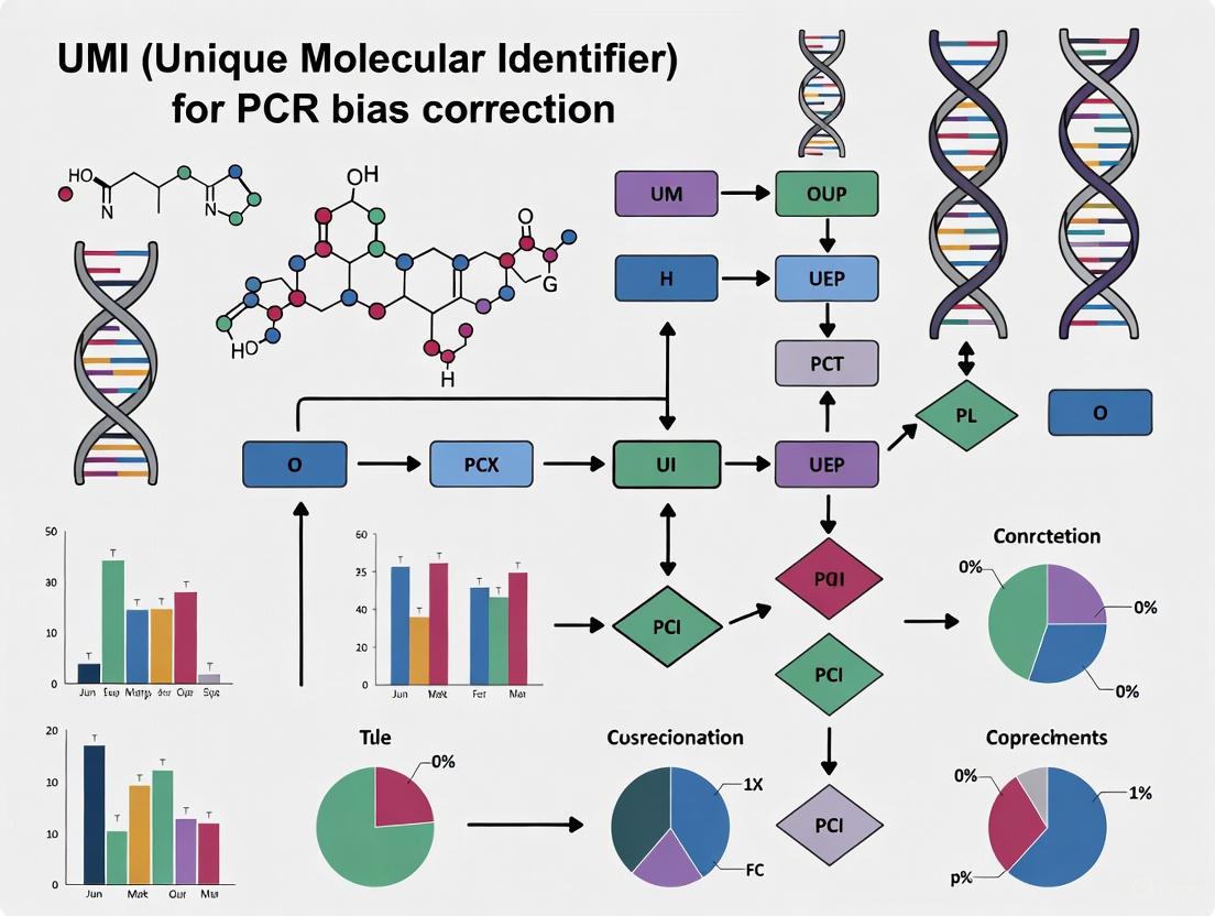
Visualizing UMI Workflows and Concepts
UMI Molecular Counting Principle
Homotrimeric UMI Error Correction
The Scientist's Toolkit: Research Reagent Solutions
Table 2: Essential Reagents and Tools for UMI-Based Sequencing
| Item | Function / Description | Example Use Case |
|---|---|---|
| Structured UMIs [5] [6] | UMI designs (e.g., homotrimeric blocks) that provide inherent error-correction capabilities. | Improving accuracy of absolute molecule counting in bulk or single-cell RNA-seq. |
| Library Prep Kits with UMIs [1] [2] | Commercial kits that incorporate UMI tagging during reverse transcription or early library construction steps. | Ensuring UMIs are added before PCR amplification in 3' RNA-Seq (e.g., QuantSeq) or single-cell protocols (e.g., 10X Genomics). |
| UMI-Tools Software [4] | A bioinformatics toolkit for handling UMI data, including error correction and deduplication. | Resolving PCR and sequencing errors in UMI sequences to generate accurate counts from sequencing data. |
| Common Molecular Identifier (CMI) [5] | A control sequence used to spike into experiments to directly measure the error rate of library prep and sequencing. | Benchmarking the performance and accuracy of different UMI protocols and correction methods. |
What are the fundamental issues that UMIs address in quantitative sequencing?
In quantitative sequencing, the core aim is to determine the original number and ratio of RNA or DNA molecules in a sample. However, nearly all sequencing protocols require a PCR amplification step to generate sufficient material for sequencing. This amplification is not perfectly neutral; it introduces two major types of distortions:
- Amplification Bias: Certain sequences are amplified more efficiently than others due to factors like GC content. This leads to the overrepresentation of some molecules in the final library, skewing the apparent abundance of sequences [4] [2] [3].
- PCR Duplicates: When a single original molecule is amplified, it produces multiple copies (duplicates) that are indistinguishable from reads derived from different, but identical, original molecules. Without a method to identify them, these duplicates lead to inaccurate quantification, making highly expressed transcripts appear even more abundant than they truly are [4] [2].
Before UMIs, a common bioinformatic approach was to remove reads that mapped to the same genomic coordinates, assuming they were PCR duplicates. This method is inefficient, especially for highly expressed genes or deep sequencing, as it often removes biological duplicates (true independent molecules from the same gene), further distorting quantification [3].
Unique Molecular Identifiers (UMIs) are short, random nucleotide sequences (typically 4-12 bases long) that provide an elegant solution to this problem [1] [7]. They are incorporated into each molecule in a library before any PCR amplification takes place. As a result, all PCR copies derived from the same original molecule inherit the same UMI sequence. After sequencing, reads that share both the same alignment coordinates and the same UMI can be confidently grouped as PCR duplicates and collapsed into a single, accurate count for the original molecule [4] [1] [2].
Table 1: Impact of PCR Amplification on Sequencing Data
| Aspect | Without UMIs | With UMIs |
|---|---|---|
| Quantification Accuracy | Distorted by amplification bias and over-counting of duplicates | High; based on counting original molecules, not reads |
| Handling of PCR Duplicates | Inefficient; can remove true biological signals | Precise; identifies and collapses technical replicates |
| Impact on Highly Expressed Genes | Severe overestimation of abundance | Accurate molecular counts |
| Rare Variant Detection | Challenging due to high background error rate | Enabled through consensus sequencing and error correction |
The following diagram illustrates how UMIs enable accurate molecular counting by tagging original molecules before amplification.
Frequently Asked Questions (FAQs) & Troubleshooting
FAQ 1: In which experimental scenarios are UMIs most critical?
UMIs provide the greatest benefit in specific, sensitive applications where accurate quantification is paramount. Their use is highly recommended in the following scenarios [3]:
- Single-cell RNA-seq (scRNA-seq) and ultra-low input samples (≤ 10 ng total RNA): These protocols start with very few molecules, necessitating a high number of PCR cycles to generate a usable library. This extensive amplification exacerbates biases and the risk of duplicate overcounting. UMIs are essential for accurate molecular counting at this scale [2] [8].
- Rare variant detection: In cancer genomics or studies of heterogeneous cell-free DNA (cfDNA), identifying true low-frequency mutations against a background of sequencing errors is challenging. UMIs allow for the creation of consensus sequences from read families, distinguishing true variants present in the original sample from errors introduced during library prep or sequencing [1] [3].
- Very deep sequencing of RNA-seq libraries (> 80 million reads per sample): As sequencing depth increases, so does the probability of sampling multiple reads from the same original molecule. UMIs enable precise deduplication, preventing the inflation of expression counts [3].
- Immune repertoire sequencing: This field requires accurately characterizing the immense diversity of B-cell and T-cell receptor genes. UMIs correct for artifactual diversity generated by PCR and sequencing errors, revealing the true clonal landscape [9].
For standard bulk RNA-seq experiments with moderate sequencing depth, the benefits of UMIs may be less pronounced, though they still serve as an excellent quality control tool for assessing library complexity [3].
FAQ 2: How do I choose the appropriate UMI length and design?
The goal of UMI design is to have a sufficiently large pool of unique identifiers to ensure it is statistically unlikely that two different original molecules will receive the same UMI by chance (a "collision").
- UMI Length: A typical UMI length is 8-12 nucleotides [7]. For a 10-nucleotide random sequence, there are 4^10 (1,048,576) possible unique combinations [2]. This is generally more than enough to tag the molecules in a typical sample. Longer UMIs reduce collision probability but use more of the sequencing read length.
- Advanced Error-Correcting Designs: Recent innovations focus on designing UMIs that are themselves resistant to errors.
- Homotrimer UMIs: This design replaces each single nucleotide in a UMI with a triplet of identical bases (e.g., 'A' becomes 'AAA'). This creates internal redundancy, allowing a "majority vote" system to correct single-base substitution errors within each triplet during bioinformatic processing. This method has been shown to correct over 96% of errors in common molecular identifiers (CMIs) across Illumina, PacBio, and Oxford Nanopore Technologies (ONT) platforms [5].
- Anchor Sequences: In droplet-based scRNA-seq, a short, fixed anchor sequence can be inserted between the cell barcode and the UMI. This helps bioinformatic tools accurately identify the start of the UMI, mitigating issues from oligonucleotide synthesis truncations [7].
FAQ 3: My molecular counts seem inflated after UMI deduplication. What could be the cause?
The inflation of molecular counts after deduplication is a classic symptom of uncorrected UMI sequencing errors [4] [7]. During PCR amplification and sequencing, nucleotide substitutions, insertions, or deletions (indels) can occur within the UMI sequence itself. A single error can transform a UMI into a new, seemingly unique identifier, causing one original molecule to be counted as two or more.
Table 2: Common Sources of UMI Errors and Their Impact
| Error Source | Error Types | Impact on Molecular Counting |
|---|---|---|
| PCR Amplification | Nucleotide substitutions that accumulate over cycles [5] | Creates artifactual UMIs, leading to overcounting |
| Sequencing | Incorrect base calls (miscalls), insertions, deletions [4] [7] | Creates artifactual UMIs, leading to overcounting |
| Oligonucleotide Synthesis | Truncations, unintended extensions [7] | Can cause misassignment of reads and inaccurate counts |
Solution: Ensure your bioinformatic pipeline includes a robust UMI error correction step. Standard deduplication, which only collapses reads with identical UMIs, is insufficient. Advanced tools use methods such as:
- Network-based clustering (e.g., UMI-tools): Groups similar UMIs (e.g., those with a Hamming distance of 1) at the same genomic locus and collapses them, inferring they originated from a common source [4].
- Directional adjacency: Conserns read counts to determine the most likely original UMI in a network of similar sequences [4].
- Homotrimer correction: Specifically designed for homotrimer UMI designs, using majority voting to correct errors within each trimer block [5].
FAQ 4: Are there any drawbacks or limitations to using UMIs?
While powerful, UMIs are not a panacea and have certain limitations:
- Not a substitute for optimal PCR: UMIs correct for duplication bias but do not eliminate the underlying biases in PCR amplification efficiency. The best practice is still to use the minimum number of PCR cycles necessary to generate your library [2].
- Computational complexity: Processing UMI data requires more sophisticated bioinformatics pipelines than standard sequencing data analysis [8].
- Read length consumption: UMIs and their associated spacers use up part of the sequencing read, which can be a consideration for short-read platforms [3].
- Limitations in error correction: While error correction algorithms are powerful, they can struggle with very complex networks of similar UMIs and may still under-correct in situations with extremely high error rates [4] [7].
Experimental Protocols & Reagent Solutions
This section outlines a key validation experiment from the literature that demonstrates the source and correction of UMI errors.
Protocol: Validating PCR as a Major Source of UMI Errors
This protocol is based on a controlled experiment published in Nature Methods [5].
1. Experimental Design:
- Use a synthetic Common Molecular Identifier (CMI)—a single, known sequence—attached to every captured RNA molecule in a pool of human and mouse cDNA.
- In the absence of errors, every transcript should be counted exactly once. The introduction of errors into the CMI sequence will cause transcripts to be overcounted, providing a direct measure of inaccuracy.
2. Library Preparation and Amplification:
- Split the CMI-tagged cDNA library into aliquots.
- Amplify each aliquot with a gradient of PCR cycles (e.g., 20, 25, 30, 35 cycles).
3. Sequencing and Analysis:
- Sequence the libraries on your platform of choice (e.g., Illumina, PacBio, ONT).
- For each sample, calculate the percentage of CMI sequences that match the expected, error-free sequence.
- Apply error-correction methods (e.g., homotrimer majority vote, UMI-tools) and measure the improvement in accurate CMI recovery.
4. Expected Outcome:
- The number of errors observed within the CMIs will increase substantially with the number of PCR cycles, demonstrating that PCR is a significant source of UMI errors.
- Robust error-correction methods (like the homotrimer approach) will correct a high percentage (>96%) of these errors, restoring accurate molecular counts [5].
Table 3: Key Research Reagent Solutions
| Reagent / Tool | Function in UMI Protocols |
|---|---|
| UMI-tools | A comprehensive bioinformatics toolkit for extracting UMIs from reads, error correction, and deduplication [4]. |
| Homotrimer UMI Primers | Oligonucleotides that synthesize UMIs using blocks of three identical nucleotides (AAA, CCC, GGG, TTT) to provide built-in error correction via majority voting [5]. |
| Common Molecular Identifier (CMI) | A non-random barcode used in validation experiments to spike into a library and directly measure the error rate introduced during library prep and sequencing [5]. |
| Cell Barcodes with Anchor Sequences | Oligonucleotides for single-cell workflows that include a fixed sequence between the cell barcode and UMI to mitigate issues from synthesis truncations [7]. |
| Sentieon UMI Workflow | A commercial, high-performance software solution for UMI extraction and consensus generation, often used in variant calling applications [10]. |
The following diagram visualizes the homotrimer error-correction method, a key experimental innovation.
Troubleshooting Guides & FAQs
UMI errors originate from three major sources throughout the experimental workflow. PCR amplification errors introduce random nucleotide substitutions that accumulate over multiple cycles. Sequencing errors occur during the sequencing process and vary by platform, including substitutions, insertions, and deletions. Oligonucleotide synthesis errors happen during UMI manufacturing, primarily involving truncation and elongation artifacts [7].
FAQ: How do UMI errors impact my experimental results?
UMI errors artificially inflate molecular counts by creating erroneous, distinct UMIs that are incorrectly interpreted as unique starting molecules. This leads to overestimation of transcript numbers in RNA-seq or molecule counts in DNA applications. In severe cases, these errors can generate false positives in differential expression analysis, with some studies reporting discordance rates of 7.8-11% for genes and transcripts between correction methods [5] [7].
Troubleshooting Guide: Addressing High UMI Error Rates
| Observation | Potential Cause | Solution |
|---|---|---|
| Inflated molecule counts with increasing PCR cycles | PCR amplification errors accumulating over cycles | Implement homotrimer UMI design for error correction; reduce PCR cycles if possible [5] |
| Platform-specific error patterns (e.g., high indel rates) | Sequencing errors inherent to platform chemistry | Apply platform-appropriate computational correction (e.g., UMI-tools for Illumina substitutions) [7] |
| Base composition bias at UMI start sites | Oligonucleotide synthesis/truncation errors | Incorporate anchor sequences in bead-based assays to demarcate UMI regions [7] |
| Persistent errors after standard correction | Complex error combinations or high error rates | Use integrated pipelines like UMIche combining multiple correction strategies [7] |
Quantitative Data on UMI Errors
Table: Sequencing Platform Error Profiles and Correction Efficacy
Data compiled from experimental validation using a Common Molecular Identifier (CMI) approach, where accuracy was measured as the percentage of correctly called CMI sequences [5].
| Sequencing Platform | Raw Accuracy (%) | Post-Homotrimer Correction (%) | Primary Error Type |
|---|---|---|---|
| Illumina | 73.36 | 98.45 | Substitutions [5] [7] |
| PacBio | 68.08 | 99.64 | Insertions/Deletions [5] [7] |
| ONT (latest chemistry) | 89.95 | 99.03 | Insertions/Deletions [5] [7] |
| ONT (older chemistry) | Substantially lower | Significant improvement | Insertions/Deletions [5] |
Table: Impact of PCR Cycles on UMI Error Rate
Experimental data from amplification of CMI-tagged cDNA libraries sequenced using Oxford Nanopore Technology [5].
| Experimental Condition | Key Finding | Implication |
|---|---|---|
| Increasing PCR cycles | Substantial increase in CMI errors [5] | PCR errors are significant source of UMI inaccuracy |
| PCR error correction | Homotrimer approach corrected significant proportion of errors [5] | Structural UMI designs mitigate PCR error impact |
| 20 vs 25 PCR cycles in single-cell | 25-cycle library had greater number of UMIs [5] | PCR errors inflate transcript counts and cause inaccurate quantification |
Experimental Protocols for UMI Error Validation
Protocol: Validating PCR Error Contribution to UMI Inaccuracy
This protocol describes the approach used to isolate and quantify PCR-derived UMI errors, as referenced in the 2024 Nature Methods study [5].
- Library Preparation: Attach a Common Molecular Identifier (CMI) to every captured RNA molecule. Using the same molecule for every RNA guarantees that, in the absence of errors, each transcript is counted only once.
- Amplification: Amplify the CMI-tagged cDNA library with increasing PCR cycles (e.g., 20, 25, 30, 35 cycles).
- Barcoding: Incorporate trimer barcodes during PCR to minimize batch effects and enable independent sequencing accuracy assessment.
- Sequencing: Split samples for sequencing across multiple platforms (Illumina, PacBio, ONT).
- Analysis: Calculate Hamming distance between observed and expected CMI sequences to measure error rates. Compare error rates across PCR cycle numbers to isolate amplification contribution.
Protocol: Evaluating UMI Error Correction Methods in Single-Cell RNA-seq
This protocol benchmarks computational versus structural UMI error correction methods [5].
- Cell Encapsulation: Encapsulate mixed-species cells (e.g., human JJN3 and mouse 5TGM1) using droplet-based systems (10X Chromium or Drop-seq).
- UMI Implementation: Compare standard monomer UMIs with homotrimer UMIs incorporated into barcoded beads.
- Amplification Series: Perform reverse transcription and split PCR products into aliquots for different amplification cycles (e.g., 20, 25, 30, 35 cycles).
- Sequencing: Sequence libraries on appropriate platforms (e.g., ONT PromethION for scale).
- Analysis Pipeline:
- Apply monomer UMI deduplication using tools like UMI-tools
- Apply homotrimer correction using majority voting and set coverage optimization
- Compare differential expression results between methods
- Quantify discordant differentially expressed genes/transcripts
Visualization of UMI Error Correction Concepts
Diagram: Homotrimer UMI Error Correction Mechanism
Homotrimer UMI Correction
Diagram: Network-Based UMI Error Correction
UMI Clustering by Edit Distance
Research Reagent Solutions
Table: Essential Materials for UMI Error Mitigation
| Reagent/Kit | Function | Application Note |
|---|---|---|
| Homotrimer UMI Primers | Structural error correction via triple modular redundancy | Replaces each nucleotide with triplet (e.g., A→AAA) for majority voting [5] [7] |
| Anchor Sequence Oligos | Demarcates barcode-UMI junction | Reduces truncation errors in bead-based assays; improves UMI identification [7] |
| Common Molecular Identifier (CMI) | Validation standard for error measurement | Same sequence attached to all molecules enables error quantification [5] |
| Platinum SuperFi II Green PCR Master Mix | High-fidelity amplification | Reduces PCR-induced errors during library preparation [11] |
| Agencourt AMPure XP Beads | Size selection and purification | Removes off-target amplification products; critical for UMI workflow cleanliness [11] |
Unique Molecular Identifiers (UMIs) are short, random oligonucleotide sequences used in next-generation sequencing to label individual DNA or RNA molecules before PCR amplification. Their primary purpose is to distinguish true biological molecules from PCR duplicates, thereby enabling accurate molecular counting and reducing amplification biases [5] [4]. However, errors introduced during library preparation, PCR amplification, and sequencing can compromise UMI effectiveness, leading to inaccurate data interpretation [7].
When errors occur within UMI sequences themselves, they create artifactual UMIs that inflate molecular counts and can generate false positive variant calls. This technical guide explores the sources, impacts, and solutions for UMI errors, providing researchers with practical troubleshooting approaches to maintain data integrity in their experiments [5] [7].
FAQ: Understanding UMI Errors and Their Consequences
Q1: What are the primary sources of UMI errors in sequencing experiments?
UMI errors originate from three major sources throughout the experimental workflow:
PCR Amplification Errors: Random nucleotide substitutions occur during PCR amplification and accumulate over multiple cycles. As each round uses previously synthesized products as templates, even low-frequency errors can propagate and become fixed. The error rate significantly increases with each PCR cycle, making this especially problematic in single-cell sequencing where limited input material necessitates extensive amplification [5] [7].
Sequencing Errors: Incorrect base calls during sequencing lead to mismatches between the readout and original template. These include substitutions, insertions, and deletions. Error profiles vary by platform: Illumina exhibits low overall error rates dominated by substitutions, while long-read platforms like PacBio and Oxford Nanopore Technologies are more susceptible to indel errors [5] [7].
Oligonucleotide Synthesis Errors: Chemical manufacturing of UMIs involves finite coupling efficiency (≈98-99% per step), leading to truncated sequences or unintended extensions. As UMI length increases, the cumulative probability of synthesis errors rises substantially [7].
Q2: How do UMI errors lead to false positives and inflated molecular counts?
UMI errors create artificial diversity that manifests as two primary data quality issues:
Inflated Molecular Counts: When errors create new, artifactual UMI sequences, multiple reads originating from the same original molecule are incorrectly counted as distinct molecules. Experiments show this inflation increases with PCR cycles—libraries subjected to 25 PCR cycles showed significantly greater UMI counts compared to those with 20 cycles, despite originating from the same sample [5].
False Positive Variant Calls: In variant detection applications, particularly at low allele frequencies (below 1%), UMI errors can be mistaken for true biological variants. This is especially problematic in circulating tumor DNA (ctDNA) analysis where true variants occur at very low frequencies similar to error rates [12] [13].
Q3: What evidence demonstrates the impact of UMI errors on differential expression analysis?
UMI errors directly impact transcriptional analyses by altering gene expression estimates:
- In single-cell RNA sequencing, differential expression analysis between conditions can show 7.8-11% discordance in regulated genes when comparing standard UMI correction versus enhanced error correction methods [5].
- More genes may appear differentially regulated after standard monomer-based UMI correction compared to advanced error-correcting methods, with examples showing marked differences in read counts for genes like TEL5 and FRG2 after proper correction [5].
- Without appropriate error correction, analyses have identified over 300 differentially regulated transcripts between libraries with different PCR cycles—artifacts that disappear when applying robust UMI error correction [5].
Troubleshooting Guide: Identifying and Resolving UMI Error Issues
Problem: Inflated Molecular Counts
Symptoms:
- Higher-than-expected unique molecular counts despite low input material
- Molecular counts that increase disproportionately with additional PCR cycles
- Discrepancies between technical replicates in molecular counting data
Solutions:
Wet-Lab Protocol: Implement Homotrimer UMI Design Synthesize UMIs using homotrimer nucleotide blocks (e.g., AAA, CCC, GGG, TTT) rather than traditional monomeric UMIs. This design incorporates built-in redundancy:
- Experimental Procedure:
- Order custom oligonucleotides with homotrimer blocks in the UMI region
- Label RNA with homotrimeric UMIs at both ends for enhanced error detection
- Process through standard library preparation appropriate for your sequencing platform (Illumina, ONT, or PacBio)
- Error Correction Mechanism:
- Process UMIs by assessing trimer nucleotide similarity
- Correct errors by adopting the most frequent nucleotide in a "majority vote" approach within each trimer block
- This approach corrects 96-99% of CMI sequences across platforms, even with increasing PCR cycles [5]
Computational Solution: Apply Network-Based Error Correction For existing data with traditional UMIs, implement graph-based clustering:
- Tool Recommendation: UMI-tools or mclUMI
- Implementation Steps:
- Extract UMI sequences from read headers
- For each genomic locus, build a network where nodes represent UMIs
- Connect edges between UMIs separated by a single edit distance
- Resolve networks using directional or adjacency methods to identify true UMIs
- This approach improves quantification accuracy in both iCLIP and single-cell RNA-seq data [4]
Problem: False Positive Variants in Low-Frequency Applications
Symptoms:
- Apparent low-frequency variants (below 1%) that don't validate orthogonally
- Variants appearing in only a subset of reads within a UMI family
- High false discovery rates in ctDNA or rare mutation detection studies
Solutions:
Wet-Lab Protocol: Incorporate Molecular Spikes for Validation Use spike-in controls with known sequences to quantify error rates:
- Experimental Procedure:
- Clone randomized synthetic DNA sequences with built-in UMIs into plasmid vectors
- Include T7 promoter and poly-A tail for RNA spike-ins
- Add spike-ins to samples before library preparation
- Sequence alongside experimental samples
- Validation Process:
- Extract spike-in UMI sequences from aligned reads
- Compare observed versus expected spike-in UMI sequences
- Calculate error rates and establish quality thresholds
- Use these metrics to normalize or filter experimental data [14]
Computational Solution: Implement UMI-Aware Variant Calling For variant detection applications, use specialized variant callers:
- Tool Recommendation: UMI-VarCal or DeepSNVMiner
- Implementation Steps:
Experimental Protocols for UMI Error Mitigation
Protocol 1: Validating UMI Counting Accuracy Using Molecular Spikes
Purpose: To experimentally quantify UMI error rates and validate counting accuracy in single-cell RNA sequencing experiments.
Materials:
- Molecular spike plasmids (5' or 3' design depending on protocol)
- In vitro transcription kit
- Cell line of interest (e.g., HEK293FT cells)
- Single-cell RNA-seq library preparation kit
- Sequencing platform access
Procedure:
- Spike-in Preparation:
- Perform in vitro transcription from molecular spike plasmids to create spike RNA
- Quantify RNA concentration and integrity
- Dilute to appropriate concentration for single-cell experiments
Sample Processing:
- Add molecular spikes to single-cell suspensions at a concentration matching expected cellular RNA abundances
- Proceed with standard single-cell RNA-seq protocol (e.g., 10x Genomics, Smart-seq3)
- Prepare sequencing libraries according to platform specifications
Data Analysis:
- Sequence libraries to appropriate depth
- Align reads to combined genome + spike reference
- Extract spike-in UMI sequences (spUMIs) from aligned reads
- Apply error correction with Hamming distance threshold (typically 1-2 nt)
- Compare observed spUMI counts to expected counts based on spike input
- Calculate counting accuracy and error rates [14]
Protocol 2: Evaluating PCR Cycle Impact on UMI Errors
Purpose: To systematically quantify how PCR amplification cycles contribute to UMI errors.
Materials:
- cDNA library with common molecular identifier (CMI)
- PCR master mix
- Trimer barcoded beads (for Drop-seq applications)
- Access to multiple sequencing platforms (Illumina, PacBio, ONT)
Procedure:
- Library Preparation:
- Attach CMI to equimolar concentrations of control cDNA (e.g., mouse/human mix)
- Split sample into aliquots for different PCR cycle conditions
- Amplify with varying PCR cycles (e.g., 20, 25, 30, 35 cycles)
- Sequencing and Analysis:
- Sequence aliquots across multiple platforms (Illumina, PacBio, ONT)
- Calculate Hamming distance between observed and expected CMI sequences
- Quantify platform-specific error rates
- Apply homotrimer error correction to assess correction efficiency
- Compare error rates before and after computational correction [5]
Table 1: UMI Error Rates Across Sequencing Platforms Before and After Correction
| Platform | Raw Accuracy (%) | After Homotrimer Correction (%) | Primary Error Type |
|---|---|---|---|
| Illumina | 73.36 | 98.45 | Substitutions |
| PacBio | 68.08 | 99.64 | Indels |
| ONT (latest) | 89.95 | 99.03 | Indels |
Source: Adapted from Nature Methods 21, 401-405 (2024) [5]
Table 2: Impact of PCR Cycles on UMI Error Rates
| PCR Cycles | Error Rate Increase | Homotrimer Correction Efficiency |
|---|---|---|
| 20 | Baseline | >96% |
| 25 | Moderate increase | >96% |
| 30 | Significant increase | >96% |
| 35 | Substantial increase | >96% |
Source: Adapted from Nature Methods 21, 401-405 (2024) [5]
Visual Workflows
Diagram 1: UMI Error Correction with Homotrimer Design
Diagram 2: Impact of UMI Errors on Molecular Counting
The Scientist's Toolkit: Research Reagent Solutions
Table 3: Essential Reagents and Tools for UMI Error Management
| Tool/Reagent | Function | Application Context |
|---|---|---|
| Homotrimer UMI Oligos | Provides built-in error correction via redundant nucleotide blocks | All sequencing applications requiring high counting accuracy |
| Molecular Spike Ins | Experimental ground truth for quantifying UMI error rates | Protocol validation and quality control |
| UMI-Tools Software | Network-based computational correction of UMI errors | Analysis of existing datasets with traditional UMIs |
| Trimer Barcoded Beads | Specialized beads for droplet-based scRNA-seq with error correction | Single-cell RNA sequencing applications |
| UMI-VarCal | Variant caller specifically designed for UMI-encoded data | Low-frequency variant detection in ctDNA |
| FGBio Toolkit | Processing UMI-encoded NGS data before variant calling | Standard workflow for UMI-aware variant detection |
FAQs: Core Concepts and Applications
Q1: What is the fundamental difference between a sample barcode and a Unique Molecular Identifier (UMI)?
Sample barcodes (or indexes) and UMIs are both short nucleotide sequences, but they serve distinct purposes and are applied differently. Sample barcodes are used to label all nucleic acids from a single sample library, enabling the pooling and subsequent computational separation of multiple samples after a single sequencing run. In contrast, UMIs are used to label each individual molecule within a single sample library before PCR amplification. This allows bioinformatics tools to distinguish between true biological duplicates and artifacts created during PCR amplification and sequencing, thereby improving quantification accuracy and variant calling [15] [1].
Q2: In which applications are UMIs considered essential?
UMIs are particularly crucial in applications where precise quantification of unique molecules or detection of rare variants is required. Key applications include:
- Single-cell RNA Sequencing (scRNA-seq): To control for amplification biases and accurately count transcript molecules from minimal input material [15] [2].
- Circulating Tumor DNA (ctDNA) Analysis and Rare Variant Detection: To distinguish true low-frequency mutations (e.g., below 1%) in oncology from errors introduced during library preparation and sequencing [16] [2].
- Minimal Residual Disease (MRD) Monitoring: Where variant calling at frequencies of 0.1% or lower is required [16].
- Any quantitative sequencing method where PCR duplicates are a significant concern, such as ChIP-seq, antibody repertoire sequencing, and karyotyping [4] [2].
Q3: Are UMIs universally beneficial for all NGS experiments?
No, the advantage of using UMIs is context-dependent. Recent research indicates that for some hybridization-based methods using DNA from high-quality, high-input sources (e.g., fresh frozen tissue), noise suppression and reliable variant calling can be achieved through read grouping based on fragment mapping positions alone, without exogenous UMIs. The significant benefit of UMIs becomes apparent when "collisions" (different original molecules sharing the same mapping position) are common, which is often the case with highly fragmented DNA like cell-free DNA (cfDNA) [16].
Q4: What are the main sources of inaccuracy when using UMIs?
The primary source of inaccuracy is PCR amplification errors, which can introduce substitutions, insertions, or deletions within the UMI sequence itself. This creates artifactual UMIs that inflate molecular counts and lead to inaccurate quantification [5]. Sequencing errors also contribute, but to a lesser extent [5] [4]. The following table summarizes the impact of these errors:
Table: Impact and Correction of UMI Errors
| Error Type | Effect on UMI Data | Common Correction Methods |
|---|---|---|
| PCR Errors (nucleotide substitutions) [5] | Creates new, incorrect UMI sequences, leading to overcounting of molecules. | Homotrimer nucleotide block design [5]; Network-based clustering (e.g., UMI-tools) [4]. |
| Sequencing Errors (base miscalling, indels) [4] | Alters the perceived UMI sequence, creating artifactual UMIs. | Hamming distance-based clustering; Majority vote consensus. |
| PCR Recombination ("jumping") [4] | Creates chimeric sequences, potentially altering both UMI and genomic alignment. | More complex network analysis; Can be mitigated by specific library prep protocols. |
Troubleshooting Guides
Issue 1: Inaccurate Transcript or Molecule Counting After UMI Deduplication
Problem: After processing UMI-tagged data, the quantitative counts of transcripts or DNA molecules are suspected to be inflated or inaccurate.
Potential Causes and Solutions:
- Cause: PCR errors generating artifactual UMIs. With each PCR cycle, the risk of errors in the UMI sequence increases, creating new, erroneous UMI sequences that are counted as unique molecules [5].
- Solution: Implement an error-correcting UMI design. Consider using homotrimeric nucleotide blocks for UMI synthesis. In this approach, each nucleotide position in the UMI is represented by a trimer. Errors can be corrected by a "majority vote" within each trimer block, significantly improving counting accuracy [5].
- Solution: Use computational tools that model UMI errors. Tools like UMI-tools employ network-based methods to cluster UMIs that are within a small Hamming distance of one another, inferring and correcting for errors [4].
- Cause: Over-amplification of the library.
Issue 2: Deciding Whether to Use UMIs in a DNA Sequencing Experiment
Problem: A researcher is unsure if the added cost and complexity of UMI incorporation are justified for their specific DNA-based NGS assay.
Solution: Follow a decision framework based on sample type and assay goal. The flowchart below outlines key decision points for determining when UMIs provide a critical advantage in DNA sequencing experiments.
Issue 3: High Computational Resource Use during UMI Processing
Problem: Bioinformatics processing of UMI-tagged data (e.g., with UMI-tools) is taking an excessively long time and consuming large amounts of memory.
Potential Causes and Solutions:
- Cause: Large network sizes for UMI error correction. The time taken to resolve UMI groups is dependent on the number of unique UMIs and their connectivity. Shorter UMIs, high sequencing depth, and high error rates all increase network size and processing time [17].
- Solution: If possible, use longer UMIs in experimental design to reduce connectivity.
- Solution: For UMI-tools, use the
--per-cellflag for single-cell data to process cells independently [17].
- Cause: Handling of chimeric or unmapped reads.
- Solution: To save memory, consider discarding chimeric read pairs (
--chimeric-reads=discard) instead of attempting to use them [17].
- Solution: To save memory, consider discarding chimeric read pairs (
The Scientist's Toolkit: Research Reagent Solutions
Table: Essential Materials for UMI-Based Experiments
| Item | Function in Experiment | Key Considerations |
|---|---|---|
| UMI-tools Software [4] | A comprehensive bioinformatics package for UMI processing, including deduplication, error correction, and counting. | Network-based methods account for sequencing errors in UMIs. Essential for accurate quantification in bulk and single-cell data. |
| Homotrimer UMI Design [5] | UMI synthesized using homotrimer nucleotide blocks to enable robust PCR error correction via a "majority vote" system. | Significantly improves accuracy of absolute molecule counting. Particularly beneficial for long-read sequencing platforms. |
| DRAGEN UMI Pipeline [18] | Illumina's integrated bioinformatics solution for UMI-based error correction and variant calling. | Optimized for Illumina sequencing data. Supports both random and non-random (e.g., TSO500) UMI designs. |
| Capture-Based Enrichment Panel [16] | A targeted panel (e.g., for oncology) used in conjunction with UMIs to enable sensitive variant calling. | The benefit of exogenous UMIs is most pronounced in capture-based assays with fragmented DNA (cfDNA) where mapping position collisions are common. |
| Error-Correcting UMI Barcoded Beads (e.g., for Drop-seq) [5] | Beads used in single-cell workflows that incorporate advanced UMI designs for improved error correction. | Enhances the accuracy of transcript counting in single-cell RNA-seq experiments by mitigating PCR errors. |
UMI Implementation Strategies: From Experimental Designs to Computational Correction Methods
Frequently Asked Questions
Q1: What is the main advantage of using homotrimer UMIs over traditional monomeric UMIs? Homotrimer UMIs incorporate built-in error correction by replacing each nucleotide in a standard UMI with a block of three identical nucleotides (e.g., A becomes AAA). This design allows for a "majority vote" system where sequencing or PCR errors affecting a single base in a trimer block can be detected and corrected, significantly improving the accuracy of molecular counting. This is particularly effective at mitigating PCR errors, which are a major source of inaccuracy [19] [5] [7].
Q2: My RNA-seq data still shows inflated transcript counts after using UMIs and standard computational tools (e.g., UMI-tools). What could be the issue? Persistent inflation of transcript counts is likely due to PCR errors that standard computational tools cannot fully correct. These tools often rely on Hamming distances and struggle with indel errors and high error rates from increased PCR cycles. Switching to a library preparation method that uses homotrimer UMIs can address this, as their structure provides inherent redundancy for correcting substitution errors and some indels, which monomeric UMIs handle poorly [19] [5].
Q3: How do I implement homotrimer UMIs in my single-cell RNA-seq experiment? You can implement homotrimer UMIs by using bespoke synthesis on beads (e.g., for Drop-seq) where the UMI region is composed of homotrimer nucleotide blocks. During data processing, a custom demultiplexing strategy is required. This involves grouping the UMI sequence into trimer blocks and applying a majority vote to each block to determine the most likely original nucleotide before proceeding with standard deduplication [19] [7].
Q4: Are homotrimer UMIs compatible with different sequencing platforms? Yes, homotrimer UMIs are compatible with major sequencing platforms, including Illumina, PacBio, and Oxford Nanopore Technologies (ONT). Experimental validation has shown that homotrimer correction significantly improves UMI accuracy on all these platforms, with correction rates achieving over 98% accuracy [19] [5].
Q5: What are "bead truncation errors" and how can they be mitigated? Bead truncation errors occur during the chemical synthesis of oligonucleotides on beads, primarily resulting in truncated sequences. This is a common issue in droplet-based single-cell methods. An effective mitigation strategy is to incorporate a short, fixed anchor sequence between the cell barcode and the UMI. This anchor acts as a positional landmark, helping computational pipelines correctly identify the start of the UMI sequence even when the oligonucleotide is incompletely synthesized [7].
Performance Comparison: Homotrimer vs. Monomer UMI Correction
The following table summarizes key experimental findings that highlight the effectiveness of homotrimer UMIs.
| Metric | Homotrimer UMI Performance | Traditional Monomer UMI Performance | Experimental Context |
|---|---|---|---|
| CMI/UMI Error Correction | Corrected 96–100% of Common Molecular Identifier (CMI) sequences [19]. | Benchmarking tools (UMI-tools, TRUmiCount) showed substantially less effective correction [19]. | Bulk cDNA with CMI, across increasing PCR cycles (10-35 cycles) [19]. |
| Impact on Differential Expression (DE) Analysis | 0 differentially expressed transcripts falsely identified due to PCR errors [19]. | Over 300 differentially regulated transcripts falsely identified between 20 vs. 25 PCR cycle libraries [19]. | Single-cell RNA-seq (JJN3 human & 5TGM1 mouse cells) with varying PCR cycles [19]. |
| Accuracy Across Sequencers | Improved CMI accuracy to 98.45% (Illumina), 99.64% (PacBio), 99.03% (ONT) [5]. | Initial accuracy was 73.36% (Illumina), 68.08% (PacBio), 89.95% (ONT) without correction [5]. | Bulk cDNA with a CMI, sequenced on multiple platforms [5]. |
| Handling of Indel Errors | Methodology can overcome indel errors due to block-based correction [19]. | A single indel can inflate Hamming distance beyond correctability [19]. | -- |
Experimental Protocol: Validating Homotrimer UMI Performance
This section outlines a key experiment from the literature that validates the homotrimer UMI approach [19] [5].
Objective: To quantify the rate of PCR errors and demonstrate the superior error correction of homotrimer UMIs compared to monomeric UMIs and standard computational tools.
Key Reagents:
- Cell Lines: JJN3 (human), 5TGM1 (mouse), RM82 (Ewing's sarcoma)
- Reagents: CLK1 splicing kinase inhibitor (e.g., SGC-CLK-1), DMSO (vehicle control)
- Library Prep: Drop-seq beads with trimer-barcoded oligonucleotides; 10X Chromium system
- Sequencing Platforms: ONT MinION/PromethION, Illumina, PacBio
Methodology:
- Library Preparation with CMI: A Common Molecular Identifier (CMI)—a single, known UMI sequence—is attached to every RNA molecule during reverse transcription. In a perfect system, this should result in a single count per molecule, and any increase indicates errors.
- Controlled PCR Amplification: The CMI-tagged library is split into aliquots and subjected to different numbers of PCR cycles (e.g., 10 to 35 cycles) to systematically increase PCR error rates.
- Sequencing: The amplified libraries are sequenced on multiple platforms (ONT, Illumina, PacBio).
- Data Analysis:
- The observed CMI sequences are compared to the expected CMI sequence.
- Homotrimer Correction: The CMI sequence is processed in blocks of three nucleotides. For each block, a majority vote is applied to correct any single-base error.
- The performance is benchmarked against computational tools like
UMI-toolsandTRUmiCount.
- Biological Validation: RM82 cells are treated with a CLK1 inhibitor versus DMSO control. Libraries are prepared and sequenced (e.g., with ONT). Differential expression analysis is performed using both monomer UMI correction (
UMI-tools) and homotrimer UMI correction to compare the results.
This experimental workflow, from library preparation to data analysis, can be visualized in the following diagram:
Homotrimer UMI Error Correction Logic
The core innovation of homotrimer UMIs is their internal redundancy. The process for correcting errors in a sequenced homotrimer UMI is as follows:
The Scientist's Toolkit: Research Reagent Solutions
| Reagent / Material | Function in Protocol |
|---|---|
| Homotrimer-barcoded Beads | Provides the physical support for oligonucleotides containing homotrimer UMI sequences during single-cell library preparation (e.g., Drop-seq) [19]. |
| Common Molecular Identifier (CMI) | A single, known UMI sequence used as an internal control to directly measure and quantify the error rate introduced during library prep and sequencing [19] [5]. |
| CLK1 Inhibitor (e.g., SGC-CLK-1) | A small molecule used to induce specific and strong splicing perturbations in cell lines (e.g., RM82), providing a robust biological signal to test quantification accuracy [19] [5]. |
| UMI-tools Software | A widely used computational package for UMI deduplication; serves as the benchmark "gold standard" for comparing the performance of new methods like homotrimer UMIs [19] [4]. |
Frequently Asked Questions
What is the primary function of a UMI in sequencing experiments? Unique Molecular Identifiers (UMIs) are short, random nucleotide sequences (barcodes) that are ligated to each DNA or RNA molecule in a sample library before any PCR amplification steps [1] [2]. Their main function is to act as a unique tag for each original molecule, enabling the bioinformatic identification and removal of PCR duplicates—identical copies generated from the same original molecule during amplification [20] [2]. This process, known as deduplication, corrects for PCR amplification biases and provides accurate, absolute counts of the original molecules, which is crucial for quantitative sequencing applications [5] [1].
What are the common sources of errors that affect UMIs? Errors that distort UMI sequences and lead to inaccurate molecular counting arise from three major sources [7]:
- PCR Amplification Errors: Random nucleotide substitutions occur during PCR and are exponentially propagated through subsequent cycles. The error rate increases with the number of PCR cycles [5] [7].
- Sequencing Errors: These are incorrect base calls (substitutions, insertions, or deletions) introduced by the sequencing platform itself. Different platforms have distinct error profiles; for example, Illumina has low overall error rates dominated by substitutions, while Oxford Nanopore Technologies (ONT) and PacBio have higher rates of insertion and deletion errors [4] [7].
- Oligonucleotide Synthesis Errors: Errors occur during the chemical manufacturing of the UMI oligonucleotides themselves, primarily involving truncations or unintended extensions due to the finite coupling efficiency of each synthesis step [7].
When are UMIs most critical for an experiment? UMIs offer the greatest benefit in experiments where input material is limited, requiring extensive PCR amplification that exacerbates biases. This is particularly critical for [2]:
- Single-cell RNA sequencing (scRNA-seq)
- Low-input RNA sequencing (≤ 10 ng total RNA)
- Detection of rare sequence variants
- Immune repertoire sequencing
- Any application requiring absolute molecular quantification
For experiments with high input amounts, the benefit of UMIs may be reduced as the number of RNA molecules can exceed the number of available UMI sequences [2].
UMI Length and Design Optimization
How long should a UMI be? The optimal UMI length balances the need for a large diversity of unique tags with practical constraints of cost, sequencing throughput, and the specific application. A UMI must have enough possible unique combinations to ensure that each original molecule in the library receives a distinct tag.
Table 1: UMI Length and Diversity
| UMI Length (Nucleotides) | Theoretical Number of Unique UMIs | Considerations and Applications |
|---|---|---|
| 10 nt | 1,048,576 (410) [2] | A common and versatile length, suitable for many applications including single-cell RNA-seq [2]. |
| 8-12 nt | 65,536 to 16,777,216 | The typical range for standard UMIs; longer UMIs within this range provide more unique identifiers, reducing the chance of "collision" where different molecules get the same UMI [7] [9]. |
| Homotrimer Design (e.g., 3x10 nt blocks) | Provides error-correction capability | This design replaces each nucleotide in a conceptual UMI with a triplet of identical bases (e.g., A becomes AAA). It introduces redundancy, allowing for "majority vote" error correction within each triplet and significantly improves accuracy, especially under high PCR cycles [5] [7]. |
What are the advanced structural designs for UMIs? Beyond simple random sequences, innovative UMI designs enhance error correction:
- Homotrimer UMIs: As described in Table 1, this design uses homotrimeric nucleotide blocks (e.g., AAA, CCC, GGG, TTT) to synthesize UMIs. Errors are corrected by adopting the most frequent nucleotide in a "majority vote" within each trimer block. This method has been shown to correct over 96% of errors in common molecular identifiers (CMIs) across Illumina, PacBio, and ONT platforms [5].
- Anchor Oligonucleotide Sequence: A short, predefined sequence is inserted between the cell barcode and the UMI region on a sequencing bead. This acts as a positional landmark to help computational pipelines reliably detect the start of the UMI, mitigating issues from oligonucleotide synthesis truncations [7].
Diagram 1: Homotrimer UMI error correction workflow. Each original molecule is tagged with a redundant UMI. After PCR and sequencing introduce errors, a majority vote within each trimer block corrects the sequence, enabling accurate molecular counting [5] [7].
UMI Placement and Adapter Integration
What are the best practices for UMI placement in a protocol? UMIs should be incorporated as early as possible in the library preparation workflow, always before the PCR amplification step [2]. The point of integration determines which parts of the process are corrected for bias.
Table 2: UMI Placement Strategies and Their Advantages
| Placement in Workflow | Method of Integration | Advantages |
|---|---|---|
| Reverse Transcription | As part of the oligo(dT) primer or random primers [2] [9] | Tags the original RNA molecule, correcting for biases in reverse transcription and all subsequent amplification steps. This is the earliest possible point of integration. |
| Second Strand Synthesis | As part of the second strand synthesis primer [2] | Corrects for biases from the second strand synthesis step onward. |
| Adapter Ligation | Incorporated directly into the library adaptor [21] [2] | A versatile method compatible with many standard protocols. Corrects for biases from the point of ligation onward. |
| Duplex Tagging | Adding UMIs at both ends of the molecule [5] [9] | Provides the highest power for error correction and consensus calling. Tolerates errors more effectively than single-end tagging. |
How can adapter design improve sequencing accuracy? Innovative adapter designs can be used to monitor and improve sequencing quality:
- Control Library Adaptors (CAPTORs): These are adaptors that encode known reference control sequences [21]. When sequenced, they provide a per-read measure of sequencing accuracy and quantitative library bias. The sequenced CAPTOR sequence is compared to its known ground-truth, generating a detailed error profile that can benchmark performance between samples, reagents, and sequencing runs [21].
- Back-to-Back Adapters: A specialized double-stranded oligonucleotide adapter with two primer sequences in a back-to-back orientation can reduce amplification bias resulting from variations in the GC content of the sample DNA fragments [22].
Diagram 2: Using CAPTORs for real-time sequencing QC. Control adaptors with known sequences are ligated to sample DNA. Sequencing these CAPTORs first provides an immediate measure of accuracy for each read and the overall run [21].
The Scientist's Toolkit
Table 3: Essential Research Reagents and Computational Tools
| Reagent / Tool | Function | Key Features |
|---|---|---|
| Homotrimer UMI Oligos [5] | Experimental reagent for error-resistant molecular tagging | Synthesized using homotrimeric nucleotide blocks (e.g., AAA, GGG) to enable majority-vote error correction during bioinformatic processing. |
| CAPTOR Adaptors [21] | Experimental reagent for internal sequencing control | Library adaptors containing a variable reference control region to measure per-read and per-run sequencing accuracy and bias. |
| UMI-tools [4] | Computational software for UMI error correction | A widely used open-source package that uses network-based methods to account for sequencing errors in UMI sequences when identifying PCR duplicates. |
| Molecular Amplification Fingerprinting (MAF) [9] | An advanced UMI strategy for ultrasensitive quantification | Incorporates distinct reverse and forward UMI tags to track molecules through cDNA synthesis and PCR, enabling an algorithm to correct amplification bias with high (98-100%) accuracy. |
Troubleshooting Common UMI Issues
How do I correct for UMI sequencing errors bioinformatically? Sequencing errors in the UMI itself can create artifactual UMIs, inflating molecular counts. Computational tools group similar UMIs to infer and correct the original sequence.
- Network-Based Clustering (e.g., UMI-tools): This method forms networks where UMIs at the same genomic locus are nodes, and edges connect UMIs separated by a single nucleotide difference (edit distance) [4]. The networks are then resolved to estimate the true number of original molecules.
- Adjacency Method: The most abundant UMI node and all nodes connected to it are removed first. This is repeated with the next most abundant node until the network is resolved. The number of steps equals the estimated number of unique molecules [4].
- Directional Method: A more sophisticated approach that uses read counts to create directional edges, reasoning that an error-derived UMI will have a lower count than its parent UMI [4].
- Markov Clustering (MCL): Tools like
mclUMIapply the Markov cluster algorithm to group similar UMI sequences, offering improved accuracy for high-error conditions without relying on fixed edit distance thresholds [7]. - Integrated Platforms (e.g., UMIche): These platforms combine multiple algorithms, such as graph-based clustering, distance-based filtering, and set cover optimization, in a multi-stage pipeline for robust error correction [7].
My molecular counts seem inflated after UMI deduplication. What could be the cause? Inflation after deduplication often points to unresolved UMI errors. Solutions include:
- Validate and Optimize Computational Correction: Ensure you are using an appropriate algorithm (e.g., directional or clustering method) for your data type and sequencing platform. Benchmark different tools and parameters [4] [7].
- Re-evaluate Experimental Conditions: High PCR cycle numbers are a major source of UMI errors. An experiment showed a substantial increase in errors within common molecular identifiers (CMIs) as PCR cycles increased from 20 to 25 [5]. Minimize PCR cycles where possible.
- Consider UMI Design: If starting a new experiment, consider implementing an error-resilient UMI design like homotrimer UMIs, which are specifically designed to correct the PCR errors that cause count inflation [5].
How can I improve UMI recovery from barcoded beads? Poor UMI recovery in droplet-based methods is frequently due to oligonucleotide synthesis truncations on the beads.
- Implement an Anchor Sequence: Introduce a short, predefined anchor sequence between the cell barcode and the UMI on the bead-bound primer. This provides a stable landmark for computational pipelines, improving the accurate identification of UMIs even when the oligonucleotide is truncated, leading to a higher fraction of usable reads [7].
Frequently Asked Questions
1. Why does UMI-tools deduplicate result in a BAM file with no reads?
This occurs when the tool is run on an unmapped BAM file. UMI-tools dedup requires mapped reads because it uses genomic alignment coordinates to group reads before examining their UMIs. If you input unmapped reads, the tool has no coordinates to process and will produce an empty or nearly empty output [23].
- Solution: Always provide a BAM file where reads have been aligned to a reference genome. If your goal is to analyze unmapped data (e.g., for metagenomic viral detection), you will need to use an alignment-free deduplication tool or map your reads to a composite reference [23] [24].
2. My UMI count seems artificially high after more PCR cycles. Is this expected? Yes, this is a documented issue. PCR errors can introduce substitutions into the UMI sequence itself, creating artifactual UMIs that inflate molecular counts. One study showed that increasing PCR cycles from 20 to 25 led to a measurable increase in UMI counts, which was attributed to these PCR errors and not an actual increase in unique molecules [5].
- Solution: Consider using error-correcting UMI designs, such as homotrimer UMIs, or computational tools that can model and correct for PCR errors within UMI sequences to generate more accurate counts [5].
3. What is the difference between "alignment-based" and "alignment-free" UMI tools? This is a fundamental distinction in how UMI clustering algorithms operate.
- Alignment-based tools (e.g., UMI-tools, DRAGEN): These tools first align reads to a reference genome. They then group reads and correct UMIs based on a combination of their alignment coordinates and UMI sequences. This is highly effective for reducing sequencing errors and amplification bias but is dependent on the reference genome, which can be a limitation for de novo applications or detecting complex indels [4] [24] [25].
- Alignment-free tools (e.g., AFUMIC, UMIc): These tools bypass the alignment step. They cluster reads based solely on UMI sequence similarity or on the similarity of the entire read sequence. This avoids reference bias and is useful for data without a reference genome, but it can struggle with "UMI collisions" where distinct molecules randomly share the same UMI [24] [26].
4. When is UMI deduplication not necessary? UMI deduplication is most critical for applications that produce high levels of PCR duplicates and require ultra-sensitive variant detection. This is typical when sequencing low-abundance or low-quality DNA, such as cell-free DNA (cfDNA), circulating tumor DNA (ctDNA), or FFPE samples, which require many PCR amplification cycles and are sequenced at very high depth [27].
For applications like sequencing genomic DNA from whole blood at standard coverages (e.g., ~100x), the proportion of duplicates is much lower (~4%), and standard duplicate marking may be sufficient [27].
Troubleshooting Guides
Issue: Inaccurate Molecular Counting Due to PCR Errors
Problem Description: Even with UMIs, the final count of unique molecules is inaccurate, often overcounted, especially after high numbers of PCR cycles. This can lead to incorrect conclusions in differential expression or variant frequency analysis [5].
Investigation & Resolution:
- Confirm the Source of Error: Check if the errors are coming from PCR amplification and not just sequencing. Experimental data shows that PCR can be a significant source of UMI error, with error rates increasing measurably with the number of cycles [5].
- Evaluate Computational Correction: Benchmark your current tool against methods that specifically account for UMI errors. The "directional" method in UMI-tools is recommended over simpler methods as it uses read count information to resolve similar UMIs [28] [4].
- Consider a Novel UMI Design: An advanced solution is to use homotrimeric nucleotide blocks to synthesize UMIs. In this design, each position in the UMI is encoded by a block of three identical nucleotides. Errors are then corrected using a 'majority vote' within each block, which has been shown to correct over 96% of errors in tested datasets [5].
Experimental Protocol for Validating UMI Accuracy:
To empirically test the accuracy of a UMI error-correction method, you can use a Common Molecular Identifier (CMI) approach [5].
- Step 1: Attach the same CMI sequence to every RNA molecule in your sample.
- Step 2: Proceed with your standard library preparation, PCR amplification, and sequencing.
- Step 3: Analyze the data. In the absence of errors, every transcript should be counted once. Errors in the CMI will cause the same transcript to be overcounted.
- Step 4: Apply your UMI error-correction tool (e.g., homotrimer correction, UMI-tools) and measure the percentage of CMIs that are correctly called. This provides a direct measure of the method's accuracy [5].
Issue: Poor Performance in Detecting Low-Frequency Variants
Problem Description: The background error rate is too high to confidently call variants with a variant allele frequency (VAF) below 1%, which is crucial for applications in oncology and liquid biopsies [27] [24].
Investigation & Resolution:
- Ensure Duplex Consensus Sequencing: For the highest accuracy, use a UMI workflow that generates duplex consensus sequences (DCS). This involves tagging both strands of a DNA molecule and generating a consensus for each strand (SSCS), then requiring that a true variant is present in the consensus of both strands. This can reduce error rates to as low as ( 7.6 \times 10^{-8} ) [24] [25].
- Adjust Supporting Read Thresholds: In your tool's parameters, increase the minimum number of reads required to form a consensus family (
--umi-min-supporting-readsin DRAGEN). For detecting variants below 1% VAF (e.g., in ctDNA), a minimum of 2 supporting reads is recommended to avoid singletons caused by late-cycle PCR errors [25]. - Use a Tool Designed for Low-Frequency Variants: Consider specialized tools like AFUMIC, which is an alignment-free framework designed for ultra-sensitive variant detection. It uses a collision-resilient UMI grouping and a consensus quality score to maximize data retention and minimize background errors, enabling detection of variants at VAFs as low as 0.01% [24].
Comparison of Quantitative Performance Data
The following table summarizes key quantitative findings from recent studies on UMI error correction, highlighting the performance of different methods.
Table 1: Performance Comparison of UMI Error Correction Methods
| Method / Tool | Reported Performance Metric | Key Finding | Source |
|---|---|---|---|
| Homotrimer UMI (with majority vote) | CMI correction accuracy after PCR | Corrected 96% - 100% of Common Molecular Identifier (CMI) errors across sequencing platforms. | [5] |
| AFUMIC | Per-base error rate reduction | Reduced the per-base error rate from ( 2.1 \times 10^{-3} ) to ( 7.6 \times 10^{-8} ). | [24] |
| AFUMIC vs. Du Novo | Consensus sequence output | 7.27-fold increase in single-strand consensus sequences (SSCS) and a 3.84-fold increase in duplex consensus sequences (DCS). | [24] |
| UMI-tools (Directional) | Impact on differential expression | Found 4.7% - 11% discordance in differentially expressed genes/transcripts compared to methods that do not properly correct UMI errors. | [5] |
| Monomer UMI (no advanced correction) | UMI count inflation with PCR cycles | A library with 25 PCR cycles had a greater number of UMIs than one with 20 cycles, demonstrating PCR-error-driven inflation. | [5] |
Research Reagent Solutions
This table outlines essential reagents and materials used in UMI-based experiments for effective PCR bias correction.
Table 2: Key Reagents for UMI-based Sequencing
| Reagent / Material | Function in UMI Workflow | |
|---|---|---|
| UMI Adapters (Random) | Short oligonucleotides with random bases that provide a unique identity to each input molecule before PCR amplification. | [27] |
| Homotrimer UMI Synthesis Oligos | Oligonucleotides synthesized using homotrimeric nucleotide blocks, enabling a 'majority vote' error-correction method for high-fidelity counting. | [5] |
| Duplex UMI Adapters | Adapters that simultaneously tag both strands of a double-stranded DNA fragment, enabling the generation of ultra-accurate duplex consensus sequences. | [24] [25] |
| Cell Barcoded Beads (e.g., 10X Chromium) | Microgels or beads containing barcoded oligonucleotides for labeling all mRNAs from a single cell in droplet-based single-cell RNA-seq. | [5] |
Workflow Diagrams
The following diagram illustrates the core decision-making process for selecting and applying UMI computational tools based on experimental goals.
Decision Workflow for UMI Tool Selection
The diagram below details the standard bioinformatic workflow for processing UMI-tagged sequencing data, from raw reads to a final count matrix or consensus BAM.
Standard UMI Data Processing Workflow
Frequently Asked Questions: UMI Troubleshooting
Q1: What is the primary source of errors in UMI sequences, and how can it be mitigated? PCR amplification, not the sequencing process itself, is a major source of errors within UMI sequences. These errors can lead to an overestimation of the number of unique molecules. Mitigation strategies include using homotrimeric nucleotide blocks for synthesis, which allow for a 'majority vote' error-correction method, and employing computational tools like UMI-tools that can model and correct these errors [5] [4].
Q2: How does UMI performance and optimal design differ across sequencing platforms? The optimal design and processing of UMIs are influenced by the specific error profiles of each sequencing platform. For example, homotrimeric UMIs have been shown to correct over 99% of common molecular identifier (CMI) errors on Illumina, PacBio, and the latest Oxford Nanopore Technologies (ONT) chemistry. Furthermore, UMIs synthesized with homotrimeric blocks are particularly suitable for long-read sequencing platforms (ONT, PacBio) as their increased length is less of a constraint, and they offer robustness against indel errors, which are more common on these platforms [5].
Q3: My molecular counts seem inflated after UMI deduplication. What could be the cause? Inflated molecular counts are frequently caused by PCR errors within the UMI sequence, which create artifactual UMIs that are mistaken for unique molecules. This is especially pronounced with high numbers of PCR cycles. To resolve this, ensure you are using an error-correcting UMI design (e.g., homotrimeric) or a bioinformatic pipeline that can cluster similar UMIs (within a 1-2 Hamming distance) that likely originated from the same source molecule [5] [4].
Q4: Can I combine UMI-based consensus sequences with other methods for higher accuracy? Yes, combining methods can yield exceptional results. The R2C2+UMI approach, for instance, integrates UMIs with a concatemeric consensus sequencing (R2C2) for Oxford Nanopore Technologies. This hybrid method can generate consensus sequences with accuracy exceeding Q50 (less than 1 error in 100,000 bases) by leveraging hundreds of subreads per original molecule, making it suitable for long amplicons like the ~1500nt 16S rRNA gene [29].
Q5: Why am I getting different biological conclusions when using different UMI correction methods? Different UMI correction methods have varying sensitivities and specificities. For instance, studies have observed discordant rates of 7.8% for differentially expressed genes when comparing standard monomeric UMI correction (e.g., UMI-tools) to a homotrimeric correction method. This occurs because inaccurate correction can either mask true signals or create false positives, highlighting the importance of selecting a robust error-correction strategy [5].
UMI Error Rates and Correction Efficiency Across Platforms
The following table summarizes key experimental data on UMI sequencing accuracy and the performance of error-correction methods across different sequencing platforms [5].
| Sequencing Platform | % of CMIs Correctly Called (Pre-Correction) | % of CMIs Correctly Called (Post Homotrimer Correction) | Key Observation |
|---|---|---|---|
| Illumina | 73.36% | 98.45% | Polymerases integral to the sequencing process may contribute to lower baseline accuracy. |
| PacBio | 68.08% | 99.64% | |
| ONT (Latest Chemistry) | 89.95% | 99.03% | Demonstrated the highest baseline accuracy in this comparison. |
| PCR-based Errors (ONT) | Decreases with more PCR cycles | 96-100% correction achieved | Confirms PCR, not sequencing, as a major error source. |
Detailed Experimental Protocols
Protocol 1: Validating UMI Error Correction Using a Common Molecular Identifier (CMI)
This protocol provides a controlled method to assess the accuracy of library preparation and sequencing by attaching an identical molecular identifier to every RNA molecule [5].
- Sample Preparation: Use an equimolar concentration of mouse and human complementary DNA (cDNA).
- CMI Attachment: Attach the same Common Molecular Identifier (CMI) to the 3' end of every captured RNA molecule.
- PCR Amplification: Amplify the CMI-tagged cDNA library.
- Platform Sequencing: Split the sample and sequence it on Illumina, PacBio, and/or ONT platforms.
- Error Analysis: Calculate the Hamming distance between the observed and the expected CMI sequence to measure raw sequencing accuracy.
- Error Correction: Apply the homotrimeric error-correction method (or another method under evaluation) to the CMI sequences.
- Validation: Compare the percentage of correctly called CMIs before and after correction to determine the method's efficacy.
Protocol 2: Evaluating PCR Cycle-Induced UMI Errors in Single-Cell RNA-seq
This protocol quantifies the impact of increasing PCR cycles on UMI error rates and transcript counting accuracy in a single-cell context [5].
- Cell Encapsulation: Encapsulate a mix of human (e.g., JJN3) and mouse (e.g., 5TGM1) cells using a system like the 10X Chromium or Drop-seq. When using Drop-seq, employ barcoded beads synthesized with homotrimeric nucleotides.
- Reverse Transcription & Template Switching: Perform reverse transcription and template switching with a CMI.
- Initial PCR: Conduct an initial set of PCR cycles (e.g., 10 cycles).
- Split and Amplify: Split the PCR product into multiple aliquots. Subject each aliquot to different numbers of additional PCR amplification cycles (e.g., resulting in total cycles of 20, 25, 30, and 35).
- Library Preparation and Sequencing: Prepare sequencing libraries and sequence on a platform such as ONT's PromethION or MinION.
- Analysis:
- Quantify the percentage of reads with accurate CMIs as PCR cycles increase.
- Apply homotrimeric correction and calculate the correction rate.
- Perform differential gene expression analysis between libraries with different PCR cycles, using both standard monomeric UMI deduplication and homotrimer-corrected deduplication. Compare the lists of differentially expressed transcripts.
Protocol 3: High-Accuracy Amplicon Sequencing with R2C2+UMI
This protocol is designed for sequencing long amplicons (e.g., ~550nt IGH, ~1500nt 16S) with very high accuracy on Oxford Nanopore Technologies platforms [29].
- Library Generation: Create Illumina-style amplicons with UMIs incorporated into the primers during reverse transcription or initial PCR.
- Circularization: Use the R2C2 method to circularize the library molecules via Gibson assembly.
- Rolling Circle Amplification (RCA): Perform RCA to generate long, linear concatemers containing multiple tandem repeats of the original library molecule.
- Sequencing: Sequence the concatemeric DNA on an ONT sequencer (e.g., PromethION).
- Computational Consensus Generation:
- Use the C3POa tool to parse concatemeric raw reads into subreads and generate accurate R2C2 consensus reads.
- Use the BC1 tool to parse R2C2 consensus reads by their UMI sequences, group similar UMIs (e.g., allowing <=1 mismatch), and generate a final polished R2C2+UMI consensus read for each original molecule. This step often uses
abpoafor alignment,raconfor error-correction, andmedakafor final polishing.
Diagram 1: R2C2+UMI Workflow for High-Accuracy Long Amplicon Sequencing
The Scientist's Toolkit: Research Reagent Solutions
| Reagent / Tool | Function / Application | Relevant Platform(s) |
|---|---|---|
| Homotrimeric UMI | A UMI synthesized in blocks of three identical nucleotides; enables a 'majority vote' error-correction method that is robust against PCR and sequencing errors. | Illumina, PacBio, ONT [5] |
| Common Molecular Identifier (CMI) | A non-random molecular tag used in validation experiments to directly measure and quantify the error rate introduced during library prep and sequencing. | All (Validation) [5] |
| UMI-tools | A bioinformatic software package that uses network-based methods to account for and correct errors in UMI sequences, improving quantification accuracy. | Primarily Illumina [4] |
| BC1 & C3POa | Computational tools specifically designed for processing R2C2+UMI data. C3POa generates consensus from concatemers, and BC1 handles UMI-based grouping and polishing. | ONT (R2C2) [29] |
| DADA2 | A standard pipeline for denoising and deduplicating amplicon sequencing data, capable of processing high-fidelity PacBio HiFi reads into Amplicon Sequence Variants (ASVs). | PacBio (HiFi) [30] |
| Spaghetti | A custom bioinformatics pipeline designed for processing Nanopore 16S rRNA data, using an Operational Taxonomic Unit (OTU)-based clustering approach. | ONT [30] |
Diagram 2: Logical Relationship Between UMI Problems and Solutions
Frequently Asked Questions (FAQs)
Q1: What is the primary cause of UMI errors, and how can I mitigate them? PCR amplification is a significant source of UMI sequence errors, which can lead to inaccurate transcript counting [19]. Sequencing errors, while present, contribute less to the overall error rate. Mitigation strategies include:
- Experimental Design: Using homotrimer nucleotide blocks to synthesize UMIs allows for a 'majority vote' error correction method, significantly improving accuracy [19].
- Computational Correction: Employ network-based computational tools like UMI-tools, which account for nucleotide miscalling and substitutions by grouping similar UMIs at the same genomic locus [4].
Q2: My single-cell RNA-seq data shows inflated UMI counts after more PCR cycles. Is this a technical artifact? Yes, an increase in UMI counts with higher PCR cycles (e.g., 25 vs. 20 cycles) is a known technical artifact caused by PCR errors creating artifactual UMIs [19]. This leads to inaccurate transcript counting and can even create false differentially expressed transcripts. Using error-correcting UMI designs (like homotrimers) or proper computational deduplication is crucial to remove this bias.
Q3: In immune repertoire sequencing, what is the best strategy to remove PCR chimeras? Intra-sample chimeras are a major challenge in bulk repertoire sequencing. An effective strategy is DUMPArts, which uses dual UMIs [31].
- Method: One UMI is introduced during reverse transcription and a second during second-strand cDNA synthesis, with minimal PCR amplification.
- Advantage: This configuration allows for the identification and removal of chimeras by analyzing the distribution of reads per UMI pair, something single-UMI strategies cannot achieve effectively [31].
Q4: How do I choose a preprocessing workflow for my scRNA-seq data? While many scRNA-seq preprocessing workflows exist (e.g., Cell Ranger, UMI-tools, kallisto bustools), a systematic benchmark found that the choice of preprocessing method is generally less impactful on final clustering results than downstream analysis steps like normalization [8]. Most performant workflow combinations produce results that agree well with known cell type labels. Your choice can be based on your specific protocol (e.g., droplet vs. plate-based) and computational resources.
Q5: When are UMIs most critical in an RNA-seq experiment? UMIs are most beneficial in low-input scenarios [2].
- Essential: Single-cell RNA-seq and low-input amounts (≤ 10 ng total RNA), where amplification bias is a major concern.
- Less Beneficial: For higher input amounts, as the number of RNA molecules can exceed the number of possible UMI sequences, reducing their effectiveness [2].
Troubleshooting Guides
Issue: Inaccurate Molecular Counting Due to UMI Sequencing Errors
Problem: Sequencing errors within the UMI sequence itself create artifactual UMIs, leading to an overestimation of the true number of original molecules [4].
Solutions:
- Computational Error Correction:
- Tool: Use
UMI-tools[4]. - Method: Apply the
directionaloradjacencynetwork-based methods within UMI-tools. These methods resolve UMI networks by connecting UMIs separated by a single edit distance and using node counts to infer the original molecules, accounting for PCR and sequencing errors [4]. - Workflow: The general process involves extracting UMIs from reads, mapping reads to the genome, and then deduplicating using the chosen network method.
- Tool: Use
- Experimental Error Correction:
- Reagent: Synthesize UMIs using homotrimeric nucleotide blocks [19].
- Method: During data processing, assess trimer nucleotide similarity. Correct errors by adopting the most frequent nucleotide in a "majority vote" approach for each trimer position. This method is particularly effective against PCR errors and can also correct for indel errors, which are difficult to fix with Hamming distance-based methods [19].
The following diagram illustrates the core logic of UMI error correction, which underlies both computational and experimental methods.
Issue: PCR Chimeras in Bulk Immune Repertoire Sequencing
Problem: PCR-mediated recombination creates chimeric sequences (intra-sample chimeras), which can constitute ~20% of reads. This leads to false antibodies, incorrect assessment of somatic hypermutation (SHM), and artifactual "shared clones" between samples [31].
Solutions:
- Strategy: Implement the DUMPArts (Dual UMIs and dual barcodes with Minimal PCR Amplification to Remove Artifacts) protocol [31].
- Experimental Protocol:
- Dual Barcoding: Label each sample with a unique dual barcode combination to remove inter-sample chimeras and index hopping artifacts.
- Dual UMI Labeling:
- First UMI: Incorporate during reverse transcription (RT) using a primer containing a random UMI sequence.
- Second UMI: Incorporate during second-strand cDNA synthesis. This step should use a template-switching oligo (TSO) or a primer containing the second UMI, followed by only one cycle of PCR extension to minimize recombination.
- Minimal PCR: Use as few PCR cycles as possible in subsequent amplification steps to further reduce chimera formation.
- Computational Analysis:
- Group reads by their dual barcode and dual UMI combination.
- Identify and filter out chimeric reads based on the distribution of the number of reads per UMI pair (RPUP). True molecules will have a coherent set of reads supporting a single sequence for a given UMI pair.
- Build consensus sequences for each UMI pair to correct for base errors and amplification bias.
The workflow for the dual UMI strategy to combat chimeras is outlined below.
Table 1: Performance Comparison of UMI Error Correction Methods
| Method / Tool | Principle | Key Performance Findings | Source |
|---|---|---|---|
| UMI-tools (directional/adjacency) | Network-based graph to resolve UMI errors | Corrects sequencing/PCR errors; improves iCLIP reproducibility & scRNA-seq clustering. 3-36% of UMI networks require resolution. | [4] |
| Homotrimer UMI | Experimental correction via trimer majority vote | Corrected ~99% of CMI errors (vs. ~73% uncorrected) on Illumina; significantly reduced discordant DEGs vs. monomeric UMIs. | [19] |
| DUMPArts | Dual UMIs + dual barcodes + minimal PCR | Removed ~15% inter-sample and ~20% intra-sample chimeric reads; enabled accurate SHM and clonal quantification. | [31] |
Table 2: Impact of PCR Cycles on UMI Accuracy in scRNA-seq
| Experimental Condition | Key Observation on UMI/Transcript Count | Implication | |
|---|---|---|---|
| 20 PCR cycles | Baseline UMI count | Represents a more accurate baseline for transcript counts. | |
| 25 PCR cycles | Increased number of UMIs vs. 20 cycles | Inflated counts are technical artifacts from PCR errors, not biological changes. | |
| 25 cycles with Homotrimer Correction | No significant differentially expressed transcripts vs. 20 cycles | Confirms that UMI errors, not biology, drove apparent differences. | [19] |
The Scientist's Toolkit: Research Reagent Solutions
Table 3: Essential Materials and Reagents for Advanced UMI Protocols
| Item | Function in UMI Protocols | Example Application | |
|---|---|---|---|
| Homotrimer UMI Oligos | Provides built-in error correction via a majority vote system for each nucleotide block. | Bulk and single-cell RNA-seq on any platform (Illumina, ONT, PacBio) where PCR error is a concern. | [19] |
| Dual-Indexed UDI Barcodes | Uniquely labels each sample with two indices to minimize cross-contamination and index hopping between multiplexed samples. | Any multiplexed NGS experiment, crucial for removing inter-sample chimeras in Rep-seq. | [32] [31] |
| Template Switching Oligo (TSO) with UMI | Facilitates both template switching during reverse transcription and the incorporation of a second UMI for dual labeling. | DUMPArts protocol for full-length immune repertoire sequencing. | [31] |
| UMI-Enabled Library Prep Kits | Commercial kits that incorporate UMIs during the initial reverse transcription step. | 3' scRNA-seq (e.g., QuantSeq-Pool), ensuring UMIs are added before any amplification. | [2] |
Solving UMI Challenges: Troubleshooting Common Issues and Optimization Strategies
Frequently Asked Questions
What are the common causes of low complexity in UMI-based sequencing libraries? Low complexity arises from issues like index hopping (sample misidentification) [33] [34], PCR amplification errors that create artifactual UMIs [5] [4] [7], and oligonucleotide synthesis errors (e.g., bead truncation) during UMI or adapter production [7]. These errors inflate molecular counts and reduce the accuracy of variant calling or gene expression quantification.
How can I tell if my library is suffering from index hopping? A key indicator is a higher-than-expected percentage of reads in the "undetermined" category after demultiplexing. You may also observe a low positive predictive value (PPV) in variant calling. Using Unique Dual Indexes (UDIs) can flag and exclude these misassigned reads during bioinformatic analysis [33] [35] [34].
My UMI counts seem inflated after high-cycle PCR. What is the cause and solution? This is a classic sign of PCR errors introducing substitutions into UMI sequences, creating new, erroneous UMIs that are counted as unique molecules [5] [7]. Solutions include:
- Experimental: Using structured UMIs, such as homotrimeric blocks, which have internal redundancy for error correction [5].
- Computational: Applying advanced bioinformatic tools (e.g., UMI-tools, DRAGEN UMI pipeline) that cluster similar UMIs to account for errors [36] [4].
Troubleshooting Guides
Problem: Index Hopping and Sample Misassignment
Issue: Index hopping occurs when library indexes are misassigned during multiplexed sequencing, leading to cross-talk between samples and compromised data integrity [33] [34].
Solutions:
- Use Unique Dual Indexes (UDIs): Replace combinatorial indexes with fully unique i5 and i7 index pairs for each sample. This allows bioinformatic pipelines to identify and filter out reads with invalid index combinations [35] [34].
- Employ UDI-UMI Adapters: Use adapters that combine UDIs with UMIs. The UDIs prevent sample misassignment, while the UMIs enable error correction and accurate molecular counting [34].
- Wet-Lab Best Practices: Thoroughly clean up and remove free adapters after the library preparation step to minimize the source of index hopping [34].
Experimental Protocol: Implementing UDI-UMI Adapters
- Materials: xGen UDI-UMI Adapters (IDT) or equivalent, library construction kit, target enrichment panel [34].
- Method:
- Prepare libraries from input DNA (e.g., 25 ng cell-line DNA or FFPE DNA) using the UDI-UMI adapters according to the manufacturer's protocol.
- Perform hybrid capture enrichment using a custom panel.
- Sequence the libraries and analyze the data with a pipeline capable of UMI consensus calling.
- Validation: Compare the results with and without UMI consensus calling. The use of UMIs should significantly reduce false-positive variant calls while maintaining high resolution [34].
Problem: PCR and Sequencing Errors in UMI Sequences
Issue: Nucleotide substitutions and indels during PCR and sequencing create erroneous UMI sequences, leading to overcounting of unique molecules and inaccurate gene expression or variant frequency estimates [5] [4] [7].
Solutions:
- Homotrimeric UMI Design: Synthesize UMIs using homotrimer nucleotide blocks (e.g., AAA, CCC). This design allows a "majority vote" correction method within each block, where the most frequent nucleotide is chosen to correct single-base errors [5].
- Bioinformatic Error Correction: Use tools that model UMI errors.
- UMI-tools: Uses a network-based approach to cluster UMIs within a single edit distance of each other [4].
- DRAGEN UMI Pipeline: Groups reads by UMI and alignment, then generates a consensus sequence. It supports different correction schemes for random and non-random UMIs [36].
- mclUMI/UMIche: Employs Markov clustering or integrated multi-step pipelines for more complex error profiles [7].
Experimental Protocol: Validating UMI Error Correction with a Common Molecular Identifier (CMI)
- Materials: A control CMI sequence attached to all RNA molecules, equimolar human and mouse cDNA, platforms for Illumina, PacBio, and ONT sequencing [5].
- Method:
- Attach the CMI to cDNA.
- Perform PCR amplification with varying cycle numbers.
- Split the sample for sequencing on different platforms.
- Calculate the Hamming distance between observed and expected CMI sequences to measure accuracy.
- Validation: The homotrimer correction method has been shown to correct over 96% of CMI errors across platforms, significantly outperforming methods that do not account for errors [5].
Problem: Oligonucleotide Synthesis and Bead Truncation Errors
Issue: Imperfect chemical synthesis of UMI-containing oligonucleotides leads to truncated sequences or base errors, which cause misassignment of reads and inflate noise before sequencing even begins [7].
Solution: Implement an Anchor Sequence Design Incorporate a short, predefined DNA sequence between the cell barcode and the UMI region on sequencing beads. This anchor acts as a positional landmark, helping computational pipelines correctly identify the start of the UMI sequence even if the preceding oligonucleotide is partially truncated [7].
Quantitative Data on UMI and UDI Performance
Table 1: Performance of Homotrimer UMI Error Correction on Different Sequencing Platforms [5]
| Sequencing Platform | % CMIs Correctly Called (Raw) | % CMIs Corrected (Homotrimer) |
|---|---|---|
| Illumina | 73.36% | 98.45% |
| PacBio | 68.08% | 99.64% |
| ONT (Latest Chemistry) | 89.95% | 99.03% |
Table 2: Impact of UDI-UMI Adapters on Variant Calling Accuracy [34]
| Sample Type | Analysis Method | Positive Predictive Value (PPV) | False-Positive Calls |
|---|---|---|---|
| Cell-line DNA (99:1 Mix) | Standard Analysis (no UMI) | 69.6% | 136 |
| Cell-line DNA (99:1 Mix) | UMI Consensus Calling | 98.6% | 4 |
| FFPE DNA (Variants <1% AF) | Standard Analysis (no UMI) | ~75%* | Not Specified |
| FFPE DNA (Variants <1% AF) | UMI Consensus Calling | ~95%* | Not Specified |
*Values estimated from graphical data.
Research Reagent Solutions
Table 3: Essential Reagents for Addressing UMI Low-Complexity Issues
| Reagent / Tool | Function | Example Products |
|---|---|---|
| Unique Dual Index (UDI) Adapters | Prevents index hopping in multiplexed sequencing by using unique i5/i7 index pairs for each sample. | Illumina DNA/RNA UD Indexes, IDT for Illumina UDI Adapters [35] [34] |
| UMI Adapters | Tags individual DNA molecules before amplification to track original fragments and remove PCR duplicates. | Twist UMI Adapter System, Takara Bio ThruPLEX Tag-seq adapters [33] [37] |
| UDI-UMI Combined Adapters | Integrates UDI and UMI functions to mitigate both index hopping and PCR biases simultaneously. | IDT xGen UDI-UMI Adapters [34] |
| Structured UMI Oligos | Implements error-resistant UMI designs (e.g., homotrimers) to correct for sequencing/PCR errors. | Custom synthesized homotrimeric UMI oligos [5] |
| Methylated UMI Adapters | Enables accurate deduplication and analysis in methylation sequencing studies. | Twist Methylated UMI Adapters [33] |
Experimental Workflow Diagrams
Workflow for UMI-Based Error-Corrected Sequencing
UDI Adapters Prevent Index Hopping
In the context of Unique Molecular Identifier (UMI) based assays, accurate quantification of nucleic acids is paramount. Polymerase Chain Reaction (PCR) is a critical step in these workflows, but the accumulation of errors during amplification can significantly bias molecular counts. This technical support center article addresses the critical balance between achieving sufficient PCR amplification and minimizing the introduction of errors that compromise data integrity in research and drug development.
FAQ: PCR Cycles and Error Accumulation
Q1: How do increasing PCR cycles specifically lead to errors that affect UMI accuracy?
Increasing the number of PCR cycles exponentially amplifies not only the target DNA but also two key sources of errors:
- Polymerase Misincorporation: Each cycle presents an opportunity for the DNA polymerase to incorporate an incorrect nucleotide. While high-fidelity enzymes minimize this, the cumulative error rate becomes significant over many cycles [38].
- Thermal Damage: Repeated exposure to high denaturation temperatures causes DNA damage, including depurination (loss of adenine or guanine) and cytosine deamination (conversion to uracil). These lesions lead to base substitution errors in subsequent amplification cycles [38]. In UMI workflows, these errors create artifactual UMIs, inflating transcript counts and reducing the accuracy of molecular quantification [5] [4].
Q2: What is the recommended range for PCR cycles to balance yield and fidelity?
For most applications, a cycle number between 25 and 35 is recommended [39]. The optimal point within this range depends on your starting template quantity. While inputs as low as 10 copies may require up to 40 cycles, it is generally advised not to exceed 45 cycles, as this leads to a high incidence of nonspecific products and errors [39]. For UMI-based applications where accurate counting is essential, using the minimum number of cycles that provides adequate yield is critical to minimize error propagation [5].
Q3: How can I troubleshoot high error rates or low yield in my PCR?
The table below summarizes common issues and solutions related to PCR optimization.
| Observation | Possible Cause | Recommended Solution |
|---|---|---|
| Sequence Errors (High Error Rate) | Low-fidelity DNA polymerase [40] | Use a high-fidelity polymerase (e.g., Q5, Pfu) [41] [40]. |
| Excessive cycle number [40] | Reduce the number of PCR cycles [40]. | |
| Unbalanced dNTP concentrations [40] | Use equimolar concentrations of dATP, dCTP, dGTP, and dTTP [40]. | |
| Suboptimal Mg²⁺ concentration [40] | Optimize Mg²⁺ concentration in 0.2-1 mM increments [42]. | |
| No or Low Amplification | Incorrect annealing temperature [42] | Recalculate primer Tm and optimize annealing temperature [40]. |
| Insufficient template quantity/quality [42] | Increase template amount within recommended ranges; assess DNA integrity [42]. | |
| Insufficient number of cycles [42] | Increase cycles within the 25-40 range [42] [39]. | |
| Non-Specific Amplification | Low annealing temperature [42] | Increase annealing temperature in 2-3°C increments [42] [39]. |
| Excess primers or DNA polymerase [42] | Optimize primer and enzyme concentrations [42]. | |
| Excessive cycle number | Reduce the number of cycles [42]. |
Experimental Protocol: Quantifying PCR Error Accumulation
This protocol leverages a Common Molecular Identifier (CMI) to directly measure error rates introduced during PCR amplification, as described in [5].
1. Principle: A known, identical barcode (CMI) is attached to every RNA molecule in a sample. In a perfect reaction, all amplified sequences will have the same CMI. Errors introduced during PCR will change the CMI sequence, creating new, erroneous barcodes and leading to an overcount of molecules.
2. Reagents and Equipment:
- cDNA sample (e.g., from human and mouse cell lines)
- CMI tagging reagents (reverse transcription primer with CMI)
- PCR master mix
- High-fidelity DNA polymerase
- Thermocycler
- Sequencing platform (Illumina, PacBio, or ONT)
3. Step-by-Step Method:
- Step 1: CMI Tagging. Perform reverse transcription and template switching to attach the common molecular identifier to the 3' end of every cDNA molecule [5].
- Step 2: Split PCR Amplification. Aliquot the CMI-tagged library and subject it to different numbers of PCR cycles (e.g., 20, 25, 30, 35 cycles) [5].
- Step 3: Sequencing. Sequence the final libraries from each condition on your preferred platform.
- Step 4: Data Analysis. For each sample, map the sequenced reads and extract the CMI sequence.
- Calculate the percentage of reads with a perfectly matching CMI.
- Calculate the Hamming distance (number of base differences) between the observed CMI sequences and the expected sequence.
- The decrease in perfect CMI matches and the increase in Hamming distance with higher PCR cycles directly quantifies error accumulation [5].
Advanced Topic: Error-Correcting UMI Designs
Beyond optimizing cycling conditions, novel UMI designs can inherently correct errors. The diagram below illustrates the concept of homotrimeric UMIs, which use a "majority vote" system for error correction.
Homotrimer UMI Error Correction Workflow
This experimental solution synthesizes UMIs using blocks of three nucleotides (homotrimers). If a single-nucleotide error occurs within a block, the consensus ("majority vote") of the three nucleotides is used to correct it. This approach has been shown to correct over 96% of CMI/UMI errors introduced by PCR, dramatically improving the accuracy of molecular counting compared to standard monomeric UMIs and computational correction tools alone [5].
The Scientist's Toolkit: Essential Reagents for High-Fidelity UMI-PCR
| Item | Function in UMI Workflow | Key Considerations |
|---|---|---|
| High-Fidelity DNA Polymerase (e.g., Q5, Pfu) | Amplifies target DNA with minimal nucleotide misincorporation [41] [40]. | Select enzymes with proofreading (3'→5' exonuclease) activity. Benchmark fidelity against Taq polymerase (e.g., >280x higher) [41]. |
| Homotrimeric UMI Barcodes | Tags individual molecules with error-correcting barcodes [5]. | Provides inherent correction for PCR and sequencing errors, crucial for accurate absolute counting [5]. |
| Hot-Start DNA Polymerase | Prevents non-specific amplification and primer-dimer formation during reaction setup [43]. | Improves specificity and yield, reducing background and competition for reagents. Essential for complex multiplexed assays [42]. |
| UMI Deduplication Tools (e.g., UMI-tools, UMI-nea) | Groups reads by UMI sequence to correct for PCR bias and count original molecules [4] [44]. | Choose tools that account for both substitution and indel errors (e.g., using Levenshtein distance) for long-read or ultra-deep sequencing data [44]. |
| PCR Additives (e.g., GC Enhancer, DMSO, Betaine) | Aids in denaturing GC-rich templates and resolving secondary structures [42] [39]. | Optimize concentration for each template; high concentrations can inhibit the polymerase [42]. |
Frequently Asked Questions (FAQs)
What are the primary sources of oligonucleotide errors in bead-based libraries? Errors primarily originate from two sources: oligonucleotide synthesis inaccuracies and PCR amplification artifacts. Solid-phase phosphoramidite synthesis has an approximately 99% coupling efficiency per cycle, leading to a significant proportion of truncated oligonucleotides. For instance, only 43.5% of 10x Chromium beads and 35% of Drop-seq beads exhibit the full, expected length [45]. Additionally, PCR amplification introduces errors into the Unique Molecular Identifier (UMI) sequences themselves, with error rates increasing with the number of PCR cycles [5].
How do synthesis errors specifically impact UMI complexity and gene expression quantification? Synthesis errors, particularly truncation, lead to a severe loss of UMI complexity and systematic bias. Truncated oligonucleotides cause sequencing to read into the poly(dT) region, resulting in a pronounced overrepresentation of thymine (T) bases at the end of UMIs [45]. This reduces the effective diversity of UMIs. Computational truncation of UMIs by just a single base has been shown to identify over 115 differentially expressed transcripts, indicating that UMI truncation compromises the accuracy of gene expression quantification [45].
What is the functional principle behind using anchor sequences to mitigate these errors? An anchor sequence is a short, predefined nucleotide sequence (e.g., 'BAGC') inserted between the cell barcode and the UMI. It provides a stable, easily identifiable landmark during computational analysis. This allows for precise pattern-matching to demarcate the start of the highly variable UMI, significantly improving the accuracy of its identification compared to methods that rely on positional guessing from the end of a PCR handle, especially in the presence of sequencing errors or truncations [45].
How do homotrimeric UMI designs correct for PCR-derived errors? Homotrimeric UMIs are synthesized using blocks of three identical nucleotides (homotrimer blocks). This design enables a 'majority vote' error-correction method. When a PCR or sequencing error occurs within a trimer block, the most frequent nucleotide in that block is adopted as the correct one. This approach can correct a significant proportion of errors within UMIs, achieving over 96% correction of Common Molecular Identifier (CMI) sequences even after 35 PCR cycles [5]. This method also offers some tolerance to indel errors, which are difficult to correct with standard monomeric UMI approaches [5].
Troubleshooting Guides
Diagnosis: Identifying Truncation and Synthesis Errors
| Observation | Underlying Cause | Experimental Confirmation |
|---|---|---|
| Overrepresentation of T bases at the 3' end of UMIs in sequence data. | Oligonucleotide truncation during synthesis, causing sequencing to read into the poly(dT) region [45]. | Sequence the bead oligonucleotides in isolation; a predictable peak size will be observed alongside a notable proportion of shorter fragments [45]. |
| Inflated UMI counts and overestimation of transcript numbers, particularly after high PCR cycles. | PCR errors creating artificial UMI diversity, leading to incorrect counting of PCR duplicates as unique molecules [5]. | Use a Common Molecular Identifier (CMI); an increase in unique CMI sequences with more PCR cycles indicates accumulating errors [5]. |
| Low UMI complexity and a high rate of read discards, especially in long-read sequencing. | Inaccurate identification of UMI start sites due to synthesis errors and the absence of a clear demarcation anchor [45]. | Analyze nucleotide distribution patterns across the UMI region; a biased distribution, particularly at the ends, suggests truncation [45]. |
Resolution: Implementing Anchor and Advanced UMI Designs
Solution 1: Incorporating an Interposed Anchor Sequence
- Principle: Introduce a fixed, short anchor sequence (e.g., 'BAGC') between the cell barcode and the UMI. A 'V' base (A, C, or G) is also added between the UMI and the poly(dT) capture handle [45].
- Experimental Workflow:
- Oligonucleotide Synthesis: Synthesize beads or capture oligonucleotides with the new design:
[PCR handle]-[Cell Barcode]-[Anchor]-[UMI]-[V-base]-[poly(dT)]. - Library Preparation & Sequencing: Proceed with standard single-cell RNA-seq protocols (e.g., 10x Chromium, Drop-seq).
- Computational Analysis: Modify the data processing pipeline to identify the UMI by pattern-matching the anchor sequence, rather than relying on a fixed positional offset.
- Oligonucleotide Synthesis: Synthesize beads or capture oligonucleotides with the new design:
- Expected Outcome: This design provides a clear landmark, leading to more accurate barcode and UMI identification, a marked improvement in UMI recovery, and a heightened feature detection rate [45].
Solution 2: Adopting Error-Correcting Homotrimer UMIs
- Principle: Replace standard monomeric random UMIs with UMIs synthesized from homotrimeric nucleotide blocks (e.g.,
AAA,CCC,GGG,TTT) [5]. - Experimental Workflow:
- Library Construction: Label RNA molecules with homotrimeric UMIs at one or both ends during library prep. This is compatible with bulk RNA-seq, single-cell RNA-seq, and various sequencing platforms (Illumina, PacBio, ONT) [5].
- Data Processing: Process UMIs by assessing trimer nucleotide similarity. Correct errors by adopting the most frequent nucleotide in each trimer block via the majority vote method [5].
- Expected Outcome: This method corrects a significant proportion of PCR-induced UMI errors (>96%), prevents inflated transcript counts, and improves the accuracy of differential expression analysis by removing false positives caused by UMI errors [5].
Diagram 1: Experimental workflow for implementing anchor sequence and homotrimer UMI designs to resolve synthesis and PCR errors.
Performance Validation: Quantitative Data
The following table summarizes key experimental results demonstrating the efficacy of anchor and homotrimer UMI designs in correcting errors.
Table 1: Performance Metrics of Error-Correcting UMI Designs
| Experimental Metric | Standard Method (No Correction) | With Anchor / Homotrimer Design | Context & Notes | Source |
|---|---|---|---|---|
| Beads with Full-Length Oligos | 10x: 43.5%; Drop-seq: 35% | N/A | Measured by sequencing beads in isolation; highlights severity of synthesis truncation. | [45] |
| CMI Accuracy (Post-PCR) | Decreases with cycle number | 96% - 100% correction | Using homotrimer correction on a Common Molecular Identifier (CMI) after 20-35 PCR cycles. | [5] |
| Differentially Expressed Transcripts | 300+ (false) | 0 (none significant) | Comparison of 20 vs. 25 PCR cycle libraries; homotrimer correction eliminated false positives. | [5] |
| UMI Identification | Relies on error-prone positional offset from PCR handle | Precise pattern-matching of anchor sequence | The anchor strategy provides a robust landmark for accurate UMI demarcation. | [45] |
The Scientist's Toolkit: Research Reagent Solutions
Table 2: Essential Materials and Reagents for Implementing Error-Resistant Designs
| Item | Function / Description | Role in Error Correction |
|---|---|---|
| Common Molecular Identifier (CMI) | A defined, non-random molecular tag attached to every RNA molecule in a validation experiment [5] [45]. | Serves as a ground truth control to precisely quantify the rate of sequencing and PCR errors, enabling benchmarking of correction methods. |
| Homotrimer UMI Synthesis | UMIs synthesized from nucleotide trimers (e.g., AAA, CCC) instead of single bases [5]. | Enables 'majority vote' error correction within each trimer block to rectify PCR and sequencing errors in the UMI sequence itself. |
| Anchor-Sequence Beads | Beads with oligonucleotides featuring a fixed anchor sequence (e.g., 'BAGC') between the barcode and UMI [45]. | Provides a clear computational landmark for precise UMI identification, mitigating issues caused by oligonucleotide truncation. |
| pGEM Control & Primers | Standardized control DNA and primers provided in sequencing kits (e.g., BigDye Terminator kits) [46]. | Helps distinguish sequencing reaction failures from problems with template quality or primer synthesis during general troubleshooting. |
Troubleshooting Guides and FAQs
Frequently Asked Questions
Q: What is the primary purpose of using UMIs in bioinformatic pipelines? A: Unique Molecular Identifiers are random oligonucleotide sequences that remove PCR amplification biases by distinguishing individual molecules in sequencing data. This enables accurate correction for biases in sampling and PCR amplification across next-generation and third-generation sequencing methods, including bulk RNA, single-cell RNA, and DNA approaches. UMIs allow for absolute counting of sequenced molecules rather than just read counts, which is crucial for precise molecular quantification [19].
Q: What are the most common sources of error in UMI-based analyses? A: The main sources of error include PCR-associated sequencing errors and sequencing platform-specific issues. PCR errors are a significant source of inaccuracy in both bulk and single-cell sequencing data, with error rates increasing substantially as PCR cycle numbers increase. Different sequencing platforms also necessitate varied PCR cycling conditions, potentially introducing UMI errors that result in inaccurate molecule counts [19].
Q: How can I identify UMI errors in my sequencing data? A: UMI errors can be identified through several methods: calculating Hamming distances between observed and expected UMI sequences, using graph networks-based computational approaches, thresholding on UMI frequency, or implementing specialized UMI designs like homotrimeric nucleotides that enable error detection through majority vote methods. Tools like FastQC can also help identify overall data quality issues that might affect UMI accuracy [19] [47].
Q: What computational tools are available for UMI processing and error correction? A: Several tools are available including UMI-tools, TRUmiCount, MIGEC for UMI consensus assembling, and homotrimeric correction approaches. specialized pipelines like ImmunoDataAnalyzer unite functionality from carefully selected immune repertoire analysis software tools and cover the whole spectrum from initial quality control up to the comparison of multiple immune repertoires, including UMI processing [19] [48].
Troubleshooting Common UMI Pipeline Issues
Problem: Inflated Molecular Counts After PCR Amplification
Symptoms: Higher than expected UMI counts after increased PCR cycles; discrepancies in differential expression analysis.
Solution: Implement homotrimeric nucleotide blocks for UMI synthesis to enable error correction.
Experimental Validation: Researchers have demonstrated that using homotrimeric UMIs provides an error-correcting solution that allows absolute counting of sequenced molecules. In experiments where libraries underwent 25 PCR cycles versus 20 cycles, the higher cycle library showed artificially inflated UMI counts without proper error correction [19].
Protocol:
- Synthesize UMIs using homotrimeric nucleotide blocks
- Process UMIs by assessing trimer nucleotide similarity
- Correct errors by adopting the most frequent nucleotide in a majority vote approach
- Validate with control samples containing known molecular quantities
Problem: Low Quality Raw Sequencing Data Affecting UMI Accuracy
Symptoms: Poor base quality scores, high N content, adapter contamination in FastQC reports.
Solution: Implement comprehensive quality control measures at the raw data stage.
Protocol:
- Run FastQC on raw FASTQ files to evaluate sequencing reads quality
- Check key metrics including quality scores, read length distributions, and GC content
- Use Trimmomatic or similar tools for quality trimming if necessary
- Remove adapter sequences if adapter content is high
- Validate that N content remains near zero across read lengths [47]
Problem: Platform-Specific UMI Errors
Symptoms: Varying UMI accuracy across different sequencing platforms (Illumina, PacBio, ONT).
Solution: Adapt UMI processing strategies to specific sequencing platforms.
Experimental Results: Studies show that 73.36%, 68.08%, and 89.95% of Common Molecular Identifiers were correctly called using Illumina, PacBio, and latest kit ONT chemistry respectively. Using homotrimeric error correction improved accuracy to 98.45%, 99.64%, and 99.03% for these platforms respectively [19].
Table 1: UMI Accuracy Across Sequencing Platforms With and Without Error Correction
| Sequencing Platform | Baseline CMI Accuracy (%) | With Homotrimeric Correction (%) |
|---|---|---|
| Illumina | 73.36 | 98.45 |
| PacBio | 68.08 | 99.64 |
| ONT (latest chemistry) | 89.95 | 99.03 |
Table 2: Impact of PCR Cycles on UMI Error Rates
| PCR Cycles | Error Rate Increase | Homotrimeric Correction Efficacy |
|---|---|---|
| 20 cycles | Baseline | >96% correction |
| 25 cycles | Substantial increase | >96% correction |
| 30 cycles | Significant increase | Maintains high correction |
| 35 cycles | Major increase | Maintains high correction |
Experimental Protocols
Protocol 1: Homotrimeric UMI Error Correction Methodology
Purpose: To correct PCR amplification errors in UMI sequencing data using homotrimeric nucleotide blocks.
Materials:
- Homotrimeric UMI-tagged libraries
- Sequencing platform (Illumina, PacBio, or ONT)
- Computational resources for analysis
Procedure:
- Label RNA with homotrimeric UMIs at either end for enhanced error detection and indel tolerance
- Sequence using preferred platform (compatible with ONT, PacBio, or Illumina)
- Process UMIs by assessing trimer nucleotide similarity
- Correct errors by adopting the most frequent nucleotide in a majority vote approach
- Validate correction efficiency using control sequences with known identities [19]
Protocol 2: Comprehensive Raw Data Quality Assessment
Purpose: To ensure high-quality input data for UMI-based analyses.
Materials:
- Raw FASTQ files from sequencing
- FastQC tool
- MultiQC for combined reports (optional)
Procedure:
- Run FastQC on all FASTQ files:
fastqc *.fastq - Examine basic statistics: filename, total sequences, sequence length, %GC content
- Analyze per-base sequence quality for degradation patterns
- Check per-tile sequence quality for flowcell abnormalities
- Review per-base sequence content for nucleotide biases
- Assess adapter content levels
- Investigate overrepresented sequences
- Combine multiple reports using MultiQC if processing multiple samples [47]
Research Reagent Solutions
Table 3: Essential Reagents and Materials for UMI-Based Studies
| Reagent/Material | Function | Application Notes |
|---|---|---|
| Homotrimeric UMI Oligonucleotides | Error-correcting molecular identifiers | Enables majority vote error correction; compatible with multiple platforms |
| Cell Barcoded Beads | Single-cell partitioning and barcoding | For single-cell RNA-seq applications (e.g., 10X Chromium, Drop-seq) |
| Reverse Transcription Master Mix | cDNA synthesis with UMI incorporation | Critical for initial molecular tagging |
| High-Fidelity PCR Polymerase | Amplification with minimal errors | Reduces introduction of errors during library amplification |
| Size Selection Beads | Library fragment purification | Ensires appropriate insert size distribution |
Experimental Workflow Visualization
Workflow for UMI Processing
UMI Error Correction Method
Frequently Asked Questions (FAQs)
1. What are the key quality control metrics for UMI-based experiments, and how are they interpreted? Quality control (QC) for UMI experiments involves tracking specific metrics to assess library health and the effectiveness of subsequent bioinformatic processing. The table below summarizes the key metrics, their descriptions, and ideal interpretations.
Table: Key Quality Control Metrics for UMI Experiments
| Metric Name | Description | Interpretation & Ideal Outcome |
|---|---|---|
| UMI Saturation | Measures the proportion of distinct molecules that have been uniquely tagged by a UMI. | High saturation indicates that the UMI complexity was sufficient for the library. Low saturation suggests over-sequencing or insufficient UMI diversity [49]. |
| Average Edit Distance | The average number of base differences between UMIs at the same genomic locus. | Should be higher than a random null distribution. An enrichment of low edit distances (e.g., 1) indicates prevalent UMI sequencing errors [4]. |
| Network Complexity | The structure of networks formed by connecting UMIs at a locus that are a single edit distance apart. | Most networks should contain a single node (one original molecule). Complex networks (multiple connected nodes) suggest PCR or sequencing errors that require resolution [4]. |
| Family Size Distribution | The number of reads supporting each unique UMI (or "family") [50]. | A balanced distribution is expected. A high number of families with very few supporting reads (e.g., 1) can indicate a high error rate or specific assay conditions [51]. |
| CMI Accuracy | The percentage of a Common Molecular Identifier (CMI) sequence that is correctly called. | Directly measures UMI sequence accuracy. High accuracy (>95%) after correction indicates effective error-correction methods [5]. |
2. My UMI correction seems to be over-zealous, leading to a loss of highly expressed transcripts. What could be the cause? This is a classic sign of insufficient UMI complexity. When the pool of available UMIs is too small for the number of molecules in the library, multiple independent molecules are incorrectly assigned the same UMI by chance. During deduplication, these are collapsed into a single molecule, leading to the under-estimation of abundant species [49]. This is particularly problematic in small RNA-seq or targeted sequencing where the underlying sequence diversity is low. For example, one study found that an 8-nucleotide UMI was insufficient for miRNA sequencing, causing under-estimation of the most abundant miRNAs by more than 20-fold. The solution is to use a UMI of sufficient length; a 12-nucleotide UMI is recommended for applications like small RNA sequencing to provide an adequately complex tag pool [49].
3. How can I determine if errors in my UMI sequences are affecting my quantification accuracy? Errors during PCR amplification or sequencing can create artifactual UMIs, inflating molecule counts. To diagnose this:
- Calculate Average Edit Distance: Compare the average edit distance between UMIs at the same genomic locus to a null distribution generated by random sampling. A significant enrichment of UMIs with a small edit distance (e.g., 1) is a strong indicator of UMI errors [4].
- Analyze UMI Networks: Form networks where nodes (UMIs) are connected if they are one edit distance apart. The presence of complex, multi-node networks suggests that errors are creating groups of related UMIs that originate from a single molecule [4].
- Benchmark with a Common Molecular Identifier (CMI): Spike-in a synthetic RNA with a known, identical molecular barcode (CMI) to every molecule. After sequencing, the percentage of inaccurately called CMIs directly quantifies the error rate introduced by your workflow [5].
4. What are the primary methods for correcting UMI errors, and how do I choose? There are both computational and experimental methods for UMI error correction, summarized in the table below.
Table: Methods for Correcting UMI Errors
| Method | Description | Use Case |
|---|---|---|
| Network-Based Clustering (e.g., UMI-tools) | Groups UMIs at a locus into networks based on edit distance and uses adjacency or directional algorithms to resolve true molecules from errors [4]. | General-purpose correction for random UMIs in bulk or single-cell RNA-seq. Improves quantification accuracy and reproducibility [4]. |
| Homotrimeric Nucleotide Design | An experimental solution where UMIs are synthesized in blocks of three identical nucleotides (e.g., "AAA"). Errors are corrected via a "majority vote" within each block, which also tolerates indels [5]. | Ideal for applications requiring absolute molecular counting across all sequencing platforms, especially when PCR cycles are high. Corrects a significant proportion of PCR errors [5]. |
| Lookup Table Correction (e.g., DRAGEN) | Uses a predefined table of valid, non-random UMIs and their nearest neighbors to correct sequences within a small Hamming distance [51]. | Best for targeted panels using predefined, non-random UMI sets (e.g., Illumina TSO). Requires a known, sparse UMI set [51]. |
| Random UMI Correction Scheme | Infers which UMIs at a position are errors based on sequence similarity, read counts, and likelihood ratios, then merges families accordingly [51]. | Suitable for assays using fully random UMIs. Accounts for errors and UMI "jumping" artifacts [51]. |
Diagram: UMI Quality Control and Correction Workflow. The process involves grouping reads, diagnosing issues via key metrics, and applying an appropriate correction method.
Experimental Protocols for Key UMI QC Assessments
Protocol 1: Quantifying UMI Error Rates Using a Common Molecular Identifier (CMI) This protocol uses a control spike-in to directly measure the error rate in your UMI sequencing workflow [5].
- Spike-in and Library Prep: Attach the same CMI sequence to the 3' end of every RNA molecule in your sample (e.g., from an equimolar mix of human and mouse cDNA). Proceed with standard library preparation, including PCR amplification.
- Sequencing: Split the final library and sequence it across your platforms of interest (e.g., Illumina, PacBio, Oxford Nanopore Technologies).
- Data Analysis:
- For each read, extract the observed CMI sequence.
- Calculate the Hamming distance between the observed CMI and the known, expected CMI sequence.
- The percentage of CMIs with a Hamming distance of 0 represents your baseline accuracy.
- Apply your chosen error-correction method (e.g., homotrimeric majority vote) and recalculate the accuracy to measure the method's efficacy [5].
Protocol 2: Assessing the Impact of PCR Cycles on UMI Errors This protocol isolates the contribution of PCR amplification to the overall UMI error rate [5].
- Library Preparation with Trimer Barcodes: Prepare a UMI-tagged cDNA library. During PCR, use trimer barcodes (included in the primer sequences) to label amplification batches, which helps control for batch effects.
- PCR Amplification: Split the initial library into multiple aliquots after a low number of PCR cycles (e.g., 10 cycles). Subject each aliquot to an increasing number of additional PCR cycles (e.g., 20, 25, 30, 35 total cycles).
- Sequencing and Analysis: Sequence all libraries. For each sample, calculate the percentage of accurately called CMIs/UMIs. Plot the accuracy against the number of PCR cycles to visualize the direct relationship between amplification and UMI errors [5].
The Scientist's Toolkit: Essential Reagents and Computational Tools
Table: Key Resources for UMI Experimentation and Analysis
| Resource Name | Type | Function |
|---|---|---|
| UMI-tools [4] | Computational Software | A comprehensive package for handling UMI data, including network-based error correction and deduplication. |
| DRAGEN UMI Pipeline [51] | Computational Pipeline (Illumina) | Performs UMI-based read grouping, consensus generation, and error correction for both random and non-random UMIs. |
| Homotrimeric UMI Design [5] | Experimental Reagent Design | A UMI synthesis method using homotrimeric nucleotide blocks to provide built-in, error-correcting capabilities. |
| Custom PCR UMI Kit (SQK-LSK109) [11] | Experimental Kit | A legacy kit for incorporating UMIs into amplicons for sequencing on Oxford Nanopore Technologies platforms. |
| ThruPLEX Tag-seq Kit [52] | Library Prep Kit | A commercial kit that provides stem-loop adapters containing degenerate bases to tag DNA fragments with UMIs. |
| Structured UMIs [6] | Experimental Reagent Design | Specially designed UMI sequences that minimize the formation of non-specific PCR products, improving assay performance. |
Validating UMI Efficacy: Performance Benchmarks Across Platforms and Methods
Frequently Asked Questions (FAQs)
Q1: What is the primary purpose of a Common Molecular Identifier (CMI) in sequencing experiments? A CMI is a known, consistent molecular tag attached to every molecule in a sample. It serves as an internal control to directly measure and assess the accuracy of your library preparation and sequencing workflow. By comparing the observed CMI sequence against the known, expected sequence, researchers can quantify the error rate introduced by PCR amplification and sequencing processes [5].
Q2: How does a CMI differ from a standard Unique Molecular Identifier (UMI)? Standard UMIs are random nucleotide sequences used to tag and distinguish individual molecules before PCR amplification, primarily for deduplication and quantitative counting [1] [53]. A CMI, in contrast, is a single, known sequence attached to all molecules. This allows it to function as a universal sentinel to directly track errors, whereas UMIs are used to track original molecules [5].
Q3: My data shows a high rate of CMI sequence errors. What is the most likely source? Experimental data indicates that PCR amplification is a significant source of errors within molecular identifiers, contributing more substantially to inaccuracies than the sequencing process itself. This was demonstrated by a controlled experiment where increasing PCR cycles led to a substantial increase in CMI errors, while sequencing errors had a negligible contribution [5].
Q4: What is a major advantage of using homotrimeric nucleotide blocks for UMIs/CMIs? Homotrimeric blocks (groups of three identical nucleotides) function as an error-correcting code. Errors within a trimer can be corrected using a 'majority vote' method—if one nucleotide is incorrect, the other two identical nucleotides reveal the original, correct base. This design simplifies error detection and correction, and shows superior performance compared to monomer-based correction tools [5].
Troubleshooting Guides
Issue: Inflated Molecular Counts Due to PCR Errors
Problem: Your data shows an unexpectedly high number of unique molecular counts after UMI deduplication, potentially skewing quantitative results.
Solution: Implement an error-correcting UMI/CMI design and validate your workflow.
- Root Cause: PCR amplification introduces errors into the random UMI sequences. Each error creates an artifactual, new UMI that bioinformatics pipelines count as a unique molecule, leading to overcounting [5] [4].
- Validation Step: Run a controlled experiment using a CMI. Attach the same CMI to all molecules in your library (e.g., on a 3' adapter). After PCR and sequencing, the percentage of reads with an incorrect CMI sequence directly measures your workflow's error rate [5].
- Corrective Action: Switch to a homotrimeric UMI design. Experimental results show that homotrimer correction can successfully correct over 96% of errors in CMI sequences, even with high PCR cycles (e.g., 35 cycles), dramatically improving counting accuracy [5].
Issue: Low Sequencing Quality for Initial UMI Bases
Problem: The first few bases of your sequencing read, which contain the UMI, have low quality scores, complicating accurate UMI extraction.
Solution: Increase sequence diversity at the start of the read.
- Root Cause: Some sequencing platforms, like Illumina's NextSeq, require high nucleotide diversity in the initial cycles for accurate base-calling. A low-complexity start can lead to systematic low-quality scores [53].
- Corrective Action: Use a pool of adapters with different, defined "UMI locator" sequences immediately following the random UMI. For example, using three different 3-nucleotide locator sequences pooled at equimolar concentrations has been shown to resolve low-quality scores by increasing initial sequence diversity [53].
Experimental Protocol: Using a CMI to Assess Workflow Accuracy
This protocol outlines how to use a Common Molecular Identifier to empirically determine the error rate introduced by your specific library preparation and sequencing pipeline.
1. Principle: By ligating an adapter containing a known CMI sequence to every captured molecule, any discrepancy between the sequenced CMI and its expected sequence represents an error introduced during PCR or sequencing. This provides a direct, quantitative measure of accuracy [5].
2. Reagents and Materials:
- Source DNA/cRNA: e.g., equimolar mix of human and mouse cDNA.
- CMI-tagged Adapter: A DNA adapter containing your chosen CMI sequence. The CMI should be of sufficient length (e.g., 9-12 bases) and placed where it will be sequenced.
- Standard library prep reagents for your platform (reverse transcription, PCR master mix, etc.).
- Access to your target sequencing platforms (e.g., Illumina, PacBio, Oxford Nanopore Technologies).
3. Step-by-Step Method: 1. Tagging: Ligate the CMI-tagged adapter to every molecule in your sample pool. 2. Amplification: Perform PCR amplification on the library. To test the impact of PCR cycles, you can split the library and amplify with different cycle numbers. 3. Sequencing: Split the final library and sequence it on your platforms of interest (e.g., Illumina, PacBio, ONT). 4. Data Analysis: * Extract the CMI sequence from each read. * Align extracted CMIs to the expected CMI sequence. * Calculate the percentage of CMIs that perfectly match the expected sequence. * Calculate the Hamming distance (number of base mismatches) for erroneous CMIs to characterize the error profile [5].
4. Expected Outcome: You will generate a quantitative error profile for your workflow. The data will show the baseline accuracy of different sequencing platforms and, crucially, reveal how increasing PCR cycles degrades this accuracy.
Table 1: Example CMI Accuracy Data from a Model Experiment
| Sequencing Platform | % Correct CMIs (Raw) | % Correct CMIs (After Homotrimer Correction) |
|---|---|---|
| Illumina | 73.36% | 98.45% |
| PacBio | 68.08% | 99.64% |
| ONT (latest chemistry) | 89.95% | 99.03% |
Source: Adapted from Nature Methods volume 21, pages 401–405 (2024) [5].
Research Reagent Solutions
Table 2: Key Reagents for CMI and Advanced UMI Experiments
| Reagent / Solution | Function | Key Considerations |
|---|---|---|
| CMI-tagged Adapter | Provides a universal, known sequence to tag all molecules for direct error measurement. | The CMI sequence should be of known length and composition. It can be incorporated into standard Illumina, ONT, or PacBio adapters [5]. |
| Homotrimeric UMI Adapters | Provides built-in error correction by grouping nucleotides in blocks of three. | Outperforms monomeric UMI designs in correcting PCR errors. The increased length is generally suitable for long-read sequencing [5]. |
| UMI-Tools Software | A computational toolkit for demultiplexing and deduplicating UMI-based sequencing data. | Incorporates network-based methods to account for UMI sequencing errors, improving quantification accuracy over naive methods [4]. |
| AmpUMI Software | An end-to-end solution for the design and analysis of UMI-based amplicon sequencing. | Helps determine the minimum UMI length required to prevent "collisions" and analyzes sequenced reads for error correction and deduplication [54]. |
| Pooled UMI Locator Adapters | A mix of adapters with different, short defined sequences next to the UMI. | Resolves low base-calling quality on some Illumina platforms caused by low sequence diversity at the start of the read [53]. |
Workflow and Conceptual Diagrams
CMI Experimental Validation Workflow
Homotrimer Majority-Vote Error Correction
Unique Molecular Identifiers (UMIs) are random oligonucleotide sequences used to label individual RNA or DNA molecules before PCR amplification, enabling the removal of duplication biases and facilitating absolute molecular counting. The accuracy of this quantification is paramount in sensitive applications like single-cell RNA sequencing and rare variant detection. However, errors introduced during PCR amplification and sequencing can corrupt UMI sequences, leading to inaccurate molecular counts. This article explores the performance of a novel homotrimer UMI design against traditional monomer UMIs, providing a technical support framework for researchers navigating UMI selection and troubleshooting.
Core Concepts: Homotrimer vs. Monomer UMIs
What are the fundamental structural differences between homotrimer and monomer UMI designs?
Monomer UMIs: Traditional UMIs are synthesized as linear sequences of single nucleotides (e.g., NNNNNN). They are simple and short but lack internal redundancy, making them vulnerable to sequencing and PCR errors. Error correction relies entirely on computational methods like Hamming distance or graph-based clustering [4] [7].
Homotrimer UMIs: This innovative design synthesizes UMIs using blocks of three identical nucleotides (e.g., AAA, CCC, GGG, TTT). This structure introduces internal redundancy, enabling a "majority vote" error correction mechanism where the most frequent nucleotide within each trimer block determines the correct base. This design offers inherent robustness against single-base substitutions and indels [19] [5] [55].
How does the "majority vote" error correction mechanism work for homotrimer UMIs?
The homotrimer design applies the principle of triple modular redundancy from fault-tolerant systems. Each nucleotide in the original UMI concept is represented by a triplet of the same base. During analysis, the sequences are processed by evaluating nucleotide similarity within each trimer block.
For example:
- A true
AAAtrimer that sequences asATAcan be corrected toAAAbecause 'A' is the majority base. - This process happens for each trimer block in the UMI, allowing for the detection and correction of substitution errors, as well as some insertions and deletions, which are challenging for monomer-based correction tools [19] [7].
Performance Benchmarking Data
How do homotrimer and monomer UMIs compare in correcting PCR amplification errors?
Experimental data demonstrates homotrimer UMIs' superior performance in correcting errors introduced during PCR amplification. In a controlled experiment using a Common Molecular Identifier (CMI), the accuracy of UMI calling was measured with increasing PCR cycles [19] [5].
Table 1: Impact of PCR Cycles on UMI Accuracy and Correction
| PCR Cycles | Monomer UMI Accuracy (%) | Homotrimer UMI Accuracy Post-Correction (%) |
|---|---|---|
| 10 | ~99.5 | ~99.9 |
| 15 | ~98 | ~99.8 |
| 20 | ~95 | ~99.5 |
| 25 | ~90 | ~99 |
The data shows that error rates in monomer UMIs substantially increase with PCR cycles, while the homotrimer approach maintains high accuracy (e.g., ~99% after 25 cycles), effectively correcting the majority of PCR-induced errors [19] [5].
What is the performance difference across major sequencing platforms?
Researchers tested both UMI designs across Illumina, PacBio, and Oxford Nanopore Technologies (ONT) platforms. The initial accuracy of Common Molecular Identifiers (CMIs) without specialized correction varied by platform, but homotrimer correction dramatically improved outcomes for all [19] [5].
Table 2: UMI Correction Performance Across Sequencing Platforms
| Sequencing Platform | % Correctly Called (No Correction) | % Correctly Called (After Homotrimer Correction) |
|---|---|---|
| Illumina | 73.36% | 98.45% |
| PacBio | 68.08% | 99.64% |
| ONT (Latest Chemistry) | 89.95% | 99.03% |
Homotrimer correction consistently achieved over 98% accuracy, significantly outperforming computational methods like UMI-tools and TRUmiCount designed for monomer UMIs, particularly on platforms like Illumina and PacBio that showed lower native accuracy [19] [5].
Experimental Protocols
Protocol for Validating UMI Performance Using a Common Molecular Identifier (CMI)
This protocol assesses the accuracy of your UMI system by attaching the same molecule to every RNA transcript, creating a ground truth where any counting beyond one represents an error [19] [5].
Key Steps:
- Tagging: Attach the CMI to the 3' end of equimolar concentrations of mouse and human cDNA.
- Amplification: Perform PCR amplification on the tagged library.
- Sequencing: Split the sample for sequencing on your target platforms (e.g., Illumina, PacBio, ONT).
- Analysis:
- Calculate the Hamming distance between observed and expected CMI sequences to measure raw accuracy.
- Apply homotrimer correction by processing the CMI as homotrimer blocks and using majority voting.
- Compare the percentage of correctly called CMIs before and after correction.
Protocol for Single-Cell RNA-seq UMI Error Assessment
This protocol evaluates the impact of PCR errors on UMI counting in a single-cell context [19] [5].
Key Steps:
- Cell Encapsulation: Encapsulate cells (e.g., JJN3 human and 5TGM1 mouse cells) using a system like 10X Chromium or Drop-seq.
- For homotrimer testing: Use beads with trimer-barcoded primers.
- Library Preparation: Conduct reverse transcription and template switching.
- Controlled PCR Amplification:
- Perform an initial set of PCR cycles (e.g., 10 cycles).
- Split the product and perform additional amplification on aliquots to different total cycle numbers (e.g., 20, 25, 30, 35).
- Sequencing and Analysis:
- Sequence the libraries.
- For monomer UMIs, deduplicate using standard tools (e.g., UMI-tools).
- For homotrimer UMIs, apply majority vote correction before deduplication.
- Compare the number of UMIs and differentially expressed genes/transcripts between libraries with different PCR cycles. Homotrimer-corrected data should show minimal artifactual differential expression.
Troubleshooting Guides
How do I resolve inflated transcript counts and UMI numbers in my single-cell data?
Problem: Inflated UMI counts per gene, leading to overestimated transcript numbers and potentially false positive differentially expressed genes.
Solution:
- Primary Cause: This is frequently caused by PCR errors accumulating over many amplification cycles, which is common in single-cell protocols [19] [56].
- Investigation:
- Check the number of PCR cycles used in your library prep. If it's high (e.g., >20), errors are likely.
- Correlate UMI inflation with PCR cycle count. Libraries with higher cycles should not have significantly more UMIs for the same number of cells.
- Remediation:
- If using homotrimer UMIs, ensure your pipeline uses the "majority vote" correction.
- If using monomer UMIs, consider re-analyzing data with more sensitive tools (e.g.,
mclUMIorUMIche), but note they may be less effective than a homotrimer design [7]. - For future experiments, incorporate homotrimer UMI designs from the start to intrinsically minimize this issue [19].
My data shows high error rates despite using UMIs. Is the issue from sequencing or PCR?
Problem: Determining the primary source of UMI errors to focus troubleshooting efforts.
Solution:
- Diagnostic Experiment:
- Amplify a CMI- or UMI-tagged library with a range of PCR cycles (e.g., 15 to 35).
- Include a trimer barcode added during PCR to track batches.
- Sequence the libraries and assess accuracy.
- Interpretation:
- If the trimer barcodes show high accuracy with little benefit from homotrimer correction, sequencing errors are likely negligible.
- If errors in the CMIs/UMIs increase substantially with more PCR cycles but are corrected effectively by the homotrimer method, PCR is the dominant error source [19] [5].
- Action:
- If PCR is the main issue, reduce the number of cycles if possible, or switch to a homotrimer UMI system.
- If sequencing is the main issue, review platform-specific quality scores and consider using a different base-calling model (e.g., super-accuracy base calling for ONT) [19].
Visualization of Workflows and Concepts
The Scientist's Toolkit
Table 3: Essential Research Reagent Solutions for UMI Studies
| Reagent / Material | Function in UMI Experiments |
|---|---|
| Homotrimer UMI Primers | Specially synthesized primers containing UMI regions composed of homotrimeric nucleotide blocks (AAA, CCC, GGG, TTT) for error-resilient molecular tagging [19] [55]. |
| Common Molecular Identifier (CMI) | A control barcode sequence attached to every RNA molecule to establish ground truth for measuring sequencing and PCR error rates [19] [5]. |
| Cell Lines (e.g., JJN3, 5TGM1) | Well-characterized human and mouse cell lines used for creating mixed-species controls in single-cell RNA-seq validation experiments [19] [5]. |
| Structured UMI Beads | Barcoded beads (e.g., for Drop-seq) featuring anchor sequences and homotrimer UMIs to mitigate synthesis truncation errors and improve barcode recovery [7]. |
| High-Fidelity Polymerase | PCR enzyme with low error rates to minimize the introduction of nucleotide substitutions during library amplification [19]. |
A technical guide for genomics researchers
This technical support center addresses frequently asked questions regarding the selection and application of Unique Molecular Identifier (UMI) error correction tools, a critical step in ensuring accurate molecular quantification in next-generation sequencing.
Frequently Asked Questions
Q1: What are the primary sources of UMI errors that require correction?
UMI errors arise from multiple stages of the sequencing workflow [7]:
- PCR Amplification Errors: Random nucleotide substitutions accumulate over multiple PCR cycles. This is a major source of error, especially in protocols with high cycle numbers like single-cell RNA-seq [5] [19].
- Sequencing Errors: Incorrect base calls during sequencing vary by platform. Illumina has low rates of substitution errors, while long-read technologies (PacBio, ONT) are more susceptible to insertions and deletions (indels) [7].
- Oligonucleotide Synthesis Errors: Chemical synthesis of UMIs can lead to truncated sequences or unintended extensions due to imperfect coupling efficiency [7].
Q2: My research requires absolute molecular counting. Which tool corrects for the most significant source of error?
Recent evidence indicates that PCR errors, not sequencing errors, are the dominant source of inaccuracy in molecular counting [5] [19]. Experiments show the number of errors in UMIs increases substantially with PCR cycles, while sequencing errors make a negligible contribution [5]. Therefore, methods designed to correct PCR-derived errors are most critical. The homotrimer method was specifically developed to address this and has been shown to correct over 99% of PCR errors in benchmarking studies, outperforming other methods that primarily model sequencing errors [5] [19].
Q3: I use long-read sequencing (ONT/PacBio). Which UMI error correction method is best suited for this data?
Long-read technologies have higher rates of indel errors. The homotrimer method is explicitly designed for indel tolerance and is compatible with ONT, PacBio, and Illumina platforms [5] [19]. In contrast, methods like UMI-tools and TRUmiCount that rely on Hamming distance cannot correct indel errors effectively, as a single indel can inflate the edit distance beyond correctability [5].
Q4: After implementing UMI error correction, I still observe inflated transcript counts in my single-cell data. What could be the cause?
In droplet-based single-cell methods, oligonucleotide synthesis errors on the beads can be a significant factor [7]. Truncation of the oligonucleotide during bead synthesis can lead to misreading of the UMI. Consider experimental solutions such as incorporating an anchor sequence—a short, predefined oligonucleotide segment between the cell barcode and the UMI. This acts as a positional landmark to help computational pipelines correctly identify the UMI start, even with truncation artifacts [7].
Quantitative Tool Comparison
The table below summarizes the key characteristics and performance metrics of the three UMI error correction methods.
| Feature | UMI-tools | TRUmiCount | Homotrimer Method |
|---|---|---|---|
| Core Approach | Network-based clustering using edit distances [4]. | Mechanistic model of PCR amplification & sequencing [57]. | Synthesis of UMIs using homotrimeric nucleotide blocks & majority vote correction [5]. |
| Primary Error Correction | Sequencing errors, substitution errors [4] [7]. | PCR chimeras ("phantom" UMIs) & molecule loss [57]. | PCR amplification errors, indels, substitutions [5] [19]. |
| Key Advantage | Widely adopted; effective for moderate error rates [7]. | Models physical PCR process; estimates efficiency & depth [57]. | Superior correction of PCR errors; tolerant to indel errors [5]. |
| Benchmarked Accuracy (CMI Correction) | ~90% (with other tools) [5]. | ~90% (with other tools) [5]. | >99% on Illumina, PacBio, and ONT platforms [5]. |
| Impact on Differential Expression | Can yield false positives (7.8-11% discordance vs. homotrimer) [5]. | Information not available in search results. | Reduces false positives; improves biological relevance of GO terms [5]. |
Experimental Protocols for Key Validation Experiments
Experiment 1: Validating Error Correction Accuracy Using a Common Molecular Identifier (CMI)
This protocol, derived from Sun et al. [5], provides a robust framework for benchmarking any UMI error correction tool.
- Library Preparation: Attach a single, known Common Molecular Identifier (CMI) to every captured RNA molecule in your sample (e.g., from an equimolar mix of human and mouse cDNA).
- Amplification and Sequencing: Perform PCR amplification. Split the sample and sequence on your platforms of interest (e.g., Illumina, PacBio, ONT).
- Data Analysis:
- Accuracy Calculation: For each sequencing read, calculate the Hamming distance between the observed CMI sequence and the expected sequence. The percentage of perfectly matched CMIs is the baseline accuracy.
- Tool Application: Run your UMI error correction tools (e.g., UMI-tools, TRUmiCount, homotrimer pipeline) on the CMI data.
- Benchmarking: Calculate the percentage of CMIs corrected to the true sequence after tool application. This directly measures each tool's correction efficiency [5].
Experiment 2: Disentangling PCR vs. Sequencing Errors
This protocol helps identify the primary source of errors in your specific workflow.
- Library Preparation: Prepare a CMI-tagged cDNA library.
- PCR Regimen: Amplify the library with an increasing number of PCR cycles (e.g., 15, 20, 25 cycles). To control for batch effects, incorporate trimer barcodes during the PCR stage.
- Sequencing: Sequence the resulting libraries on a platform like ONT MinION.
- Analysis:
- The high-accuracy trimer barcodes will reveal the baseline sequencing error rate.
- By comparing the error rate in the CMIs across different PCR cycles, you can directly quantify the contribution of PCR amplification to the total error burden. This experiment has demonstrated that PCR errors increase substantially with cycle count, while sequencing errors are minimal [5].
The Scientist's Toolkit: Research Reagent Solutions
| Item | Function | Application in UMI Research |
|---|---|---|
| Homotrimer UMI Barcodes | UMI synthesized in blocks of three identical nucleotides (e.g., AAA, CCC) [5]. | Provides internal redundancy for majority-vote error correction, especially against PCR errors and indels. |
| Anchor Sequence | A short, predefined oligonucleotide segment [7]. | Inserted between cell barcode and UMI on sequencing beads; mitigates bead synthesis truncation errors. |
| Common Molecular Identifier (CMI) | A single, known molecular barcode attached to all molecules [5]. | Serves as a ground truth control for benchmarking the accuracy of UMI error correction tools. |
| Trimer Barcodes | Barcodes made from homotrimer blocks for sample multiplexing [5]. | Used experimentally to track batches and independently assess sequencing accuracy with high fidelity. |
Workflow Diagrams for UMI Error Correction
The diagrams below illustrate the core concepts and workflows of the discussed methods.
Diagram 1: UMI-tools vs. Homotrimer Computational Workflows. The homotrimer method adds a pre-processing step to correct errors within the UMI sequence itself before deduplication.
Diagram 2: Troubleshooting UMI Errors. This diagram maps specific types of UMI errors to their most effective solutions, guiding researchers to the right tool or design modification.
For further details on the experimental data behind these comparisons, please refer to the primary research articles [5] [19].
Troubleshooting Guides
Guide 1: Addressing Inaccurate Molecular Counting from PCR Errors
Problem Statement: Despite using UMIs, your data shows inflated transcript or unique molecule counts, leading to inaccurate differential expression results or false positive rare variants.
Underlying Cause: PCR amplification errors introduce mutations within the UMI sequences themselves, creating artifactual, new UMIs that are incorrectly counted as unique molecules [5]. This effect is exacerbated with increasing PCR cycle numbers [5].
Symptoms:
- A significant increase in UMI counts after higher PCR cycles, even when analyzing the same sample [5].
- Detection of "differentially expressed" genes in experiments where no biological difference is expected (e.g., a sample split and amplified with different PCR cycles) [5].
- In single-cell RNA-seq, over-estimation of transcripts per cell and altered cell clustering.
Solutions:
- Wet-Lab Solution: Homotrimeric UMI Design. Synthesize UMIs using homotrimer nucleotide blocks (e.g., 'ATG' as a single unit). During analysis, apply a "majority vote" correction to each trimer block, which effectively corrects for single-nucleotide substitution errors [5].
- Outcome: This method has been shown to correct 96-100% of errors in common molecular identifiers (CMIs), even after 35 PCR cycles, and removes false positive differentially expressed transcripts [5].
- Computational Solution: Advanced UMI Deduplication Tools. Use network-based bioinformatic tools like UMI-tools that account for sequencing errors in UMI sequences [4].
- Methodology: These tools build networks where nodes represent UMIs and edges connect UMIs separated by a single nucleotide difference (edit distance of 1). They then resolve these networks to deduce the original molecules using methods like "directional" or "adjacency" clustering, which consider UMI count information to distinguish true molecules from errors [4].
- Benchmarking: Homotrimer correction has been shown to outperform monomer-based UMI-tools and TRUmiCount in error correction [5].
Verification Experiment:
- Protocol: Split a single cDNA library aliquot and subject each portion to a different number of PCR cycles (e.g., 20 vs. 25 cycles) [5].
- Expected Result: Without proper UMI error correction, you will observe hundreds of significantly different transcripts between the aliquots. With proper correction (e.g., homotrimeric), no significant differences should be detected [5].
Guide 2: Resolving Persistent False Positive Rare Variants
Problem Statement: In targeted sequencing for rare variant detection (e.g., in cancer or population genetics), you encounter false positive variant calls that obscure true biological signals.
Underlying Cause: The combined effects of PCR amplification errors and sequencing errors can create artifactual variants that are not present in the original sample. Standard UMI approaches that do not correct for UMI errors are insufficient to eliminate these [5] [2].
Symptoms:
- Inability to reproduce rare variant calls in technical replicates.
- Background noise of low-frequency variants that do not correlate with phenotype.
- Failure to distinguish true rare mutations from errors introduced during library preparation [2].
Solutions:
- Create Read Families using UMIs. Group sequencing reads that originate from the same original DNA/RNA molecule by their UMI sequence [2].
- Apply Consensus Filtering. Within each read family, a true mutation should appear in the majority of reads. Random errors will be present in only a small subset of reads.
- Cross-Validate Across UMIs. A true variant should be found in multiple independent read families (different UMIs) mapping to the same genomic locus. This helps exclude systematic errors from steps like reverse transcription [2].
Verification Workflow: The diagram below illustrates the bioinformatic workflow for distinguishing true rare variants from technical errors using UMI-based read families.
Guide 3: Correcting for Inefficient Rare Variant or Resisto-Virulome Detection
Problem Statement: In metagenomic or whole-exome studies, you fail to detect rare elements of interest, such as low-abundance antimicrobial resistance (AMR) genes or rare genetic variants, because they are "drowned out" by dominant sequences.
Underlying Cause: Standard shotgun sequencing is highly inefficient for targeting rare genomic elements, which can account for less than 1% of all sequenced DNA [58]. This makes deep sequencing prohibitively expensive and unreliable for accessing the "rare resistome-virulome" or very low-frequency genetic variants.
Symptoms:
- In metagenomic resistome studies, failure to detect clinically relevant resistance genes (e.g., extended-spectrum betalactamases) that are known to be present.
- In genetic association studies, lack of power to detect gene-trait associations driven by rare variants due to insufficient on-target data.
Solution: Bait-Capture Enrichment with UMIs.
- Methodology: Use a target enrichment system (e.g., MEGaRICH) that uses biotinylated cRNA baits to hybridize and capture DNA sequences of interest (e.g., AMR genes, virulence factors, or exonic regions) from a metagenomic or genomic sample before sequencing [58].
- Role of UMIs: Incorporate UMIs before enrichment and PCR to correct for amplification biases introduced during the process. This allows for accurate quantification and identification of rare haplotypes [58].
- Outcome: This method can identify over 1000 additional low-abundance gene accessions compared to non-enriched metagenomic sequencing, providing access to a more diverse and compositionally different rare element pool [58].
Key Research Reagent Solutions:
| Reagent / Method | Function in Experiment | Key Consideration |
|---|---|---|
| Homotrimeric UMI [5] | Enables error correction via "majority vote" on trimer blocks. | Ideal for long-read sequencing; length is less limiting. |
| Bait-capture System (e.g., MEGaRICH) [58] | Selectively enriches target DNA sequences from a complex sample. | Requires pre-designed baits; essential for rare element detection. |
| Common Molecular Identifier (CMI) [5] | A known sequence tag attached to every molecule to benchmark error rates. | Serves as a positive control for quantifying library prep and sequencing accuracy. |
| Network-based UMI Tools (e.g., UMI-tools) [4] | Bioinformatically resolves PCR/sequencing errors in UMI sequences. | Crucial for accurate deduplication with standard monomeric UMIs. |
Frequently Asked Questions (FAQs)
Q1: My analysis shows high UMI counts but low gene coverage. What might be wrong? This often indicates a high rate of PCR duplication. A large number of sequencing reads are derived from a small number of original molecules, suggesting potential over-amplification during library preparation or insufficient starting material. Investigate your PCR cycle numbers and consider using UMIs to accurately quantify the library complexity.
Q2: Why should I use UMIs if I'm already doing targeted sequencing? In targeted sequencing, the probability of independent molecules having identical start and end coordinates is high. Without UMIs, these are incorrectly flagged as PCR duplicates and removed, leading to under-counting. UMIs allow you to distinguish true biological duplicates from technical PCR duplicates, ensuring accurate quantification [2].
Q3: How do PCR errors specifically lead to false conclusions in differential expression analysis? PCR errors within UMIs create new, artifactual molecular barcodes. During bioinformatic analysis, these are counted as additional unique molecules, inflating transcript counts [5]. When comparing conditions, this inflation can be misinterpreted as true biological up-regulation, leading to false positive differentially expressed genes.
Q4: Are some sequencing platforms better for UMI-based rare variant detection? The primary source of UMI errors is PCR, not the sequencing platform itself [5]. However, the latest chemistry on platforms like Oxford Nanopore Technologies (ONT) has shown high base-calling accuracy for UMI/CMI sequences. The critical factor is implementing a robust error-correction method (like homotrimers) that is effective across platforms.
Q5: What is the consequence of ignoring UMI errors in rare variant association studies? In rare variant meta-analyses, methods that fail to control for technical artifacts can exhibit severely inflated type I error rates (false positives), especially for binary traits with low prevalence [59]. Using methods that properly account for these errors (e.g., via saddlepoint approximation) is essential for reliable results.
Troubleshooting Guides and FAQs
Frequently Asked Questions
Q1: Our differential expression analysis is identifying immune-related genes as significant, even in control vs. control comparisons. What could be causing these false discoveries? A1: This is a documented issue where highly expressed genes, including immune-related genes, are often falsely identified as differentially expressed. One study found that when methods like DESeq2 and edgeR were applied to permuted data (where no true differences exist), they still identified spurious DEGs enriched for immune-related GO terms [60]. This occurs primarily due to violation of statistical model assumptions and the presence of outliers [60]. Solution: Validate your findings with a non-parametric method like the Wilcoxon rank-sum test, which has demonstrated better FDR control in population-level studies [60].
Q2: How much can PCR errors actually inflate our transcript counts in UMI-based experiments? A2: PCR errors can significantly impact accuracy. Recent research shows that increasing PCR cycles from 20 to 25 in single-cell RNA-seq experiments led to inflated UMI counts, creating the false appearance of additional transcripts [5]. Without proper error correction, this led to hundreds of falsely identified differentially expressed transcripts [5]. Solution: Implement homotrimeric UMI designs that can correct over 96% of PCR errors, effectively eliminating false differential expression findings [5].
Q3: We're using UMIs, but our molecular counts still seem inaccurate for low-expression genes. Why? A3: This is a common limitation of UMI technologies. Handling very low-frequency clones remains challenging, and error-free consensus calling requires high sequencing depth for each UMI [9]. Stochastic effects are more pronounced at low copy numbers, leading to high variation in amplification ratios [61]. Solution: Ensure sufficient sequencing depth and consider molecular amplification fingerprinting (MAF) approaches that use dual UMI tagging for improved low-frequency variant detection [9].
Q4: Which differential expression methods best control false discovery rates in single-cell RNA-seq? A4: Pseudobulk methods that aggregate cells within biological replicates before applying statistical tests (like edgeR, DESeq2, or limma to pseudobulk data) significantly outperform methods that compare individual cells [62]. In comprehensive benchmarking, pseudobulk methods more accurately recapitulated ground truth from bulk RNA-seq and avoided the bias toward highly expressed genes that plagues single-cell methods [62].
Q5: How do I choose between different UMI error correction algorithms? A5: The choice depends on your error profile and sequencing depth. Below is a comparison of major approaches:
Table: UMI Error Correction Algorithm Performance Characteristics
| Algorithm Type | Key Principle | Best For | Limitations |
|---|---|---|---|
| Network-based (e.g., UMI-tools) | Forms networks of UMIs within edit distance; resolves based on connectivity [4] | Standard bulk RNA-seq; situations with moderate error rates | Struggles with complex networks originating from multiple true molecules [4] |
| Homotrimeric Correction | Uses trimer nucleotide blocks with majority voting for error correction [5] | High-accuracy applications; long-read sequencing; PCR-heavy protocols | Requires specific UMI design; longer oligonucleotides [5] |
| Directional Adjacency | Connects UMIs based on edit distance and count differences; assumes errors have lower counts [4] | Data with clear count differentials between true and erroneous UMIs | May incorrectly merge true low-count molecules |
| Seed-based Methods | Identifies abundant "seed" UMIs; corrects low-abundance UMIs by mapping to seeds [9] | Data with clear high-frequency and low-frequency UMI separation | Dependent on sufficient coverage for seed identification |
Quantitative Analysis of Error Correction Performance
Table: Experimental Performance of UMI Error Correction Methods
| Method | Error Correction Rate | Impact on FDR | Experimental Context |
|---|---|---|---|
| Homotrimeric UMI Design | 96-100% of CMI sequences corrected [5] | Eliminated all significant differentially regulated transcripts in negative control [5] | Single-cell RNA-seq with 20-35 PCR cycles |
| UMI-tools (Network-based) | Lower correction efficiency compared to homotrimeric design [5] | Identified hundreds of false DEGs in permuted data [60] | iCLIP and single-cell RNA-seq data sets |
| Molecular Amplification Fingerprinting (MAF) | 98-100% error correction for clonal variants [9] | 99% accuracy in estimating clonal frequencies [9] | Antibody repertoire sequencing with spike-in standards |
| Commercial Library Kits with UMIs | Duplicate rates dramatically decreased [63] | Enabled detection of 1% VAF variants with high sensitivity [63] | PCR-based targeted sequencing using 6.25-50 ng input DNA |
Experimental Protocols for Validation
Protocol 1: Validating FDR Control in Differential Expression Analysis
This protocol helps researchers verify whether their DEG analysis is properly controlling false discovery rates.
- Generate Permuted Datasets: Randomly shuffle condition labels across samples to create negative control datasets where no true differential expression should exist [60].
- Apply DEG Methods: Run your differential expression pipeline (DESeq2, edgeR, etc.) on these permuted datasets.
- Calculate Actual FDR: For each method, compute the percentage of datasets where significant DEGs are identified. At a target FDR of 5%, well-controlled methods should identify significant genes in approximately 5% of permuted datasets [60].
- Check for Biases: Examine whether identified "false positive" genes are enriched for highly expressed genes or specific functional categories, which indicates systematic bias [60] [62].
Protocol 2: Quantifying UMI Error Correction Efficiency Using Common Molecular Identifiers
This method adapts an experimental approach from recent research to directly measure UMI error rates [5].
- Design CMI-tagged Controls: Attach a common molecular identifier (CMI) - a known, identical sequence - to all RNA molecules in your sample instead of random UMIs.
- Library Preparation and Sequencing: Process samples through your standard library prep with varying PCR cycles (e.g., 15, 20, 25 cycles).
- Sequence and Analyze: Sequence the libraries and analyze the proportion of reads with correct vs. erroneous CMI sequences.
- Apply Correction Algorithms: Implement your UMI error correction method and calculate the percentage of corrected CMIs. Effective methods should achieve >95% correction efficiency [5].
Research Reagent Solutions
Table: Essential Research Reagents for UMI-Based Studies
| Reagent/Category | Function in UMI Experiments | Key Considerations |
|---|---|---|
| Homotrimeric UMI Oligonucleotides | Provides error-correcting barcodes that resist PCR errors [5] | Requires custom synthesis; compatible with Illumina, PacBio, and ONT platforms [5] |
| Common Molecular Identifiers (CMI) | Experimental controls for quantifying error rates and correction efficiency [5] | Should be designed with the same length and composition as your standard UMIs |
| UDI (Unique Dual Index) Primers | Sample multiplexing while UMIs tag individual molecules [1] | Prevents index hopping; can be used alongside UMIs for different purposes |
| Commercial UMI Library Kits (e.g., Qiagen HASTP) | Integrated workflow for PCR-based targeted sequencing with UMIs [63] | Show variable performance in library complexity and coverage uniformity [63] |
| Spike-in RNA Standards | Controls for quantification accuracy and detection of technical biases [9] | Particularly important for validating low-frequency variant detection |
Workflow and Relationship Diagrams
Diagram 1: UMI error correction impacts DEG analysis accuracy.
Diagram 2: Relationship between PCR errors and FDR inflation.
Conclusion
UMI technologies have evolved from simple barcoding tools to sophisticated systems integrating both experimental and computational strategies for unprecedented accuracy in molecular counting. The emergence of error-resistant designs like homotrimer UMIs and advanced computational platforms represents a significant advancement in combating PCR amplification biases, particularly crucial for single-cell transcriptomics and rare variant detection. As sequencing scales toward millions of cells, proper UMI implementation becomes increasingly critical for generating biologically meaningful data. Future directions point toward further integration of molecular redundancy in UMI designs, adaptive computational methods that leverage consensus strategies, and platform-agnostic solutions that maintain accuracy across evolving sequencing technologies. For biomedical research and drug development, these advancements enable more reliable biomarker discovery, accurate expression profiling in rare cell populations, and enhanced detection of low-frequency mutations—ultimately leading to more robust and reproducible research outcomes.
References
- 1. Unique Molecular Identifiers (UMIs) - Sequencing [https://www.illumina.com/techniques/sequencing/ngs-library-prep/multiplexing/unique-molecular-identifiers.html]
- 2. What are Unique Molecular Identifiers (UMIs) and Why do ... [https://www.lexogen.com/blog/rna-lexicon-what-are-unique-molecular-identifiers-umis-and-why-do-we-need-them/]
- 3. What are UMIs and why are they used in high-throughput ... [https://dnatech.ucdavis.edu/faqs/what-are-umis-and-why-are-they-used-in-high-throughput-sequencing]
- 4. UMI-tools: modeling sequencing errors in Unique Molecular ... [https://pmc.ncbi.nlm.nih.gov/articles/PMC5340976/]
- 5. Correcting PCR amplification errors in unique molecular ... [https://www.nature.com/articles/s41592-024-02168-y]
- 6. Digital sequencing is improved by using structured unique ... [https://pubmed.ncbi.nlm.nih.gov/40001095/]
- 7. Rethinking UMI Errors in the Era of High-Resolution ... [https://sctrends.org/p/rethinking-umi-errors-in-the-era]
- 8. Benchmarking UMI-based single-cell RNA-seq preprocessing ... [https://genomebiology.biomedcentral.com/articles/10.1186/s13059-021-02552-3]
- 9. UMIs in antibody and immune repertoire sequencing [https://pipebio.com/blog/umis]
- 10. Frequently Asked Questions (FAQs) - BALSAMIC documentation [https://balsamic.readthedocs.io/en/latest/FAQs.html]
- 11. Ligation sequencing amplicons - custom PCR UMI (SQK- ... [https://nanoporetech.com/document/custom-pcr-umi]
- 12. UMI-Gen: A UMI-based read simulator for variant calling ... [https://pmc.ncbi.nlm.nih.gov/articles/PMC7484502/]
- 13. Benchmarking UMI-aware and standard variant callers for low ... [https://bmcgenomics.biomedcentral.com/articles/10.1186/s12864-024-10737-w]
- 14. Molecular spikes: a gold standard for single-cell RNA ... [https://www.nature.com/articles/s41592-022-01446-x]
- 15. Understanding Single Cell technologies: Barcodes and UMIs [https://help.geneiousbiologics.com/hc/en-us/articles/4781289585300-Understanding-Single-Cell-technologies-Barcodes-and-UMIs]
- 16. Benefits of applying molecular barcoding systems are not ... [https://pmc.ncbi.nlm.nih.gov/articles/PMC10163729/]
- 17. FAQ — UMI-tools documentation - Read the Docs [https://umi-tools.readthedocs.io/en/latest/faq.html]
- 18. Unique Molecular Identifiers (UMI) Support [https://support-docs.illumina.com/SW/DRAGEN_v40/Content/SW/DRAGEN/UMISupport.htm]
- 19. Correcting PCR amplification errors in unique molecular ... [https://pmc.ncbi.nlm.nih.gov/articles/PMC10927542/]
- 20. How can unique molecular identifiers (UMIs) help to ... [https://www.ecseq.com/support/ngs/how-can-unique-molecular-identifiers-help-to-reduce-quantitative-biases]
- 21. Library adaptors with integrated reference controls improve ... [https://pmc.ncbi.nlm.nih.gov/articles/PMC9616880/]
- 22. Ligation-mediated PCR with a back-to-back adapter ... [https://www.sciencedirect.com/science/article/abs/pii/S0003269717302087]
- 23. Issues with UMI Tools deduplicate [https://help.galaxyproject.org/t/issues-with-umi-tools-deduplicate/13428]
- 24. Alignment-free unique molecular identifier clustering ... [https://pmc.ncbi.nlm.nih.gov/articles/PMC12452285/]
- 25. Unique Molecular Identifiers [https://support-docs.illumina.com/SW/DRAGEN_v310/Content/SW/DRAGEN/UMIs.htm]
- 26. UMIc: A Preprocessing Method for UMI Deduplication and ... [https://pmc.ncbi.nlm.nih.gov/articles/PMC8193862/]
- 27. Unique Molecular Identifiers (UMIs) [https://nonacus.com/blog-unique-molecular-identifiers-unmask-low-frequency-variants/]
- 28. Tools for dealing with Unique Molecular Identifiers — UMI ... [https://umi-tools.readthedocs.io/]
- 29. R2C2+UMI: Combining concatemeric consensus ... [https://pmc.ncbi.nlm.nih.gov/articles/PMC10473586/]
- 30. Comparative analysis of Illumina, PacBio, and nanopore ... [https://www.frontiersin.org/journals/microbiomes/articles/10.3389/frmbi.2025.1587712/full]
- 31. Dual UMIs and Dual Barcodes With Minimal PCR ... [https://www.frontiersin.org/journals/immunology/articles/10.3389/fimmu.2021.778298/full]
- 32. Challenges in UMI-UDI Data Analysis for NGS Workflows [https://www.revvity.com/blog/challenges-analyzing-data-umi-udis-next-generation-sequencing]
- 33. The Little Sequences That Could: How UDIs and UMIs ... [https://www.twistbioscience.com/blog/science/NGS-UDI-UMI-Adapters]
- 34. Unique dual index adapters reduce index hopping | IDT [https://www.idtdna.com/pages/technology/next-generation-sequencing/dual-index-adapters-reduce-index-hopping]
- 35. Understanding unique dual indexes (UDI) and associated ... [https://knowledge.illumina.com/library-preparation/general/library-preparation-general-reference_material-list/000002344]
- 36. Unique Molecular Identifiers [https://support-docs.illumina.com/SW/DRAGEN_v39/Content/SW/DRAGEN/UMIs.htm]
- 37. Using Unique Molecular Identifiers (UMIs) in NGS ... [https://www.takarabio.com/about/bioview-blog/tips-and-troubleshooting/using-umis-in-ngs-experiments?srsltid=AfmBOoqoqpDFS7hz8z6Ya9QWCLqGK6QYVVyHFgotyKJq52oXFvaWBTwC]
- 38. A QUANTITATIVE MODEL OF ERROR ACCUMULATION ... [https://pmc.ncbi.nlm.nih.gov/articles/PMC1544370/]
- 39. PCR Cycling Parameters—Six Key Considerations for ... [https://www.thermofisher.com/us/en/home/life-science/cloning/cloning-learning-center/invitrogen-school-of-molecular-biology/pcr-education/pcr-reagents-enzymes/pcr-cycling-considerations.html]
- 40. PCR Troubleshooting Guide [https://www.neb.com/en-us/tools-and-resources/troubleshooting-guides/pcr-troubleshooting-guide?srsltid=AfmBOorSdG1Ebyfn-je87GQ0Etk01I5kpG3BKEuiJxN5dkQmib1BpuO6]
- 41. DNA Amplification, PCR & qPCR [https://www.neb.com/en-us/applications/dna-amplification-pcr-and-qpcr?srsltid=AfmBOooX7DZJANM-EsS-OeFq-I8bLEe8WzyMkd_lwGiv2lZuR6DjZoc_]
- 42. PCR Troubleshooting Guide | Thermo Fisher Scientific - US [https://www.thermofisher.com/us/en/home/life-science/cloning/cloning-learning-center/invitrogen-school-of-molecular-biology/pcr-education/pcr-reagents-enzymes/pcr-troubleshooting.html]
- 43. PCR Amplification | An Introduction to PCR Methods [https://www.promega.com/resources/guides/nucleic-acid-analysis/pcr-amplification/]
- 44. UMI-nea: a fast, robust tool for reference-free ... [https://pmc.ncbi.nlm.nih.gov/articles/PMC12453673/]
- 45. Enhancing single-cell transcriptomics using interposed ... [https://www.nature.com/articles/s42003-025-07474-5]
- 46. Sanger Sequencing Support—Troubleshooting [https://www.thermofisher.com/us/en/home/technical-resources/technical-reference-library/capillary-electrophoresis-applications-support-center/sanger-sequencing-support/sanger-sequencing-support-troubleshooting.html]
- 47. 3. Raw data processing [https://www.sc-best-practices.org/introduction/raw_data_processing.html]
- 48. ImmunoDataAnalyzer: a bioinformatics pipeline for processing ... [https://pmc.ncbi.nlm.nih.gov/articles/PMC8734366/]
- 49. Insufficiently complex unique-molecular identifiers (UMIs) ... [https://www.nature.com/articles/s41598-020-71323-0]
- 50. fgbio Metrics Descriptions - Fulcrum Genomics - GitHub Pages [http://fulcrumgenomics.github.io/fgbio/metrics/latest/]
- 51. Unique Molecular Identifiers | DRAGEN [https://help.dragen.illumina.com/product-guide/dragen-v4.4/dragen-dna-pipeline/unique-molecular-identifiers]
- 52. Using Unique Molecular Identifiers (UMIs) in NGS ... [https://www.takarabio.com/about/bioview-blog/tips-and-troubleshooting/using-umis-in-ngs-experiments?srsltid=AfmBOoqb8Zt0YnaBh8HQcS9Gl8heAASwmPF9PlRdYVlEVaWp3W5xwBjB]
- 53. Elimination of PCR duplicates in RNA-seq and small RNA-seq ... [https://bmcgenomics.biomedcentral.com/articles/10.1186/s12864-018-4933-1]
- 54. AmpUMI: design and analysis of unique molecular ... [https://pmc.ncbi.nlm.nih.gov/articles/PMC6022702/]
- 55. New research improves accuracy of molecular ... [https://www.rna-seqblog.com/new-research-improves-accuracy-of-molecular-quantification-in-high-throughput-rna-sequencing/]
- 56. The impact of PCR duplication on RNAseq data generated ... [https://genomebiology.biomedcentral.com/articles/10.1186/s13059-025-03613-7]
- 57. correctly counting absolute numbers of molecules using ... [https://pubmed.ncbi.nlm.nih.gov/29672674/]
- 58. Enrichment allows identification of diverse, rare elements in ... [https://microbiomejournal.biomedcentral.com/articles/10.1186/s40168-017-0361-8]
- 59. Scalable and accurate rare variant meta-analysis with Meta ... [https://www.nature.com/articles/s41588-025-02403-y]
- 60. Exaggerated false positives by popular differential expression ... [https://genomebiology.biomedcentral.com/articles/10.1186/s13059-022-02648-4]
- 61. Quantifying molecular bias in DNA data storage [https://www.nature.com/articles/s41467-020-16958-3]
- 62. Confronting false discoveries in single-cell differential ... [https://www.nature.com/articles/s41467-021-25960-2]
- 63. Performance evaluation of commercial library construction kits ... [https://bmcgenomics.biomedcentral.com/articles/10.1186/s12864-019-5583-7]