Standardizing Liquid Biopsy for Clinical Use: Current Protocols, Challenges, and Future Directions in Precision Oncology
This article provides a comprehensive overview of the critical efforts to standardize liquid biopsy protocols for clinical application in oncology.
Standardizing Liquid Biopsy for Clinical Use: Current Protocols, Challenges, and Future Directions in Precision Oncology
Abstract
This article provides a comprehensive overview of the critical efforts to standardize liquid biopsy protocols for clinical application in oncology. Aimed at researchers, scientists, and drug development professionals, it explores the foundational need for standardization across the entire testing workflow—from pre-analytical sample collection to analytical processes and post-analytical reporting. The content delves into the methodological nuances of analyzing key biomarkers like ctDNA, CTCs, and exosomes; addresses major troubleshooting and optimization challenges such as sensitivity in early-stage cancer and analyte isolation; and examines rigorous validation frameworks and comparative performance of emerging assays. By synthesizing insights from ongoing international initiatives and clinical trials, this article serves as a strategic resource for advancing robust, reproducible, and clinically actionable liquid biopsy testing.
The Imperative for Standardization: Establishing a Common Framework in Liquid Biopsy
The Clinical Limitations of Traditional Tissue Biopsy and the Rise of Liquid Biopsy
FAQs: Liquid Biopsy in Clinical Research
Q1: What are the primary clinical limitations of traditional tissue biopsy that liquid biopsy aims to address? Traditional tissue biopsy, while the gold standard for tumor diagnosis, has several significant limitations. It is an invasive surgical procedure that can cause patient damage, is difficult to perform for serial monitoring, and is challenging to obtain in sufficient quantity and quality for comprehensive profiling [1]. Furthermore, tissue biopsies often fail to capture tumor heterogeneity, as the sample is taken from a specific area of the tumor and may miss mutations present in other parts of the tumor or metastatic sites [2] [1]. This spatial and temporal heterogeneity is a major hurdle in precision oncology, as tumors evolve over time and in response to therapy [3].
Q2: Under what specific clinical scenarios is liquid biopsy considered a superior option? Liquid biopsy is particularly valuable in scenarios where tissue biopsy is infeasible or contraindicated. Current guidelines support its use when there is insufficient tumor tissue available for testing or when a tissue biopsy is considered unsafe due to the patient's clinical condition [4]. It is also recognized as a complementary tool for monitoring treatment response, detecting the emergence of drug resistance mutations, and identifying Minimal Residual Disease (MRD)—a key indicator of cancer recurrence that traditional imaging often misses [5] [1] [6].
Q3: How concordant are the results from liquid and tissue biopsies, and what does discordance imply? Studies show that concordance between tissue and liquid biopsies for detecting actionable genomic alterations is not perfect, but their combination is powerful. The phase II ROME trial found that tissue and liquid biopsies identified the same actionable alterations in 49.2% of cases [2] [7]. Actionable alterations were found exclusively in tissue in 34.7% of cases and exclusively in liquid biopsy in 16% of cases [2]. This discordance can arise from factors like differences in tumor shedding, the location of metastatic sites, and technical detection limits. Crucially, patients with concordant findings in both biopsies who received tailored therapy had the best outcomes, highlighting the complementary nature of the two approaches [2].
Q4: What is the evidence for using liquid biopsy in Minimal Residual Disease (MRD) detection? Evidence for liquid biopsy in MRD detection is rapidly accumulating. The VICTORI study in colorectal cancer demonstrated that an ultrasensitive ctDNA assay detected all post-resection clinical recurrences before they were visible on imaging, with half of these recurrences detected at least six months prior [8] [6]. In a phase II study of early-stage, dMMR solid cancers, ctDNA-guided administration of pembrolizumab after surgery resulted in 86.4% of ctDNA-positive patients clearing their disease and remaining recurrence-free at two years [8]. This shows ctDNA's potential to intercept cancer at the first molecular signs of relapse.
Q5: What are the key technical and standardization challenges facing liquid biopsy implementation? Key challenges include the low abundance of circulating tumor DNA (ctDNA), especially in early-stage cancers, which can lead to reduced sensitivity and false-negative results [9]. There is also a lack of standardized protocols across different platforms and laboratories [10] [9]. Another significant challenge is distinguishing tumor-derived mutations from clonal hematopoiesis (non-cancerous mutations from blood cells), which can lead to false-positive interpretations [9]. Initiatives like the European Liquid Biopsy Society (ELBS) are actively working on establishing standard protocols, external quality assessment (EQA) schemes, and validated reporting standards to overcome these hurdles [10].
Troubleshooting Common Experimental Challenges
| Challenge | Root Cause | Proposed Solution |
|---|---|---|
| Low ctDNA Yield | Low tumor burden (early-stage cancer); low tumor shedding [9]. | Use high-sensitivity assays (e.g., ddPCR, personalized NGS); optimize blood draw volume and plasma processing; test at later timepoints (e.g., 4 weeks post-surgery) [5] [8] [6]. |
| Discordant Tissue vs. Liquid Biopsy Results | Tumor heterogeneity; spatial sampling differences; test failure; clonal hematopoiesis [2] [9] [7]. | Employ a combined biopsy approach; use a molecular tumor board for interpretation; confirm liquid biopsy findings with orthogonal techniques; use assays that filter out clonal hematopoiesis [2] [10] [7]. |
| Inconsistent CTC Capture & Analysis | CTC heterogeneity (EpCAM-negative cells); low CTC count; suboptimal isolation protocol [1] [10]. | Utilize label-free or multi-marker enrichment technologies (e.g., Parsortix); participate in ring trials (e.g., CellSearch-EQA) for protocol harmonization; implement standardized SOPs across sites [10]. |
| Inadequate Detection Sensitivity for MRD | ctDNA concentration below standard NGS detection limits [9] [6]. | Implement tumor-informed, ultra-sensitive NGS assays (e.g., NeXT Personal); utilize novel methods like MUTE-Seq for wild-type DNA depletion; use fragmentomics patterns to enhance signal [8] [6]. |
Standardized Experimental Protocols for Key Applications
Protocol for Combined Tissue and Liquid Biopsy Analysis (Based on ROME Trial)
Application: Comprehensive molecular profiling for therapy selection in advanced solid tumors. Methodology: [2] [7]
- Sample Collection: Collect paired fresh-frozen or FFPE tissue biopsy and two 10mL blood samples in Streck Cell-Free DNA BCT or similar ctDNA-preserving tubes.
- DNA Extraction:
- Tissue: Macrodissection of tumor-rich areas (>20% tumor content), followed by DNA extraction using a kit such as QIAamp DNA FFPE Tissue Kit.
- Liquid Biopsy: Plasma separation via double centrifugation (1600×g for 20min, then 16,000×g for 20min). ctDNA extraction using the QIAamp Circulating Nucleic Acid Kit.
- Next-Generation Sequencing (NGS):
- Perform NGS on both sample types using validated comprehensive gene panels (e.g., FoundationOne CDx for tissue, FoundationOne Liquid CDx for plasma).
- Sequencing Parameters: Ensure minimum mean coverage of >500x for tissue and >5,000x for plasma to detect low-frequency variants.
- Bioinformatic Analysis: Use validated pipelines for variant calling (SNVs, Indels, CNVs, fusions). Filter against population databases and clonal hematopoiesis panels.
- Interpretation & Integration: Results are reviewed by a multidisciplinary molecular tumor board to interpret concordant and discordant findings and recommend tailored therapies.
Protocol for Ultrasensitive MRD Detection (Based on VICTORI Study)
Application: Detection of minimal residual disease in post-operative solid tumor patients. Methodology: [8] [6]
- Baseline Tumor Sequencing: Perform whole-exome or whole-genome sequencing (WGS) of the primary tumor tissue to identify patient-specific somatic variants.
- Personalized Panel Design: Select up to 16-50 top-ranked, clonal somatic variants (SNVs, indels) to create a patient-specific NGS panel for tracking.
- Post-Treatment Blood Collection: Draw blood at predefined timepoints (e.g., 4 weeks post-surgery, then every 3-6 months). The 4-week timepoint is recommended to avoid confounding cfDNA from surgical inflammation.
- ctDNA Extraction and Sequencing: Extract ctDNA from plasma and prepare libraries for ultra-deep sequencing using the patient-specific panel.
- Bioinformatic Analysis & MRD Calling:
- Sequence to ultra-high depth (>100,000X).
- Use a customized bioinformatic pipeline (e.g., NeXT Personal) to detect and aggregate the patient-specific variants.
- A sample is called MRD-positive if the aggregated variant signal exceeds a pre-determined statistical significance threshold (e.g., a limit of detection of 2 parts per million).
Diagram 1: Workflow for tumor-informed, ultrasensitive MRD detection.
Research Reagent Solutions
| Reagent / Material | Function in Liquid Biopsy | Key Considerations |
|---|---|---|
| Cell-Free DNA Blood Collection Tubes (e.g., Streck BCT, PAXgene) | Preserves blood sample integrity by stabilizing nucleated cells and preventing ctDNA degradation during transport and storage. | Critical for preventing false-positive results from background leukocyte genomic DNA release. Must be processed within a validated time window (e.g., 3-7 days) [10] [6]. |
| ctDNA Extraction Kits (e.g., QIAamp Circulating Nucleic Acid Kit) | Isolation of high-quality, short-fragment ctDNA from plasma. | Optimized for low analyte input and high recovery of short DNA fragments (~170 bp) characteristic of ctDNA [1]. |
| NGS Library Prep Kits (e.g., for FoundationOne CDx, AVENIO) | Preparation of sequencing libraries from low-input, fragmented ctDNA. | Must be compatible with degraded/fragmented DNA. Often include unique molecular identifiers (UMIs) to correct for PCR errors and enable ultra-sensitive detection [2] [6]. |
| Targeted Panels (Tissue-specific, MRD-specific) | Hybrid capture or amplicon-based enrichment of cancer-associated genes for sequencing. | Can be fixed (for general profiling) or personalized (for MRD). Panel size and content should be chosen based on clinical or research question [8] [6]. |
| CTC Enrichment Systems (e.g., CellSearch, Parsortix) | Immunomagnetic or size-based isolation of rare circulating tumor cells from whole blood. | CellSearch is FDA-cleared for CTC enumeration. Parsortix enables harvest of viable CTCs for downstream molecular and functional analysis [1] [10]. |
Diagram 2: Information obtained from different liquid biopsy biomarkers.
Liquid biopsy has emerged as a transformative tool in precision oncology, enabling non-invasive detection and monitoring of cancer through the analysis of circulating tumor DNA (ctDNA), circulating tumor cells (CTCs), and other biomarkers in blood and other biofluids [1]. Unlike traditional tissue biopsies, liquid biopsy provides a minimal-invasive alternative that facilitates real-time monitoring of tumor dynamics and heterogeneity [11]. However, the clinical integration of liquid biopsy faces significant challenges, primarily due to the lack of standardized procedures across testing phases [12] [13].
The absence of harmonized protocols has resulted in substantial inter-laboratory variability, compromising the reliability and reproducibility of test results [12]. Standardization is particularly crucial because an estimated 46-68% of errors in liquid biopsy testing occur during the pre-analytical phase alone [13]. Without robust standardization, clinicians cannot confidently base critical therapeutic decisions on liquid biopsy results, potentially affecting patient outcomes.
International organizations and consortia have recognized these challenges and are actively working to establish consensus standards. The International Society of Liquid Biopsy (ISLB), Blood Profiling Atlas in Cancer Consortium (BloodPAC), SPIDIA4P, and CANCER-ID are among the initiatives driving collaborative efforts among academia, industry, and regulatory bodies to develop standardized protocols across all phases of liquid biopsy testing [14] [15] [13]. This technical support center aims to provide researchers and clinicians with practical guidance to navigate these standardization challenges, with a specific focus on troubleshooting common issues encountered during liquid biopsy experiments.
Pre-analytical Phase: Standardization Challenges & Solutions
Critical Pre-analytical Variables
The pre-analytical phase encompasses all steps from patient sample collection to processing and storage prior to analysis. This phase is particularly vulnerable to standardization failures, as numerous variables can significantly impact sample quality and analytical outcomes [13].
Table 1: Key Pre-analytical Variables and Their Impact on Liquid Biopsy Analysis
| Pre-analytical Variable | Key Considerations | Impact on Downstream Analysis |
|---|---|---|
| Blood Collection Tube Selection | EDTA tubes vs. specialized cell-free DNA BCTs | Specialized tubes prevent cell lysis and genomic DNA contamination, enabling longer storage [15] [16] |
| Time to Processing | 2-6 hours for EDTA tubes; up to 14 days for specialized BCTs | Delayed processing with EDTA tubes increases genomic DNA contamination, reducing ctDNA detection sensitivity [15] [16] |
| Sample Volume | Minimum 10mL blood; 4mL plasma for routine applications; 8-20mL for MRD detection | Insufficient volume reduces assay sensitivity and may lead to false negatives, especially in low tumor fraction cases [15] |
| Centrifugation Protocol | Two-step protocol: initial low-speed spin followed by high-speed centrifugation | Incomplete cellular removal contaminates plasma with genomic DNA; improper forces affect cell-free DNA yield [15] |
| Plasma Storage Conditions | Aliquot in low-binding tubes; store at -80°C immediately; keep on ice during handling | Improper storage causes nucleic acid degradation and compromises sample integrity [15] |
Frequently Asked Questions: Pre-analytical Phase
Q1: What is the maximum time window for processing blood samples collected in EDTA tubes for ctDNA analysis? A: Blood collected in EDTA tubes should be processed within 2-6 hours of draw [16]. Exceeding this window leads to leukocyte lysis and release of genomic DNA, which dilutes the already scarce ctDNA and significantly reduces assay sensitivity. For studies requiring longer processing windows, specialized cell-free DNA blood collection tubes (e.g., Streck Cell-Free DNA BCT or PAXgene Blood ccfDNA Tube) can preserve samples for up to 14 days at room temperature [15].
Q2: Why is plasma preferred over serum for cell-free DNA analysis? A: Plasma is strongly recommended over serum because the clotting process in serum collection causes significant leukocyte lysis, releasing substantial amounts of genomic DNA [15]. This dramatically increases the background DNA levels, making the detection of low-frequency tumor-derived DNA fragments more challenging and reducing assay sensitivity and specificity.
Q3: How can I visually assess sample quality during processing? A: During plasma separation, visually inspect the sample for hemolysis, indicated by a reddish-pink discoloration of the plasma layer [11]. Hemolyzed samples should be discarded for ctDNA analysis, as they contain elevated levels of genomic DNA from ruptured blood cells. The proper plasma fraction should appear clear and light yellow [11].
Troubleshooting Guide: Pre-analytical Phase
Problem: Low yield of cell-free DNA after extraction
- Potential Cause 1: Inadequate blood volume processed.
- Solution: Ensure a minimum of 10mL whole blood is collected. For minimal residual disease (MRD) detection, consider increasing volume to 8-20mL blood [15].
- Potential Cause 2: Improper plasma separation technique.
- Solution: Implement a standardized two-step centrifugation protocol: initial spin at 1,600×g for 10 minutes at room temperature to separate plasma, followed by a second high-speed centrifugation at 3,000×g for 10-20 minutes to remove residual cells and debris [15] [11].
- Potential Cause 3: Inefficient DNA extraction method.
- Solution: Validate extraction kits for your specific application. Studies show variations in performance between kits, with QIAamp Circulating Nucleic Acid Kit demonstrating higher recovery rates compared to some alternatives [15].
Problem: Genomic DNA contamination in cell-free DNA preparation
- Potential Cause 1: Exceeding processing time for EDTA tubes.
- Solution: Process EDTA samples within 4 hours or switch to specialized cell-free DNA BCTs if longer processing times are unavoidable [15] [16].
- Potential Cause 2: Disturbing the cellular layer during plasma collection.
- Solution: When aspirating plasma, leave approximately 5mm of plasma above the buffy coat layer to avoid disturbing cellular components [11]. Use serological pipettes or filtered pipette tips for careful aspiration.
Problem: Inconsistent results between replicate samples
- Potential Cause 1: Improper sample storage conditions.
- Solution: Aliquot plasma in low-binding tubes immediately after processing and freeze upright at -80°C. Avoid multiple freeze-thaw cycles by creating single-use aliquots [15].
- Potential Cause 2: Variation in blood collection techniques.
- Solution: Standardize phlebotomy procedures across all collection sites. Use butterfly needles to reduce shear stress on blood cells, and gently invert collection tubes immediately after draw to prevent clotting [15].
Analytical Phase: Standardization Challenges & Solutions
Critical Analytical Variables
The analytical phase involves the technical processing and analysis of purified analytes, requiring rigorous standardization of methods, equipment, and validation procedures to ensure accurate and reproducible results [14].
Table 2: Key Analytical Standardization Requirements for ctDNA Testing
| Analytical Component | Standardization Requirement | Quality Control Measures |
|---|---|---|
| ctDNA Quantification | Fluorometric or qPCR-based methods | Establish minimum input requirements; monitor fragment size distribution [15] |
| Assay Validation | Determine accuracy, precision, sensitivity, specificity, and robustness | Follow FDA-reviewed validation protocols like those from BloodPAC [14] |
| Limit of Detection (LOD) | Define minimum variant allele frequency detectable | Validate for each mutation type using contrived samples; MRD assays require exceptional sensitivity [14] |
| Tumor Fraction Estimation | Assess variant allele frequency, copy number variations, or fragmentation patterns | Essential for interpreting negative results and preventing false negatives [15] |
Frequently Asked Questions: Analytical Phase
Q1: What are the minimal validation requirements for a new NGS-based ctDNA assay? A: The BloodPAC consortium has developed FDA-reviewed validation protocols that recommend comprehensive assessment of five key parameters: (1) limits of detection (LOD) to determine the lowest variant allele frequency reliably detectable; (2) accuracy and precision studies; (3) reproducibility across operators and lots; (4) analytical specificity; and (5) robustness under varying conditions [14]. These protocols provide a standardized framework for test developers to ensure analytical validity.
Q2: How should tumor fraction be estimated, and why is it important? A: Tumor fraction can be estimated through various approaches, including variant allele frequency of somatic mutations, copy number variations, or genome-wide fragmentation patterns [15]. Estimating tumor fraction is particularly crucial when reporting negative results, as it helps distinguish true negatives from false negatives due to insufficient tumor DNA in the sample. This assessment directly impacts clinical decision-making regarding the need for re-testing.
Q3: What quality control measures are needed for input ctDNA? A: Laboratories should establish strict quality thresholds for input DNA, including accurate quantification using fluorometric or qPCR-based methods, evaluation of fragment size distribution, and assessment of genomic DNA contamination [15]. Poor-quality input DNA can lead to low library yields, sequencing artifacts, and reduced assay sensitivity, ultimately compromising clinical interpretation of results.
Troubleshooting Guide: Analytical Phase
Problem: High variability in mutation detection between replicates
- Potential Cause 1: Inconsistent DNA input quantities.
- Solution: Precisely quantify input DNA using standardized methods (fluorometric or qPCR-based). Establish minimum quality thresholds for DNA integrity and purity [15].
- Potential Cause 2: Inadequate limit of detection validation.
- Solution: Comprehensively validate assay LOD using contrived samples with known mutation frequencies. BloodPAC protocols provide specific guidance on LOD determination for ctDNA assays [14].
Problem: Incomplete library preparation or low conversion rates
- Potential Cause 1: Poor DNA quality with genomic DNA contamination.
- Solution: Implement rigorous quality control checks post-extraction, including fragment size analysis. Use automated extraction systems where possible to improve reproducibility [15].
- Potential Cause 2: Suboptimal DNA input quantity.
- Solution: Determine optimal input DNA range during assay validation and adhere strictly to these specifications. Consider increasing input volume for samples with very low DNA concentrations.
Problem: Discordant results between different assay platforms
- Potential Cause 1: Varying analytical sensitivities across platforms.
- Solution: Clearly establish and document the specific LOD for each assay platform. Understand that different technologies may have different detection capabilities, particularly at very low variant allele frequencies [14].
- Potential Cause 2: Differences in target regions or detection methods.
- Solution: Ensure thorough understanding of each platform's technical specifications, including covered genomic regions, bait efficiency, and amplification biases. Cross-validate assays when implementing new platforms.
Post-analytical Phase: Standardization Challenges & Solutions
Critical Post-analytical Variables
The post-analytical phase encompasses result interpretation, reporting, and data storage. Standardization challenges in this phase primarily revolve around consistent interpretation and reporting of complex molecular data within appropriate clinical contexts [12].
Data Interpretation and Reporting Standards: Effective post-analytical standardization requires establishing clear guidelines for variant calling, clinical interpretation, and report formatting. This includes standardized approaches for distinguishing somatic from germline variants, determining clinical actionability based on evidence levels, and structuring reports to ensure clarity for clinicians [12] [15].
Quality Assurance and Proficiency Testing: Regular participation in external quality assessment (EQA) schemes, also known as proficiency testing (PT), is essential for verifying analytical performance and ensuring inter-laboratory consistency [12]. Accreditation based on international standards such as ISO15189 (Europe) or CLIA/CAP (United States) provides the most robust framework for maintaining quality in the post-analytical phase [12].
Frequently Asked Questions: Post-analytical Phase
Q1: What accreditation standards are most relevant for liquid biopsy laboratories? A: The most recognized accreditation standards include ISO15189 in Europe and CLIA/CAP in the United States [12]. These accreditation frameworks ensure laboratories implement appropriate quality management systems, personnel qualifications, proficiency testing, and method validation procedures. Accreditation is considered the optimal approach for establishing the reliability of liquid biopsy testing in clinical settings.
Q2: How should negative ctDNA results be interpreted and reported? A: Negative results must be interpreted in the context of the assay's sensitivity and the estimated tumor fraction [15]. Reports should clearly indicate the assay's limit of detection and include information about sample adequacy. If tumor fraction is low, the report should note the possibility of false-negative results due to insufficient tumor DNA in the sample, which may necessitate re-testing with a new sample.
Q3: What external quality assessment programs are available for liquid biopsy? A: Multiple organizations offer EQA schemes for ctDNA testing, including the International Society of Liquid Biopsy and SPIDIA4P [12] [13]. Regular participation in these programs allows laboratories to identify potential deficiencies, implement corrective actions, and demonstrate analytical competency to regulatory bodies and clinical users.
Troubleshooting Guide: Post-analytical Phase
Problem: Inconsistent variant interpretation between analysts
- Potential Cause 1: Lack of standardized variant classification criteria.
- Solution: Implement standardized variant classification guidelines based on established frameworks (e.g., AMP/ASCO/CAP guidelines). Regularly update classification systems as new evidence emerges.
- Potential Cause 2: Insufficient clinical context for interpretation.
- Solution: Ensure reports integrate necessary clinical information (cancer type, prior treatments, test indication) to enable biologically and clinically relevant interpretation.
Problem: Clinician confusion regarding test results and clinical implications
- Potential Cause 1: Inconsistent report format and structure.
- Solution: Standardize report templates to include key elements: test purpose, methods including limitations, results with clear interpretation, and specific clinical implications when evidence exists.
- Potential Cause 2: Inadequate communication of assay limitations.
- Solution: Clearly state analytical sensitivity, specificity, and limitations in every report. Provide educational support to clinical users on test interpretation.
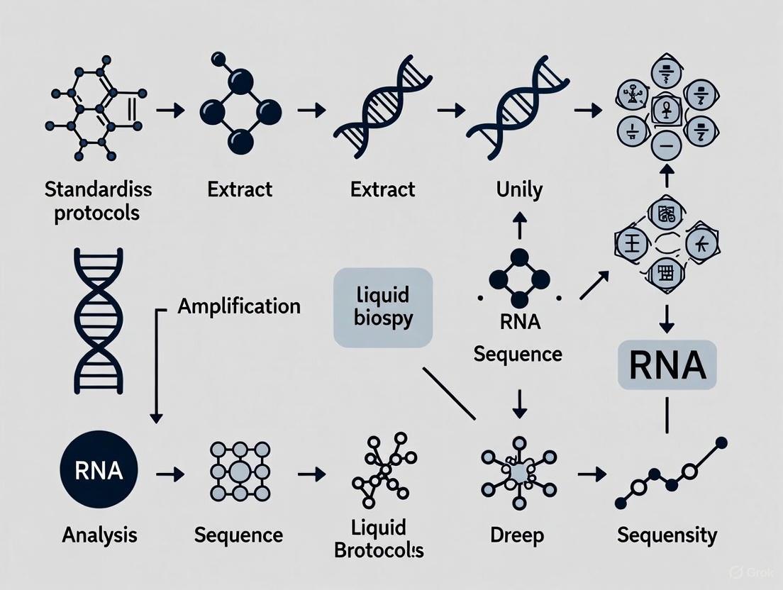
Visual Workflows and Standardization Diagrams
Liquid Biopsy Standardization Workflow
Diagram 1: Comprehensive Liquid Biopsy Standardization Workflow covering all testing phases with critical decision points and feedback mechanisms.
Pre-analytical Decision Pathway
Diagram 2: Pre-analytical Decision Pathway illustrating critical quality control checkpoints from blood collection to sample storage.
Research Reagent Solutions for Liquid Biopsy
Table 3: Essential Research Reagents and Materials for Standardized Liquid Biopsy Workflows
| Reagent Category | Specific Examples | Function & Application |
|---|---|---|
| Blood Collection Tubes | EDTA tubes; Streck Cell-Free DNA BCT; PAXgene Blood ccfDNA Tube | Sample collection and preservation; specialized tubes prevent cell lysis enabling extended processing windows [15] [16] |
| Nucleic Acid Extraction Kits | QIAamp Circulating Nucleic Acid Kit; QIAsymphony DSP Circulating DNA Kit; Maxwell RSC ccfDNA Plasma Kit | Isolation of high-quality cell-free DNA from plasma; varying performance characteristics require careful validation [15] |
| DNA Quantification Methods | Qubit Fluorometer; TapeStation; qPCR-based assays | Accurate quantification and quality assessment of extracted DNA; essential for determining input quality [15] |
| DNA Stabilization Reagents | Various proprietary formulations in specialized BCTs | Stabilize blood cells to prevent lysis and genomic DNA release during storage and transport [16] |
| Quality Control Materials | Reference standards; contrived samples; EQA/PT materials | Validation of assay performance; monitoring of analytical sensitivity and specificity [12] [14] |
The standardization of pre-analytical, analytical, and post-analytical phases is fundamental to realizing the full clinical potential of liquid biopsy. While significant challenges remain, international consortia and professional societies have made substantial progress in developing consensus standards and validation frameworks [12] [14] [15]. The troubleshooting guides and FAQs provided in this technical support center address the most common practical issues researchers encounter, offering evidence-based solutions derived from current literature and consensus recommendations.
Successful implementation of liquid biopsy in clinical practice requires ongoing collaboration across laboratories, manufacturers, and regulatory bodies to refine standardization protocols. By adhering to established guidelines, participating in proficiency testing programs, and maintaining rigorous quality control throughout all testing phases, laboratories can ensure the reliability and reproducibility necessary for liquid biopsy to fulfill its promise in personalized cancer care [12] [15] [13].
The clinical implementation of liquid biopsy (LB) relies on global efforts to standardize technologies, validate assays, and create collaborative networks. Three initiatives at the forefront of these efforts are the European Liquid Biopsy Society (ELBS), the International Society of Liquid Biopsy (ISLB), and the CANCER-ID project. These organizations represent coordinated attempts to overcome the technical and regulatory hurdles preventing widespread adoption of liquid biopsy in clinical practice [17] [18].
CANCER-ID (2015-2019) was an EU Innovative Health Initiative project that established foundational protocols for liquid biopsy clinical validation [19]. The European Liquid Biopsy Society (ELBS) directly evolved from CANCER-ID to maintain its network and expand its mission [10]. The International Society of Liquid Biopsy (ISLB), founded in 2017, operates as an independent professional organization with a global membership base [20]. Together, these initiatives address the critical need for standardized, reproducible liquid biopsy methods that can be implemented across diverse clinical and research settings.
Table 1: Core Characteristics of Major Liquid Biopsy Initiatives
| Initiative | Primary Focus | Founded | Key Outputs | Membership |
|---|---|---|---|---|
| CANCER-ID | Technology standardization & clinical validation | 2015 (completed 2019) | Standardized protocols, technology benchmarking, clinical feasibility studies | 38 partners from 13 countries [19] |
| ELBS | Clinical implementation & stakeholder collaboration | 2019 (evolved from CANCER-ID) | Guidelines, white papers, ring trials, regulatory engagement | 93 institutions (55 academic, 38 industry) across 21 countries [10] |
| ISLB | Multidisciplinary education & global knowledge exchange | 2017 | Annual congress, educational programs, professional networking | International memberships for healthcare professionals [20] |
Frequently Asked Questions: Technical Guidance for Researchers
Pre-analytical Considerations
What are the most critical pre-analytical variables affecting liquid biopsy results? Pre-analytical variables represent the most significant source of variability in liquid biopsy analysis [17]. Blood collection tube selection, processing time, temperature control, and centrifugation protocols can dramatically impact analyte integrity. For circulating tumor DNA (ctDNA) analysis, even moderate exercise before blood withdrawal or food intake can increase background levels of genomic DNA, reducing assay sensitivity [21]. The BLOODPAC consortium has developed Minimum Technical Data Elements (MTDEs) that provide comprehensive guidelines for blood sample collection, handling, and storage to maintain analyte integrity [22].
How should samples be processed for CTC preservation? Circulating Tumor Cell (CTC) analysis requires strict adherence to sample processing protocols due to the extreme rarity and fragility of these cells. The ELBS CTC Working Group has established an external quality assessment (EQA) ring trial using the CellSearch system to validate processing methods across multiple sites [10]. Recommendations include processing blood samples within specific time windows (typically 24-72 hours depending on preservative tubes), using standardized centrifugation forces to prevent cell loss, and implementing fixatives that maintain both morphological and molecular integrity for downstream analysis [10].
Analytical Challenges
What sensitivity thresholds are achievable for ctDNA detection? Sensitivity requirements vary substantially based on clinical context. For early-stage cancer or minimal residual disease (MRD) detection, where ctDNA can represent <0.01% of total cell-free DNA, extremely sensitive methods are required [21]. The CANCER-ID project found that reliable detection at variant allele frequencies (VAF) below 0.5% remains challenging, with FDA-evaluated commercial assays showing inconsistent performance below this threshold [21]. Techniques such as optimized library preparation, error-suppression sequencing, and unique molecular identifiers can improve sensitivity, but require rigorous validation in each laboratory [21].
How can researchers validate new CTC capture technologies? Technology validation should follow a tiered approach comparing new methods against established platforms. The ELBS recommends: (1) Initial spike-in experiments using cultured tumor cell lines in healthy donor blood; (2) Analytical validation assessing specificity, sensitivity, and reproducibility across multiple operators and sites; (3) Clinical validation using patient samples with comparison to CellSearch as the FDA-cleared standard [10]. The ELBS CTC Working Group is creating a comprehensive inventory of technologies, analytes, and biomarkers used by member laboratories to inform future ring trials and validation studies [10].
Data Analysis and Reporting
What metrics should be included in ctDNA assay reports? The ELBS ctDNA Working Group conducted a dedicated workshop on quality assessment and reporting, establishing consensus recommendations for diagnostic reports [10]. Essential components include: patient clinical features, sample quality metrics (including collection-to-processing time), assay specifications (including limit of detection), and detailed variant reporting. The workshop also addressed how to report challenging variants, negative results, and incidental findings [10]. Standardized reporting templates are available through ELBS publications for research and clinical use.
How should laboratories approach bioinformatic processing of ctDNA sequencing data? Bioinformatic pipelines must account for the exceptionally low VAF in ctDNA applications. The CANCER-ID project developed error-suppression algorithms and validation frameworks that differentiate true tumor-derived mutations from technical artifacts [19]. Key considerations include: implementing duplex sequencing to reduce errors, establishing tumor-informed baselines when possible, using healthy control samples to identify sequencing artifacts, and applying machine learning approaches to fragmentation patterns that can enhance specificity [21].
Standardized Experimental Protocols
CTC Enrichment and Analysis (CANCER-ID Protocol)
The CANCER-ID consortium established standardized protocols for CTC enrichment and characterization, with specific focus on non-small cell lung carcinoma and breast cancer [19].
Materials and Equipment:
- Blood collection tubes (EDTA or CellSave preservative tubes)
- Density gradient medium (e.g., Ficoll-Paque)
- EpCAM-based enrichment system (e.g., CellSearch Profile Kit)
- Immunofluorescence staining reagents (CK, CD45, DAPI)
- Automated fluorescence microscope or flow cytometer
- Nucleic acid extraction kit (for downstream molecular analysis)
Procedure:
- Collect 10mL whole blood into approved preservative tubes
- Process samples within 96 hours of collection (maintain at room temperature)
- Enrich CTCs using immunomagnetic separation (EpCAM-based capture)
- Fix cells and perform immunofluorescence staining (Pan-CK+, CD45-, DAPI+)
- Identify and enumerate CTCs using automated imaging system
- Islect individual CTCs for downstream molecular analysis (optional)
- For molecular analysis, use whole genome amplification followed by NGS library preparation
Troubleshooting:
- Low CTC yields may indicate improper sample storage or processing delays
- High background leukocyte contamination suggests suboptimal enrichment
- Poor RNA quality from CTCs may require immediate processing after enrichment
- For molecular analysis of single CTCs, the CANCER-ID validated Ampli1 workflow provides robust whole genome amplification for copy number alteration analysis [19]
ctDNA Analysis for Molecular Residual Disease
The ELBS ctDNA Working Group has established protocols for sensitive ctDNA detection in MRD settings [10], building on the CANCER-ID foundations [19].
Materials and Equipment:
- Streck Cell-Free DNA Blood Collection Tubes or equivalent
- Plasma preparation tubes (e.g., PPT tubes)
- High-speed centrifuge capable of 16,000 × g
- Cell-free DNA extraction kit (silica membrane or magnetic bead-based)
- Library preparation kit for NGS (e.g., hybrid capture-based)
- Unique molecular identifiers (UMIs)
- High-throughput sequencer
- Bioinformatics pipeline with error suppression
Procedure:
- Collect 10mL blood into ctDNA-specific preservative tubes
- Process within 6 hours of collection (centrifuge at 800 × g for 10 minutes)
- Transfer plasma to new tube and centrifuge at 16,000 × g for 10 minutes
- Extract cell-free DNA from 4-5mL plasma using validated kit
- Quantify cfDNA using fluorometric method (e.g., Qubit)
- Prepare sequencing library incorporating UMIs
- Hybrid capture targeting patient-specific mutations or tumor-informed panel
- Sequence to minimum 10,000x raw coverage (30,000x recommended for MRD)
- Bioinformatic processing with UMI consensus calling and error suppression
Troubleshooting:
- Low cfDNA yield may indicate hemolysis or improper processing
- For ultra-low VAF detection (<0.01%), increase plasma input volume
- High genomic DNA contamination suggests cell lysis during processing
- Inconsistent variant calls may require improved error suppression or duplicate removal
Visual Workflows for Liquid Biopy Standardization
Figure 1: This workflow illustrates the comprehensive pathway from research project to clinically implemented standardized protocol, as established by initiatives like CANCER-ID and ELBS. The process emphasizes rigorous validation at each phase to ensure reproducibility across sites [19] [10].
Figure 2: Global initiatives employ a multi-faceted strategy to address the complex challenges of liquid biopsy implementation. This includes simultaneous progress in standardization, validation, education, and regulatory engagement to achieve clinical adoption [19] [10] [14].
Essential Research Reagents and Materials
Table 2: Essential Research Reagents for Liquid Biopsy Analysis
| Reagent Category | Specific Examples | Function | Considerations |
|---|---|---|---|
| Blood Collection Tubes | CellSave tubes (CTC), Streck cfDNA tubes (ctDNA) | Cellular preservation, prevention of genomic DNA contamination | Tube selection depends on target analyte; processing time varies by preservative [10] |
| Nucleic Acid Extraction Kits | Silica-membrane kits, Magnetic bead-based kits | Isolation of high-quality ctDNA/CFDNA | Recovery efficiency critical for low-abundance targets; validate with spike-in controls [19] |
| CTC Enrichment Systems | CellSearch system, Parsortix system, Microfluidic chips | CTC capture and isolation | Technology selection depends on enrichment principle (e.g., EpCAM, size-based) [10] |
| Library Preparation Kits | Hybrid capture panels, Amplicon-based panels | NGS library construction for ctDNA | Unique Molecular Identifiers (UMIs) essential for error correction [21] |
| Reference Standards | Seraseq ctDNA reference materials, Horizon multiplex standards | Assay validation and quality control | Critical for establishing sensitivity and limit of detection [14] |
| Cell Culture Reagents | Tumor cell lines (e.g., MCF-7, PC-3) | Spike-in controls for CTC recovery studies | Essential for establishing and validating CTC capture efficiency [10] |
The coordinated efforts of ELBS, ISLB, and CANCER-ID have significantly advanced the field of liquid biopsy toward clinical implementation. Through protocol standardization, technology validation, education, and regulatory engagement, these initiatives address the critical pre-analytical, analytical, and post-analytical challenges that researchers face. The troubleshooting guides and experimental protocols provided here synthesize their collective expertise to support researchers in generating robust, reproducible liquid biopsy data. As the field continues to evolve, these foundational resources will remain essential for validating new technologies and applications in cancer diagnostics and monitoring.
Frequently Asked Questions (FAQs)
Q1: What are the primary biological factors that limit the sensitivity of liquid biopsy in early-stage cancer detection?
The main biological factors are the low abundance of analytes and tumor heterogeneity. In early-stage disease, the tumor burden is minimal, leading to a very low concentration of circulating tumor DNA (ctDNA) in the bloodstream. ctDNA can represent less than 0.1% of the total cell-free DNA (cfDNA), making it challenging to distinguish from normal background cfDNA [23] [1]. Furthermore, tumors are genetically heterogeneous, meaning that a single blood draw may not capture the full spectrum of molecular alterations present in different parts of the tumor or its metastases [24].
Q2: How does tumor heterogeneity impact the genetic profile obtained from a liquid biopsy compared to a tissue biopsy?
Tumor heterogeneity leads to discrepancies between liquid and tissue biopsies. A single tissue biopsy provides a snapshot of a specific lesion and may miss subclonal populations present elsewhere [24]. In contrast, liquid biopsy is thought to provide a more comprehensive profile by sampling DNA released from multiple tumor sites. However, studies comparing post-mortem tissue with pre-mortem liquid biopsies show that while liquid biopsy captures a significant portion (33-92%) of tissue-identified variants, it can also miss some mutations detected in specific lesions and detect unique variants not found in the analyzed tissues [24]. This confirms that while liquid biopsy effectively captures heterogeneity, it should be used alongside tissue biopsies for the most complete genetic profiling.
Q3: What are the major technical sources of variability in liquid biopsy workflows, particularly for ctDNA analysis?
Technical variability arises across the entire workflow. Key challenges include:
- Pre-analytical Phase: A lack of standardized protocols for blood collection, sample processing, and plasma storage can significantly impact results [17].
- Analytical Phase: The extremely low concentration of ctDNA requires ultra-sensitive detection methods, which are prone to artifactual signals from ultra-deep sequencing amplification [25]. Furthermore, somatic mutations originating from clonal hematopoiesis (an age-related process in blood cells) can be mistaken for tumor-derived mutations, leading to false positives [25].
- Post-analytical Phase: Variations in bioinformatics pipelines and a lack of standardized reporting make it difficult to compare results across different laboratories and platforms [25].
Q4: Why can CTC-based analyses sometimes provide a different molecular profile than ctDNA-based analyses?
ctDNA and Circulating Tumor Cells (CTCs) represent biologically distinct components. ctDNA is primarily released from apoptotic or necrotic tumor cells, reflecting the genetics of dying cell populations [25]. In contrast, CTCs are intact, viable cells that can originate from actively proliferating tumor regions and have the potential to metastasize [25]. Therefore, CTCs may harbor genomic information from more aggressive, treatment-resistant clones that are under-represented in the ctDNA pool. This makes CTCs a superior source for analyzing the genomic landscape of viable and metastatic tumor cells [25].
Troubleshooting Common Experimental Challenges
Challenge 1: Low Abundance of ctDNA
| Symptom | Possible Cause | Recommended Solution |
|---|---|---|
| Inconsistent mutation detection between replicates. | ctDNA concentration below the limit of detection (LOD) of the assay. | Use blood collection tubes designed to stabilize nucleated blood cells to prevent genomic DNA contamination [26]. |
| High background noise obscures true variants. | Input DNA quantity or quality is suboptimal. | Employ ultra-sensitive sequencing technologies, such as error-corrected NGS or digital droplet PCR (ddPCR), which can detect mutant allele fractions as low as 0.01% [26]. |
| Failure to detect known tumor mutations in plasma. | Tumor may not shed sufficient DNA into circulation, common in early-stage disease. | Increase plasma input volume for DNA extraction to ensure sufficient cfDNA mass for analysis. |
Challenge 2: Incomplete Capture of Tumor Heterogeneity
| Symptom | Possible Cause | Recommended Solution |
|---|---|---|
| Liquid biopsy reveals fewer mutations than a multi-region tissue biopsy. | A single blood draw may not capture all tumor subclones, especially those in immune-privileged or poorly perfused sites. | Implement serial sampling to monitor clonal evolution over time, as this can capture emerging resistant subclones that were initially missed [23] [24]. |
| Discrepant mutation profiles between primary tumor liquid biopsy and a metastatic lesion. | Spatial heterogeneity, where different metastases have unique genomic profiles. | Adopt a multi-analyte approach. Combine ctDNA with analysis of CTCs and extracellular vesicles (EVs) to get a more comprehensive view of the tumor ecosystem [23] [3]. |
| Detection of mutations of unknown origin. | Variants may come from clonal hematopoiesis or subclones not sampled in the original tissue biopsy. | Use paired white blood cell sequencing to identify and filter out mutations originating from clonal hematopoiesis [25]. |
Challenge 3: Technical Variability and Lack of Standardization
| Symptom | Possible Cause | Recommended Solution |
|---|---|---|
| Poor reproducibility of variant allele frequency (VAF) between different labs. | Differences in sequencing platforms, bioinformatics pipelines, and variant-calling algorithms. | Incorporate validated reference standards (e.g., synthetic ctDNA controls) into each run to enable performance evaluation and cross-platform calibration [25]. |
| Inconsistent CTC recovery rates. | Suboptimal enrichment technique or loss of CTCs that have undergone epithelial-mesenchymal transition (EMT) and downregulated epithelial markers like EpCAM. | Explore label-free isolation methods (e.g., based on size or deformability) or methods using novel tumor-specific markers (e.g., SP70) to improve capture efficiency of heterogeneous CTC populations [23] [25]. |
| High false-positive rate in ctDNA mutation calling. | Inadequate error suppression in NGS workflows or contamination from lysed white blood cells. | Implement unique molecular identifiers (UMIs) to tag original DNA molecules and correct for PCR amplification and sequencing errors [26]. |
Experimental Protocols for Key Methodologies
Protocol: Isolation of CTCs using an Immunomagnetic Enrichment Workflow
This protocol outlines the process for isolating Circulating Tumor Cells from whole blood using antibody-coated magnetic beads.
Title: CTC Immunomagnetic Enrichment Workflow
Procedure:
- Blood Collection: Draw blood into tubes containing EDTA or specialized cell preservatives (e.g., Streck tubes) to prevent clotting and stabilize cells. Process within a strict time window (e.g., within 4-96 hours depending on the tube) [23] [25].
- Plasma Removal and RBC Lysis: Centrifuge blood to separate plasma (which can be used for cfDNA extraction). subject the remaining cell pellet to red blood cell (RBC) lysis using an ammonium chloride solution or similar.
- Immunomagnetic Labeling: Resuspend the remaining white blood cell (WBC) and CTC pellet in a buffer containing immunomagnetic beads conjugated to an antibody against a target of interest (e.g., EpCAM for epithelial cancers, or a novel marker like SP70 for broader specificity) [23] [25].
- Incubation and Separation: Incubate the mixture with gentle rotation to allow beads to bind to CTCs. Then, place the tube in a magnetic stand. The magnet will retain the bead-bound CTCs while the unbound WBCs remain in the supernatant.
- Washing: Carefully remove and discard the supernatant. Wash the captured cells several times with buffer while the tube is in the magnetic stand to remove nonspecifically bound cells.
- CTC Elution and Analysis: Resuspend the magnetically captured cells in an appropriate medium for downstream applications, such as immunofluorescence staining, genomic DNA/RNA extraction, or single-cell sequencing [23].
Protocol: Targeted Sequencing of ctDNA using Error-Corrected NGS
This protocol describes a method for detecting low-frequency mutations in ctDNA using next-generation sequencing with error suppression.
Procedure:
- cfDNA Extraction: Extract cfDNA from 2-4 mL of plasma using a commercially available silica-membrane or magnetic bead-based kit optimized for low-concentration samples. Precisely quantify the yield using a fluorescent assay (e.g., Qubit).
- Library Preparation with UMIs: Construct sequencing libraries from the extracted cfDNA. A critical step is the ligation of Unique Molecular Identifiers (UMIs), also known as molecular barcodes, to each original DNA fragment prior to PCR amplification [26].
- Target Enrichment: Hybridize the library to biotinylated probes designed to capture a panel of cancer-related genes. Pull down the target sequences using streptavidin-coated magnetic beads, then wash and amplify the enriched library.
- Sequencing: Pool the libraries and sequence on a high-throughput sequencer to achieve high coverage (often >10,000x) to confidently detect low-frequency variants.
- Bioinformatic Analysis:
- Demultiplexing: Assign sequences to samples based on index barcodes.
- UMI Consensus Building: Group reads that originate from the same original DNA molecule using their UMI. Create a consensus sequence for each molecule to eliminate errors introduced during PCR and sequencing.
- Variant Calling: Align consensus reads to a reference genome and call somatic variants using specialized algorithms designed for low variant-allele-frequency detection.
Research Reagent Solutions
The following table lists key reagents and materials essential for robust liquid biopsy experiments.
| Research Reagent | Function & Application | Key Considerations |
|---|---|---|
| Cell-Free DNA Blood Collection Tubes (e.g., Streck, PAXgene) | Stabilizes nucleated blood cells to prevent release of genomic DNA and preserve ctDNA profile for up to several days. | Critical for multi-center studies; prevents false positives from lysed WBCs [26]. |
| Immunomagnetic Beads (e.g., anti-EpCAM, anti-SP70) | Isolate CTCs from whole blood based on surface marker expression. | Anti-EpCAM beads may miss CTCs undergoing EMT; novel markers like SP70 can improve capture breadth [25]. |
| Unique Molecular Identifiers (UMIs) | Short DNA barcodes ligated to each original DNA molecule to enable bioinformatic error correction. | Essential for achieving high sensitivity and specificity in ctDNA NGS assays; reduces background noise [26]. |
| Validated Reference Standards | Synthetic ctDNA controls with known mutations at defined allele frequencies. | Used for assay validation, quality control, and inter-laboratory calibration to ensure result consistency [25]. |
| Methylation-Sensitive Restriction Enzymes | Digest unmethylated DNA in assays focusing on tumor-specific methylation patterns in ctDNA. | Useful for early detection and cancer type classification, as methylation changes are abundant and early events in carcinogenesis [26]. |
Visualizing Tumor Heterogeneity and the Multi-Analyte Approach
The following diagram illustrates how tumor heterogeneity manifests and how a multi-analyte liquid biopsy approach can provide a more complete picture.
Title: Liquid Biopsy Captures Tumor Heterogeneity
The Impact of Standardization on Diagnostic Accuracy and Reimbursement
Liquid biopsy has emerged as a transformative tool in precision oncology, enabling non-invasive detection and monitoring of cancers through the analysis of circulating tumor biomarkers such as circulating tumor DNA (ctDNA), circulating tumor cells (CTCs), and extracellular vesicles [1] [27]. Unlike traditional tissue biopsies, liquid biopsies provide a dynamic snapshot of tumor heterogeneity and can be performed repeatedly to monitor treatment response and disease progression [28]. However, the clinical utility of these tests depends heavily on standardized procedures across the entire testing workflow, from sample collection to data interpretation [15].
Standardization directly impacts two critical aspects of liquid biopsy implementation: diagnostic accuracy and reimbursement. Variability in pre-analytical, analytical, and post-analytical processes can lead to inconsistent results, reduced test sensitivity and specificity, and ultimately, limited clinical adoption and reimbursement [29] [15]. This technical support guide addresses the key challenges and solutions for implementing standardized liquid biopsy protocols to ensure reliable results and facilitate reimbursement in both research and clinical settings.
Troubleshooting Guides & FAQs
Pre-Analytical Phase Standardization
FAQ: What are the most critical pre-analytical factors affecting liquid biopsy results?
The most critical pre-analytical factors include blood collection tube selection, sample processing timelines, plasma separation protocols, and cfDNA extraction methods. Inconsistencies in any of these areas can introduce genomic DNA contamination or degrade ctDNA, significantly impacting downstream analysis [15].
Troubleshooting Guide: Managing Pre-Analytical Variability
Table: Pre-Analytical Standards and Impact on Data Quality
| Process Step | Standardized Protocol | Deviation Impact | Recommended QC Check |
|---|---|---|---|
| Blood Collection | Use cfDNA-specific BCTs (e.g., Streck, PAXgene) | Genomic DNA contamination; false variants | Document tube type & draw order |
| Processing Time | ≤4h for EDTA tubes; ≤14d for stabilized tubes | Leukocyte lysis; reduced cfDNA yield | Record collection-to-spin interval |
| Plasma Separation | Two-step centrifugation: 1,600-2,000×g then 16,000×g | Cellular contamination; assay interference | Check for visible hemolysis |
| Plasma Storage | Aliquot in low-bind tubes at -80°C | cfDNA degradation; lower assay sensitivity | Measure cfDNA yield & fragment size |
| Sample Volume | ≥10mL blood; 4-20mL plasma based on application | Insensitive detection; false negatives | Quantify input cfDNA for each assay |
Experimental Protocol: Optimal Plasma Processing for ctDNA Analysis
Blood Collection: Draw a minimum of 10mL whole blood into cfDNA-specific blood collection tubes using a 21-gauge butterfly needle to minimize shear stress [15]. Invert tubes 8-10 times immediately after collection.
Plasma Separation:
- First centrifugation: 1,600-2,000 × g for 10-20 minutes at 4°C within 4 hours of collection for EDTA tubes or within stability period for specialized BCTs
- Carefully transfer supernatant to new tube without disturbing buffy coat
- Second centrifugation: 16,000 × g for 10 minutes at 4°C to remove residual cells [15]
Plasma Storage: Aliquot plasma into low-binding tubes (300μL-2mL aliquots) and store at -80°C. Avoid repeated freeze-thaw cycles.
cfDNA Extraction: Use validated extraction kits (e.g., QIAamp Circulating Nucleic Acid Kit) with appropriate plasma volumes (4-20mL based on application). For minimal residual disease detection, higher plasma volumes (8-20mL) are recommended due to low ctDNA fraction [15].
Analytical Phase Standardization
FAQ: How does standardization improve analytical sensitivity and specificity?
Standardized analytical protocols ensure consistent detection of low-frequency variants by establishing clear limits of detection (LOD) and controlling for interfering substances. For example, the Tempus xF liquid biopsy assay demonstrated 93.75% sensitivity for SNVs at 0.25% variant allele frequency (VAF) when using 30ng input DNA, highlighting how standardized input requirements affect sensitivity [28].
Troubleshooting Guide: Addressing Analytical Challenges
Table: Analytical Validation Parameters for ctDNA Assays
| Parameter | Standardized Requirement | Impact of Non-Standardization | Validation Approach |
|---|---|---|---|
| Input DNA Quality | Minimum 30ng cfDNA; fragment size 20-50bp | Reduced sensitivity; false negatives | Fluorometric/qPCR quantification + fragment analysis |
| Limit of Detection | ≥0.25% VAF for SNVs; ≥0.5% for indels | Missed low-frequency variants | Serial dilution studies with reference standards |
| Clonal Hematopoiesis | Matched normal buffy coat analysis | False-positive somatic calls | Paired ctDNA-white blood cell sequencing [28] |
| Assay Reproducibility | >95% inter-assay concordance | Inconsistent clinical results | Replicate testing across operators & instruments [28] |
| Tumor Fraction Estimation | Bioinformatic estimation (e.g., OTTER algorithm) | False-negative interpretation | Multiple estimation methods correlation [28] |
Experimental Protocol: Analytical Validation of ctDNA Assays
Input DNA Quantification and QC:
- Quantify cfDNA using fluorometric methods (Qubit) rather than UV spectrophotometry
- Assess fragment size distribution via Bioanalyzer/TapeStation; expect peak ~160-170bp
- Establish minimum input requirements (typically 10-30ng) for your specific assay [28]
Limit of Detection (LOD) Determination:
- Prepare serial dilutions of reference standards with known variant allele frequencies
- Test each dilution in replicates (n≥3) to establish the lowest VAF with ≥95% detection rate
- Validate for different variant types (SNVs, indels, CNVs, fusions) separately [28]
Specificity and Interference Testing:
Post-Analytical Phase and Data Interpretation
FAQ: How can standardized reporting improve clinical utility and reimbursement?
Standardized reporting frameworks, including tumor fraction estimation and clear interpretation guidelines, provide clinical context for test results and demonstrate clinical value to payers. For example, reporting the estimated tumor fraction helps clinicians distinguish true negative results from false negatives due to low tumor DNA shedding, enabling more informed clinical decisions [28] [15].
Troubleshooting Guide: Data Interpretation Challenges
Challenge: Differentiating clonal hematopoiesis from tumor-derived variants Solution: Sequence matched white blood cell DNA or use bioinformatic filters based on variant patterns and chromatin organization [28]
Challenge: Interpreting negative results when clinical suspicion remains high Solution: Report estimated tumor fraction and define reliable "detection thresholds" based on validation data [15]
Challenge: Variant reporting consistency across laboratories Solution: Implement standardized variant classification frameworks (e.g., AMP/ASCO/CAP guidelines) and reporting templates
The Scientist's Toolkit: Essential Research Reagent Solutions
Table: Key Reagents and Materials for Standardized Liquid Biopsy Research
| Reagent/Material | Function | Standardization Consideration | Example Products |
|---|---|---|---|
| cfDNA Blood Collection Tubes | Preserves cfDNA & prevents white blood cell lysis | Tube type affects processing timeline; must document and standardize | Streck Cell-Free DNA BCT, PAXgene Blood ccfDNA Tube |
| cfDNA Extraction Kits | Isolate cell-free DNA from plasma | Different kits yield varying quantity/quality; must validate and consistently use one system | QIAamp Circulating Nucleic Acid Kit, Maxwell RSC ccfDNA Plasma Kit |
| DNA Quantitation Assays | Measure cfDNA concentration & quality | Fluorometric methods preferred over spectrophotometry for accuracy | Qubit dsDNA HS Assay, TapeStation Genomic DNA Assay |
| Reference Standards | Validate assay performance & LOD | Essential for inter-laboratory comparison and quality control | Seraseq ctDNA Reference Materials, Horizon Multiplex I cfDNA Reference |
| Library Preparation Kits | Prepare sequencing libraries | Different efficiencies for cfDNA; require optimization for input amount | Illumina TruSeq DNA PCR-Free, Swift Accel-NGS 2S Plus |
| Hybrid Capture Panels | Target enrichment for sequencing | Design impacts genomic coverage & mutation detection capability | IDT xGen Panels, Twist Human Core Exome |
| Positive Controls | Monitor assay performance | Include in each run to detect technical failures | Custom synthetic ctDNA controls, cell line-derived controls |
Standardization Impact on Diagnostic Accuracy and Reimbursement
The relationship between standardization, diagnostic accuracy, and reimbursement is interdependent and multifaceted. Standardized protocols directly improve test performance, which in turn provides the evidence base needed for favorable reimbursement decisions.
Diagnostic Accuracy Improvements through Standardization
Standardization directly enhances key diagnostic accuracy parameters:
Sensitivity: Controlled pre-analytical conditions and optimized DNA input requirements improve detection of low-frequency variants. The Tempus xF assay demonstrated 94.8% sensitivity for SNVs when compared to orthogonal methods using standardized protocols [28].
Specificity: Standardized bioinformatic pipelines for clonal hematopoiesis identification reduce false positive results. Implementing dynamic filtering approaches decreased false positives by 11.45% in validation studies [28].
Reproducibility: Inter-assay concordance of 96.83% across instruments was achieved through standardized analytical protocols, enabling consistent results across laboratories and over time [28].
Reimbursement Implications
Standardization creates the evidence base required for favorable reimbursement decisions through several mechanisms:
Regulatory Compliance: Adherence to standards like those proposed by the International Society of Liquid Biopsy (ISLB) facilitates regulatory approvals [15] [30]. The FDA's Breakthrough Device designation, which can influence reimbursement, often requires demonstrated analytical validity through standardized validation [31].
Demonstrated Clinical Utility: Standardized tests generate reproducible real-world evidence that payers increasingly demand. Medicare Advantage plans and private insurers are incorporating value-based care models that require proof of clinical effectiveness [31].
Economic Value Proposition: Standardization reduces variable results that lead to repeated testing and unnecessary treatments, creating the cost-effectiveness data needed for positive coverage decisions [32] [31].
The implementation of standardized liquid biopsy protocols represents a critical pathway for advancing personalized cancer care while ensuring sustainable reimbursement models. Through continued refinement of pre-analytical, analytical, and post-analytical standards, the field can realize the full potential of liquid biopsy for cancer diagnosis, monitoring, and treatment selection.
Analytes and Workflows: A Technical Deep Dive into Liquid Biopsy Components
The clinical application of circulating tumor DNA (ctDNA) analysis hinges on the implementation of robust, standardized protocols. The International Society of Liquid Biopsy (ISLB) emphasizes that ensuring reliable and reproducible ctDNA testing necessitates standardization across pre-analytical, analytical, and post-analytical phases [30]. This technical support center addresses the specific experimental hurdles researchers encounter, providing troubleshooting guides and FAQs to support the development of precise and accurate liquid biopsy protocols essential for drug development and clinical research.
Technical FAQs & Troubleshooting Guides
Pre-analytical Phase: Sample Collection and Processing
Q1: What are the critical pre-analytical factors affecting cfDNA yield and quality? Pre-analytical variables including sample collection tube type, processing conditions, storage temperature, and extraction methodology significantly impact the yield, integrity, and quality of isolated cfDNA [33]. Inefficient handling can lead to genomic DNA contamination from white blood cell lysis or degradation of the target cfDNA.
Troubleshooting Guide: Low cfDNA Yield
- Problem: Consistently low cfDNA concentration after extraction.
- Potential Causes & Solutions:
- Cause: Incomplete plasma separation during centrifugation.
- Solution: Implement a two-step centrifugation protocol (e.g., 10 minutes at 1,700 × g, followed by 10 minutes at 20,000 × g) [34].
- Cause: Suboptimal sample storage leading to degradation.
- Solution: Ensure plasma is frozen at -80°C if not processed immediately. Validate sample stability for your specific storage conditions; cfDNA in plasma can be stable at 4°C or room temperature for up to 48 hours in certain preservative tubes, but this requires verification [33].
- Cause: Inefficiency of the extraction chemistry itself.
- Solution: Validate and potentially switch to a magnetic bead-based extraction method, which has been demonstrated to provide high cfDNA recovery rates and consistent fragment size distribution [33].
- Cause: Incomplete plasma separation during centrifugation.
Analytical Phase: Mutation Detection and Platform Selection
Q2: How do I choose the right mutation detection platform for my research question? The choice depends on the required sensitivity, breadth of genomic coverage, sample throughput, and cost [34]. The table below summarizes a cross-platform comparison based on KRAS mutation detection studies.
Table 1: Comparison of ctDNA Mutation Detection Platforms
| Platform | Key Principle | Sensitivity (LoD) | Breadth of Target | Key Performance Metrics (from KRAS studies) | Best Applications |
|---|---|---|---|---|---|
| Droplet Digital PCR (ddPCR) [35] [34] | Partitioning of sample into thousands of droplets for endpoint PCR | ~0.1% | Low (Typically 1-5 mutations per assay) | Sensitivity: ~47-93%; Specificity: ~77% [35] | High-sensitivity detection and absolute quantification of known hot-spot mutations. |
| BEAMing [35] [34] | PCR on magnetic beads in emulsion droplets | ~0.03% | Medium (e.g., Panels of 34 mutations) [35] | Sensitivity: ~93%; Specificity: ~69% [35] | Ultra-sensitive detection for longitudinal monitoring of a predefined set of mutations. |
| Next-Generation Sequencing (NGS) [35] [36] [37] | Massively parallel sequencing of templated libraries | ~0.1% - 0.5% (Varies with depth) | High (数十 to hundreds of genes) | Sensitivity: ~73%; Specificity: ~77% [35] | Comprehensive genomic profiling, discovery of novel alterations, and resistance mechanism screening. |
| CAPP-Seq [37] | A specific NGS method using a selector of biotinylated oligonucleotides for hybrid capture | High (~0.01% reported) | High (Customizable selector) | Not directly compared in provided results | Cost-effective, personalized profiling for monitoring a patient-specific set of mutations. |
Q3: Our NGS workflow for ctDNA has a high false-positive rate. How can we improve specificity? False positives in NGS often arise from sequencing errors or PCR artifacts. The implementation of Unique Molecular Identifiers (UMIs) is critical. UMIs are short random sequences ligated to each original DNA fragment prior to PCR amplification. Bioinformatic consensus building based on UMIs allows distinction of true mutations from amplification or sequencing errors [36] [37]. Furthermore, employing a bioinformatic "blocked list" of variants known to be recurrent artifacts can enhance accuracy [36].
Troubleshooting Guide: High Variability in NGS Results
- Problem: Inconsistent variant allele frequency (VAF) measurements between replicates or runs.
- Potential Causes & Solutions:
- Cause: Inconsistent input cfDNA quantity or quality.
- Solution: Pre-quantify cfDNA using a fluorescence-based method (e.g., Qubit) to ensure uniform input mass across runs. Assess fragment size distribution via bioanalyzer or TapeStation to confirm sample quality [33].
- Cause: Inefficient or non-uniform library preparation.
- Solution: Use library preparation kits optimized for low-input and fragmented DNA. Incorporate UMI adapters to improve quantitative accuracy [36].
- Cause: Insufficient sequencing depth.
- Solution: Increase raw sequencing depth. Note that after UMI deduplication, the effective depth is typically only ~10% of the raw depth. Achieving 99% detection probability for a 0.1% VAF variant requires an effective depth of ~10,000x, which necessitates a raw depth of ~100,000x [36].
- Cause: Inconsistent input cfDNA quantity or quality.
Post-analytical Phase: Data Interpretation
Q4: How can we distinguish true somatic tumor mutations from clonal hematopoiesis (CH) variants? Mutations detected in ctDNA can originate from malignant cells or from clonal hematopoiesis of indeterminate potential (CHIP). To mitigate false attribution:
- Bioinformatic Filtering: Compare the ctDNA mutation profile against a database of genes commonly associated with CHIP (e.g., DNMT3A, TET2, ASXL1).
- Paired Analysis: Sequence matched peripheral blood cells (buffy coat) from the same blood draw. True CHIP mutations will be present in the cellular DNA, while true somatic tumor mutations will not [38].
Essential Experimental Protocols
Protocol: Standardized cfDNA Extraction from Plasma Using Magnetic Beads
This protocol is adapted from a validated, high-throughput magnetic bead-based system [33].
Principle: Silica-coated magnetic beads bind cfDNA in the presence of a chaotropic salt (e.g., guanidine hydrochloride). cfDNA is purified through wash steps and eluted in a low-salt buffer.
Workflow Diagram:
Key Steps:
- Input: Use 1-6 mL of cell-free plasma obtained via double centrifugation.
- Lysis/Binding: Mix plasma with a lysis/binding buffer containing chaotropic salts and magnetic beads. Incubate to allow cfDNA to bind to the beads.
- Washing: Place the tube on a magnet to capture beads. Discard the supernatant. Wash the bead-bound DNA twice with a wash buffer.
- Elution: Elute the purified cfDNA in a small volume (e.g., 50-100 µL) of TE buffer or nuclease-free water.
- QC: Quantify yield using a fluorescence-based method and assess fragment size distribution (expecting a peak at ~167 bp) via Agilent TapeStation or Bioanalyzer [33].
Protocol: Core Workflow for ctDNA Mutation Detection by NGS
Principle: cfDNA is converted into an NGS library, enriched for target regions (e.g., by hybrid capture or amplicon generation), and sequenced at high depth. Bioinformatics pipelines then identify somatic variants against a reference genome.
Workflow Diagram:
Key Steps:
- Library Preparation: Convert fragmented cfDNA into a sequencing library. This involves end-repair, A-tailing, and ligation of sequencing adapters. Crucially, these adapters must contain UMIs [36].
- Target Enrichment: Enrich the library for genomic regions of interest. This can be achieved via:
- Sequencing: Sequence the enriched libraries on an NGS platform. The required depth is a function of desired sensitivity; for low VAFs (0.1-0.5%), raw depths of 15,000x or higher are typical for commercial panels [36].
- Bioinformatic Analysis:
- Demultiplexing: Assign reads to samples.
- UMI Consensus Building: Group reads originating from the same original DNA molecule and generate a consensus sequence to correct for errors.
- Variant Calling: Identify somatic mutations against a reference genome.
- Filtering: Apply filters to remove sequencing artifacts, germline polymorphisms, and CHIP-associated variants [36] [38].
The Scientist's Toolkit: Essential Research Reagents
Table 2: Key Reagents for ctDNA Analysis Workflows
| Reagent / Material | Function | Key Considerations |
|---|---|---|
| Cell-free DNA BCT Tubes (e.g., Streck) [34] | Blood collection tubes with preservatives to stabilize nucleated blood cells and prevent gDNA release. | Critical for maintaining pre-analytical sample integrity, especially during sample transport. |
| Magnetic Bead-based cfDNA Kits (e.g., from Qiagen, nRichDX) [33] | To isolate high-quality cfDNA from plasma with high recovery and minimal gDNA contamination. | Look for kits validated for high-throughput automation. Assess performance by recovery rate and fragment size profile. |
| cfDNA/ctDNA Reference Standards (e.g., from Seraseq, Horizon Discovery, AcroMetrix) [33] | Synthetic or cell-line derived controls with known variant allele frequencies. | Used for assay validation, quality control, and monitoring of sensitivity, specificity, and limit of detection. |
| UMI Adapter Kits | For incorporation of unique molecular identifiers during NGS library preparation. | Essential for error correction and accurate quantification in NGS-based ctDNA assays [36]. |
| Target Enrichment Panels | Biotinylated probes or primer sets for enriching specific genomic regions. | Choice between hybrid-capture (breadth, uniformity) and amplicon (speed, input) depends on application [36] [37]. |
Technology Comparison: CellSearch vs. Parsortix
The selection of an appropriate CTC enrichment technology is fundamental to experimental success. The table below provides a technical comparison of two widely used systems.
Table 1: Comparative Analysis of CellSearch and Parsortix Technologies
| Feature | CellSearch System | Parsortix System |
|---|---|---|
| Enrichment Principle | Immunoaffinity (positive selection) based on EpCAM expression [39] [40] | Size-based and deformability (6.5 µm cassette) [41] [39] |
| FDA Status | Approved for CTC enumeration in metastatic breast, prostate, and colorectal cancer [40] | Cleared for CTC enrichment from metastatic breast cancer patient blood [41] [39] |
| Captured CTC Phenotype | EpCAM-positive, epithelial CTCs [42] [40] | EpCAM-positive, EpCAM-negative, and mesenchymal CTCs; capable of capturing CTC clusters [41] [39] |
| Key Advantage | Standardized, reproducible enumeration; strong clinical prognostic validation [43] [40] | Phenotype-independent capture; viable cells for downstream culture/analysis [41] [40] |
| Key Limitation | Inability to detect CTCs that have undergone EMT and downregulated EpCAM [42] [39] | Potential loss of smaller CTCs; requires user-validated downstream analysis [41] |
| Typical Recovery Rate (from spike-in studies) | Varies significantly with EpCAM expression (e.g., 70% for EpCAMhigh, ~1% for EpCAMlow cells) [41] | Consistent recovery (~50%) independent of EpCAM expression [41] |
| Cell Viability Post-Enrichment | Cells are fixed, not viable [40] | Maintains cell viability for functional assays and culture [41] [40] |
Troubleshooting Guides & FAQs
FAQ 1: My CTC yields are lower than expected. What are the potential causes and solutions?
Low CTC yield is a common challenge. The solution depends on the technology you are using.
If using CellSearch: The most likely cause is the presence of CTCs that have undergone Epithelial-to-Mesenchymal Transition (EMT) and have downregulated or lost EpCAM expression [42] [40]. To address this:
- Solution A: Validate your patient cohort. This technology is most reliable for cancers known to have high EpCAM expression (e.g., prostate, breast) [42].
- Solution B: Corroborate your findings with a marker-independent method, such as a size-based enrichment system, to determine the fraction of EpCAM-negative CTCs you may be missing [39].
If using Parsortix: The primary cause could be an inappropriate cassette size or the nature of the cancer type.
- Solution A: Ensure you are using the correct cassette (e.g., 6.5 µm) for your target cells. Verify that the CTCs in your cancer type are larger than the cassette pore size [41].
- Solution B: Process a larger blood volume to increase the absolute number of cells captured, as the recovery rate is consistent but the starting count may be low [41].
General Considerations:
FAQ 2: How can I improve the purity of my enriched CTC sample for downstream molecular analysis?
High sample purity is critical for sensitive downstream applications like single-cell sequencing.
- Strategy A: Integrated Workflows. For the Parsortix system, perform the immunostaining and identification in-cassette before harvest. This allows for precise localization and picking of individual CTCs, avoiding contamination from white blood cells (WBCs) during the harvest step [41].
- Strategy B: Negative Depletion. Combine your primary enrichment method with a subsequent negative depletion step using anti-CD45 magnetic beads to remove residual WBCs from your enriched sample [44] [41].
- Strategy C: Optimized Staining and Washing. Implement stringent washing steps during your immunofluorescence protocol. Use high-quality, pre-titrated antibodies to minimize non-specific binding and background noise [39].
FAQ 3: What are the best practices for isolating viable CTCs for cell culture or functional assays?
The Parsortix system is designed for this purpose, as it isolates cells without fixation [40].
- Practice 1: Maintain Sterility. Perform all processing steps, from blood draw to cell harvest, under sterile conditions using sterile disposables and buffers to prevent microbial contamination.
- Practice 2: Gentle Harvest. Follow the manufacturer's protocol for gentle cell harvest from the Parsortix cassette to maximize cell viability. Use collection media containing serum or other viability-enhancing supplements [41].
- Practice 3: Use a Co-culture System. For establishing CTC cultures, plate the harvested cells directly into specialized co-culture systems, such as with cancer-associated fibroblasts or in conditioned media, to mimic the tumor microenvironment and support the growth of these rare cells [40].
Experimental Protocols
Protocol 1: CTC Enrichment and In-Cassette Staining using the Parsortix System
This protocol is optimized for the subsequent molecular characterization of CTCs [41] [39].
Key Reagents:
- Parsortix PR1 System with 6.5 µm separation cassettes
- K2EDTA blood collection tubes
- Phosphate Buffered Saline (PBS)
- 4% Paraformaldehyde (PFA)
- Permeabilization Buffer (e.g., Inside Perm)
- Primary Antibodies (e.g., anti-Pan-Cytokeratin, anti-EpCAM, anti-CD45)
- Fluorescently-labeled Secondary Antibodies (if needed)
- Nucleic acid stain (DAPI or Hoechst 33342)
- Mounting Medium
Methodology:
- Blood Collection and Loading: Collect peripheral blood into K2EDTA tubes. Process within 4 hours of blood draw. Load up to 7.5 mL of whole blood into the Parsortix instrument using the prescribed cassette [39].
- Enrichment Cycle: Run the instrument's standard enrichment program. This program pumps blood through the cassette, where CTCs are captured based on their larger size and rigidity compared to hematopoietic cells.
- In-Cassette Fixation and Permeabilization: After enrichment, fix the captured cells by flowing 4% PFA through the cassette for 15 minutes at room temperature. Remove PFA and wash with PBS. Follow by flowing permeabilization buffer for 10-15 minutes [39].
- Immunofluorescence Staining: Dilute primary antibodies in an appropriate buffer and incubate within the cassette for 1-2 hours at room temperature or overnight at 4°C. Wash with PBS. Incubate with fluorescent secondary antibodies (if using an indirect method) and DAPI/Hoechst for 1 hour in the dark. Perform a final PBS wash [39].
- Microscopy and Analysis: Remove the cassette from the instrument. Carefully disassemble the cassette and mount the membrane on a glass slide. Image using a fluorescence microscope. CTCs are typically identified as DAPI+, Cytokeratin+, CD45- cells with intact morphology [39].
Protocol 2: Functional Analysis: Development of a CTC-Derived Xenograft (CDX) Model
CDX models provide a powerful tool for studying metastasis and drug response in vivo [40].
Key Reagents:
- Immunocompromised mice (e.g., NSG)
- Matrigel
- RPMI-1640 culture medium
- Insulin syringe (29G)
Methodology:
- CTC Enrichment for Viable Cells: Enrich CTCs from a large blood volume (or leukapheresis product) using a method that preserves viability, such as the Parsortix system or negative depletion cocktails (e.g., RosetteSep) [41] [40].
- Cell Preparation: Harvest the enriched cells gently. Resuspend the cell pellet in a 1:1 mixture of cold PBS and Matrigel. Keep the suspension on ice to prevent gelation.
- Mouse Inoculation: Using an insulin syringe, inject the cell suspension (up to 200 µL) subcutaneously into the flank of an anesthetized immunocompromised mouse. For orthotopic metastasis models, intracardiac or intrafemoral injection may be used [40].
- Monitoring and Tumor Assessment: Monitor mice regularly for tumor growth by palpation and caliper measurements. The time for xenograft formation can vary from months to over a year.
- Model Validation: Once a tumor forms, it can be excised and analyzed. Validate the CDX model by confirming that it genetically and histologically matches the patient's original tumor or metastases through techniques like genomic sequencing and immunohistochemistry [40].
The Scientist's Toolkit: Essential Research Reagents
Table 2: Key Reagent Solutions for CTC Workflows
| Reagent / Kit | Function / Application | Key Consideration |
|---|---|---|
| CellSearch Profile Kit | Immunomagnetic enrichment and staining of EpCAM+ CTCs from whole blood [40]. | Standardized for enumeration; cells are fixed. |
| Parsortix Separation Cassette | Captures CTCs from whole blood based on size and deformability [41]. | Choice of pore size (e.g., 6.5 µm) is critical. |
| RosetteSep CTC Enrichment Cocktail | Negative selection; uses an antibody cocktail to deplete hematopoietic cells from whole blood [44] [41]. | Preserves cell viability; phenotype-independent. |
| EasySep CD45 Depletion Kit | Negative immunomagnetic selection for removing CD45+ white blood cells from pre-enriched samples [41]. | Increases sample purity for downstream assays. |
| Inside Stain Kit (Miltenyi) | Permeabilization buffer for intracellular staining (e.g., cytokeratins) within Parsortix cassettes [39]. | Compatible with in-cassette workflows. |
| CellTracker Dyes (e.g., CMFDA) | Fluorescent cytoplasmic dyes for pre-labeling cultured tumor cells for spike-in control experiments [41]. | Essential for determining recovery rates during method validation. |
Signaling Pathways and Experimental Workflows
The following diagrams illustrate the core technology principles and a key biological process affecting CTC detection.
Diagram 1: CTC Enrichment Workflow Comparison
This diagram outlines the fundamental operational differences between the two main enrichment technologies.
Diagram 2: Epithelial-Mesenchymal Transition (EMT) Impact on CTC Detection
This diagram shows how the biological process of EMT directly influences the efficacy of EpCAM-dependent detection methods.
The Emerging Role of Exosomes and Extracellular Vesicles (EVs) in Multi-Analyte Approaches
FAQs & Troubleshooting Guides
FAQ 1: What are the primary challenges in quantifying and characterizing individual EVs, and what techniques can overcome them?
Answer: The primary challenges in EV analysis include their small size (30-200 nm), heterogeneity, low refractive index, and the presence of non-vesicular contaminants like protein aggregates and lipoproteins [45] [46]. These factors make precise quantification and characterization difficult.
- Recommended Technique: Single-EV Analysis Techniques. Methods like Multiplexed Analysis of EV (MASEV) and simplified flow cytometry protocols using instruments dedicated to small particles (e.g., Apogee A50 Micro) are designed to address these issues [45] [46]. MASEV uses bioorthogonal chemistry to perform multiple rounds of staining and destaining, allowing for the profiling of up to 15 different biomarkers on individual vesicles [46].
- Key Consideration: Techniques like Nanoparticle Tracking Analysis (NTA) can overestimate EV counts because they cannot distinguish EVs from other similarly-sized particles [45]. Combining specific fluorescent labeling with single-particle analysis provides more accurate characterization.
FAQ 2: My EV samples are contaminated with protein aggregates. How can I distinguish vesicles from contaminants?
Answer: Contamination by protein aggregates is a common issue that can skew quantification and functional analysis.
- Recommended Protocol: Dual-Fluorescent Labeling. A simplified flow cytometry protocol recommends double-labeling EV preparations with both protein-specific dyes (e.g., Carboxyfluoresceinsuccinimidyl ester, CFSE) and lipid-specific dyes (e.g., FM dyes) [45].
- Workflow: Isolate EVs via ultracentrifugation with a sucrose cushion purification step. Resuspend the pellet and incubate with both CFSE and a lipophilic dye. The unbound dye does not require removal by additional ultracentrifugation for this specific protocol.
- Interpretation: True EVs will be positive for both protein and lipid signals. In contrast, protein aggregates will only stain with the protein-specific dye, and micelles formed from free lipophilic dyes will only stain with the lipid-specific dye. This enables clear separation and accurate enumeration of EVs [45].
FAQ 3: How can I achieve multiplexed biomarker profiling from a single, small-volume clinical sample?
Answer: Achieving deep phenotypic profiling from limited samples is crucial for clinical translation.
- Recommended Technique: Iterative Multiplexing with Bioorthogonal Chemistry. The MASEV technology is specifically designed for this purpose [46].
- Workflow:
- EV Capture: Adhere EVs to a silanized coverglass within a custom flow chamber.
- Staining Cycle: Incubate with a panel of antibodies conjugated to fluorochromes via a cleavable linker.
- Imaging: Acquire multi-channel fluorescence images.
- Destaining: Add "tetrazine scissors" to cleave the fluorophores from the antibodies with >99% efficiency.
- Repetition: Repeat the staining and destaining cycle for a new panel of antibodies (up to 5 cycles demonstrated).
- Key Advantage: This method allows the interrogation of thousands of individual EVs for 15 or more biomarkers from a single sample, revealing rare EV subtypes and detailed biomarker composition with high diagnostic potential [46].
- Workflow:
FAQ 4: What are the critical standardization guidelines I must follow for my EV research to ensure reproducibility and clinical relevance?
Answer: Adherence to international guidelines is essential for rigorous and reproducible EV research.
- Primary Guideline: The Minimal Information for Studies of Extracellular Vesicles (MISEV) guidelines, updated by the International Society for Extracellular Vesicles (ISEV) in 2023 (MISEV2023), are the cornerstone of EV standardization [47].
- Key Recommendations: MISEV2023 provides detailed guidance on:
- Nomenclature: Recommends classifying EVs based on physical characteristics (size, density) and biochemical composition (e.g., CD9/CD81/CD63-positive) rather than presumed cellular origin [47].
- Characterization: Mandates demonstrating the presence of EV-associated proteins (e.g., tetraspanins) and the absence of common contaminants from non-vesicular compartments [47].
- Reporting: Encourages the use of the EV-TRACK knowledge base to document all experimental details related to EV isolation and characterization, which increases transparency and reproducibility [47].
Technical Specifications & Data
Table 1: Comparison of Key EV Analysis Techniques
| Technique | Principle | Key Metrics | Throughput | Key Advantages | Key Limitations |
|---|---|---|---|---|---|
| ELISA | Antigen-antibody reaction with enzymatic signal generation [48] | Concentration of specific EV subpopulations | Medium to High (96-well plate) | High specificity for EV subtypes; quantitative with standards; amenable to automation [48] | Measures specific subpopulations, not total EV; multi-step, time-consuming; potential for cross-reactivity [48] |
| Nanoparticle Tracking Analysis (NTA) | Tracking Brownian motion to determine size and concentration [45] | Particle size distribution, concentration | Medium | Rapid size analysis; label-free option [45] | Cannot distinguish EVs from other particles (e.g., protein aggregates, lipoproteins); low dynamic range [45] |
| Flow Cytometry (Dedicated) | Scattering and fluorescence of single particles in a flow stream [45] | Particle count, size estimation, biomarker positivity | High | High-throughput single-particle analysis; multi-parameter data [45] | Requires specialized instrument for small particles; potential for swarm detection (multiple particles) [45] |
| MASEV | Iterative staining/destaining with bioorthogonal chemistry on surface-captured EVs [46] | Single-EV biomarker profile for 15+ targets | Low to Medium (imaging-based) | High multiplexing depth from single sample; reveals EV heterogeneity and rare subtypes [46] | Technically complex; requires custom chemistry and setup; lower throughput than conventional flow cytometry [46] |
Table 2: Essential Research Reagent Solutions for EV Analysis
| Reagent / Material | Function | Example Application |
|---|---|---|
| Tetraspanin Antibodies (e.g., anti-CD9, CD63, CD81) | Detection of common EV surface markers; used for characterization and immunocapture [48] [47] | ELISA, flow cytometry, MASEV, Western Blot |
| Lipophilic Dyes (e.g., PKH67, FM dyes) | Labeling of the EV lipid bilayer membrane [45] | Flow cytometry to distinguish EVs from protein contaminants |
| Protein Stain (e.g., CFSE) | Labeling of protein components on/within EVs [45] | Flow cytometry, used in conjunction with lipophilic dyes |
| Bioorthogonal Linker (C2-symmetric TCO moiety) | Links antibodies to fluorophores in a cleavable manner for iterative staining [46] | Core component of the MASEV multiplexing technology |
| Tetrazine Scissors (HK-Tz) | Cleaves the fluorophore from the antibody post-imaging [46] | Destaining step in the MASEV cycling process |
| Size Exclusion Chromatography Columns | Isolation of EVs from biofluids based on size [46] | Sample preparation for downstream analyses like MASEV or NTA |
Experimental Workflow Visualizations
MASEV Multiplexing Technique
EV Technique Selection Guide
Blood Collection Tube Comparison and Protocols for Plasma Processing and Storage
Frequently Asked Questions (FAQs)
FAQ 1: What is the fundamental difference between plasma and serum, and why does it matter for liquid biopsy?
Plasma and serum are both liquid components of blood, but they are obtained through different processes that significantly impact their composition. Plasma is collected by mixing blood with an anticoagulant (e.g., EDTA, heparin, or citrate) to prevent clotting, thus preserving clotting factors within the liquid component. Serum, in contrast, is the clear, yellowish fluid that remains after blood has clotted and the clot (including blood cells and clotting proteins like fibrinogen) has been removed [49].
For liquid biopsy, which often focuses on analyzing circulating tumor DNA (ctDNA) and extracellular RNA (exRNA), this distinction is critical. The clotting process in serum preparation can lead to the entrapment of tumor-derived components in the clot and the release of cellular RNA by cells under stress, potentially altering the biomarker profile compared to plasma [50] [1].
FAQ 2: For time-sensitive research, which blood collection tube should I use to minimize processing delays?
For urgent or STAT analyses, plasma separator tubes (PST), which contain lithium heparin, or rapid serum tubes (RST) are recommended to reduce turnaround time [49].
- PST (Plasma Tubes): These tubes contain an anticoagulant and allow for immediate centrifugation after collection, separating plasma without a clotting wait time [49].
- RST (Rapid Serum Tubes): These tubes are designed to accelerate the clotting process using thrombin-based activators, typically allowing for serum separation within 5 to 10 minutes, much faster than the 30 minutes required for standard serum tubes [49].
FAQ 3: My liquid biopsy experiment focuses on extracellular RNA (exRNA). What are the critical pre-analytical factors I must control?
The stability and accurate profiling of exRNA are highly susceptible to pre-analytical variables. A comprehensive study (exRNAQC) evaluated these factors and found [50]:
- Blood Collection Tube Type: The choice of tube significantly impacts exRNA concentration, detected gene numbers, and replicability. Some preservation tubes failed to stabilize exRNA effectively.
- RNA Purification Method: The method used for RNA purification causes substantial variations in performance, including differences in RNA yield and observed transcriptome complexity.
- Blood Processing Time: The time interval between blood draw and processing is a critical factor.
- Interactions: Crucially, there are significant interactions between these variables (e.g., a tube type may perform well with one purification method but poorly with another). The study emphasizes the need for strict standardization and full reporting of these pre-analytical conditions [50].
FAQ 4: How does the choice of anticoagulant in plasma tubes affect downstream metabolomics analysis?
The anticoagulant can significantly alter the metabolic profile. Research comparing NMR-based metabolomics data across different tubes revealed [51]:
- Heparin plasma and EDTA plasma tubes performed closest to serum, with only 3 and 5 significantly different metabolites out of 50, respectively.
- Fluoride tubes showed differences in 11 metabolites.
- ACD and Citrate plasma tubes differed significantly for approximately half of the metabolites assessed. These differences were largely due to significant interfering peaks from the anticoagulants themselves, which can obscure the biological signal [51].
FAQ 5: What are the consequences of centrifuging serum collection tubes before complete clot formation?
Centrifuging serum tubes before the clot has fully formed (typically before 30 minutes) is a major pre-analytical error. It can lead to:
- Residual Fibrin: Incomplete clot formation can leave residual fibrin or fibrinogen in the serum supernatant.
- Instrument Interference: This fibrin can interfere with automated instrument operation and testing.
- Inaccurate Results: The presence of fibrin can lead to clinically inaccurate results, for example, false increases in cardiac troponin I measurements [49].
Troubleshooting Guides
Problem 1: Low Yield or Poor Quality of Cell-Free Nucleic Acids (cfDNA/ctDNA/exRNA)
| Possible Cause | Recommended Action | Preventive Measures for Future |
|---|---|---|
| Inappropriate collection tube | Validate tube type for your specific analyte. For exRNA, some specialized preservation tubes may be required [50]. | Select and validate tubes based on the target analyte (cfDNA, exRNA) and ensure they are compatible with your downstream purification method [50]. |
| Delayed processing | Note the deviation in processing time; data may be compromised. | Process samples according to established protocols. For plasma, centrifugation should ideally occur within 30 minutes to a few hours of collection. Adhere to a standardized processing time interval [50] [52]. |
| Incomplete mixing of blood with anticoagulant | The sample may clot and must be discarded. | After collection, gently invert tubes according to the manufacturer's instructions (e.g., 8 times for EDTA tubes) to ensure proper mixing and prevent clotting [51]. |
| Hemolysis | Visually inspect sample for pink/red color. Hemolyzed samples can invalidate many tests and affect exRNA profiles [50]. | Use proper phlebotomy technique, avoid using a small needle, and ensure tubes are filled to the correct volume to prevent vacuum-related rupture of red blood cells [49]. |
Problem 2: Inconsistent or Irreproducible Results in Biomarker Quantification
| Possible Cause | Recommended Action | Preventive Measures for Future |
|---|---|---|
| Improper sample storage | If improperly stored, repeat the experiment with correctly aliquoted and stored samples. | Aliquot samples into single-use portions to avoid freeze-thaw cycles. Store and transport plasma/serum at -20°C or lower for long-term preservation [53]. |
| Variability in RNA purification methods | Re-assess the RNA purification method's performance metrics for your specific application. | Choose a purification method that demonstrates high reproducibility, sensitivity, and RNA yield for your desired analyte (miRNA vs. mRNA). The performance of these methods varies significantly [50]. |
| Interaction between tube type and purification method | Review the exRNAQC study data on interactions between your tube and purification method [50]. | During method development, conduct a paired validation of the blood collection tube and RNA purification method to ensure compatibility and optimal performance [50]. |
Problem 3: Interference in Downstream Analytical Assays (e.g., NMR, PCR)
| Possible Cause | Recommended Action | Preventive Measures for Future |
|---|---|---|
| Anticoagulant interference | For metabolomics, avoid ACD and citrate tubes if possible, as the anticoagulants produce interfering peaks in NMR [51]. | Match the tube type to the test requirements. For metabolomics, heparin or EDTA plasma are preferable, as they show fewer interferences compared to serum [51]. |
| Cellular contamination | Re-centrifuge the plasma/serum sample to remove residual cells or platelets. | For platelet-poor plasma, centrifuge at 2,000 x g for 15 minutes [53]. Ensure a clean transfer of the supernatant using a pipette, avoiding the cell pellet. |
Data Presentation
Table 1: Comparison of Common Blood Collection Tubes for Research
| Tube Type (Top Color) | Additive | Primary Use / Sample Type | Key Advantages | Key Limitations / Considerations |
|---|---|---|---|---|
| Serum Tube (Red) | No anticoagulant (may contain clot activator) | Serum | Broad assay compatibility; considered gold standard for many chemistry tests [49] [51]. | Requires 30-min clot formation time; risk of fibrin interference if processed early; lower yield vs. plasma; clotting may entrap biomarkers [49] [50]. |
| Serum Separator Tube (SST) (Red/Black) | Clot activator + gel separator | Serum | Gel barrier provides clean serum separation from clot [49]. | Same clotting time requirement as standard serum tubes [49]. |
| Rapid Serum Tube (RST) | Thrombin-based clot activator | Serum | Fast clotting (5-10 min); reduces hemolysis and false positives for some tests like troponin [49]. | Still requires a short clotting period compared to plasma tubes. |
| EDTA (Lavender) | EDTA (Anticoagulant) | Plasma | Excellent for preserving cell-free DNA and hematology; minimal interference in metabolomics [53] [51] [1]. | Chelating agent may interfere with metal-dependent assays. |
| Heparin (Green) | Heparin (Anticoagulant) | Plasma | Can be centrifuged immediately; good for chemistry; closest metabolomic profile to serum [49] [51]. | Heparin can inhibit PCR and reverse transcription; potential endotoxin contamination [53]. |
| Citrate (Blue) | Citrate (Anticoagulant) | Plasma | Standard for coagulation studies [49] [52]. | Significant interference in NMR metabolomics due to anticoagulant peaks; requires volume adjustment for testing [51]. |
| Sodium Fluoride (Grey) | Potassium Oxalate/Sodium Fluoride | Plasma | Fluoride inhibits glycolytic enzymes, stabilizing glucose [53]. | Shows multiple metabolite differences in metabolomics profiles compared to serum [51]. |
Table 2: Impact of Pre-analytical Variables on Extracellular RNA (exRNA) Analysis
| Pre-analical Variable | Impact on exRNA Profiling | Key Findings from exRNAQC Study [50] |
|---|---|---|
| Blood Collection Tube | Significant impact on concentration, detected gene numbers, and replicability. | Performance varied widely; some preservation tubes failed to stabilize exRNA. Not all tubes are equal for exRNA analysis. |
| RNA Purification Method | Major cause of variation in yield, sensitivity, and observed transcriptome complexity. | Methods differed significantly in performance. A higher plasma input volume generally increased detected mRNAs and reduced replicate variability. |
| Processing Time Interval | Critical for exRNA stability and profile integrity. | Longer processing times can degrade labile RNA species and alter profiles. |
| Interaction of Variables | The effect of one variable (e.g., tube) depends on another (e.g., purification method). | Critical interactions were identified, highlighting that optimal performance requires compatible pairs of tubes and methods. |
Experimental Protocols
Detailed Protocol: Standard Plasma Preparation from EDTA Tubes
This protocol is adapted from standard procedures for processing blood for plasma-based assays and liquid biopsy [53] [52].
Principle: To obtain platelet-poor plasma suitable for downstream molecular analysis (e.g., ctDNA, exRNA extraction) by centrifugation of anticoagulated whole blood.
Materials and Reagents:
- Blood Collection Tubes: K2EDTA tubes (Lavender top) [53].
- Equipment: Refrigerated centrifuge, micro-pipettes with sterile tips, sterile polypropylene tubes (e.g., 15 mL conical tubes), -80°C freezer.
- Personal Protective Equipment (PPE): Lab coat, gloves, safety glasses.
Procedure:
- Collection and Mixing: Collect whole blood via venipuncture into K2EDTA tubes. Immediately after collection, invert the tube gently but thoroughly 8-10 times to ensure complete mixing of the blood with the anticoagulant [51].
- Initial Centrifugation:
- Place the tubes in a refrigerated centrifuge (maintained at 2-8°C).
- Centrifuge at 1,000-2,000 x g for 10 minutes to separate plasma from cellular components [53].
- Plasma Transfer:
- Using a sterile Pasteur pipette or micro-pipette, carefully transfer the supernatant (plasma) into a new sterile polypropylene tube.
- Critical Step: Avoid disturbing the buffy coat (white layer of white blood cells) or the red blood cell pellet at the bottom. Transfer the plasma cleanly to minimize cellular contamination [53].
- Secondary Centrifugation (for Platelet-Poor Plasma):
- To further deplete platelets, which can be a source of RNA, centrifuge the transferred plasma again at 2,000 x g for 15 minutes [53].
- Transfer the resulting supernatant into a new sterile tube. This is the platelet-poor plasma.
- Aliquoting and Storage:
- Immediately aliquot the plasma into 0.5 mL portions in cryovials.
- Maintain samples at 2-8°C during handling.
- Store aliquots at -20°C or lower (recommended -80°C for long-term storage). Avoid freeze-thaw cycles, as they can degrade labile biomarkers [53].
Quality Control:
- Visually inspect the plasma for hemolysis (pink/red color), icterus (yellow), or lipemia (milky). Note that these can invalidate certain tests [53].
Workflow Visualization
Diagram 1: Plasma and Serum Processing Workflow. This flowchart outlines the critical steps and decision points for processing blood into serum or plasma, including an optional step for obtaining platelet-poor plasma for sensitive liquid biopsy applications [49] [53].
The Scientist's Toolkit: Research Reagent Solutions
| Essential Material | Function in Liquid Biopsy Research | Key Considerations |
|---|---|---|
| K2EDTA Blood Collection Tubes | Preferred anticoagulant for cell-free DNA and extracellular RNA studies. Inhibits clotting by chelating calcium. | Widely used for liquid biopsy; minimizes DNAse/RNAse activity. Avoid for calcium-dependent assays [50] [1]. |
| Cell-Free DNA BCT Tubes | Specialized tubes containing preservatives that stabilize nucleated blood cells, preventing lysis and release of genomic DNA. | Critical for preserving the integrity of ctDNA profiles when processing is delayed beyond 24-48 hours. |
| Platelet Preparation Tubes | Tubes with a specialized gel for simplified separation of platelets. | Useful for studying tumor-educated platelets (TEPs), a emerging liquid biopsy biomarker [3] [1]. |
| RNA Stabilization Reagents | Reagents (e.g., TRIzol, Qiazol) that immediately lyse cells and inactivate RNases upon sample mixing. | Essential for stabilizing the exRNA transcriptome at the point of collection, especially when using non-stabilizing blood tubes [50]. |
| Size-Selection Magnetic Beads | Beads for solid-phase reversible immobilization (SPRI) to selectively purify nucleic acids by size. | Crucial for enriching for short, fragmented ctDNA (~160-180 bp) and exRNA, removing contaminating genomic DNA and other debris [50]. |
| Synthetic Spike-In RNA/DNA Controls | Non-human, synthetic nucleic acids added to the sample at the start of processing. | Allows for normalization and quality control across different samples, protocols, and batches, correcting for technical variation in extraction and sequencing [50]. |
The clinical standardization of liquid biopsy protocols is a critical step in translating promising biomarker research into routine patient care. The European Liquid Biopsy Society (ELBS) network, a partnership of academic and private institutions, highlights that despite breakthroughs, implementing these discoveries into clinical practice remains a significant challenge [10]. This technical support center provides standardized troubleshooting guides and FAQs to help researchers and clinicians overcome technical hurdles, ensuring the reliability and reproducibility of cell-free DNA (cfDNA) analyses within this broader framework of clinical standardization [10]. The ELBS emphasizes establishing standard protocols and external quality assessment (EQA) schemes, objectives that these resources are designed to support [10].
Frequently Asked Questions (FAQs)
Sample Collection and Pre-Analytics
Q1: What is the recommended blood collection tube for cfDNA studies, and how should samples be stored before processing? We recommend using blood collection tubes with a stabilization solution, such as the PAXgene Blood ccfDNA Tube, especially if the blood cannot be processed promptly. These tubes prevent the release of intracellular genomic DNA and maintain constant cfDNA levels. If using non-stabilizing tubes like EDTA or ACD-A, samples should be processed within a few hours, storing them at 2-8°C in the meantime. Stabilizing tubes allow for storage at room temperature for up to 10 days. For long-term storage, plasma should be generated quickly and stored at -20°C, avoiding repeated freeze-thaw cycles [54].
Q2: What is the optimal plasma volume for downstream cfDNA mutation detection? The required plasma volume depends on your scientific question. For detecting rare mutations, increased plasma volume directly increases assay sensitivity. We recommend harnessing the maximum sample processing capacity of your isolation kit. For example, the EZ1&2 ccfDNA Kit can process up to 8 mL of plasma, which is ideal for high-sensitivity applications like digital PCR (dPCR) or Next-Generation Sequencing (NGS) [54].
cfDNA Isolation and Quantification
Q3: My cfDNA yields are lower than expected. What could be the cause? Low yields can stem from several pre-analytical and analytical factors:
- Isolation Kit Choice: Different kits have significantly different extraction efficiencies. One study found yield variations of up to 4.3-fold between kits [55]. Spin-column-based kits like the QIAamp Circulating Nucleic Acid Kit (QiaS) may provide higher yields than some magnetic bead-based methods [55].
- Sample Processing Delays: Failure to process non-stabilized blood samples within the recommended timeframe can lead to white blood cell lysis and contamination of the plasma with high-molecular-weight genomic DNA, diluting the cfDNA fraction.
- Improper Centrifugation: Incomplete removal of cellular components during plasma generation can lead to the same contamination issue. Ensure the recommended two-step centrifugation protocol (e.g., 1600× g for 10 min, then 6000× g for 10 min) is followed precisely [55].
Q4: How should I quantify and assess the quality of isolated cfDNA?
- Quantification: Use a fluorometric method like the Qubit Fluorometer with the dsDNA HS Assay. This is specific for double-stranded DNA and provides accurate concentrations for low-abundance samples, unlike spectrophotometric methods [55].
- Quality/Fragment Sizing: Use a fragment analyzer like the Bioanalyzer with the High-Sensitivity DNA Kit. This confirms that the isolated DNA has the expected mono-nucleosomal size profile (~130-170 bp) and assesses the degree of genomic DNA contamination [55].
Downstream Analysis and Troubleshooting
Q5: My downstream dPCR or NGS results show poor efficiency. Could my sample contain inhibitors? Yes, poor sample quality from contaminants or PCR inhibitors is a common issue. Digital PCR (dPCR) is inherently more tolerant of inhibitors than quantitative PCR (qPCR). Because dPCR relies on end-point amplification and Poisson statistics across thousands of partitions, it can provide accurate absolute quantification even in the presence of inhibitors that would compromise qPCR efficiency. Using optimized sample preparation kits is the best strategy to obtain amplifiable, inhibitor-free cfDNA [54].
Q6: What strategies can I use to achieve high specificity (<0.1%) for rare mutation detection in a high wild-type background? For dPCR, using LNA (Locked Nucleic Acid) Mutation Assays is highly effective. LNA-containing primers and probes increase the melting temperature (Tm) and improve the discrimination power between wild-type and mutant sequences. This allows for the design of shorter amplicons, which is ideal for the highly fragmented nature of cfDNA [54].
Troubleshooting Common Experimental Issues
| Problem | Potential Causes | Recommended Solutions |
|---|---|---|
| Low cfDNA Yield | Suboptimal isolation kit, plasma volume too low, sample processing delays, improper centrifugation | Increase plasma input volume; switch to a higher-yield kit (e.g., QIAamp Circulating Nucleic Acid Kit); ensure blood is processed within 1-2 hours (non-stabilized tubes) or use stabilizing tubes; follow validated centrifugation protocols [55] [54]. |
| Poor Purity (gDNA contamination) | Inefficient plasma generation, leukocyte lysis due to delayed processing, hemolysis | Implement a validated two-step centrifugation protocol; use blood collection tubes with stabilizers; avoid freeze-thaw cycles; check fragment profile with Bioanalyzer [55]. |
| Inconsistent dPCR/NGS Results | PCR inhibitors, low template input, suboptimal assay design, high "dead volume" in dPCR system | Use recommended sample prep kits to remove inhibitors; maximize template addition volume; use LNA-enhanced assays for dPCR; for dPCR, select a system with a high analyzed volume (e.g., 21.6 µL in QIAcuity 26K) [54]. |
| Insufficient Analytical Sensitivity | Plasma input volume too low, total analyzed DNA volume is too small, mutant allele frequency is below LOD | Use maximum possible plasma volume (e.g., 8 mL); for dPCR, use a system that allows for a large template addition volume to increase the number of mutant molecules analyzed [54]. |
Research Reagent Solutions
| Item | Function | Example Products |
|---|---|---|
| Blood Collection Tubes (Stabilizing) | Prevents leukocyte lysis and preserves in vivo cfDNA concentration for up to 10 days at room temperature. | PAXgene Blood ccfDNA Tube, Streck Cell-Free DNA BCT [54]. |
| cfDNA Extraction Kits (Spin-Column) | Can provide higher yields; well-suited for processing lower sample volumes. | QIAamp Circulating Nucleic Acid Kit (QiaS), NucleoSpin Plasma XS (MNaS) [55]. |
| cfDNA Extraction Kits (Magnetic Bead) | Faster, easier to automate, and more amenable to high-throughput workflows. | MagMAX Cell-Free DNA Isolation Kit (TFiM), QIAamp MinElute ccfDNA Mini Kit (QiaM), cfPure Cell-Free DNA Extraction Kit (BChM) [55]. |
| Automated Extraction Systems | Maximizes reproducibility, reduces hands-on time, and minimizes inter-operator variability crucial for clinical standardization. | MagNA Pure 24 System using MagNA Pure 24 Total NA Isolation Kit (RocA) [55]. |
| Fluorometric Quantification | Provides highly accurate concentration measurements for low-abundance dsDNA samples. | Qubit Fluorometer with dsDNA HS Assay [55]. |
| Fragment Analyzer | Assesses cfDNA size distribution and quality, confirming successful isolation of the mono-nucleosomal fraction. | Agilent Bioanalyzer with High-Sensitivity DNA Kit [55]. |
| dPCR Master Mix | A proprietary, optimized reagent mix for consistent and efficient amplification in digital PCR partitions, requiring no end-user optimization. | QIAcuity 4x Probe Master Mix [54]. |
Experimental Protocol: Comparing cfDNA Isolation Kits
This protocol is adapted from a published methodology comparing six commercial cfDNA kits [55].
1. Plasma Sample Preparation:
- Collect blood in stabilized tubes or K3E EDTA tubes.
- Critical Step: Process non-stabilized blood within 60 minutes of draw.
- Perform initial centrifugation at 1600× g for 10 min at 20°C to separate plasma from blood cells.
- Transfer the upper plasma layer to a new tube and perform a second centrifugation at 6000× g for 10 min at 20°C to remove any remaining cells or debris.
- Aliquot plasma and store at -80°C within 30 minutes of the second centrifugation.
2. cfDNA Isolation:
- Thaw frozen plasma samples at room temperature.
- Isolate cfDNA from 1 mL of plasma per kit's instructions, using the kits listed in the Reagent Solutions table. Perform all isolations in duplicate.
- Use the elution volumes as specified by the manufacturer (typically 30-100 µL).
3. cfDNA Quantification and Sizing:
- Quantify the double-stranded DNA concentration in duplicate using the Qubit Fluorometer and dsDNA HS Assay.
- Analyze the fragment size profile for representative samples using the Agilent 2100 Bioanalyzer with the High-Sensitivity DNA Kit.
4. Data Analysis:
- Calculate mean cfDNA concentrations and standard deviations from duplicate measurements.
- Perform statistical analysis (e.g., one-way ANOVA with post-hoc Tukey test) to identify significant differences in yield between the isolation kits (p < 0.05 considered significant) [55].
Workflow Diagram: From Blood Draw to Analysis
The following diagram outlines the complete cfDNA analysis workflow, highlighting key decision points and potential sources of variability.
cfDNA Analysis Workflow
This workflow maps the critical steps from sample collection to data generation, identifying key points where protocol standardization is essential to minimize variability and ensure reproducible results. Adherence to standardized protocols in the Pre-Analytical Phase is fundamental for reliable downstream analysis.
Overcoming Implementation Hurdles: Sensitivity, Specificity, and Real-World Workflow Solutions
Addressing Low Sensitivity in Early-Stage Cancer and Low-Shedding Tumors
FAQ: Understanding the Core Challenge
Why is detecting early-stage cancer and low-shedding tumors so challenging for liquid biopsy?
The primary challenge is the extremely low concentration of tumor-derived material in the bloodstream during the initial stages of disease. In early-stage cancers, the tumor burden is minimal, leading to limited release of circulating tumor DNA (ctDNA) and circulating tumor cells (CTCs). The ctDNA can constitute less than 0.1% of the total cell-free DNA (cfDNA) in a patient's plasma, a level that often borders on the technical detection limits of many analytical platforms [1] [56]. Furthermore, the half-life of ctDNA is short (approximately 1-2.5 hours), giving a narrow window for detection [1] [56]. This combination of a low signal (ctDNA) and a high background (cfDNA from healthy cells) creates a significant signal-to-noise problem. The issue is compounded in "low-shedding" tumors, which, due to their biological characteristics, release less genetic material into the circulation regardless of stage [57].
FAQ: Tackling Pre-Analytical and Analytical Limitations
What are the key methodological strategies to improve detection sensitivity?
Enhancing sensitivity requires a multi-pronged approach targeting both pre-analytical sample handling and the analytical technology itself. The table below summarizes the core challenges and the corresponding strategic solutions being implemented in advanced research settings.
Table 1: Key Challenges and Strategic Solutions for Improving Detection Sensitivity
| Challenge | Strategic Solution | Brief Rationale |
|---|---|---|
| Low ctDNA fraction & high background noise [56] [58] | Noise Reduction Techniques | Molecular barcoding corrects for errors introduced during sequencing, lowering background. |
| Limited genetic material [57] | Multi-modal Biomarker Analysis | Combining mutations with methylation or fragmentation provides more detectable signals. |
| Low absolute amount of ctDNA [56] | High-Efficiency Sequencing | Increases the probability of sampling rare mutant fragments; essential for low-frequency variants. |
| Tumor material diluted in blood [56] | Alternative Biofluid Sources | Peritoneal fluid, CSF, or urine may have higher local concentrations of tumor DNA. |
Beyond these strategic approaches, specific technical methodologies are critical for implementation:
- Molecular Barcoding (Unique Molecular Identifiers, UMIs): This involves tagging individual DNA molecules with unique barcodes before amplification and sequencing. This allows bioinformatic correction of PCR and sequencing errors, distinguishing true low-frequency variants from technical artifacts [58].
- Multi-Target Assays: Instead of relying on a single mutation, next-generation sequencing (NGS) panels simultaneously interrogate hundreds to thousands of genomic regions for mutations, methylation changes, and copy number variations. This increases the likelihood of detecting a tumor-derived signal [57] [58].
- Optimized Sample Processing: Using larger plasma volumes (e.g., 10-20 mL of blood) and specialized kits designed for optimal cfDNA extraction are crucial pre-analytical steps to maximize the yield of available ctDNA [17].
Experimental Protocol: ctDNA Enrichment and Ultra-Deep Sequencing for Low-Frequency Variants
This protocol outlines a methodology for detecting low-frequency mutations in ctDNA using noise-reduction techniques and high-coverage sequencing.
Application: Sensitive detection of somatic mutations (e.g., SNVs, indels) in plasma for minimal residual disease (MRD) monitoring or early-cancer detection studies. Principle: Leveraging molecular barcodes and ultra-deep sequencing to suppress technical noise and enable the identification of true variants present at frequencies as low as 0.01%-0.1%.
Materials:
- Cell-Free DNA BCT Tubes: For blood collection, stabilizing nucleated blood cells and preventing background cfDNA release.
- Magnetic Bead-based cfDNA Extraction Kits: For high-efficiency isolation of cfDNA from plasma.
- Library Preparation Kit with UMI Integration: Kits designed to attach dual-indexed UMIs to each DNA fragment.
- Hybridization Capture Probes: Biotinylated probes for a targeted gene panel relevant to the cancer type(s) under study.
- Next-Generation Sequencer: Platform capable of high-output sequencing (e.g., Illumina NovaSeq).
Procedure:
- Sample Collection & Processing:
- Collect peripheral blood (recommended: 20-30 mL) into stabilizing tubes.
- Process within 6 hours of collection. Centrifuge at 1600 × g for 20 min to separate plasma, followed by a 16,000 × g centrifugation for 20 min to remove residual cells.
- Store plasma at -80°C if not processed immediately.
cfDNA Extraction & QC:
- Extract cfDNA from 4-10 mL of plasma using a magnetic bead-based kit, eluting in a low-volume elution buffer (e.g., 30-50 µL).
- Quantify cfDNA using a fluorometer specific for dsDNA. Confirm fragment size distribution (~167 bp peak) using a Bioanalyzer or TapeStation.
Library Construction & Target Enrichment:
- Construct sequencing libraries from 20-100 ng of cfDNA. The kit should enzymatically or ligationally add UMIs to both ends of each original DNA molecule.
- Amplify the library and perform hybrid capture using a customized bait panel. Include matched white blood cell (WBC) DNA as a control to filter out clonal hematopoiesis (CHIP)-related variants.
Sequencing & Data Analysis:
- Sequence the enriched libraries to a high mean coverage depth (recommended: >10,000x).
- Process the data through a bioinformatic pipeline that:
- Groups sequencing reads by their UMI to generate consensus reads, eliminating errors not present in the original molecule.
- Aligns consensus reads to the reference genome.
- Calls variants against a matched normal (WBC) sample to exclude germline and CHIP mutations.
- Applies statistical models to distinguish true somatic variants from residual noise.
The Scientist's Toolkit: Essential Research Reagent Solutions
Table 2: Key Reagents and Materials for High-Sensitivity Liquid Biopsy Research
| Item | Function in the Protocol |
|---|---|
| Cell-Free DNA BCT Tubes | Preserves sample integrity during transport/storage by preventing leukocyte lysis, which reduces wild-type cfDNA background [10]. |
| Magnetic Bead-based cfDNA Kits | Isolate short-fragment cfDNA with high efficiency and reproducibility, crucial when working with limited starting material [17]. |
| UMI-Adapter Kits | Uniquely tags each original DNA molecule for subsequent error correction, a cornerstone of noise reduction [58]. |
| Targeted Hybridization Panels | Enriches for disease-relevant genomic regions, enabling ultra-deep sequencing of a focused gene set and improving cost-efficiency [57] [58]. |
| Methylation Conversion Reagents | (e.g., Bisulfite) Converts unmethylated cytosines to uracils, allowing for the detection of cancer-specific hypermethylation patterns, an epigenetic marker [56] [57]. |
Experimental Workflow: A Multi-Faceted Approach
The following diagram illustrates the integrated, multi-parameter workflow required to overcome the challenge of low sensitivity, moving from sample collection to a final, analytically verified result.
Frequently Asked Questions (FAQs)
What is the most significant source of biological false positives in liquid biopsy for cancer detection? Clonal hematopoiesis of indeterminate potential (CHIP) is a major source of biological false positives. CHIP occurs when hematopoietic stem cells acquire somatic mutations and expand clonally, releasing these mutations into the bloodstream via cell-free DNA (cfDNA). When detected in liquid biopsies, these mutations can be misinterpreted as coming from a solid tumor, leading to false-positive cancer signals [59]. This is particularly challenging because the majority of cfDNA in healthy individuals (over 80%) originates from hematopoietic cells [59].
Which genes are most commonly mutated in CHIP? Mutations in certain genes are frequently associated with CHIP. The most common mutations occur in genes involved in epigenetic regulation, with DNMT3A being the most prevalent. The following table summarizes the key genes and their approximate prevalence in CHIP:
Table 1: Common Driver Genes in Clonal Hematopoiesis
| Gene | Primary Functional Category | Prevalence in CHIP |
|---|---|---|
| DNMT3A | Epigenetic regulator | Most common (e.g., mutated in 52 of 164 healthy samples in one study) [59] |
| TET2 | Epigenetic regulator | Very common [59] [60] [61] |
| ASXL1 | Epigenetic regulator | Very common [61] |
| JAK2 | Signaling protein | Less common [60] [61] |
| TP53 | DNA damage response | Less common (though one study found only one TP53 mutation in healthy cfDNA) [59] [60] |
| SRSF2, SF3B1 | Spliceosome components | Less common [60] [61] |
How does age affect the risk of CHIP-related false positives? The incidence of CHIP increases dramatically with age, making it a crucial factor in assay design and interpretation [59] [61]. The prevalence is very low (less than 1%) in the population under 40 years old but rises to approximately 10-20% in individuals over 70 years of age [60] [61]. This age-dependent increase means that liquid biopsy assays for early cancer detection in older populations must incorporate robust CHIP-filtering strategies.
What technical factors besides CHIP contribute to false positives? Technical or analytical noise is another critical source of false positives. This includes errors that occur during library preparation (e.g., from DNA damage), incorrect base calling by the sequencing platform, and other artifacts [59]. The background error rate of your sequencing assay is a key performance metric that must be minimized to reliably detect low-frequency true variants.
Troubleshooting Guides
Issue: Suspected CHIP Interference in Liquid Biopsy Results
Problem: Variants are detected in plasma cfDNA at low allele frequencies, but their origin is uncertain. They could be from a true solid tumor or from clonal hematopoiesis.
Solution: Implement a Multi-Factor CHIP-Filtering Strategy
A single approach is often insufficient. The most reliable method involves a combination of the following strategies:
Sequencing of Matched White Blood Cell (WBC) DNA: This is the gold standard for identifying CHIP variants [59] [62].
- Protocol: Isolate DNA from a patient's peripheral blood cells (buffy coat). Sequence this WBC DNA using the same NGS panel and to the same depth (or deeper) as the plasma cfDNA [59] [62].
- Analysis: Any somatic variant found in both the plasma cfDNA and the matched WBC DNA is likely of clonal hematopoietic origin and should be filtered out from the tumor report.
- Critical Consideration: Standard germline exome sequencing (typically at 415x depth) is insufficient for this purpose. To detect CHIP variants at 0.1% frequency with 95% sensitivity, an original sequencing depth of nearly 3000x is required [59].
Bioinformatic Filtering Using CHIP-Associated Gene Lists: Filter out variants found in genes commonly associated with CHIP (e.g., DNMT3A, TET2, ASXL1), especially if they are not typical early drivers of the solid tumor in question [59].
Functional Annotation Filtering: Prioritize variants that are known oncogene activating events (e.g., specific KRAS codon 12 mutations). While most oncogenic mutations in plasma are from solid tumors, some exceptions exist, so this filter should be used with caution and in conjunction with WBC sequencing [59].
Issue: High Background Noise Obscuring Low-Frequency Variants
Problem: The background error rate of the NGS assay is too high, creating noise that masks true low-frequency circulating tumor DNA (ctDNA) signals and increases false positives.
Solution: Implement Error-Controlled Next-Generation Sequencing
Utilize molecular barcoding (Unique Molecular Identifiers - UMIs) to distinguish true biological variants from technical artifacts [59] [58].
- Protocol: Duplex Sequencing using Molecular Barcodes
- Library Preparation: During library construction, label each original DNA template molecule with a unique random barcode (UMI). Both single-stranded and double-stranded (duplex) barcoding methods exist [59] [62].
- Amplification & Sequencing: Amplify the library and perform deep sequencing.
- Bioinformatic Consensus: Group sequencing reads that originate from the same original DNA molecule using their shared UMI. Generate a consensus sequence for each molecule. True variants will appear in the majority of reads from the same original molecule, while random PCR or sequencing errors will not be reproducible and are filtered out [62].
Table 2: Comparison of Error-Correction Methods
| Method | Principle | Reported Background Error Rate | Pros/Cons |
|---|---|---|---|
| Endogenous Duplex Barcoding | Uses barcodes on both strands to form a duplex consensus [59]. | ~2×10⁻⁷ errors per base [59]. | Pro: Very low error rate.Con: Low conversion efficiency (e.g., 6% of original templates), reducing sensitivity [59]. |
| Single-Strand Molecular Barcoding | Uses barcodes on a single strand to form a consensus [59]. | ~1.5×10⁻⁵ errors per base (as in CAPP-Seq) [59]. | Pro: Higher conversion efficiency (e.g., 40% with TEC-Seq), better sensitivity [59].Con: Higher background error rate than duplex method. |
The Scientist's Toolkit: Essential Research Reagents & Materials
Table 3: Key Reagents for False Positive Mitigation in Liquid Biopsy
| Item | Function | Example Products / Methods |
|---|---|---|
| Cell-Free DNA Blood Collection Tubes | Stabilizes blood cells to prevent genomic DNA contamination and preserve cfDNA profile after blood draw. | Roche Cell-Free DNA Collection Tubes [62] |
| cfDNA Extraction Kits | Isolate high-quality, short-fragment cfDNA from plasma samples. | QIAamp Circulating Nucleic Acid Kit (Qiagen) [62] |
| UMI-Adapters for NGS | Provides unique molecular identifiers for error correction and accurate quantification of original DNA molecules. | xGEN Dual Index UMI Adapters (Integrated DNA Technologies) [62] |
| Targeted Hybrid-Capture Panels | Enriches for cancer-related genes prior to sequencing, allowing for deeper coverage of relevant regions. | Custom panels (e.g., from Twist Biosciences) [62] |
| White Blood Cell (WBC) DNA Extraction Kits | Isolates DNA from the cellular fraction of blood for matched CHIP screening. | QIAamp DNA Mini Kit (Qiagen) [62] |
| Droplet Digital PCR (ddPCR) | Provides absolute quantification of specific variants; useful for orthogonal validation of low-frequency NGS calls. | Bio-Rad ddPCR systems [63] |
Liquid biopsy analyzes circulating tumor DNA (ctDNA) and other biomarkers in the blood to provide a minimally invasive method for genomic profiling. However, detecting complex genomic alterations such as Copy Number Variations (CNVs), gene fusions, and Microsatellite Instability (MSI) presents unique technical challenges compared to identifying simple single nucleotide variants (SNVs) [64].
The table below summarizes the core capabilities and key challenges associated with detecting these complex alterations in ctDNA.
Table 1: Core Capabilities and Challenges for Detecting Complex Alterations in Liquid Biopsy
| Alteration Type | Core Capability in Liquid Biopsy | Key Technical Challenge |
|---|---|---|
| CNVs | Detection of gene amplifications and losses [64] [65]. | Differentiating tumor-derived signals from background noise and normal genomic variation; requires high sequencing depth and sophisticated bioinformatics [64]. |
| Fusions | Detection of some gene fusions via DNA analysis [64] [65]. | Comprehensive fusion detection often requires RNA analysis; DNA-based methods can miss breakpoints in large intronic regions [64]. |
| MSI | Evaluation of microsatellite instability status is a feature of most liquid biopsy tests [64] [65]. | Requires sufficient ctDNA fraction and specific bioinformatic algorithms to analyze repetitive sequences [64]. |
A primary, universal limitation is the dependence on sufficient tumor DNA shedding into the bloodstream. Cancers such as renal, brain, and thyroid tumors are known to shed less DNA, making any alteration detection more difficult. Furthermore, the short fragment length of ctDNA (approximately 20–50 base pairs) adds a layer of complexity for assays designed to detect larger structural changes [1].
Troubleshooting Common Experimental Issues
FAQ 1: Our liquid biopsy assay consistently fails to detect CNVs that were previously confirmed by tissue-based testing. What are the primary factors we should investigate?
Low sensitivity for CNV detection is frequently linked to insufficient tumor content or assay-related limitations. Key factors to troubleshoot include:
- Tumor Fraction and Shedding: The fraction of ctDNA in the total cell-free DNA (cfDNA) is critical. A low tumor fraction (often below 0.5%) can render CNV signals indistinguishable from baseline noise [64]. Investigate the patient's cancer type, as some (e.g., colorectal, lung, breast) shed DNA more readily than others (e.g., brain, renal) [64].
- Sequencing Depth and Bioinformatic Analysis: CNV detection requires high sequencing depth and robust bioinformatic pipelines to normalize coverage and distinguish somatic copy-number changes from germline variants and technical artifacts [65]. Verify that your sequencing depth is adequate for the expected tumor fraction.
- Assay Limit of Detection (LOD): Confirm the validated LOD of your assay for CNVs. Recent assays have demonstrated the ability to detect CNVs down to ~2.11 copies for amplifications and ~1.80 copies for losses, but performance varies significantly between platforms [65].
FAQ 2: We are getting inconsistent results when tracking fusion genes over time in a patient monitoring study. How can we improve reliability?
Inconsistency in fusion detection can arise from biological and technical sources.
- Dynamic Shedding and Tumor Evolution: ctDNA levels fluctuate with tumor burden and treatment response [64]. A negative result may reflect successful treatment and low disease burden rather than assay failure. Consistency is best evaluated when the disease is active and shedding detectable ctDNA.
- Assay Design and Capture Efficiency: DNA-based liquid biopsy tests are not optimized for all fusion types [64]. Ensure your assay's design covers the specific intronic regions where breakpoints for your target fusion occur. For comprehensive fusion screening, consider supplementing DNA-based testing with RNA-based methods (e.g., from extracellular vesicles), which can more directly detect expressed fusion transcripts [10] [1].
- Analytical Sensitivity: Note the LOD for fusions, which is typically higher than for SNVs. For example, one validated assay reported a fusion LOD of 0.30% variant allele frequency (VAF) [65]. Low-shedding tumors may produce fusion signals below this threshold.
FAQ 3: What is the most critical pre-analytical step to ensure accurate MSI detection from plasma?
The most critical step is ensuring high-quality plasma extraction and preventing genomic DNA contamination.
- Specimen Choice and Processing: Plasma (not serum) is the preferred specimen because the clotting process in serum tubes can release wild-type DNA from leukocytes, diluting the tumor-derived signal and increasing the false-negative risk [66].
- Rapid Processing and cfDNA Preservation: Blood samples should be processed rapidly—ideally within a few hours—to prevent leukocyte lysis. The use of specialized blood collection tubes that stabilize nucleases and protect cfDNA is highly recommended to maintain the integrity of the ctDNA fragmentome, which is crucial for MSI analysis [6].
Advanced Experimental Protocols
This section details a validated methodology for comprehensive genomic profiling of complex alterations, based on a recently published study [65].
Protocol: Sensitive Comprehensive Genomic Profiling (CGP) for CNVs, Fusions, and MSI in Plasma
1. Objective: To perform tumor-naive (without a matched tumor tissue sample) CGP from plasma, detecting SNVs/Indels, CNVs, fusions, and MSI with high sensitivity, particularly in low-shedding tumors.
2. Key Reagents and Equipment
Table 2: Research Reagent Solutions and Essential Materials
| Item/Category | Function/Explanation |
|---|---|
| ctDNA Isolation Kits | Designed to efficiently purify short-fragment cfDNA/ctDNA from large volumes of plasma. |
| Hybridization Capture Probes | Biotinylated oligonucleotide probes targeting a defined gene panel (e.g., 84 genes) for enrichment of genomic regions of interest. |
| Next-Generation Sequencer | Platform for high-throughput sequencing (e.g., Illumina NovaSeq). |
| Digital Droplet PCR (ddPCR) | Used as an orthogonal method for independent validation of detected variants. |
| Bioinformatic Pipelines for CNV | Specialized algorithms to normalize sequencing depth and call copy-number alterations from noisy ctDNA data. |
| MSI Caller Algorithms | Bioinformatics tools that analyze sequencing data at microsatellite loci to detect instability. |
3. Workflow Diagram
4. Step-by-Step Methodology
- Step 1: Sample Acquisition and Processing. Collect peripheral blood into cfDNA-stabilizing tubes. Process within the validated time window (e.g., within 6 hours) through double centrifugation to isolate platelet-free plasma. Store plasma at -80°C if not used immediately [65] [66].
- Step 2: cfDNA Extraction. Extract cfDNA from a minimum of 4-5 mL of plasma using commercially available kits optimized for short-fragment DNA recovery. Quantify yield using a fluorescence-based method sensitive to low DNA concentrations [65].
- Step 3: Library Preparation and Target Enrichment. Convert cfDNA into a sequencing library. Use a hybridization capture-based approach with a comprehensive biotinylated probe set (e.g., covering 84 genes) to enrich for genomic regions of interest. This method is more effective for capturing the diversity of alterations, including fusions and CNVs, compared to amplicon-based approaches [65].
- Step 4: Next-Generation Sequencing. Sequence the enriched libraries on a high-throughput platform to achieve a high uniform depth (e.g., >20,000x raw coverage) to ensure sufficient statistical power for detecting low-frequency variants and subtle copy-number changes [65].
- Step 5: Bioinformatic Analysis. Process the sequencing data through a dedicated pipeline:
- Alignment and QC: Map reads to a reference genome and perform quality control metrics.
- Variant Calling: Use different algorithmic modules for different variant types. Call SNVs/Indels, CNVs (using depth-of-coverage and statistical models), fusions (by identifying discordant read pairs and split reads), and MSI (by analyzing length variations in microsatellite loci) [65].
- Step 6: Analytical Validation. Confirm a subset of findings (e.g., SNVs, CNVs) using an orthogonal technology like ddPCR to ensure analytical validity [65].
5. Key Performance Metrics from Validated Protocol [65]
Table 3: Quantitative Performance Metrics for a High-Sensitivity CGP Assay
| Parameter | SNVs/Indels | CNVs | Fusions | MSI |
|---|---|---|---|---|
| Limit of Detection (LOD) at 95% | 0.15% VAF | 2.11 copies (gain)1.80 copies (loss) | 0.30% VAF | Reported with high accuracy |
| Clinical Validation (vs. on-market assays) | 51% more pathogenic variants detected | 109% more CNVs detected | N/A | N/A |
| Impact | 91% of additional actionable variants found below 0.5% VAF | 45% fewer null reports | N/A | N/A |
Roadmap for Clinical Standardization
Integrating sensitive and reliable assays for CNVs, fusions, and MSI into standardized clinical liquid biopsy protocols requires a multi-faceted effort. Key focus areas include:
- Reference Standards and Quality Control: Developing well-characterized reference materials for CNVs and fusions is essential for calibrating different assays and platforms. Initiatives like the European Liquid Biopsy Society (ELBS) are working on establishing external quality assessment (EQA) schemes and standardized protocols (ISO-15189) to ensure reproducibility and accuracy across laboratories [10].
- Harmonized Reporting: Standardizing the content and structure of diagnostic reports is critical. Reports should clearly state patient clinical features, sample quality, assay specifications, and how challenging findings (e.g., variants of unknown significance, potential CHIP variants) were managed [10].
- Combined Biomarker Approaches: Acknowledging that no single test is perfect, the future of clinical standardization lies in leveraging the complementary strengths of different biomarkers. Combining liquid biopsy with tissue-based analysis or analyzing multiple analytes (e.g., ctDNA, CTCs, extracellular vesicles) can provide a more complete genomic picture and improve patient outcomes [6].
Ring Trials and External Quality Assessment (EQA) Programs for Performance Verification
Troubleshooting Guide: Addressing Common EQA Performance Issues
FAQ: Our laboratory received a poor EQA result. What is the systematic process for investigation and corrective action?
A structured, step-by-step approach is essential for investigating and correcting poor EQA performance. The process should consist of three main phases: investigating the source of the problem, implementing corrective actions, and checking the effectiveness of those actions [67].
Investigation and Corrective Action Workflow:
Systematic Investigation Checklist:
When investigating poor EQA performance, use this comprehensive checklist to identify potential root causes [67] [68]:
Specimen Handling
- Samples received in good condition
- Samples stored and prepared appropriately
- Sample integrity is acceptable
Clerical and Administrative
- Correct result entered and transmitted
- Proper use of decimal points and units
- Calculations performed correctly
- Appropriate conversion factors applied
Technical and Analytical
- Internal Quality Control (IQC) performance at time of EQA testing
- Calibration status and frequency
- Instrument maintenance performed
- Reagent preparation, storage, and stability
- Operator training and competence
FAQ: What are the most common causes of poor EQA performance, and how are they categorized?
EQA performance errors can be systematically categorized into three main types, each with distinct characteristics and common examples [67]:
Table 1: Categorization and Examples of Common EQA Errors
| Error Category | Characteristics | Common Examples |
|---|---|---|
| Clerical Errors | Mistakes in documentation or data handling; often easily correctable | - Transcription errors- Incorrect units used- Calculation/conversion errors- Testing wrong sample |
| Systematic Errors | Consistent bias in results (always high or low); affects accuracy | - Reagent/calibrator issues- Instrument calibration drift- Improper method classification- Sample/reagent handling problems |
| Random Errors | Unpredictable fluctuations in results; affects precision | - Poor pipetting technique- Bubbles in reagent or sample- Temperature fluctuations- Inexperienced operator technique |
Advanced EQA Challenges in Liquid Biopsy
FAQ: What are the specific EQA challenges for liquid biopsy methods like ctDNA testing?
Liquid biopsy EQA presents unique challenges due to the nature of the samples and analytical techniques. Specific challenges include [69] [70] [30]:
Sample Commutability: EQA samples for liquid biopsy must behave like native patient samples in all measurement procedures. Processed control materials may not reflect actual patient sample behavior due to matrix effects, potentially leading to misleading EQA results [69] [68].
Low Variant Allele Frequency (VAF): EQA schemes must evaluate a laboratory's ability to detect low-frequency mutations, with some schemes testing VAF as low as 0.45%. This requires highly sensitive methods and rigorous validation [71].
Pre-analytical Variables: Factors such as blood collection tubes, sample storage conditions, transportation time, and cfDNA extraction methods can significantly impact results, making standardization challenging [30].
Material Availability: Obtaining sufficient volumes of commutable EQA materials with pathological analyte concentrations for numerous participating laboratories is ethically and practically challenging, especially for rare diseases [69] [70].
Target Value Assignment: Assigning reliable target values for genomic variants in ctDNA EQA schemes is complex. While reference materials are available for some common mutations, for many novel or rare variants, peer-group consensus values must be used [68].
FAQ: How does sample commutability affect EQA results, and why is it particularly challenging for liquid biopsy?
Commutability refers to the ability of an EQA sample to behave like a native clinical patient sample across different measurement procedures. Lack of commutability can significantly compromise EQA result interpretation [69] [68].
Commutability Impact Diagram:
Practical Commutability Challenges for EQA Providers [69]:
Ethical Constraints: Obtaining large volumes of native patient samples with pathological analyte concentrations for EQA schemes is ethically problematic, especially for rare diseases or conditions where patients cannot provide large blood volumes.
Resource Intensity: Proper commutability assessment requires testing with at least 30 native patient samples and multiple measurement procedures, creating enormous practical and financial challenges for EQA providers.
Material Processing Effects: EQA materials often require processing (pooling, spiking, stabilization, lyophilization) to ensure sufficient volume, stability, and homogeneity. These processing steps can introduce non-commutability.
Method-Dependent Effects: Different measurement procedures may be affected differently by matrix modifications, making it difficult to establish universally applicable EQA materials.
Research Reagent Solutions for Liquid Biopsy EQA Studies
Table 2: Essential Research Reagents and Materials for Liquid Biopsy EQA Studies
| Reagent Category | Specific Examples | Function in EQA Studies | Key Considerations |
|---|---|---|---|
| Reference Materials | Certified reference materials (CRMs) for specific mutations; commercially available ctDNA controls | Provide target values with metrological traceability; used for accuracy assessment | Commutability with patient samples; coverage of relevant mutations and VAF levels |
| Blood Collection Systems | Cell-free DNA blood collection tubes (e.g., Streck, PAXgene) | Standardize pre-analytical phase; preserve sample integrity for transport | Stability of nucleases; allowable storage time; compatibility with downstream assays |
| Nucleic Acid Extraction Kits | Silica-membrane based kits; magnetic bead-based systems | Isolate cell-free DNA from plasma with high efficiency and reproducibility | Extraction efficiency; fragment size bias; removal of PCR inhibitors |
| Library Preparation Kits | Hybridization-capture kits; amplicon-based panels | Prepare sequencing libraries from low-input cfDNA | Capture efficiency; technical artifacts; coverage uniformity; duplicate rates |
| Quality Control Assays | Fluorometric quantitation (Qubit); fragment analyzers (Bioanalyzer) | Assess DNA quantity, quality, and fragment size distribution | Sensitivity for low concentrations; accuracy in fragment size distribution |
| Positive Control Materials | Synthetic cfDNA mimics; engineered cell line derivatives | Monitor assay performance and limit of detection | Defined mutation spectrum; variant allele frequencies; commutable behavior |
Methodologies for EQA Scheme Implementation
FAQ: What are the key methodological considerations when establishing an EQA scheme for liquid biopsy?
Establishing a robust EQA scheme requires careful methodological planning across multiple phases [72] [68]:
Sample Design and Validation:
- EQA samples should represent current laboratory practice and clinical scenarios
- Samples must be homogeneous, stable, and available in sufficient quantities
- Target values should be established using reference methods when available
- For genomic EQA, samples should cover clinically relevant variants and VAF levels
Assessment Criteria Development:
- Define acceptance limits based on clinical requirements, not just statistical performance
- For regulated tests, use established regulatory limits (e.g., CLIA requirements)
- Consider biological variation and clinical decision thresholds when setting limits
Data Analysis and Reporting:
- Use appropriate statistical methods (e.g., robust statistics for peer-group comparison)
- Provide educational feedback alongside performance assessment
- Enable longitudinal performance tracking for continuous improvement
Liquid Biopsy Specific Considerations [30] [71]:
- Include pre-analytical phase assessment (sample collection, processing, storage)
- Evaluate analytical sensitivity for low VAF detection
- Assess ability to detect different variant types (SNVs, indels, CNVs, fusions)
- Validate performance across different sample types (plasma, CSF, urine)
Performance Evaluation and Continuous Improvement
FAQ: How should laboratories interpret EQA results and implement continuous improvement?
Proper interpretation of EQA results requires understanding key performance indicators and their implications for laboratory quality [68]:
EQA Performance Metrics and Interpretation:
Continuous Improvement Implementation [72] [67]:
- Trend Analysis: Monitor performance across multiple EQA cycles to identify emerging issues before they become critical failures
- Root Cause Analysis: For unsatisfactory performance, conduct thorough investigations using standardized checklists and documentation
- Corrective Actions: Implement appropriate corrective actions based on root cause identification, which may include:
- Staff retraining and competency assessment
- Equipment maintenance, calibration, or replacement
- Process modifications and additional quality controls
- Reagent or method changes with proper validation
- Effectiveness Verification: Monitor subsequent EQA performance and internal quality control data to verify the effectiveness of corrective actions
Successful EQA participation is demonstrated by organizations that maintain consistent performance across multiple testing sites, such as the reported example of BGI Group's three laboratories all achieving perfect scores in the 2025 NCCL national tumor ctDNA gene mutation high-throughput sequencing detection EQA [71].
Developing Actionable Diagnostic Reports and Managing Incidental Findings
Standardized Data Elements for Liquid Biopsy Reporting
To ensure consistency, comparability, and reliability in liquid biopsy research and clinical reporting, standardizing data elements is a foundational step. The following table summarizes the key contextual data elements recommended by consortia like BLOODPAC for inclusion in liquid biopsy reports and databases [73].
| Category | Data Element | Description & Purpose |
|---|---|---|
| Patient Context | Age, Ethnicity, Race, Sex at Birth, Self-identified Gender [73] | Demographic traits that provide meaningful insights into disease detection and health outcomes. |
| Patient Context | BMI, Tobacco Smoking History and Amount, Fasting Status [73] | Lifestyle and clinical factors known to influence liquid biopsy detection and analyte concentrations. |
| Patient Context | Clinically Validated Comorbidities [73] | Other health conditions that can affect analyte levels and test interpretation. |
| Clinical Context | Tumor Type, Stage, Grade [73] | Defines the type, timing, and extent of a patient's disease. |
| Clinical Context | Date of Initial Diagnosis [73] | Helps determine the patient's position in their treatment journey. |
| Clinical Context | Evidence of Metastatic Disease (from clinicopathological sampling and imaging) [73] | Critical for correlating liquid biopsy findings with overall disease burden. |
Defining and Classifying Actionable Findings
What is an "Actionable" finding?
An actionable finding is one that has medical or personal decision-making utility, notably when additional diagnostic or preventive measures are needed or when alternative treatment is available [74]. In the context of radiology, which provides a useful analogy for liquid biopsy, actionable findings are those that require additional, non-routine communication with the referring physician due to their urgency or unexpectedness [75].
Framework for Categorizing Findings
A critical step in managing findings is to systematically categorize them. The following workflow, adapted from radiology guidelines and ethical frameworks for incidental findings, provides a logical pathway for handling results from a liquid biopsy analysis.
Troubleshooting Guides & FAQs
Pre-analytical and Analytical Challenges
Q1: Our liquid biopsy results show high variability between replicate samples. What could be the cause?
| Potential Issue | Diagnostic Steps | Solution |
|---|---|---|
| Pre-analytical Degradation | Review time from blood draw to plasma processing. | Process samples within a strict time window (e.g., 1-2 hours) or use specialized cell-stabilizing blood collection tubes to prevent cfDNA dilution and degradation [76]. |
| Inconsistent cfDNA Isolation | Check the efficiency and purity of cfDNA extraction. | Validate and standardize the cfDNA isolation protocol (e.g., using silica membrane columns or magnetic beads) across all personnel to ensure consistent yield and purity [30]. |
| Low Assay Sensitivity | Determine the variant allele fraction (VAF) and limit of detection (LOD). | Optimize the assay (e.g., using more sensitive PCR techniques or increasing sequencing depth in NGS) to reliably detect variants at low VAFs (e.g., <0.1%) [76]. |
Q2: How should we handle a potentially actionable finding from a research-grade, non-CLIA validated assay?
| Step | Action | Rationale |
|---|---|---|
| 1 | Do not report immediately. | Research results are not held to the same clinical standards. Returning them directly may be unethical and cause patient harm due to potential false positives/negatives [74]. |
| 2 | Confirm finding in a CLIA-certified lab. | If the finding is deemed potentially actionable, the test must be repeated in a CLIA-certified (or equivalent) laboratory to ensure analytical validity before even considering a return of results [74]. |
| 3 | Follow the pre-established protocol. | The action plan for such scenarios, including who communicates the result and how, should be predefined in the study protocol and approved by the ethics committee [77] [74]. |
Managing Incidental Findings
Q3: A research liquid biopsy test, intended to monitor a known lung cancer, unexpectedly reveals a molecular signature highly suggestive of a secondary, unrelated malignancy. How should this incidental finding be managed?
This scenario highlights the ethical and logistical challenges of incidental findings in research [77]. The management process for such a finding is complex and should follow a carefully designed workflow.
Q4: Our study uses a novel radiopharmaceutical. How do we handle indeterminate findings on the associated low-dose CT scan?
This is a common challenge with research imaging protocols where CT is used for attenuation correction but is not of diagnostic quality [77].
| Consideration | Recommendation |
|---|---|
| Ethical Dilemma | Informing the participant may cause undue distress and lead to unnecessary follow-up radiation exposure. Not informing them could miss a significant finding [77]. |
| Proactive Solution | The informed consent process must transparently explain this specific risk. The study protocol should pre-define a management pathway, which may involve a blinded review by a radiologist to establish the finding's clarity and a plan for confirmatory diagnostic imaging only if certain criteria are met [77]. |
| Resource Impact | Account for the potential cost and logistical burden of follow-up diagnostic imaging in the research budget, as it can strain tight resources and delay primary outcomes [77]. |
The Scientist's Toolkit: Essential Reagents & Materials
The following table details key reagents and materials essential for conducting robust liquid biopsy analyses, based on current methodologies.
| Item | Function | Technical Notes |
|---|---|---|
| Cell-Stabilizing Blood Collection Tubes | Preserves blood cells and prevents lysis during transport and storage, protecting cfDNA from dilution by genomic DNA and nuclease degradation [76]. | Critical for multi-center studies where immediate processing is not feasible. |
| Nucleic Acid Extraction Kits | Isolate and purify cell-free DNA (cfDNA) from plasma samples. | Kits based on silica membranes or magnetic beads are common. Efficiency and purity are paramount for downstream assay success [30]. |
| PCR Reagents for ddPCR or dPCR | Enable ultra-sensitive detection and absolute quantification of specific low-frequency mutations. | Ideal for tracking known mutations with fast turnaround time and lower cost than NGS, but limited in the number of variants analyzed simultaneously [76]. |
| NGS Library Preparation Kits | Prepare sequencing libraries from low-input cfDNA for broad genomic profiling. | Methods include hybrid capture or targeted PCR amplification. Must be optimized for low-input, low-VAF samples and to minimize PCR errors [76]. |
| Reference Standard Materials | Act as positive controls to validate assay performance, sensitivity, and specificity. | Commercially available synthetic or cell-line-derived controls with known mutation profiles are essential for assay qualification and quality control [30]. |
Benchmarking Performance: Analytical Validation, Clinical Concordance, and Agnosticity
Troubleshooting Guide & FAQs
FAQ 1: Our liquid biopsy assay is producing false-positive variant calls. What are the potential causes and solutions?
False positives can often be attributed to clonal hematopoiesis (CH) or inadequate germline filtering. CH results in age-related mutations in hematopoietic cells that are detected in cell-free DNA but are not tumor-derived [28]. To address this:
- Implement Matched Normal Sequencing: Use buffy coat DNA from the patient's blood sample as a germline comparator to identify and filter out constitutional variants [28].
- Apply Bioinformatic Filters: Utilize dynamic filtering methodologies that leverage variant allele frequency (VAF) and sequencing context to distinguish somatic tumor mutations from CH-related mutations. One study reported that such filtering reduced discordant variant calls by over 11% [28].
- Annotate Known CH Genes: Be particularly cautious with variants found in genes frequently affected by clonal hematopoiesis, such as TP53, DNMT3A, TET2, and ASXL1.
FAQ 2: How can we improve the sensitivity of our ctDNA assay for detecting low-frequency variants?
The limit of detection (LoD) is critically dependent on several factors [9]. To enhance sensitivity:
- Optimize Input Material: Ensure sufficient input of cell-free DNA (cfDNA). Assays are often validated with specific input masses, such as 10-30 ng [28]. Low input can dramatically reduce sensitivity.
- Increase Sequencing Depth: Employ ultra-deep sequencing to improve the confidence of detecting low VAF variants. The Tempus xF assay, for instance, achieves a unique median read depth of over 4,500x [28].
- Use High-Quality Reference Materials: For LoD establishment, use contrived samples or reference materials titrated to known variant allele frequencies. The BloodPAC protocols provide guidance on using multi-analyte ctDNA controls and Seraseq reference materials for this purpose [78] [14].
- Validate with Dilution Studies: Establish your assay's LoD by testing dilution series from fusion-positive cell lines or synthetic controls. One RNA assay established an LoD ranging from 21 to 85 supporting reads by diluting positive cell lines [79].
FAQ 3: What are the critical pre-analytical steps to ensure reproducible liquid biopsy results?
Pre-analytical variables are a major source of variability. Standardization is key to achieving reproducibility [78] [30].
- Standardize Blood Collection Tubes: Use validated blood collection tubes (e.g., Streck cfDNA tubes) that preserve cell-free DNA and prevent lysis of blood cells, which would release genomic DNA and contaminate the sample.
- Control Processing Time and Temperature: Establish strict protocols for the time and temperature between blood draw and plasma processing. Studies show samples should be processed within 48 hours when stored at 4°C [78].
- Optimize cfDNA Extraction: Implement a standardized, high-throughput cfDNA extraction system, such as magnetic bead-based cartridge systems, which have demonstrated high cfDNA recovery rates and consistent fragment size distribution [78].
- Monitor gDNA Contamination: Analyze extracted cfDNA using a fragment analyzer (e.g., Agilent TapeStation) to ensure the DNA is predominantly the mononucleosomal size (~167 bp) and lacks high molecular weight genomic DNA contamination [78].
The following tables summarize key performance metrics from validation studies of various liquid biopsy assays, providing benchmarks for your own validation work.
Table 1: Limit of Detection (LoD) and Sensitivity Across Assay Types
| Assay Name / Type | Analyte | Variant Type | Input Mass | Established LoD (VAF) | Sensitivity |
|---|---|---|---|---|---|
| Tempus xF [28] | ctDNA | SNVs | 30 ng | 0.25% | 93.75% (45/48) |
| Indels | 30 ng | ≥ 0.5% | 95.83% (23/24) | ||
| CNVs | 10 ng | ≥ 0.5% | 100.00% (8/8) | ||
| Rearrangements | 30 ng | ≥ 1.0% | 90.00% (9/10) | ||
| FoundationOneRNA [79] | RNA | Fusions | 300 ng | 21-85 supporting reads | N/A |
Table 2: Precision and Accuracy Metrics
| Performance Characteristic | FoundationOneRNA (Fusion Detection) [79] | Tempus xF (All Variants) [28] |
|---|---|---|
| Positive Percent Agreement (PPA) / Sensitivity | 98.28% | 94.8% (vs. orthogonal NGS, 30 ng input) |
| Negative Percent Agreement (NPA) / Specificity | 99.89% | 100% for SNVs, Indels, Rearrangements (≥0.25% VAF) |
| Reproducibility / Precision | 100% (10/10 fusions) | 100% intra- and inter-assay concordance for SNVs |
Detailed Experimental Protocols
Protocol 1: Establishing Limit of Detection (LoD)
This protocol outlines the process for determining the lowest variant allele frequency an assay can reliably detect, based on established methods [79] [28].
- Source Positive Controls: Obtain cell lines or synthetic reference materials with known, validated mutations (e.g., Seraseq ctDNA reference material) [78].
- Create Dilution Series: Titrate the positive material into a wild-type background (e.g., DNA-free plasma or plasma-like matrix) to generate a series of samples with VAFs spanning the expected LoD (e.g., from 2% down to 0.1%) [79] [28].
- Replicate Testing: Analyze each dilution level with multiple replicates (e.g., 10-20 replicates per level) to determine the detection rate at each VAF [79].
- Data Analysis: Use a statistical model (e.g., probit analysis) to identify the VAF at which the assay achieves ≥95% detection rate. This is your verified LoD [14].
Protocol 2: Determining Assay Precision (Repeatability and Reproducibility)
This protocol assesses the assay's ability to produce consistent results across multiple runs, operators, and days [79] [28].
- Sample Selection: Select a minimum of 3 clinical samples or contrived controls with known variants that span the assay's reportable range (e.g., high, medium, and low VAF) [79].
- Experimental Design:
- Repeatability: A single operator tests each sample in multiple replicates (e.g., 3-5) in a single run.
- Reproducibility: Multiple operators test the same samples across different days (e.g., 3 days with 3 replicates per day for a total of 9 replicates per sample) [79].
- Analysis: Calculate the percent concordance for variant detection across all replicates. The target for precision is typically ≥99% concordance [79] [28].
Protocol 3: Conducting Orthogonal Validation for Accuracy
This protocol validates the accuracy of a new liquid biopsy assay by comparing its results to a validated reference method [79] [28].
- Sample Cohort: Acquire a set of well-characterized clinical samples (e.g., residual patient specimens from routine testing) or reference standards [79] [28].
- Reference Methods: Test all samples in parallel with the new assay and one or more orthogonal methods. Common orthogonal methods include:
- Statistical Analysis: Calculate Positive Percent Agreement (PPA), Negative Percent Agreement (NPA), and overall concordance. For example, the FoundationOneRNA assay demonstrated a PPA of 98.28% and an NPA of 99.89% compared to orthogonal NGS assays [79].
Workflow Visualization
Liquid Biopsy Assay Validation Workflow
Critical Pre-analytical Steps
The Scientist's Toolkit
Table 3: Essential Research Reagent Solutions for Liquid Biopsy Validation
| Reagent / Material | Function | Key Considerations |
|---|---|---|
| Cell-Free DNA Blood Collection Tubes (e.g., Streck) | Stabilizes nucleated blood cells to prevent genomic DNA contamination and preserves cfDNA. | Critical for reproducible pre-analytics; enables sample stability for up to 48 hours at room temperature [78]. |
| Reference Standards (e.g., Seraseq ctDNA, Multi-analyte ctDNA controls) | Provides a known quantity of specific variants for establishing LoD, accuracy, and precision. | Choose materials with a well-characterized variant profile and known VAF to serve as a ground truth for validation [78] [14]. |
| Magnetic Bead-Based cfDNA Extraction Kits | Isolates and purifies high-quality cfDNA from plasma samples. | Look for systems validated for high cfDNA recovery rates, consistent fragment size distribution, and minimal gDNA contamination [78]. |
| Hybrid-Capture NGS Library Prep Kits | Enables target enrichment for specific genomic regions of interest prior to sequencing. | Select panels covering clinically relevant genes. FoundationOneRNA and Tempus xF are examples of hybrid-capture based assays [79] [28]. |
| Process Match Controls | Monitors reagent stability and overall workflow quality from library construction to sequencing. | Run alongside patient samples to detect batch-to-batch variability and ensure consistent assay performance [79]. |
The foundation of modern precision oncology is undergoing a fundamental transformation, shifting from tissue-based classification to molecular alteration-focused diagnosis. The tumor-agnostic principle represents this paradigm shift, where therapies and diagnostic assays target specific molecular biomarkers regardless of their tissue of origin. This approach acknowledges that certain driver mutations and fusions can propel cancer growth across diverse tumor types, enabling biomarker-defined patient populations to benefit from targeted therapies regardless of their cancer diagnosis.
Regulatory approvals of tissue-agnostic therapies have paved the way for complementary diagnostic assays that can identify these biomarkers across the entire spectrum of solid tumors. The validation of pan-cancer assays, particularly liquid biopsies that analyze circulating tumor DNA (ctDNA), presents unique challenges and opportunities for the diagnostic community. This technical resource center provides evidence-based guidance, troubleshooting assistance, and standardized protocols to support researchers and clinicians in implementing and validating these transformative technologies.
Quantitative Evidence: Analytical Performance of Pan-Cancer Assays
Key Evidence Supporting Tumor-Agnostic Assay Validation
Table 1: Evidence Supporting Tumor-Agnostic Analytical Validation of Liquid Biopsy Assays
| Evidence Category | Key Finding | Supporting Data | Implications for Validation |
|---|---|---|---|
| Precision Performance | Minimal variability between tumor types | Median absolute pairwise difference of 0.94% for reproducibility and 0.91% for repeatability across 335 disease ontologies [80] | Supports unified precision validation across cancer types |
| Concordance with Tissue | High agreement between liquid and tissue biopsy | 76% sensitivity for Tier I variants; 14.3% increase in actionable variants with ctDNA vs tissue testing alone [81] | Confirms liquid biopsy as viable first-line test |
| Coverage Uniformity | Consistent sequencing depth across cancers | Similar coverage distribution observed across tumor types in 31,247 clinical samples [80] | Validates universal coverage requirements regardless of cancer origin |
| Analytical Sensitivity | Reliable detection of low-frequency variants | 96.92% sensitivity for SNVs/Indels at 0.5% allele frequency in reference standards [82] | Supports pan-cancer claims for variant detection |
| Clinical Actionability | High prevalence of actionable biomarkers across cancers | 8.4% of samples harbored tumor-agnostic biomarkers across 26 cancer types; 62.3% showed clinically actionable findings [83] | Demonstrates clinical utility across diverse malignancies |
Prevalence of Tissue-Agnostic Biomarkers Across Cancer Types
Table 2: Distribution of Key Tumor-Agnostic Biomarkers in Real-World Cohorts
| Biomarker | Overall Prevalence | Highest Prevalence Cancer Types | Therapeutic Implications |
|---|---|---|---|
| TMB-High | 6.6% of cases [83] | Lung (15.4%), Endometrial (11.8%), Esophageal (11.1%) [83] | Eligibility for immune checkpoint inhibitors |
| MSI-High/dMMR | 1.4% of cases [83] | Endometrial (5.9%), Gastric (4.7%), Unknown Primary (4%) [83] | Response to pembrolizumab and other immunotherapies |
| NTRK Fusions | 0.2% of cases [84] | NSCLC (1.1%), Pancreatic, Gastric, Colorectal (rare cases) [84] [83] | Targeted therapy with larotrectinib or entrectinib |
| BRAF V600E | Variable across cancers | Melanoma (22.7%), Thyroid, Colorectal, CNS tumors [83] | Dabrafenib/trametinib combination therapy |
| HRD Status | 34.9% of cases [83] | Breast (50%), Colon (49%), Lung (44.2%), Ovary (42.2%) [83] | Potential benefit from PARP inhibitors |
| RET Fusions | Rare (<0.3% typically) | Lung cancer (primary site) [83] | Selective RET inhibitors |
Troubleshooting Guides & FAQs: Addressing Technical Challenges
Pre-Analytical Phase Challenges
Q: What are the minimum blood collection and processing requirements to ensure reliable pan-cancer ctDNA analysis?
A: The International Society of Liquid Biopsy (ISLB) has established that reliable ctDNA testing requires standardization across pre-analytical, analytical, and post-analytical phases [30]. Key requirements include:
- Collection Tubes: Use specialized ctDNA preservation tubes (e.g., Streck, PAXgene) to prevent white blood cell lysis and genomic DNA contamination
- Processing Time: Centrifuge within 2-4 hours of collection when using conventional EDTA tubes; extended stability to 3-5 days with preservation tubes
- Plasma Yield: Minimum of 2-4 mL plasma, ideally from 10 mL whole blood
- Centrifugation Protocol: Initial centrifugation at 800-1600× g for 10 minutes, followed by secondary centrifugation at 16,000× g for 10 minutes to remove residual cells
Q: How does sample quality differ between cancer types, and does this impact pan-cancer validation?
A: While ctDNA fragment size and distribution show minor variations between cancer types, multiple studies confirm that the analytical performance of well-validated assays remains consistent. The "DNA-is-DNA" principle demonstrates that once ctDNA is extracted, its behavior in NGS workflows is independent of tumor origin [80]. However, ctDNA fraction (variant allele frequency) varies significantly between patients and cancer types, which should be considered during assay validation.
Analytical Phase Challenges
Q: What validation approaches demonstrate that an assay performs consistently across different cancer types?
A: Three complementary approaches provide robust evidence:
- Precision Studies: Evaluate reproducibility and repeatability across multiple tumor types. Recent data shows median absolute pairwise differences of <1% for both reproducibility and repeatability across 335 disease ontologies [80]
- Coverage Analysis: Demonstrate uniform sequencing coverage distribution across genomic regions of interest in diverse cancer types
- Reference Materials: Use commercially available reference standards with predetermined variant allele frequencies spiked into normal plasma to establish sensitivity claims
Q: How do we address the challenge of detecting rare fusions (NTRK, RET) across diverse cancers?
A: Rare fusion detection requires:
- RNA Sequencing: DNA-only workflows may miss important fusion events; incorporate RNA sequencing when possible
- Hybrid Capture Designs: Use overlapping bait designs that capture both known and novel fusion partners
- Analytical Validation: Validate using contrived samples with known fusion status across multiple cancer backgrounds
- Orthogonal Confirmation: Establish protocols for confirming positive results with orthogonal methods, especially for low-frequency fusions
Post-Analytical Phase & Clinical Implementation
Q: What evidence supports clinical actionability of pan-cancer liquid biopsy results?
A: Real-world evidence demonstrates significant clinical utility:
- Actionability Rates: Comprehensive genomic profiling identifies actionable biomarkers in 62.3% of cases, with 8.4% harboring established tumor-agnostic biomarkers [83]
- Therapeutic Impact: ctDNA testing identified Tier I variants (linked to approved therapies) in 12.7% of cases in an Asian cohort [83]
- Complementary Value: ctDNA testing increased actionable variant detection by 14.3% compared to tissue testing alone [81]
Q: How do we address variability in clinical benefit across tumor types for the same biomarker?
A: While analytical performance is tumor-agnostic, clinical response shows some tissue-specific variability. For example:
- TMB-High Tumors: Melanoma and NSCLC show significantly longer time on treatment with pembrolizumab (4.9 months for NSCLC) compared to SCLC (2.4 months) [84]
- MSI-High/dMMR Tumors: Colorectal cancer shows longer time on treatment (6.3 months) compared to prostate cancer (3.0 months) [84]
- Implementation Strategy: Report both the biomarker result and tissue-specific clinical evidence to guide treatment decisions
Experimental Protocols for Pan-Cancer Assay Validation
Comprehensive Analytical Validation Protocol
Objective: Establish analytical performance claims across multiple cancer types using a unified validation approach.
Materials & Methods:
- Sample Selection: Include a minimum of 30 unique cancer types representing both common and rare malignancies
- Reference Materials: Use commercially available reference standards with variants at 0.5%, 1%, and 5% allele frequencies
- Clinical Samples: Procure banked samples from diverse cancer types with orthogonal validation data
- Replication Scheme: Process each sample across multiple operators, instruments, and days to assess reproducibility
Validation Parameters:
- Precision: Calculate pairwise differences between tumor types for reproducibility and repeatability
- Sensitivity: Establish limit of detection for variant types (SNVs, Indels, CNVs, fusions) across cancer backgrounds
- Specificity: Determine false positive rates using normal donor plasma samples
- Coverage Uniformity: Assess sequencing depth consistency across genomic regions and cancer types
Acceptance Criteria:
- ≤2% variability in precision metrics between any two cancer types
- ≥95% sensitivity for Tier I variants at 0.5% allele frequency
- ≥99% specificity across all variant types
- Consistent coverage distribution (CV <15%) between cancer types
Workflow: Pan-Cancer Liquid Biopsy Analysis
Quality Control Checkpoints Throughout Workflow
Table 3: Critical Quality Control Parameters for Pan-Cancer Validation
| Workflow Stage | QC Parameter | Acceptance Criteria | Corrective Action |
|---|---|---|---|
| Sample Receipt | Blood Collection Tube | Appropriate preservative tube used | Reject samples in improper containers |
| Plasma Processing | Hemolysis Index | ≤10% hemolysis | Flag samples for potential gDNA contamination |
| cfDNA Extraction | DNA Yield | ≥10 ng total cfDNA | Repeat extraction or use larger plasma volume |
| Library Preparation | Fragment Size | Peak at ~160-170 bp | Assess degradation; repeat if abnormal profile |
| Target Enrichment | On-Target Rate | ≥70% for hybrid capture | Optimize bait concentration or hybridization conditions |
| Sequencing | Total Reads | ≥50M paired-end reads | Additional sequencing if below threshold |
| Variant Calling | Tumor Fraction | ≥0.5% for sensitivity claims | Report limitations for low tumor fraction samples |
Essential Research Reagent Solutions
Table 4: Key Reagents for Pan-Cancer Liquid Biopsy Assays
| Reagent Category | Specific Examples | Function | Technical Considerations |
|---|---|---|---|
| Blood Collection Tubes | Streck Cell-Free DNA BCT, PAXgene Blood ccfDNA Tubes | Preserve blood sample integrity | Choose based on required stability period (3-5 days typically) |
| Nucleic Acid Extraction Kits | QIAamp Circulating Nucleic Acid Kit, MagMAX Cell-Free DNA Isolation Kit | Isolate high-quality cfDNA | Evaluate yield, fragment size preservation, and inhibitor removal |
| Library Preparation | KAPA HyperPrep, Illumina DNA Prep | Convert cfDNA to sequencing libraries | Assess input DNA requirements and conversion efficiency |
| Hybrid Capture Reagents | IDT xGen Lockdown Probes, Twist Pan-Cancer Panel | Enrich target genomic regions | Optimize bait concentration and hybridization conditions |
| Reference Standards | Seraseq ctDNA Reference Materials, Horizon Multiplex I cfDNA | Assay validation and QC | Select materials with clinically relevant variant frequencies |
| QC & Quantification | Agilent TapeStation, Qubit dsDNA HS Assay | Assess quality and quantity | Implement multiple methods for comprehensive QC |
Signaling Pathways & Biomarker Relationships
The evidence supporting tumor-agnostic assay validation continues to grow, with multiple studies demonstrating that well-validated liquid biopsy assays can perform consistently across diverse cancer types. The "DNA-is-DNA" principle establishes that once ctDNA is extracted from blood, its behavior in analytical workflows is independent of tumor origin [80]. This foundational understanding enables more efficient validation approaches that focus on analytical performance rather than tissue-specific claims.
Successful implementation requires rigorous attention to pre-analytical standardization, robust analytical validation across cancer types, and clinical interpretation that considers both the molecular result and tissue-specific evidence. As the field evolves, ongoing harmonization efforts led by organizations like the International Society of Liquid Biopsy will be crucial for establishing universal standards that ensure reliable, reproducible results across laboratories and patient populations [30].
The future of tumor-agnostic testing will likely expand to include additional biomarker classes and technologies, but the core principle remains: molecular alterations that drive cancer can be effectively detected and targeted across traditional histological boundaries, enabling more personalized and effective cancer care.
For researchers and clinicians advancing the clinical standardization of liquid biopsy protocols, comprehensive genomic profiling (CGP) represents a transformative tool for precision oncology. The transition towards liquid biopsy-based CGP addresses critical limitations of tissue biopsies, including invasive sampling procedures, tumor heterogeneity, and inadequate tissue quantity [85]. However, variable performance of existing assays, particularly at low variant allele frequencies (VAFs), has highlighted an urgent need for rigorous, head-to-head comparisons to establish standardized performance benchmarks. This technical resource center provides evidence-based troubleshooting guides and experimental protocols to support robust validation of next-generation CGP assays against established market alternatives, fostering improved reproducibility and reliability in liquid biopsy research.
Frequently Asked Questions (FAQs)
Q1: What are the key sensitivity limitations of current on-market CGP liquid biopsy assays?
Current commercial CGP liquid biopsy assays exhibit significant performance limitations, particularly in detection sensitivity at low variant allele frequencies (VAFs) and for specific variant types. The majority of available assays maintain a limit of detection (LOD) above 0.2% VAF, while over 25% of somatic mutations in non-small cell lung cancer (NSCLC) are detected below this threshold [85]. This creates a substantial detection gap in clinical practice.
Additional limitations include:
- Copy Number Variant (CNV) Detection: Sensitivity for CNVs decreases dramatically (to 28-35%) in samples with low tumor fraction compared to high tumor fraction samples (20-35%) where concordance with tissue assays is stronger [85].
- Challenges with Low-Shedding Tumors: Assays often fail to reliably detect alterations in tumors that shed low levels of circulating tumor DNA (ctDNA), potentially missing clinically actionable variants [85] [86].
- Differentiation Limitations: Many existing assays cannot reliably differentiate between clinically actionable focal "driver" amplifications and broad chromosomal aneuploidies, which lack specific therapeutic targets [86].
Q2: How do newer CGP assays demonstrate improved performance in direct comparisons?
Prospective head-to-head validation studies provide the most compelling evidence for performance improvements in newer CGP assays. In one such study comparing a novel assay (Northstar Select) against six commercially available liquid biopsy assays from four CLIA/CAP laboratories, the new technology demonstrated markedly enhanced sensitivity across multiple variant classes [85] [86].
Table 1: Key Performance Metrics from a Prospective Head-to-Head Validation Study
| Performance Metric | New-Generation Assay | On-Market Comparators | Improvement |
|---|---|---|---|
| SNV/Indel Detection | 51% more pathogenic variants detected | Baseline | 51% increase |
| CNV Detection | 109% more CNVs detected | Baseline | 109% increase |
| Null Reports | 11% of reports with no pathogenic findings | 20% of reports with no pathogenic findings | 45% reduction |
| Limit of Detection (SNV) | 0.15% VAF | Typically >0.2% VAF | ~25% improvement |
| CNV LOD (Amplification) | 2.11 copies | 2.46-3.83 copies | 14-45% improvement |
| Actionable Variants <0.5% VAF | 91% of additional actionable variants below 0.5% VAF | Typically miss variants below 0.5% VAF | Significant detection improvement |
The study, which analyzed 182 patients across more than 17 solid tumor types, found that the majority (91%) of additional clinically actionable SNV/indels detected by the new assay were found below 0.5% VAF, where other assays typically fail to reliably detect alterations [85] [86]. This enhanced sensitivity was orthogonally confirmed via digital droplet PCR (ddPCR), demonstrating 98% concordance [86].
Q3: What methodologies ensure valid head-to-head comparison studies?
Robust experimental design is crucial for generating meaningful head-to-head comparison data. The following protocol outlines key methodological considerations:
Table 2: Essential Research Reagent Solutions for CGP Assay Validation
| Reagent Category | Specific Examples | Function in Validation |
|---|---|---|
| CGP Assay Kits | Northstar Select, TruSight Oncology 500, oncoReveal | Primary analytical tools for genomic profiling comparison |
| Reference Materials | Contrived samples with known variants at different VAFs (0.06%-0.35%) | Establish limit of detection and analytical sensitivity |
| Orthogonal Validation Tools | Digital droplet PCR (ddPCR) systems | Confirm true positives and verify novel assay findings |
| Sample Processing Components | Buffy coat separation materials, cfDNA extraction kits | Control for clonal hematopoiesis and ensure sample quality |
| Hybridization & Enrichment Reagents | Target-specific probes and capture reagents | Enable targeted sequencing of genomic regions of interest |
Experimental Protocol for Head-to-Head CGP Assay Comparison:
Study Population & Sample Collection:
- Recruit patients with advanced solid tumors across multiple cancer types (e.g., NSCLC, colorectal cancer, breast cancer) to ensure representation of various genomic landscapes and ctDNA shedding patterns [85] [86].
- Collect blood samples from the same venipuncture at the time of sampling for comparator assays to eliminate biological variation [85].
Sample Processing & Analysis:
- Process samples using standardized protocols for plasma separation, cfDNA extraction, and quantification.
- Analyze all samples in parallel using the new-generation and on-market CGP assays according to manufacturer specifications.
- Include reference samples and confirmed negative controls to establish assay specificity and limit of blank [85].
Data Analysis & Validation:
- Perform bioinformatic analysis using respective pipelines for each assay.
- Focus concordance analysis on different VAF levels (e.g., above and below 0.5% VAF) [85].
- Conduct orthogonal validation of discordant findings using complementary technologies like ddPCR to confirm true positives/false negatives [86].
- Perform matched buffy coat testing to identify and account for clonal hematopoiesis variants [86].
Outcome Measures:
- Calculate detection rates for different variant types (SNVs, indels, CNVs, fusions).
- Determine positive predictive agreement (PPA) and negative predictive agreement (NPA) at different VAF thresholds.
- Compare null report rates (samples with no pathogenic or actionable alterations).
- Assess clinical actionability of additional variants detected [85] [86].
Q4: How does integrated DNA+RNA sequencing improve fusion detection?
While many CGP assays focus exclusively on DNA-based detection, integrating RNA sequencing significantly enhances the identification of clinically relevant gene fusions, particularly in challenging tumor types like central nervous system (CNS) malignancies [87].
Technical Advantages of DNA+RNA Dual-Approach:
- Superior Fusion Detection: DNA-based analysis alone does not always reveal fusion genes, especially when breakpoints occur in non-coding regions or when fusion events are complex. RNA sequencing directly captures expressed fusion transcripts [87].
- Comprehensive Biomarker Profile: Combined DNA/RNA profiling enables simultaneous detection of mutations, copy number variations, and fusion events from a single sample, maximizing informational yield from limited specimens [87].
- Therapeutic Relevance: Many targeted therapies (e.g., TRK, ALK, ROS1, RET inhibitors) specifically target fusion-driven cancers, making comprehensive fusion detection critical for patient eligibility [87].
In one study evaluating 210 glioma patients, a dual-omics DNA/RNA NGS panel detected fusion genes in 16.7% of cases, including diagnostically and therapeutically relevant fusions involving NTRK1, NTRK2, FGFR1, FGFR3, and MET genes that might have been missed by DNA-only approaches [87].
Q5: What technical innovations enable improved sensitivity in new-generation assays?
Next-generation CGP assays employ several technological advancements to overcome the sensitivity limitations of earlier platforms:
Single-Molecule Counting Technology: Proprietary approaches like Quantitative Counting Template (QCT) technology enable precise molecule counting at the single-molecule level, significantly improving detection sensitivity and reducing background noise [86].
Enhanced Bioinformatic Pipelines: Novel computational algorithms better distinguish true biological signals from sequencing artifacts and technical noise, particularly for challenging variant types like CNVs and fusion events [85] [86].
Optimized Hybridization & Capture: Improved probe design and hybridization conditions increase target enrichment efficiency, leading to more uniform coverage and enhanced detection of low-frequency variants [85].
Integrated Workflow Solutions: Newer assays feature streamlined workflows with reduced hands-on time and simplified processes, minimizing technical variability and improving reproducibility [88].
These innovations collectively enable significantly lower limits of detection (0.15% VAF for SNVs compared to >0.2% for earlier assays) and improved performance across all variant classes, particularly in challenging low-shedding tumor scenarios [85] [86].
Q6: How do updated CGP platforms address workflow efficiency?
Recent updates to established CGP platforms focus significantly on improving workflow efficiency and operational characteristics while maintaining or enhancing analytical performance:
Key Workflow Improvements in Updated Platforms:
- Reduced Turnaround Time: Streamlined processes from sample to analysis decrease overall turnaround time, enabling faster results for clinical decision-making [88].
- Integrated Biomarker Analysis: Newer versions incorporate additional biomarker assessments (e.g., homologous recombination deficiency status) without requiring separate testing or additional samples [88].
- Minimized Sample Requirements: Enhanced sensitivity allows for reliable analysis from lower input DNA quantities, critical for samples with limited material [88].
- Sustainable Design & Automation: Updated kits feature reduced packaging, color-coded tubes for error reduction, and compatibility with automated workstation platforms to increase throughput and reproducibility [88].
These improvements address practical implementation challenges while maintaining the comprehensive genomic analysis required for advanced cancer characterization, potentially increasing the accessibility and adoption of CGP technologies in diverse laboratory settings [88].
Clinical validation establishes that a liquid biopsy test accurately and reliably correlates with established clinical endpoints, including tissue biopsy findings and patient health outcomes such as survival and treatment response. For researchers and drug development professionals, rigorous validation is the cornerstone for integrating liquid biopsy into clinical trials and standardized protocols. This process confirms that the assay detects what it claims to detect and that the results have proven clinical utility for decision-making in oncology [1] [10].
The European Liquid Biopsy Society (ELBS) consortium, with members from academia and industry, is actively working to advance liquid biopsy from a research tool to routine clinical practice by establishing standard protocols and addressing challenges in validation [10]. This technical support center provides troubleshooting guides and detailed methodologies to support your research within this broader framework of clinical standardization.
Key Performance Metrics & Quantitative Data
Understanding and calculating key performance metrics is fundamental to validating a liquid biopsy assay. The following table summarizes quantitative data from recent validation studies, providing benchmarks for your research.
Table 1: Analytical and Clinical Performance Metrics from Recent Studies
| Assay/Variant Type | Study Context | Key Performance Metrics | Clinical Correlation / Impact |
|---|---|---|---|
| Northstar Select (CGP Assay) [89] | Retrospective (674 samples) & Prospective (182 patients) | - SNV/Indels LOD: 0.15% VAF- CNV LOD: 2.11 copies (gain), 1.80 copies (loss)- Fusion LOD: 0.30% VAF | - Identified 51% more pathogenic SNV/indels vs. on-market assays- Identified 109% more CNVs- Resulted in 45% fewer null reports |
| PhasED-Seq (ctDNA-MRD in LBCL) [90] | Pooled analysis of 5 prospective studies (137 patients) | - Clinical Sensitivity: 86%- Clinical Specificity: 91% | - 2-year PFS: 97% for MRD-negative vs. 29% for MRD-positive patients at end-of-treatment |
| LD-WGS for CNV (HCC) [91] | 64 HCC patients (266 samples) | - Tumor Fraction (TFx) quantified from cfDNA | - TFx changes correlated with tumor burden and TACE treatment response- Predicted patient PFS and OS |
Abbreviations: CGP: Comprehensive Genomic Profiling; LOD: Limit of Detection; VAF: Variant Allele Frequency; CNV: Copy Number Variation; SNV/Indel: Single Nucleotide Variant/Insertion-Deletion; MRD: Minimal Residual Disease; LBCL: Large B-Cell Lymphoma; PFS: Progression-Free Survival; OS: Overall Survival; HCC: Hepatocellular Carcinoma; TACE: Transarterial Chemoembolization; LD-WGS: Low-Depth Whole Genome Sequencing.
Troubleshooting Guides & FAQs
A. Pre-Analytical Phase
Q1: Our ctDNA yield is consistently low, leading to assay failures. What are the potential causes and solutions?
Low ctDNA yield can stem from pre-analytical variables. Implement the following troubleshooting steps:
- Verify Blood Collection Tubes: Ensure you are using approved cell-free DNA blood collection tubes (e.g., Streck, PAXgene) that stabilize nucleated blood cells and prevent lysis, which dilutes ctDNA with genomic DNA [10].
- Optimize Processing Time: Centrifuge and separate plasma from whole blood within the recommended timeframe (usually within 2-6 hours of draw) to minimize cell lysis. Adhere strictly to a standardized protocol [10].
- Consider Tumor Burden: A low yield may be biologically accurate for low-shedding tumors or early-stage disease. Use ultra-sensitive assays (LOD < 0.5% VAF) and correlate with clinical imaging to confirm [89] [92].
Q2: How can we ensure sample quality and stability for multi-center clinical trials?
Standardization across sites is critical for valid results.
- Develop a SOP: Create a detailed Standard Operating Procedure (SOP) for blood collection, handling, processing, and plasma storage. The ELBS consortium focuses on developing such ISO-15189 standardized protocols [10].
- Centralized Processing and Biobanking: If possible, establish a central lab for plasma processing and long-term storage at -80°C to minimize inter-site technical variability.
- Utilize Quality Control Metrics: Quantify cfDNA concentration and fragment size distribution (e.g., via Bioanalyzer) upon extraction. A peak at ~167 bp indicates high-quality cfDNA [1].
B. Analytical Phase
Q3: How do we address discordant results between liquid and tissue biopsy?
Discordance is not always an error; it may provide critical biological insight. Follow this analytical and clinical decision tree to systematically investigate the cause.
Q4: Our assay sensitivity is insufficient for detecting low-frequency variants in MRD studies. What can we do?
Enhancing sensitivity for MRD detection requires both technical and methodological improvements.
- Switch to Ultra-Sensitive Technology: Move beyond standard NGS to technologies specifically designed for low-VAF detection. The PhasED-Seq assay, for example, demonstrated superior prognosis in lymphoma by detecting ctDNA missed by other methods [90].
- Implement Duplex Sequencing: This technique tags and sequences both strands of DNA independently, dramatically reducing sequencing errors and enabling reliable detection of variants at frequencies as low as 0.01%.
- Utilize Tumor-Informed Assays: For MRD, the highest sensitivity is achieved by designing patient-specific assays (like Foresight CLARITY) based on the mutational profile of the patient's primary tumor [90].
- Increase Sequencing Depth: While costly, sequencing to a high depth (e.g., >10,000x) statistically improves the ability to detect very low VAF variants present in only a few DNA fragments.
C. Post-Analytical & Clinical Validation Phase
Q5: What are the essential components of a clinically actionable liquid biopsy report for a molecular tumor board?
A diagnostic report must be clear, comprehensive, and structured to aid clinical decision-making. The ELBS ctDNA Working Group has developed consensus recommendations on diagnostic reporting [10]. Essential components include:
- Patient Clinical Features: Cancer type, stage, prior therapies, and the specific clinical question being asked (e.g., treatment selection, MRD monitoring).
- Sample Quality Metrics: cfDNA concentration, tumor fraction estimate (if available), and QC pass/fail status.
- Assay Specifications: Genes and genomic regions covered, validated limit of detection (LOD), and the sequencing technology used.
- Reporting of Results: Clearly list all detected genomic alterations (SNVs, Indels, CNVs, Fusions), their VAF, and functional impact. It is equally important to report negative results and manage unexpected or incidental findings as per guidelines [10].
Q6: How do we design a study to clinically validate a liquid biopsy assay for predicting patient outcomes?
A robust clinical validation study links the assay result to a hard clinical endpoint.
- Define the Clinical Endpoint: The strongest endpoints are Overall Survival (OS) and Progression-Free Survival (PFS), as used in the LBCL study where MRD negativity predicted 97% 2-year PFS [90].
- Choose a Comparator: Perform a head-to-head comparison with the current standard of care. For therapy selection, this is tissue biopsy; for MRD, it is imaging (e.g., PET/CT). The study should demonstrate superior or non-inferior performance [89] [90].
- Establish Timepoints: For MRD, key timepoints are after initial cycles of therapy and at the end of treatment. The prognostic value is often strongest at the end of therapy [90].
- Prospective Enrollment: Whenever possible, design a prospective study with predefined statistical goals to avoid the biases inherent in retrospective analyses.
Experimental Protocols for Key Applications
A. Protocol: Validating a Liquid Biopsy Assay Against Tissue Biopsy
This protocol outlines a head-to-head comparison study design, as used in the validation of the Northstar Select assay [89].
Objective: To determine the positive percent agreement (PPA) and negative percent agreement (NPA) of a liquid biopsy assay for detecting actionable genomic variants, using tissue biopsy as the reference standard.
Workflow:
- Patient Cohort: Recruit patients with matched tissue and blood samples. Tissue samples should be fresh or archival FFPE blocks with confirmed tumor content >20%. Blood samples (e.g., 2x10mL in cfDNA tubes) are collected prior to, or concurrently with, tissue biopsy.
- DNA Extraction:
- Tissue: Extract genomic DNA from macro-dissected tumor areas using a commercial kit. Quantify by fluorometry.
- Plasma: Isect cfDNA from plasma using a silica-membrane or bead-based kit optimized for low concentrations. Elute in a low TE buffer.
- Library Preparation & Sequencing:
- For both DNA sources, prepare sequencing libraries using a targeted NGS panel covering relevant cancer genes.
- Use unique molecular identifiers (UMIs) to correct for PCR and sequencing errors.
- Sequence on a high-throughput platform (e.g., Illumina NovaSeq) to a minimum depth of 10,000x for plasma and 500x for tissue.
- Bioinformatic Analysis:
- Align sequences to a reference genome (e.g., GRCh37).
- Use UMI-aware pipelines to call SNVs, Indels, CNVs, and Fusions.
- For tissue, subtract germline variants using a matched normal sample or population databases.
- Statistical Analysis:
- Calculate PPA (Sensitivity) = [True Positives / (True Positives + False Negatives)] x 100.
- Calculate NPA (Specificity) = [True Negatives / (True Negatives + False Positives)] x 100.
- Report confidence intervals for all estimates.
B. Protocol: Correlating ctDNA Levels with Patient Survival
This protocol is based on studies that linked ctDNA-MRD status to progression-free survival (PFS) [91] [90].
Objective: To assess the prognostic value of ctDNA levels measured during and after cancer treatment.
Workflow:
- Study Design: A prospective, longitudinal cohort study.
- Sample Collection: Collect plasma samples at predefined timepoints:
- T0: Baseline (before treatment)
- T1: During treatment (e.g., after 2 cycles of chemotherapy)
- T2: End of treatment
- T3: During post-treatment surveillance (e.g., every 3-6 months)
- ctDNA Analysis:
- For defined targets: Use a tumor-informed, patient-specific assay (e.g., PhasED-Seq) or a fixed-panel assay with high sensitivity (LOD < 0.1% VAF) to quantify ctDNA [90].
- For agnostic analysis: Use low-pass whole-genome sequencing (LP-WGS) to estimate tumor-derived copy number alterations and calculate a tumor fraction (TFx), as done in the HCC study [91].
- Patient Follow-Up: Clinically monitor all patients according to standard guidelines. Record dates of disease progression (based on radiological imaging) and death.
- Statistical Analysis:
- Define ctDNA positivity (e.g., MRD-positive) using a pre-specified VAF threshold.
- Use the Kaplan-Meier method to plot PFS and OS curves, comparing ctDNA-positive and ctDNA-negative groups at each timepoint.
- Perform a log-rank test to determine the statistical significance of differences in survival curves.
- Use a Cox proportional-hazards model to evaluate ctDNA status as an independent prognostic factor, adjusting for other clinical variables (e.g., stage, performance status).
The Scientist's Toolkit: Research Reagent Solutions
Table 2: Essential Reagents and Kits for Liquid Biopsy Research
| Item | Function/Application | Examples / Key Features |
|---|---|---|
| cfDNA Blood Collection Tubes | Stabilizes blood cells for up to several days at room temperature, preserving cfDNA profile. | Streck Cell-Free DNA BCT, PAXgene Blood ccfDNA Tube |
| cfDNA Extraction Kits | Isolation of high-quality, short-fragment cfDNA from plasma/serum. | QIAamp Circulating Nucleic Acid Kit, MagMAX Cell-Free DNA Isolation Kit |
| Library Prep Kits for NGS | Preparation of sequencing libraries from low-input, fragmented cfDNA. | Illumina TruSight Oncology 500 ctDNA, AVENIO ctDNA Kits (Integrated UMIs) |
| Targeted Panels | Hybrid capture or amplicon-based panels for enriching cancer-associated genes. | Comprehensive panels (e.g., covering 84+ genes for SNV, CNV, Fusion, MSI) [89] |
| Digital PCR Systems | Absolute quantification of known low-frequency mutations; orthogonal validation. | Bio-Rad ddPCR System, Thermo Fisher QuantStudio Absolute Q Digital PCR System [93] |
| Reference Standards | Controls for assay validation, quality assessment, and cross-lab comparability. | Seraseq ctDNA Mutation Mix, Horizon Multiplex I cfDNA Reference Standard |
Standardization and Future Directions
The path to full clinical standardization of liquid biopsy protocols requires a concerted effort from the global research community. Key initiatives include participating in External Quality Assessment (EQA) schemes, such as the ring trials for the CellSearch system organized by the ELBS, to ensure reproducibility and robustness across laboratories [10]. Furthermore, consensus-building workshops, like the ELBS 'ctDNA Expert Workshop on Quality Assessment and Reporting', are critical for developing guidelines on reporting negative results, managing incidental findings, and defining the essential components of a diagnostic report [10]. Continued collaboration between academia, industry, and regulatory bodies is essential to foster the implementation of validated liquid biopsy tools in clinical practice and drug development.
Liquid biopsy has emerged as a transformative tool in oncology clinical trials, enabling non-invasive monitoring of treatment response and detection of resistance mechanisms. This approach analyzes circulating tumor-derived biomarkers, primarily circulating tumor DNA (ctDNA) and circulating tumor cells (CTCs), providing real-time insights into tumor dynamics. As the field advances toward clinical standardization, understanding technical protocols, analytical challenges, and troubleshooting methodologies becomes paramount for researchers and drug development professionals implementing these assays in trial settings.
Technical Performance & Analytical Validation
Before deploying liquid biopsy assays in clinical trials, rigorous validation of their technical performance is essential. The following table summarizes key analytical performance metrics from validated platforms.
Table 1: Analytical Performance Metrics of Liquid Biopsy Assays
| Assay Characteristic | Performance Standard | Clinical Trial Consideration |
|---|---|---|
| SNV/Indel Sensitivity | 93.75-95.83% at 0.25-0.5% VAF with 30ng input DNA [28] | Sufficient for monitoring residual disease and emerging mutations |
| CNV Sensitivity | 100% at ≥0.5% VAF with 10ng input DNA [28] | Critical for tracking gene amplifications in resistance |
| Rearrangement Sensitivity | 90% at ≥1% VAF with 30ng input DNA [28] | Important for detecting fusion oncogenes |
| Specificity | 96.2-100% across variant types [28] | Minimizes false positives in minimal residual disease detection |
| Concordance with Tissue | 68.18% for SNVs/Indels; 57.89% for CNVs [28] | Informs about tumor heterogeneity and complementary value |
| Technical Sensitivity | Down to 1% for SNPs and InDels [94] | Standard for many commercial research-use only panels |
Frequently Asked Questions & Troubleshooting Guides
Pre-Analytical Phase
Q1: What blood collection tubes are recommended for liquid biopsy studies, and what is the maximum allowable time between blood draw and processing?
A: For cell-free DNA (cfDNA) stabilization, blood should be collected in specialized tubes containing stabilizing buffers, such as Streck tubes or similar BCTs (Blood Collection Tubes with stabilizer) [94]. For CTC analysis, the CellSearch system is the FDA-approved standard for enumeration in metastatic breast, colorectal, and prostate cancer [23]. For cfDNA studies, samples should be shipped to the testing facility within 24 hours of blood draw when stored in BCT Streck tubes at room temperature. Plasma samples must be shipped on dry ice [94]. Delayed processing can lead to genomic DNA contamination from leukocyte lysis, reducing assay sensitivity.
Q2: What are the minimum input requirements for ctDNA assays, and how should samples with low DNA yield be handled?
A: Most validated ctDNA assays require a minimum of 10-30 ng of input cfDNA [28]. Low DNA yield is a common challenge, particularly in early-stage disease or low-shedding tumors. When DNA concentration is suboptimal:
- Concentrate the sample using specialized cfDNA concentration kits
- Use library preparation kits specifically designed for low-input cfDNA, which incorporate unique molecular identifiers (UMIs) to improve error correction [95]
- Consider whole genome amplification approaches, though this may introduce bias
- Document the input mass clearly as it affects variant detection sensitivity
Analytical Phase
Q3: How can we distinguish true somatic tumor variants from clonal hematopoiesis (CH) in ctDNA results?
A: Clonal hematopoiesis represents a significant challenge, as mutations in hematopoietic lineage cells can be mistaken for tumor-derived mutations, particularly in genes like TP53, GNAS, IDH2, and KRAS [28]. To address this:
- Matched normal sequencing: Include buffy coat DNA from the same blood draw as a germline comparator [28]
- Variant allele frequency (VAF) analysis: CH variants often appear at consistent VAFs across multiple timepoints, unlike tumor-derived variants that change with treatment [28]
- Dynamic filtering: Implement computational methods like Bayesian filters that integrate VAF, sequencing quality metrics, and variant characteristics [28]
- Variant signature: CH mutations typically show different mutational signatures than solid tumor mutations
Q4: What limitations affect copy number variation (CNV) detection in liquid biopsy, and how can these be mitigated?
A: CNV detection in ctDNA faces several technical challenges:
- Tumor heterogeneity can make it difficult to correctly identify all relevant CNVs [94]
- Excessive contamination with normal DNA can lead to coverage differences too small for reliable detection [94]
- Low tumor fraction reduces the signal-to-noise ratio for CNV calling
Mitigation strategies include:
- Using control samples (plasma from multiple other patients/individuals) as references [94]
- Increasing sequencing depth to improve signal detection
- Implementing specialized algorithms like the Off-Target Tumor Estimation Routine (OTTER) for targeted-panel sequencing to estimate tumor fraction and improve CNV calling [28]
- Applying GC-bias correction and other normalization techniques
Post-Analytical Phase
Q5: How should we interpret negative liquid biopsy results, particularly in patients with known advanced disease?
A: A negative result does not necessarily rule out the presence of tumor or mutations. Key considerations include:
- Tumor fraction assessment: Foundation Medicine's ctDNA tumor fraction metric with a threshold of 1%+ helps distinguish whether tumor-derived DNA is present in sufficient quantity for detection [96]. An "informative negative" occurs when tumor fraction is adequate, increasing confidence in the negative result.
- Clinical correlation: Negative results should always be correlated with clinical and radiological findings
- Technical follow-up: Patients negative for mutations in liquid biopsy, particularly when clinical suspicion remains high, should be reflexed to tumor tissue testing if feasible [96]
- Serial monitoring: A single negative result may be less informative than tracking changes over time
Q6: What strategies can improve sensitivity for detecting minimal residual disease (MRD) and early recurrence?
A: MRD detection represents the ultimate sensitivity challenge for liquid biopsy. Enhancement strategies include:
- Tumor-informed approaches: Designing patient-specific assays based on mutations identified in the primary tumor
- Multi-analyte profiling: Combining ctDNA with other markers like CTCs or exosomes
- Methylation analysis: Assessing epigenetic changes in ctDNA, which may provide greater sensitivity than mutational analysis alone [97]
- Ultra-deep sequencing: Increasing sequencing depth to >50,000x for target regions
- Personalized dosing: Adjusting the assay's limit of detection based on the expected VAF of MRD
Standardized Experimental Protocols
Protocol 1: Hybridization Capture-Based ctDNA Profiling
This protocol is adapted from the Tempus xF and FoundationOne Liquid CDx assays, which utilize hybrid-capture technology [96] [28].
Workflow Overview:
Step-by-Step Methodology:
Sample Collection and Processing
- Collect 10-20mL whole blood in Streck BCT or similar cfDNA stabilization tubes
- Process within 24 hours of collection: centrifuge at 1600-2000 × g for 10 minutes at 4°C to separate plasma
- Transfer plasma to a fresh tube and perform a second centrifugation at 16,000 × g for 10 minutes to remove residual cells
- Aliquot and store plasma at -80°C if not proceeding immediately
cfDNA Extraction
- Use commercial cfDNA extraction kits (Qiagen, Norgen, or similar)
- Elute in a minimal volume (20-40μL) of provided elution buffer
- Quantify using fluorometric methods (Qubit) - expected yield: 1-50ng/mL plasma
- Assess fragment size distribution using Bioanalyzer or TapeStation - expect peak at ~167bp
Library Preparation
- Use cfDNA-specific library prep kits (e.g., xGen cfDNA & FFPE DNA Library Prep Kit) that incorporate UMIs [95]
- Input: 10-30ng cfDNA (do not exceed 50ng to maintain library complexity)
- Perform limited-cycle PCR to amplify libraries (typically 8-12 cycles)
- Clean up libraries using bead-based purification
Target Enrichment via Hybrid Capture
Sequencing and Data Analysis
- Sequence on Illumina platform to achieve minimum 10,000x raw coverage (≥3,000x unique molecular coverage)
- Process data through bioinformatics pipeline:
Protocol 2: Circulating Tumor Cell Enrichment and Analysis
This protocol incorporates elements from the FDA-approved CellSearch system and emerging technologies [23] [98].
Workflow Overview:
Step-by-Step Methodology:
Sample Collection and Preservation
- Collect blood in CellSave or similar CTC preservation tubes
- Process within 48-96 hours of collection (varies by system)
- Maintain at room temperature; do not refrigerate or freeze
CTC Enrichment
- Immunomagnetic (CellSearch) approach:
- Incubate blood with anti-EpCAM antibody conjugated to magnetic ferrofluid beads [23]
- Apply magnetic field to separate labeled cells
- Transfer to cartridge for staining
- Size-based (ScreenCell) approach:
- Pass blood through microporous membrane filter [23]
- Capture larger CTCs while allowing blood cells to pass through
- Fix cells on filter for analysis
- Emerging technologies:
- Protein corona disguised immunomagnetic beads (PIMBs) for improved specificity [23]
- Microfluidic devices for label-free capture
- Immunomagnetic (CellSearch) approach:
Immunofluorescence Staining and Identification
- Stain with fluorescently labeled antibodies:
- Anti-cytokeratin (CK-8, 18, 19) for epithelial markers
- Anti-CD45 to exclude leukocytes
- DAPI for nuclear staining
- Include appropriate controls: healthy donor blood, buffer controls
- Criteria for CTC identification: CK+/DAPI+/CD45-
- For epithelial-mesenchymal transition (EMT) detection, include mesenchymal markers (vimentin, N-cadherin) [23]
- Stain with fluorescently labeled antibodies:
Downstream Applications
- Enumeration: Count CTCs per volume of blood (prognostic value)
- Molecular characterization:
- Single-cell sequencing for genomic analysis
- RNA analysis for gene expression profiling
- Protein-level assessment of therapeutic targets (e.g., HER2, PD-L1) [98]
- Functional studies:
- In vitro culture for drug sensitivity testing
- Mouse xenograft models to study metastatic potential
The Scientist's Toolkit: Essential Research Reagents & Materials
Table 2: Key Reagents and Materials for Liquid Biopsy Research
| Category | Specific Product/Technology | Research Application | Technical Notes |
|---|---|---|---|
| Blood Collection | Streck Cell-Free DNA BCT [94] | cfDNA stabilization | Room temperature storage & shipping |
| Blood Collection | CellSave Preservation Tubes [23] | CTC stabilization | Maintain cell viability for enumeration |
| DNA Extraction | QIAamp Circulating Nucleic Acid Kit [95] | cfDNA isolation | High recovery of low concentration DNA |
| Library Prep | xGen cfDNA & FFPE DNA Library Prep Kit [95] | NGS library construction | UMI integration for error correction |
| Target Enrichment | Agilent SureSelect Hybridization [94] [28] | Gene panel capture | 120nt RNA baits for comprehensive coverage |
| CTC Enrichment | CellSearch System [23] [98] | CTC enumeration | FDA-approved; EpCAM-based capture |
| CTC Enrichment | ScreenCell Devices [23] | Size-based CTC isolation | Label-free approach; preserves cell viability |
| Analysis Software | OTTER Algorithm [28] | Tumor fraction estimation | Uses off-target reads for estimation |
| Reference Standards | Seraseq ctDNA Reference Materials [10] | Assay validation | Multiplexed mutations at known VAFs |
The implementation of liquid biopsy in clinical trials requires meticulous attention to pre-analytical, analytical, and post-analytical phases. Standardization initiatives led by organizations like the European Liquid Biopsy Society (ELBS) are addressing key challenges through working groups focused on CTCs, ctDNA, and extracellular vesicles [10]. These efforts include establishing reference standards, external quality assessment programs, and standardized reporting frameworks.
For researchers, successful deployment demands:
- Understanding the limitations and appropriate applications of different liquid biopsy modalities
- Implementing rigorous validation protocols with orthogonal confirmation
- Adopting standardized workflows to enable cross-trial comparisons
- Incorporating novel bioinformatics approaches to distinguish tumor-derived signals from biological noise
As standardization efforts mature, liquid biopsy will increasingly fulfill its potential as a robust tool for monitoring treatment response and detecting resistance in clinical trials, ultimately accelerating oncology drug development.
Conclusion
The clinical standardization of liquid biopsy protocols is no longer a future goal but an ongoing necessity to fully realize the potential of precision oncology. Synthesizing the key intents reveals that success hinges on collaborative, international efforts to establish unified standards across the entire testing spectrum—from standardized blood draws to validated reporting formats. The future of the field lies in technological innovation to enhance sensitivity, the integration of multi-analyte and multi-omic approaches for a comprehensive tumor profile, and the widespread adoption of artificial intelligence for data interpretation. For researchers and drug developers, the path forward involves actively participating in consortium-led ring trials, embracing tumor-agnostic validation strategies where applicable, and designing clinical trials that incorporate serial liquid biopsies to dynamically monitor disease. By solidifying these standardized frameworks, liquid biopsy will transition from a complementary tool to a cornerstone of cancer diagnosis, monitoring, and personalized treatment selection, ultimately improving patient outcomes globally.
References
- 1. Liquid biopsy in cancer: current status, challenges and ... [https://www.nature.com/articles/s41392-024-02021-w]
- 2. Patient Outcomes May Improve With Tailored Treatment ... [https://www.aacr.org/about-the-aacr/newsroom/news-releases/patient-outcomes-may-improve-with-tailored-treatment-guided-by-tissue-plus-liquid-biopsies-vs-individually/]
- 3. Liquid biopsy – a narrative review with an update on current ... [https://pmc.ncbi.nlm.nih.gov/articles/PMC12333414/]
- 4. Genetic Liquid Biopsy in the Management of Cancer and ... [https://guidelines.carelonmedicalbenefitsmanagement.com/genetic-liquid-biopsy-in-the-management-of-cancer-and-cancer-surveillance-2025-11-15/]
- 5. The prospects and limitations of liquid biopsy utilization for ... [https://pmc.ncbi.nlm.nih.gov/articles/PMC11937291/]
- 6. Cancer in a Drop: Liquid biopsy insights from AACR 2025 [https://pmc.ncbi.nlm.nih.gov/articles/PMC12223554/]
- 7. AACR Results Seek Solutions to Precision Medicine ... [https://www.ajmc.com/view/aacr-results-seek-solutions-to-precision-medicine-challenges]
- 8. Leveraging Liquid Biopsy to Improve Cancer Care | Blog [https://www.aacr.org/blog/2025/05/21/breakthroughs-in-the-blood-leveraging-liquid-biopsy-to-improve-cancer-care/]
- 9. Challenges and limitations of liquid biopsy in precision ... [https://www.sciencedirect.com/science/article/pii/B9780443274961000038]
- 10. Fostering the implementation of liquid biopsy in clinical practice [https://pmc.ncbi.nlm.nih.gov/articles/PMC12100835/]
- 11. 用于下游无循环DNA应用的标准化液体活检前分析方案 [https://www.jove.com/cn/t/64123/a-standardized-liquid-biopsy-preanalytical-protocol-for-downstream]
- 12. Pre-analytical conditions and implementation of quality ... [https://pubmed.ncbi.nlm.nih.gov/37518938/]
- 13. The Need for Standardization in Liquid Biopsy Sample Prep [https://www.clinicallab.com/liquid-biopsy-sample-prep-lacks-standardization-22921]
- 14. FDA-Reviewed Validation Protocols for Liquid Biopsy [https://www.bloodpac.org/publications/press-release-bloodpac-publishes-fda-reviewed-validation-protocols-for-liquid-biopsy]
- 15. International society of liquid biopsy (ISLB) perspective on ... [https://pmc.ncbi.nlm.nih.gov/articles/PMC12164225/]
- 16. Liquid Biopsy Preservation Solutions for Standardized Pre ... [https://pmc.ncbi.nlm.nih.gov/articles/PMC6290703/]
- 17. A scoping review of factors influencing the implementation of ... [https://jeccr.biomedcentral.com/articles/10.1186/s13046-025-03322-w]
- 18. ELBS – European Liquid Biopsy Society - Goals [https://www.uke.de/english/departments-institutes/institutes/tumor-biology/european-liquid-biopsy-society-elbs/about-us/goals/index.html]
- 19. CANCER-ID | IHI Innovative Health Initiative - European Union [http://www.ihi.europa.eu/projects-results/project-factsheets/cancer-id]
- 20. ISLB || International Society of Liquid Biopsy [https://islb.info/]
- 21. The changing face of circulating tumor DNA (ctDNA) profiling [https://pmc.ncbi.nlm.nih.gov/articles/PMC11006350/]
- 22. BLOODPAC's collaborations with European Union liquid ... [https://pmc.ncbi.nlm.nih.gov/articles/PMC12475853/]
- 23. Liquid Biopsy: The Challenges of a Revolutionary ... [https://pmc.ncbi.nlm.nih.gov/articles/PMC12154446/]
- 24. Dissecting Tumor Heterogeneity by Liquid Biopsy—A ... [https://www.mdpi.com/1422-0067/26/15/7614]
- 25. Liquid biopsy in oncology: navigating technical hurdles ... [https://www.degruyterbrill.com/document/doi/10.1515/cclm-2025-0271/html?srsltid=AfmBOoooaL6MZIZt3OHUbW9UVydNkUJYBl6xM5fdnNQbYCmchExgotC6]
- 26. Multi-analyte liquid biopsy approaches for early detection ... [https://www.frontiersin.org/journals/oncology/articles/10.3389/fonc.2025.1622984/full]
- 27. The Liquid Biopsy Consortium: Challenges and ... [https://pmc.ncbi.nlm.nih.gov/articles/PMC10591039/]
- 28. Validation of a liquid biopsy assay with molecular and ... [https://www.nature.com/articles/s41698-021-00202-2]
- 29. Standardizing in vitro diagnostics tasks in clinical trials [https://pmc.ncbi.nlm.nih.gov/articles/PMC4876285/]
- 30. International society of liquid biopsy (ISLB) perspective on ... [https://www.sciencedirect.com/science/article/pii/S2950195425000177]
- 31. US Healthcare Reimbursement 2025: Innovation, Value ... [https://info.idrmedical.com/blog/us-healthcare-reimbursement-2025-innovation-value-policy-trends]
- 32. Pricing & Reimbursement Laws and Regulations 2025 – USA [https://www.globallegalinsights.com/practice-areas/pricing-reimbursement-laws-and-regulations/usa/]
- 33. Standardized Workflow and Analytical Validation of Cell ... [https://pmc.ncbi.nlm.nih.gov/articles/PMC12293894/]
- 34. Performance of four platforms for KRAS mutation detection ... [https://www.nature.com/articles/s41598-020-64822-7]
- 35. Cross-platform comparison for the detection of RAS ... [https://pmc.ncbi.nlm.nih.gov/articles/PMC5940402/]
- 36. Real-World Technical Hurdles of ctDNA NGS Analysis [https://pmc.ncbi.nlm.nih.gov/articles/PMC12564474/]
- 37. Circulating tumor DNA to monitor treatment response in ... [https://www.nature.com/articles/s41698-025-00876-y]
- 38. Clinical applications of cell-free DNA-based liquid biopsy ... [https://www.sciencedirect.com/science/article/pii/S1936523325002505]
- 39. Circulating tumour cells are a prognostic indicator in ... [https://www.nature.com/articles/s41416-025-03227-7]
- 40. Circulating tumor cell-derived preclinical models [https://www.nature.com/articles/s41419-023-06059-6]
- 41. A comparative study of circulating tumor cell isolation and ... [https://pmc.ncbi.nlm.nih.gov/articles/PMC12234383/]
- 42. Circulating tumor cells: from new biological insights to ... [https://www.nature.com/articles/s41392-024-01938-6]
- 43. A pilot study in metastatic prostate cancer patients [https://www.sciencedirect.com/science/article/pii/S305047592500260X]
- 44. Strategies for enrichment of circulating tumor cells - PMC [https://pmc.ncbi.nlm.nih.gov/articles/PMC8797280/]
- 45. Simplified protocol for flow cytometry analysis of ... [https://pmc.ncbi.nlm.nih.gov/articles/PMC4382613/]
- 46. Multiplexed analysis of EV reveals specific biomarker ... [https://www.nature.com/articles/s41467-023-36932-z]
- 47. Standardized Reporting of Research on Exosomes to Ensure ... [https://pmc.ncbi.nlm.nih.gov/articles/PMC12344122/]
- 48. Extracellular Vesicles: Current Analytical Techniques for ... [https://pmc.ncbi.nlm.nih.gov/articles/PMC7357140/]
- 49. Not All Blood Collection Tubes are Created Equal [https://myadlm.org/cln/cln-industry-insights/2025/not-all-blood-collection-tubes-are-created-equal-choosing-the-right-tube-for-the-right-test]
- 50. Blood collection tube and RNA purification method ... [https://pmc.ncbi.nlm.nih.gov/articles/PMC12081633/]
- 51. A Comparison of Serum and Plasma Blood Collection ... [https://www.frontiersin.org/journals/molecular-biosciences/articles/10.3389/fmolb.2021.682134/full]
- 52. H21 | Collection, Transport, and Processing of Blood ... [https://clsi.org/shop/standards/h21/]
- 53. Blood Plasma and Serum Preparation [https://www.thermofisher.com/us/en/home/references/protocols/cell-and-tissue-analysis/elisa-protocol/elisa-sample-preparation-protocols/blood-plasma-serum-preparation.html]
- 54. Top 10 FAQs on sensitive mutation analysis of cfDNA by ... [https://www.qiagen.com/us/knowledge-and-support/knowledge-hub/science-matters/pcr-solutions/top-10-faqs-on-sensitive-mutation-analysis-of-cfdna-by-dpcr]
- 55. and Quantification of Plasma Cell-Free DNA Using Different... Isolation [https://pmc.ncbi.nlm.nih.gov/articles/PMC9601152/]
- 56. Challenges and proposed solutions to the adoption of cell ... [https://pmc.ncbi.nlm.nih.gov/articles/PMC12362566/]
- 57. Liquid biopsy into the clinics: Current evidence and future ... [https://pmc.ncbi.nlm.nih.gov/articles/PMC11863819/]
- 58. Strategies for improving detection of circulating tumor DNA ... [https://www.sciencedirect.com/science/article/pii/S0305737223000889]
- 59. a source of biological noise in cell-free DNA analyses [https://pmc.ncbi.nlm.nih.gov/articles/PMC6442654/]
- 60. Clonal hematopoiesis [https://en.wikipedia.org/wiki/Clonal_hematopoiesis]
- 61. Clonal Hematopoiesis: Impact on Health and Disease - PMC [https://pmc.ncbi.nlm.nih.gov/articles/PMC12167641/]
- 62. Clinical utility of liquid biopsy next-generation sequencing ... [https://www.nature.com/articles/s41598-025-13667-z]
- 63. Unlocking the Potential of Liquid Biopsy - PubMed Central - NIH [https://pmc.ncbi.nlm.nih.gov/articles/PMC12346894/]
- 64. Liquid Biopsy FAQ [https://www.jax.org/education-and-learning/clinical-and-continuing-education/clinical-topics/tumor-testing/liquidbx]
- 65. Validation of a liquid biopsy assay with increased sensitivity for clinical comprehensive genomic profiling - PubMed [https://pubmed.ncbi.nlm.nih.gov/40980342/]
- 66. Technical and Regulatory Considerations for Taking Liquid ... [https://pmc.ncbi.nlm.nih.gov/articles/PMC6360476/]
- 67. Poor Troubleshooting /QC Performance in the Laboratory | PDF EQA [https://www.slideshare.net/slideshow/troubleshooting-eqa-performance/32529244]
- 68. Interpretation of EQA results and EQA-based trouble shooting [https://pmc.ncbi.nlm.nih.gov/articles/PMC5382861/]
- 69. Experiences and challenges for EQA providers in ... [https://pmc.ncbi.nlm.nih.gov/articles/PMC11324535/]
- 70. Challenges of External Quality Assessment | RMHP [https://www.dovepress.com/navigating-quality-assessment-hurdles-in-clinical-laboratory-services--peer-reviewed-fulltext-article-RMHP]
- 71. 捷报!华大基因三地满分通过2025年NCCL全国肿瘤游离 ... [https://www.bgi.com/news/2025102001]
- 72. The Significance of External Quality Assessment Schemes for... [https://pmc.ncbi.nlm.nih.gov/articles/PMC9367251/]
- 73. Pursuit of Standardized Data! [https://www.ascpt.org/Journals/CTS/Translational-Bytes/View/ArticleId/28713/Pursuit-of-Standardized-Data]
- 74. Actionable - Return of Individual Research Results [https://mrctcenter.org/return-of-individual-results/how-to-return-irr/how-to-return/actionable/]
- 75. Assessment of actionable findings in radiology reports [https://www.sciencedirect.com/science/article/pii/S0720048X20302989]
- 76. The 'Liquid' Biopsy [https://www.cap.org/member-resources/articles/the-liquid-biopsy]
- 77. Managing incidental findings in total body PET/CT studies [https://pmc.ncbi.nlm.nih.gov/articles/PMC12095698/]
- 78. Standardized Workflow and Analytical Validation of Cell ... [https://pubmed.ncbi.nlm.nih.gov/40710315/]
- 79. Analytical validation (accuracy, reproducibility, limit of ... [https://pmc.ncbi.nlm.nih.gov/articles/PMC12431237/]
- 80. Analytical validation and sequencing coverage studies ... [https://journals.plos.org/plosone/article?id=10.1371/journal.pone.0329392]
- 81. Validation and Clinical Utility of a Pan-Cancer Circulating ... [https://pubmed.ncbi.nlm.nih.gov/41015180/]
- 82. Analytical Validation of a Pan-Cancer NGS Assay for In- ... [https://www.sciencedirect.com/science/article/pii/S152515782500220X]
- 83. High clinical actionability of a pan-cancer tissue-based ... [https://www.nature.com/articles/s41698-025-01126-x]
- 84. Real-world evidence provides clinical insights into tissue ... [https://www.nature.com/articles/s41467-025-57941-0]
- 85. Validation of a liquid biopsy assay with increased sensitivity ... [https://pmc.ncbi.nlm.nih.gov/articles/PMC12447423/]
- 86. BillionToOne's Northstar Select® Demonstrates Superior ... [https://www.billiontoone.com/news-media/billiontoones-northstar-select-r-demonstrates-superior-sensitivity-in-prospective-head-to-head-validation-study-publication]
- 87. DNA+RNA双组学NGS更全面检测融合基因,助力胶质瘤精准 ... [https://www.medsci.cn/article/show_article.do?id=06e18486400f]
- 88. 因美纳升级旗舰检测产品,加速全面肿瘤基因组分析可及 [https://caclp.com/article/?detail_13563.html]
- 89. Validation of a liquid biopsy assay with increased ... [https://www.sciencedirect.com/science/article/pii/S2950195425000384]
- 90. Landmark Study Highlights Prognostic Value of Liquid ... [https://www.targetedonc.com/view/landmark-study-highlights-prognostic-value-of-liquid-biopsy-in-large-b-cell-lymphoma]
- 91. 液体活检-第十九期|《不同DNA 测序平台的标准化比较》 - 华为云 [https://bbs.huaweicloud.com/blogs/430810]
- 92. 液体活检技术:“滴血验癌”走向成熟还有多远? [https://www.stdaily.com/web/gdxw/2025-08/22/content_388958.html]
- 93. 液体活检市场规模,份额|全球增长报告[2032] [https://www.fortunebusinessinsights.com/zh/liquid-biopsy-market-102506]
- 94. FAQs Next Generation Sequencing [https://eurofinsgenomics.eu/en/eurofins-genomics/product-faqs/next-generation-sequencing/inview-liquid-biopsy-oncoprofiling/]
- 95. Liquid Biopsy with NGS | IDT [https://www.idtdna.com/pages/research-area/cancer/cancer-research/cancer-liquid-biopsy]
- 96. Knowledge Center [https://www.foundationmedicine.com/resources/knowledge-center]
- 97. A guide to liquid biopsy and non-invasive diagnosis [https://frontlinegenomics.com/a-guide-to-liquid-biopsy-and-non-invasive-diagnosis/]
- 98. Liquid Biopsy Information for Patients [https://www.proteanbiodx.com/patients/liquid-biopsy]