The 3MIC Ex Vivo Model: Visualizing and Targeting the Tumor Microenvironment for Metastasis Research
This article explores the 3D Microenvironment Chamber (3MIC), a transformative ex vivo model designed to directly observe and perturb the early metastatic process.
The 3MIC Ex Vivo Model: Visualizing and Targeting the Tumor Microenvironment for Metastasis Research
Abstract
This article explores the 3D Microenvironment Chamber (3MIC), a transformative ex vivo model designed to directly observe and perturb the early metastatic process. We detail how the 3MIC spontaneously generates ischemic gradients like hypoxia and acidosis, enabling the study of tumor cell migration, invasion, and stromal interactions with unprecedented clarity. The content covers foundational principles, step-by-step methodology, troubleshooting for robust results, and validation against established models. Aimed at cancer researchers and drug development professionals, this guide illustrates the 3MIC's application in dissecting pro-metastatic cues and testing therapies within a physiologically relevant context, offering a powerful tool to bridge the gap between traditional in vitro and in vivo studies.
Unveiling the 3MIC: Principles and Purpose in Metastasis Research
The Critical Challenge of Observing Nascent Metastases
The direct observation of nascent metastases has been virtually impossible due to their stochastic emergence deep within tumor tissues, where ischemic conditions such as hypoxia, nutrient starvation, and acidosis create pro-metastatic environments [1]. These microenvironments drive critical phenotypic changes, including increased cell migration, enhanced invasion, and epithelial marker loss, yet their inaccessibility has hampered direct study. Traditional models, including in vivo imaging and 3D organoids, face prohibitive costs or technical barriers in visualizing these buried cellular events [1]. The 3D Microenvironment Chamber (3MIC) represents a transformative ex vivo approach, designed to overcome these limitations by enabling unprecedented spatial and temporal resolution of early metastatic processes within a controlled, tunable 3D context [1].
The 3MIC Technology: Principles and Advantages
The 3MIC system engineers a geometry that spontaneously generates reproducible metabolic gradients, mimicking the ischemic conditions of solid tumors. Unlike its predecessor MEMIC, which was limited to 2D cultures, the 3MIC supports 3D tumor structures by employing a dense monolayer of "consumer cells" grown upside down on a coverslip. These cells create nutrient and oxygen sinks, while a single opening connects to a media reservoir acting as a source [1]. This design enables easy imaging of ischemic cells and their interactions.
Key advantages of the 3MIC platform include:
- Direct Visualization: Allows real-time observation of metastatic features like cell migration, ECM degradation, and stromal interactions [1].
- Tunable Microenvironment: Permits controlled introduction of stromal components (macrophages, endothelial cells) and precise manipulation of metabolic conditions (acidosis, nutrient levels) [1].
- Drug Testing Application: Serves as a platform for evaluating anti-metastatic drugs under different metabolic contexts, providing insights into how local conditions affect therapeutic efficacy [1].
- Complementarity: Functions as a bridge between simplified 2D cultures and complex in vivo models, offering controlled conditions without sacrificing biological relevance [2].
Key Experimental Insights from the 3MIC Model
Metabolic Drivers of Metastasis
Research using the 3MIC platform has directly demonstrated that multiple ischemic conditions collectively drive metastatic progression. While hypoxia's role was previously acknowledged, the 3MIC revealed that medium acidification is one of the strongest pro-metastatic cues, potently inducing cell migration and invasion [1]. The system has also illuminated the reversibility of metastatic phenotypes, suggesting that pro-metastatic changes can occur without permanent genetic alteration, a finding with significant implications for therapeutic intervention [1].
Stromal Interactions
The 3MIC enables precise study of tumor-stroma crosstalk by coculturing tumor cells with stromal components. Data show that macrophages and endothelial cells significantly enhance the pro-metastatic effects of ischemia, synergistically increasing tumor cell invasiveness [1]. This provides a valuable model for dissecting the molecular mechanisms of these interactions.
Table 1: Quantitative Pro-Metastatic Effects of Microenvironmental Cues Observed in 3MIC Models
| Environmental Cue | Observed Effect on Tumor Cells | Key Findings |
|---|---|---|
| Medium Acidification | Strong induction of migration and invasion | One of the most potent pro-metastatic signals identified [1] |
| Hypoxia | Increased migratory and invasive behavior | Works in concert with other stressors rather than alone [1] |
| Nutrient Starvation | Promotes metastatic features | Part of the combined ischemic stress [1] |
| Macrophage Co-culture | Enhanced pro-metastatic effects of ischemia | Synergistic interaction with metabolic stress [1] |
| Endothelial Cell Co-culture | Enhanced pro-metastatic effects of ischemia | Synergistic interaction with metabolic stress [1] |
Experimental Protocols for 3MIC Applications
Protocol: Establishing the Core 3MIC System
This protocol outlines the assembly of the fundamental 3MIC culture system for observing nascent metastatic features [1].
Materials:
- Consumer cells (e.g., fibroblasts)
- Tumor cells of interest
- Appropriate culture media
- Extracellular matrix (ECM) components (e.g., Collagen I, Matrigel)
- 3MIC chamber apparatus
Method:
- Chamber Preparation: Sterilize the 3MIC chamber before use.
- Consumer Cell Seeding: Plate a dense monolayer of consumer cells on the upper coverslip of the chamber. These cells will be inverted and serve as the primary nutrient and oxygen sink to establish metabolic gradients.
- Tumor Cell Embedding: Suspend tumor cells within a 3D ECM hydrogel (e.g., a collagen-Matrigel mix) and polymerize within the main chamber compartment.
- Culture Initiation: Fill the reservoir with complete culture medium and connect it to the chamber opening to act as the nutrient source.
- Gradient Formation: Incubate the assembled system for 24-48 hours to allow spontaneous formation of stable metabolic gradients (oxygen, nutrients, pH).
- Monitoring: Confirm gradient establishment using fluorescent reporters or microsensors for pH (e.g., SNARF-1) or hypoxia (e.g., pimonidazole).
Protocol: Assessing Drug Response Under Metabolic Stress
This application note describes leveraging the 3MIC to evaluate anti-metastatic drug efficacy across different metabolic contexts [1].
Materials:
- Established 3MIC culture with tumor spheroids
- Anti-metastatic drug candidates
- Live-cell imaging setup
- Immunofluorescence staining reagents for metastatic markers
Method:
- Model Establishment: Set up the 3MIC as in Protocol 4.1, incorporating tumor cells expressing fluorescent reporters for motility (e.g., LifeAct-GFP) or invasion.
- Drug Application: After metabolic gradients are established, introduce the drug candidate into the media reservoir at the desired concentration.
- Live Imaging: Use time-lapse microscopy to track tumor cell behavior (migration speed, invasion distance, spheroid dispersal) in response to the drug over 24-72 hours.
- Endpoint Analysis: Fix and stain the cultures to assess metastatic markers (e.g., MMP activity via FRET probes, E-cadherin/Vimentin for EMT).
- Data Correlation: Correlate drug effects with the position of cells along the metabolic gradient (e.g., comparing highly ischemic vs. well-nourished regions).
Protocol: Integrating Stromal Components
This protocol details the incorporation of stromal cells, such as macrophages, to study their influence on metastasis within the ischemic niche [1].
Materials:
- Primary macrophages or macrophage cell line
- Fluorescent labels for stromal and tumor cells
Method:
- Stromal Cell Preparation: Pre-label macrophages with a cell tracker dye (e.g., CellTracker Red) distinct from the tumor cell label.
- Integration Methods:
- Option A (Pre-mixing): Mix labeled macrophages with tumor cells prior to embedding in the 3D ECM.
- Option B (Layered Co-culture): Seed macrophages on top of the polymerized tumor cell-ECM layer.
- Culture and Imaging: Proceed with culture establishment and live imaging as in previous protocols.
- Interaction Analysis: Quantify parameters like the percentage of tumor cells directly interacting with macrophages, the co-migration of cell pairs, and macrophage-induced changes in tumor cell invasion.
Data Visualization and Analysis
The complex, multi-parametric data generated by the 3MIC system requires robust visualization and analytical approaches. Effective data visualization clarifies complex datasets, reveals trends, and communicates results [3]. The following workflow outlines the path from raw image data to quantitative insight.
Table 2: Key Visualization Methods for Metastasis Research Data
| Visualization Type | Primary Application in Metastasis Research | Key Advantage |
|---|---|---|
| Kaplan-Meier Curve | Analyzing time to metastatic event in intervention studies [4] | Handles censored data; visualizes survival probability over time |
| Heat Map | Displaying molecular marker patterns (e.g., methylation, protein expression) across cell populations or conditions [3] [5] | Reveals patterns and clusters in complex multidimensional data |
| Violin Plot | Showing distribution of continuous metrics (e.g., migration speed, invasion depth) across experimental groups [4] | Combines box plot summary with detailed distribution shape |
| Forest Plot | Displaying effect sizes of multiple variables (e.g., genetic, clinical) on metastatic risk [4] | Allows comparison of multiple subgroup effects simultaneously |
Research Reagent Solutions
Table 3: Essential Research Reagents for 3MIC Metastasis Studies
| Reagent/Category | Specific Examples | Function in Experimental Design |
|---|---|---|
| Metabolic Reporters | pH-sensitive fluorophores (SNARF-1), hypoxia probes (pimonidazole) | Visualize and quantify metabolic gradients (acidosis, hypoxia) within the 3MIC [1] |
| Cell Lineage Reporters | Fluorescent proteins (GFP, RFP) for tumor and stromal cells | Enable live tracking of cell migration, invasion, and heterotypic interactions [1] |
| Extracellular Matrix | Collagen I, Matrigel, synthetic hydrogels | Provide a 3D structural scaffold that mimics in vivo tissue context and permits invasion [1] [2] |
| Stromal Components | Primary macrophages, cancer-associated fibroblasts (CAFs), endothelial cells | Model the tumor microenvironment to study paracrine signaling and cell-assisted invasion [1] |
| Molecular Probes | FRET-based MMP activity sensors, immunofluorescence antibodies for EMT markers | Enable functional readouts of proteolytic activity and phenotypic switching at the single-cell level [1] |
The ischemic tumor niche is a critical pathological compartment within solid tumors, characterized by oxygen and nutrient deprivation due to inadequate vascular supply. This niche emerges deep within tumor tissues where the demand for resources outstrips supply, creating conditions of hypoxia and nutrient starvation [6]. Within this specialized microenvironment, tumor cells face metabolic stress that drives the acquisition of aggressive, pro-metastatic features. The ischemic niche is not merely a passive consequence of poor perfusion but an active driver of tumor progression, influencing cellular migration, invasion, and survival strategies [6]. Understanding and experimentally recreating this niche is therefore paramount for advancing our knowledge of metastasis and developing effective therapeutic interventions.
The ischemic niche shares functional characteristics with the hypoxic tumor niche found in glioblastoma, which features either non-functional or regressed vasculature leading to necrotic areas surrounded by palisading tumor cells [7]. In the broader context of tumor microenvironment (TME) research, the ischemic niche represents a dynamic interface where tumor cells interact with stromal components under metabolic stress, activating adaptive pathways that promote invasion and treatment resistance [8] [7]. The development of ex vivo models that faithfully capture these conditions provides an invaluable platform for direct observation and perturbation of early metastatic processes.
Quantitative Characterization of the Ischemic Niche
Recreating the ischemic niche requires precise quantification of its defining biophysical and metabolic parameters. The table below summarizes the core characteristics that must be experimentally established and maintained in an ex vivo model system.
Table 1: Key Quantitative Parameters Defining the Ischemic Tumor Niche
| Parameter Category | Specific Metric | Target Range/Description | Measurement Technique |
|---|---|---|---|
| Metabolic Conditions | Extracellular pH | Acidic (pH ~6.5-6.8); identified as a strong pro-metastatic cue [6] | pH sensor / fluorescent dye |
| Oxygen Concentration | Hypoxia (< 0.1-1% O₂) [7] | Oxygen sensor / chemical probes | |
| Nutrient Availability | Glucose deprivation, nutrient starvation [6] | Biochemical assay | |
| Cellular Responses | Migration Capacity | Increased migration velocity and persistence [6] | Time-lapse imaging tracking |
| Invasion Potential | Enhanced ECM degradation and 3D invasion [6] | Invasion assay in 3D matrix | |
| Metabolic Shifts | Upregulation of glycolytic pathways, oxidative stress | Seahorse analyzer, ROS probes | |
| Stromal Interactions | CAF Activation | Presence of FAPHigh SMAHigh CAF subsets [8] | Immunofluorescence / flow cytometry |
| Endothelial Plasticity | Dysfunctional, regressed, or co-opted vasculature [7] | Microscopy of vascular networks | |
| Immune Modulation | Recruitment of MDSCs, alternative macrophage polarization [8] | Cytokine array, cell profiling |
Experimental Protocol: Establishing the Ischemic Niche Using the 3MIC Ex Vivo Model
The 3MIC (3D Model of the Ischemic Niche) ex vivo system enables researchers to directly visualize and perturb the emergence of metastatic features by spontaneously generating ischemic-like conditions within tumor spheroids [6]. The following protocol provides a detailed methodology for its implementation.
Materials and Equipment
Research Reagent Solutions:
- Tumor Cells: Appropriate cancer cell line(s) for the research question (e.g., MC38, KPC, or patient-derived organoids).
- Stromal Components: Fibroblasts (to model CAFs), endothelial cells, or other relevant stromal cells [8] [9].
- 3D Scaffold: A mixture of Collagen I and Matrigel (e.g., Corning, 354230) to provide a physiologically relevant extracellular matrix [6].
- Culture Medium: Standard cell culture medium (e.g., DMEM/RPMI) without phenol red for imaging compatibility.
- Acidification Buffer: A sterile, concentrated HEPES-based buffer or similar for precise pH control.
Equipment:
- U-bottomed low-adhesion 96-well plates or microfluidic devices for spheroid formation.
- Confocal or multiphoton live-cell imaging microscope with environmental chamber.
- Standard cell culture incubator (37°C, 5% CO₂).
- Anaerobic chamber or hypoxia workstation (for pre-conditioning experiments).
Step-by-Step Procedure
Part A: Generation of Tumor-Stromal Spheroids
- Cell Preparation: Harvest and count tumor cells and stromal cells (e.g., at a 1:1 ratio for stromal-rich models). Centrifuge and resuspend the cell pellet in a cold, neutral-pH culture medium.
- 3D Matrix Embedding: Mix the cell suspension with cold, growth factor-reduced Matrigel and neutralized Collagen I to a final concentration of 3-5 mg/mL for each matrix component. Pipette gently to avoid introducing air bubbles.
- Spheroid Seeding: Plate 50-100 µL of the cell-matrix mixture into each well of a U-bottomed 96-well plate. Ensure even distribution of cells.
- Polymerization: Incubate the plate at 37°C for 30-45 minutes to allow the 3D matrix to polymerize fully.
- Medium Overlay: Carefully add 100-150 µL of standard culture medium on top of the polymerized matrix.
Part B: Induction and Monitoring of Ischemic Conditions
- Ischemia Initiation: Place the sealed culture plate into the live-cell imaging microscope's environmental chamber (maintained at 37°C and 5% CO₂). Do not change the medium, allowing the spheroids to consume nutrients and acidify their local environment over 24-72 hours.
- Parameter Validation (24-hour timepoint):
- pH Measurement: Use a ratiometric pH-sensitive fluorescent dye (e.g., SNARF-1) according to the manufacturer's instructions. Confirm a drop in extracellular pH to the target range of 6.5-6.8.
- Hypoxia Staining: Add a hypoxia probe (e.g., Pimonidazole) to the culture for 2-3 hours before fixation for endpoint analysis. For live monitoring, transduce cells with a HIF-1α reporter construct.
- Phenotypic Quantification (48-72 hour timepoint):
- Migration/Invasion: Acquire time-lapse images every 30-60 minutes for 12-24 hours. Track the distance individual cells migrate from the spheroid core into the surrounding matrix using image analysis software (e.g., ImageJ with TrackMate plugin).
- Cell Viability: Use a live/dead viability/cytotoxicity kit (e.g., Calcein AM for live cells, Ethidium homodimer-1 for dead cells) to visualize and quantify necrosis within the spheroid core and survival in the invasive front.
Data Analysis and Interpretation
- Migration Analysis: Calculate mean migration speed and directionality for at least 50 cells per condition from three independent experiments.
- Invasion Index: Quantify the area of the spheroid core and the total area covered by invading cells. The Invasion Index = (Total Area - Core Area) / Core Area.
- Statistical Testing: Compare metrics between control (normal pH, high nutrients) and ischemic conditions using an unpaired t-test or ANOVA with post-hoc testing. A significant increase in migration speed and invasion index under acidic, nutrient-poor conditions confirms a successful recapitulation of the pro-metastatic ischemic niche.
Visualizing the Core Principle and Workflow
The following diagrams, generated with Graphviz using the specified color palette, illustrate the core concepts and experimental workflow for modeling the ischemic niche.
Diagram 1: The Ischemic Niche Drives Metastasis.
Diagram 2: 3MIC Experimental Workflow.
The Scientist's Toolkit: Essential Research Reagents
The table below catalogs key reagents and their functional roles in modeling the ischemic tumor niche, drawing from the protocols and research reviewed.
Table 2: Essential Research Reagent Solutions for Ischemic Niche Modeling
| Reagent / Material | Function / Application | Specific Example / Context |
|---|---|---|
| Growth Factor-Reduced Matrigel | Provides a biologically active 3D scaffold for spheroid formation and cell invasion studies. | Used in the 3MIC model to support spontaneous formation of ischemic conditions [6]. |
| Acidification-Indicator Dyes | Enable real-time, non-invasive monitoring of extracellular acidification, a key pro-metastatic cue. | Ratiometric dye SNARF-1; confirms pH drop to ~6.5-6.8 in the 3MIC model [6]. |
| Hypoxia-Activated Probes | Label and identify hypoxic regions and cells within 3D cultures and tumor spheroids. | Pimonidazole hydrochloride; forms protein adducts in hypoxic cells (<1.5% O₂) [7]. |
| Live-Cell Imaging Dyes | Allow simultaneous tracking of cell viability, death, and migration in live specimens. | Calcein AM (live, green) and Ethidium homodimer-1 (dead, red) for viability/cytotoxicity. |
| Cancer-Associated Fibroblasts (CAFs) | Model critical stromal-cell interactions; FAPHigh SMAHigh subsets promote invasion and metastasis. | Co-culture with CAFs to study ECM remodeling and pro-invasive paracrine signaling [8]. |
| Cytokine/Antibody Panels | Characterize and manipulate the immune and secretory profile of the ischemic niche. | Panels to quantify VEGF, IL-6, CXCL8, and other factors secreted under stress [8] [9]. |
Key Components of the 3MIC Architecture
The 3D Microenvironment Chamber (3MIC) is an ex vivo model of the tumor microenvironment, specifically engineered to overcome a central challenge in cancer research: the direct observation of nascent metastases [10]. In solid tumors, metastatic cells emerge from deep ischemic regions characterized by hypoxia, nutrient starvation, and acidosis [1]. These conditions are critical drivers of metastasis but are virtually impossible to access and image in vivo or within traditional 3D culture systems [6]. The 3MIC architecture addresses this by creating a system where tumor cells spontaneously generate and experience these ischemic-like conditions, all while being readily accessible for live-cell imaging and perturbation [11]. This allows researchers to directly visualize and study the transition of primary tumor cells into migratory, invasive, metastatic-like cells with unprecedented spatial and temporal resolution [12].
The fundamental operating principle of the 3MIC is the controlled generation of metabolic gradients within a 3D cell culture [1]. The chamber is designed so that a dense population of cells has restricted access to nutrients and oxygen, mimicking the diffusion-limited environment of a solid tumor.
The table below outlines the core components of the 3MIC assembly and their primary functions.
Table 1: Core Components of the 3MIC Assembly
| Component Name | Primary Function | Key Characteristics |
|---|---|---|
| Main Chamber | Houses the 3D cell culture and enables gradient formation. | Small volume chamber, sealed on multiple sides to restrict resource access [1]. |
| Media Reservoir | Acts as a source of fresh nutrients and oxygen. | Large volume connected to one side of the main chamber, establishing a diffusion sink [1]. |
| Coverslip | Serves as a mounting point for "consumer cells". | Positioned at the top of the chamber; cells are grown upside-down on it [1]. |
| Consumer Cell Layer | Consumes oxygen and nutrients to establish metabolic gradients. | A dense monolayer of cells (not the primary experimental cells) grown on the coverslip [1]. |
| 3D Matrix | Provides a physiologically relevant context for tumor spheroid growth and invasion. | Extracellular matrix (ECM) material (e.g., Collagen, Matrigel) within the main chamber [10]. |
Figure 1: The 3MIC operational workflow. Nutrient and oxygen diffusion from the media reservoir creates a gradient across the main chamber, establishing ischemic conditions for tumor spheroids embedded in the 3D matrix.
Key Architectural Features and Design Rationale
The 3MIC's design incorporates several critical features that enable its unique functionality in modeling the tumor microenvironment.
Unique Geometry for Visualization and Gradient Formation
The most innovative aspect of the 3MIC is its geometrical configuration [1]. The chamber is designed to be optically accessible, allowing standard live-cell microscopy to be performed easily. Crucially, the "consumer cells" are grown on a coverslip at the top of the chamber, creating a dense, metabolically active layer that depletes resources. This setup ensures that the experimental tumor cells embedded in the 3D matrix below are subjected to a predictable and reproducible gradient of ischemia, with the most severe conditions located farthest from the media source. This makes the deeply ischemic cells, which are normally buried and unobservable, as easy to image as well-nurtured cells [10].
Spontaneous Metabolic Gradient Generation
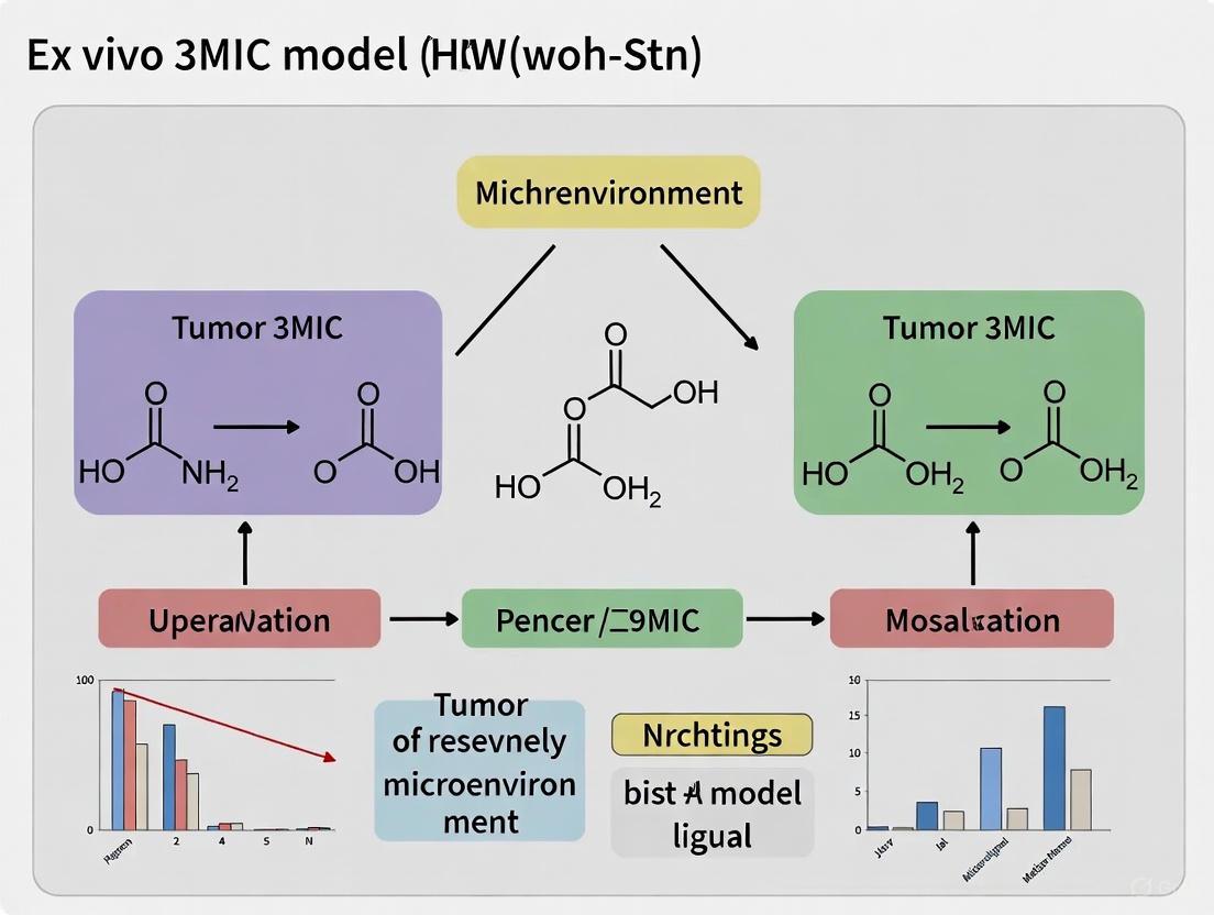
Unlike systems that require external control to create gradients, the 3MIC leverages the metabolic activity of the cells within the chamber to spontaneously generate ischemic conditions [6]. As the consumer and tumor cells respire and consume nutrients, they create a depletion zone. Metabolic by-products, such as lactic acid, simultaneously accumulate, leading to medium acidification [10]. This self-generating system closely mirrors the in vivo situation where gradients form naturally due to high cellular density and insufficient vascularization.
Modularity for Incorporation of Stromal Components
The 3MIC architecture is inherently flexible. In addition to tumor cells, researchers can incorporate key stromal cells known to facilitate metastasis, such as macrophages and fibroblasts, into the 3D matrix [11]. This modularity allows for the dissection of the individual and combined roles of cell-autonomous responses to ischemia and paracrine interactions with the stroma in driving metastatic progression [10] [1].
Experimental Applications and Key Findings
The 3MIC platform has been successfully applied to investigate core questions in metastasis and therapy resistance, yielding quantitative insights into these processes.
Investigating Pro-Metastatic Cues
Using the 3MIC, researchers confirmed that ischemic conditions robustly increase cell migration and invasion [6]. A key finding was that medium acidification, often a consequence of hypoxia and glycolysis, is one of the strongest pro-metastatic cues, directly driving the emergence of migratory features [10] [11]. The system also visualizes the loss of epithelial features and degradation of the extracellular matrix (ECM) by tumor cells [1].
Evaluating Drug Responses in Different Microenvironments
The 3MIC enables the testing of anti-cancer drugs on tumor cells experiencing different metabolic conditions within the same experiment. For instance, it was shown that chemotherapy drugs like Taxol, which are effective against tumor cells under normal conditions, failed to act on resource-deprived cells within the 3MIC [11] [12]. This suggests that the ischemic microenvironment itself can confer intrinsic drug resistance, providing a potential explanation for the resilience of metastatic disease.
The quantitative data from these core applications is summarized in the table below.
Table 2: Quantitative Findings from Key 3MIC Experiments
| Experimental Paradigm | Measured Outcome | Key Quantitative Result |
|---|---|---|
| Ischemia vs. Normoxia | Cell Migration & Invasion | Significant increase in migratory speed and ECM invasion under ischemic conditions [6]. |
| pH Modulation | Metastatic Feature Acquisition | Medium acidification identified as a primary driver of cell migration and invasion [10]. |
| Drug Treatment (e.g., Taxol) | Drug Efficacy | Drug effectiveness was markedly reduced against tumor cells in the nutrient/oxygen-starved region [11] [12]. |
| Stromal Co-culture | Enhancement of Invasion | Presence of macrophages or fibroblasts further increased pro-metastatic effects of ischemia [10]. |
Figure 2: Signaling pathways in metastasis. Ischemic conditions and stromal interactions drive pro-metastatic cellular changes, with acidification being a key intermediate.
Detailed Experimental Protocols
This section provides a step-by-step guide for a standard experiment using the 3MIC to study metastasis.
Protocol 1: Assembling the 3MIC Chamber
- Chamber Fabrication: Fabricate the 3MIC chamber using 3D printing technology as described in the original design [11] [12].
- Coverslip Preparation: Sterilize the glass coverslip that will form the top of the chamber.
- Seeding Consumer Cells: Seed a dense monolayer of "consumer cells" (e.g., a readily available cell line like fibroblasts) onto the sterilized coverslip. Culture the cells until they form a confluent layer.
- Chamber Assembly: Invert the coverslip with the attached consumer cells and carefully mount it to the top of the main chamber, creating a sealed unit where the consumer cells are now facing inward and upside-down.
- Matrix and Tumor Cell Embedding: Prepare a suspension of tumor cells (as single cells or pre-formed spheroids) in an appropriate 3D matrix material, such as a collagen or Matrigel solution. Gently inject the cell-matrix mixture into the main chamber of the assembled 3MIC.
- Polymerization and Media Addition: Allow the 3D matrix to fully polymerize under controlled conditions (e.g., 37°C for 30 minutes). Once set, connect the media reservoir filled with standard culture medium to the open side of the main chamber.
Protocol 2: Live-Cell Imaging of Metastatic Features
- Microscope Setup: Place the assembled 3MIC on the stage of a live-cell imaging microscope enclosed in a humidified chamber at 37°C and 5% CO₂.
- Image Acquisition Planning: Define multiple positions within the 3MIC for imaging, ensuring to capture regions expected to experience severe ischemia (far from the media source) and well-nourished regions (close to the media source).
- Time-Lapse Imaging: Initiate a time-lapse experiment, acquiring images of the tumor spheroids at regular intervals (e.g., every 15-30 minutes) over a period of 24-72 hours.
- Image Analysis: Use image analysis software to quantify pro-metastatic behaviors, including:
- Cell Migration: Track the speed and trajectory of individual cells or the leading edge of a spheroid.
- Morphological Changes: Analyze changes in cell shape, such as the transition from a rounded to an elongated, mesenchymal morphology.
- Matrix Degradation: If using a fluorescently tagged matrix, quantify the area of degradation around tumor spheroids.
Protocol 3: Drug Testing in Metabolic Gradients
- 3MIC Establishment: Set up the 3MIC with tumor cells as described in Protocol 1 and allow the metabolic gradients to establish over 24-48 hours.
- Drug Application: Introduce the anti-cancer drug of interest at a specific concentration into the media reservoir. A vehicle control should be run in a parallel 3MIC.
- Viability and Efficacy Assessment:
- Live/Dead Staining: After an appropriate incubation period (e.g., 24-72 hours), add a fluorescent live/dead cell viability stain to the media.
- Endpoint Imaging: Acquire fluorescence images across the metabolic gradient within the 3MIC.
- Quantification: Quantify the ratio of dead to live cells in both the ischemic and normoxic regions of the chamber and compare to the vehicle control.
The Scientist's Toolkit: Research Reagent Solutions
Successful implementation of the 3MIC system relies on a set of key reagents and materials.
Table 3: Essential Research Reagents and Materials for the 3MIC
| Reagent/Material | Function in the 3MIC | Specific Application Notes |
|---|---|---|
| Consumer Cells | To consume nutrients and oxygen, establishing metabolic gradients. | Often a robust, fast-growing cell line (e.g., fibroblasts). Must form a dense, confluent monolayer [1]. |
| Tumor Cell Line | The primary experimental subject for studying metastatic transition. | Can be used as single cells or pre-formed spheroids. Should be stably expressing a fluorescent protein for visualization [10]. |
| Extracellular Matrix (ECM) | Provides a 3D physiological context for cell growth, migration, and invasion. | Common choices include Type I Collagen or Basement Membrane Extract (e.g., Matrigel). Concentration and polymerization conditions are critical [10]. |
| Live-Cell Imaging Media | Sustains cell viability during long-term imaging without causing background fluorescence. | Phenol-free medium, buffered with HEPES, supplemented with appropriate serum or growth factors. |
| Fluorescent Viability Stains | To assess cell death in response to drug treatments or ischemic stress. | Used in Protocol 3 (e.g., Calcein-AM for live cells, Propidium Iodide for dead cells). |
| Metabolic Probes | To visualize and quantify metabolic gradients (e.g., oxygen, pH). | Examples include pH-sensitive fluorescent dyes (e.g., SNARF) or hypoxia probes (e.g., Pimonidazole) [10]. |
Metabolic Gradients as Drivers of Metastatic Features
Metastasis is the primary cause of cancer-related mortality, yet observing its earliest stages remains profoundly challenging. The initiation of metastasis is driven by microenvironmental conditions—such as hypoxia, nutrient starvation, and metabolic waste accumulation—that arise deep within tumor tissues. These ischemic conditions are difficult to access and visualize in vivo, creating a critical technical barrier to understanding the initial steps of metastatic progression. The 3D Microenvironment Chamber (3MIC) has been developed as an ex vivo model to overcome these limitations, enabling direct observation and perturbation of tumor cells as they acquire pro-metastatic features under controlled, gradient-forming conditions [1].
This application note details the use of the 3MIC platform to investigate how metabolic gradients serve as organizational cues and drivers of metastasis. We provide validated protocols for establishing metabolic gradients, quantifying emergent metastatic behaviors, and testing therapeutic interventions within a spatially-defined context that mimics the in vivo tumor microenvironment.
Key Findings: Metabolic Regulation of Metastasis
Research using the 3MIC and related models has revealed that metabolic gradients are not merely byproducts of tumor growth but are active instructors of cellular behavior and organization within the tumor ecosystem.
Spatial Organization by Metabolic Gradients
The altered metabolism of cancer cells establishes predictable gradients of extracellular metabolites that convey positional information to cells in the tumor microenvironment, much like morphogen gradients organize embryonic tissues [13].
- Gradient Formation: Metabolites consumed and secreted within the tumor microenvironment form concentration gradients relative to the vasculature. Hypoxia, for instance, typically saturates approximately 100 μm from the nearest blood vessel [13].
- Macrophage Patterning: Tumor-associated macrophages (TAMs) differentiate into distinct subpopulations based on local metabolic conditions. Macrophages in well-nourished, perivascular areas express the Mannose Receptor, C type 1 (MRC1), while those in hypoxic, nutrient-deprived regions express Arginase 1 (ARG1) [13].
- Inductive Signal: Lactate and hypoxia act as synergistic cues to induce ARG1 expression in macrophages. Lactate alone is insufficient to trigger a maximal response, indicating that multiple metabolic parameters combine to dictate cell fate [13].
Medium Acidification as a Potent Pro-Metastatic Cue
Within the 3MIC system, tumor spheroids spontaneously generate metabolic gradients, allowing direct observation of nascent metastatic features.
- Strong Inducer: Data from the 3MIC indicate that medium acidification is one of the strongest pro-metastatic cues, significantly increasing cell migration and invasion [1].
- Reversible Phenotypes: The acquisition of migratory and invasive properties in response to ischemia is reversible, suggesting that metastatic features can arise without permanent clonal selection [1].
- Stromal Augmentation: Interactions with stromal cells, including macrophages and endothelial cells, further enhance the pro-metastatic effects of ischemia [1].
Metabolic Flexibility in the Metastatic Cascade
Metastasizing cells exhibit dynamic metabolic rewiring, characterized by metabolic plasticity (using one metabolite for multiple purposes) and metabolic flexibility (using different metabolites to fulfill the same requirement) at different stages of the metastatic cascade [14].
Table 1: Metabolites Regulating Key Steps of Metastasis
| Metabolite | Primary Function | Role in Metastasis | Experimental Evidence |
|---|---|---|---|
| Lactate | Glycolytic end product | Promotes invasion, survival in circulation, and colonization; synergizes with hypoxia to polarize macrophages. | In vivo and MEMIC models show lactate gradients pattern macrophage ARG1 expression [14] [13]. |
| 2-Hydroxyglutarate (2-HG) | Oncometabolite | Induces EMT via epigenetic silencing of anti-metastatic miRNAs and activation of ZEB1. | IDH1/2 mutant cancers show elevated 2-HG and reversible EMT; exogenous 2-HG induces EMT in wildtype cells [15]. |
| Succinate/Fumarate | TCA cycle intermediates | Inhibit α-KG-dependent dioxygenases, leading to epigenetic changes that promote EMT. | SDH/FH mutations cause succinate/fumarate accumulation, driving EMT in PCC, PGL, and ovarian cancers [15]. |
| Acetyl-CoA | Central metabolic hub | Substrate for protein acetylation and epigenetic regulation; influences metastatic potential. | Deregulated acetyl-CoA metabolism reported in multiple cancers, contributing to malignant phenotypes [15]. |
Experimental Protocols
Protocol 1: Establishing the 3MIC for Visualizing Nascent Metastases
The 3MIC is designed to recreate the metabolic gradients of a tumor, placing ischemic cells in an easily observable plane [1].
Research Reagent Solutions
- Consumer Cells: A dense monolayer of cells (e.g., cancer-associated fibroblasts) to act as nutrient and oxygen sinks.
- Tumor Spheroids: Fluorescently-labeled tumor cells of interest, cultured as 3D spheroids.
- Stromal Cells: Optional addition of macrophages or endothelial cells for coculture studies.
- Extracellular Matrix (ECM): A defined ECM, such as Matrigel or collagen, to support 3D growth and invasion.
- Imaging-Compatible Culture Vessel: A chamber with a coverslip bottom for high-resolution live-cell imaging.
Procedure
- Plate Consumer Cells: Seed a dense monolayer of "consumer cells" upside down on a coverslip at the top of the chamber. These cells will consume nutrients and oxygen, creating a sink [1].
- Embed Tumor Cells: Suspend tumor spheroids within an ECM hydrogel and pipet into the main chamber volume, ensuring contact with the consumer cell layer [1].
- Establish Metabolic Gradients: Add culture medium to the reservoir connected to the chamber's opening. This opening acts as a source of fresh nutrients and oxygen. Incubate to allow metabolic gradients to form spontaneously over 24-48 hours [1].
- Live-Cell Imaging: Place the chamber on a confocal or two-photon microscope. Ischemic, pro-metastatic cells are accessible for high-resolution imaging. Monitor processes like migration, ECM degradation, and stromal interactions over time [1].
Protocol 2: Mapping Metabolic Gradients and Cell Phenotypes
This protocol combines the 3MIC with fluorescence lifetime imaging (FLIM) to correlate cellular metabolic states with phenotypic outcomes.
Research Reagent Solutions
- Metabolic Reporters: Use the auto-fluorescent metabolic co-enzymes NADH and FAD [16].
- Hypoxia Reporters: Chemical probes (e.g., Pimonidazole) or cell lines engineered with HIF-responsive elements (e.g., GFP-HRE) [13].
- Immunostaining Antibodies: Antibodies against phenotypic markers (e.g., ARG1, MRC1 for macrophages; E-cadherin, vimentin for EMT).
- Two-Photon Microscope: Equipped with time-correlated single photon counting (TCSPC) electronics for FLIM.
Procedure
- Culture and Gradient Formation: Set up the 3MIC as in Protocol 1, optionally including reporter cell lines.
- FLIM Acquisition: After 24-48 hours, acquire NADH and FAD fluorescence intensity and lifetime images using a two-photon microscope.
- Excitation Wavelengths: 750 nm for NADH, 890 nm for FAD [16].
- Calculate Redox Ratio: For each pixel, compute the optical redox ratio as NADH intensity / FAD intensity [16].
- Analyze Lifetimes: Fit fluorescence decay curves to a two-component exponential model to determine the mean lifetime (τm) of NADH and FAD, which reflects protein-binding status and metabolic activity [16].
- Post-Hoc Analysis: Fix the sample and perform immunostaining for relevant phenotypic markers (ARG1, MRC1, etc.).
- Data Correlation: Correlate the spatial patterns of the optical redox ratio and metabolite gradients (e.g., hypoxia) with the expression of phenotypic markers from immunohistochemistry.
Table 2: Key Parameters for Ex Vivo Metabolic Imaging
| Parameter | Description | Technical Notes |
|---|---|---|
| Optical Redox Ratio | NADH fluorescence intensity divided by FAD fluorescence intensity. | A higher ratio typically indicates a more glycolytic phenotype. Statistically identical to in vivo measurements for up to 24h in cultured tissue [16]. |
| NADH Mean Lifetime (τm) | The average time NADH remains in the excited state. | Remains stable for the first 8 hours in live culture. Increases in frozen-thawed samples, indicating loss of viability [16]. |
| Cell Viability | Percentage of live cells within the culture. | Should be >90% in high-quality ex vivo preparations [17]. |
| ATP Content | Indicator of energy charge. | In viable liver tissue cultures, reaches ~5 µmol/g of protein [17]. |
Signaling Pathways and Workflow Visualizations
Metabolic Regulation of Metastasis
3MIC Experimental Workflow
Implementing the 3MIC: Protocols and Practical Applications
Step-by-Step Guide to Assembling the 3MIC Chamber
The 3D Microenvironment Chamber (3MIC) is an innovative ex vivo model designed to recreate the complex and ischemic conditions of a tumor microenvironment, enabling the direct observation of nascent metastatic features [1] [11]. Metastasis initiation predominantly occurs within deep tumor regions characterized by nutrient and oxygen scarcity, conditions that are notoriously difficult to access and observe in vivo or with traditional 3D models [1]. The 3MIC overcomes this technical hurdle through its unique geometry, which spontaneously generates metabolic gradients, allowing researchers to directly visualize and perturb how tumor cells acquire migratory and invasive properties under controlled, ischemia-like conditions [1] [11]. This protocol provides a detailed guide for assembling the 3MIC, a crucial tool for any research program focused on understanding the early stages of cancer metastasis and testing novel anti-metastatic therapies.
Principle of the 3MIC
The fundamental principle of the 3MIC is to physically confine a dense cellular sample, restricting its access to nutrients and oxygen from all sides except one. This opening acts as a source of fresh media, while the cells inside the chamber consume these resources, thereby functioning as a sink [1]. This setup reliably creates a gradient of ischemic conditions—including hypoxia, nutrient starvation, and medium acidification—from the source to the deepest part of the chamber. Unlike its 2D predecessor (MEMIC), the 3MIC supports 3D cultures, which are essential for modeling key metastatic features like cell invasion and complex tumor-stroma interactions [1]. Its design is optimized for live-cell imaging, making ischemic cells at the core of the culture as easy to observe as well-nourished cells at the periphery.
Materials and Equipment
Research Reagent Solutions
Table 1: Essential materials and reagents for assembling and using the 3MIC.
| Item | Function/Description |
|---|---|
| Consumer Cells | A dense monolayer of cells grown upside-down on a coverslip; they consume nutrients to establish metabolic gradients within the chamber [1]. |
| Tumor Cells | The cells of interest (e.g., cancer cell lines), typically prepared as spheroids or in a 3D matrix, which are placed in the main chamber to study metastatic behavior [1] [11]. |
| Stromal Cells | Optional addition of partner cells such as macrophages or fibroblasts to study tumor-stroma interactions under ischemic conditions [1] [11]. |
| Extracellular Matrix (ECM) | A 3D hydrogel (e.g., Collagen I, Matrigel) to support the tumor cells and enable invasive migration [1]. |
| Cell Culture Medium | Appropriate medium for the tumor and consumer cells; the large reservoir connected to the chamber's opening serves as the source [1]. |
| 3D Printing Resin | A biocompatible resin used to fabricate the custom-designed chamber body [11]. |
| Coverslip | Serves as the transparent top window of the chamber, allowing for high-resolution live microscopy [1]. |
Equipment and Hardware
- 3D Printer: For fabricating the chamber with the specific geometry.
- Upright Microscope with Live-Cell Imaging Capabilities: Equipped with environmental control (e.g., temperature at 37°C).
- Standard Cell Culture Facility: Including biosafety cabinet, CO₂ incubator, and centrifuge.
Assembly Procedure
Chamber Fabrication and Preparation
- 3D Design: The chamber is designed with a small, central well for the 3D tumor culture and a single opening on one side that will connect to a large media reservoir.
- Printing: Fabricate the chamber using a high-resolution 3D printer and a biocompatible resin. Sterilize the printed chamber thoroughly, such as by UV exposure or ethanol wash, before use [11].
Seeding the Consumer Cell Layer
- Culture Consumer Cells: Expand an adequate number of consumer cells (e.g., fibroblasts or other dense cell type) in standard 2D culture.
- Prepare Coverslip: Sterilize a glass coverslip that will form the top of the chamber.
- Seed Cells on Coverslip: Trypsinize the consumer cells and seed them at a high density onto the sterilized coverslip to form a confluent monolayer.
- Incubate: Allow the cells to adhere and form a stable, dense monolayer. This "consumer cell layer" will be positioned at the top of the chamber, facing downwards.
Preparing the 3D Tumor Cell Culture
- Generate Spheroids: Form tumor cell spheroids using your method of choice (e.g., hanging drop, ultra-low attachment plates).
- Suspend in Matrix: Gently mix the spheroids with a cold, liquid ECM solution (e.g., collagen).
- Optionally Add Stromal Cells: To model tumor-stroma interactions, mix stromal cells like macrophages directly into the ECM solution with the tumor spheroids [1].
Final Chamber Assembly
- Load Tumor-Matrix Mixture: Pipette the tumor cell-spheroid-ECM mixture into the central well of the 3D-printed chamber body.
- Position Consumer Layer: Invert the prepared coverslip with the consumer cell monolayer and carefully lower it onto the chamber body, ensuring the monolayer faces the inside of the chamber. The consumer cells will now be at the top, "upside down" [1].
- Seal the Chamber: Securely seal the coverslip to the chamber body to prevent leaks and contamination, and to ensure a restricted environment.
- Connect Media Source: Connect the chamber's opening to a large reservoir of fresh, pre-warmed culture medium, which will act as the sole source of nutrients and oxygen.
Key Workflows and Experimental Setup
The diagram below illustrates the logical workflow for assembling the 3MIC chamber and initiating an experiment.
Establishing Metabolic Gradients and Experimental Timeline
Once assembled and connected to the media reservoir, the chamber must be incubated to allow metabolic gradients to form spontaneously. The following table outlines a typical experimental timeline.
Table 2: Experimental timeline for a 3MIC assay.
| Time Point | Key Process | Observation & Analysis |
|---|---|---|
| Day 0 | Chamber final assembly and connection to media source. | - |
| Day 1-2 | Establishment of stable metabolic gradients (hypoxia, nutrient starvation, acidosis). | Begin live imaging to track tumor cell morphology and initial migration [1] [11]. |
| Day 3-5 | Acquisition of metastatic features: increased migration, ECM degradation, stromal interactions. | Quantify migration speed, invasion distance, and changes in spheroid morphology [1]. |
| Day 5-7 | Drug perturbation studies (if applicable). | Introduce anti-metastatic drugs to the media reservoir and assess changes in metastatic behavior [11]. |
Applications and Perturbation Protocols
Drug Testing in the 3MIC
The 3MIC is uniquely suited for testing how local metabolic conditions affect drug efficacy. For example, studies have shown that resource-deprived tumor cells inside the 3MIC can be protected from certain chemotherapies, potentially mirroring the treatment resistance seen in metastases [11].
Protocol:
- Establish Gradients: Allow the 3MIC to incubate until metabolic gradients are fully established (typically 48-72 hours).
- Administer Drug: Introduce the drug candidate directly into the media reservoir at the desired concentration. The drug will diffuse into the chamber, creating a gradient that parallels the metabolic conditions.
- Image and Analyze: Use live-cell imaging to monitor and compare drug responses in tumor cells located in different regions of the chamber (e.g., well-nourished vs. ischemic). Key metrics include cell viability, migration arrest, and spheroid disintegration.
Incorporating Stromal Cells
To investigate tumor-stroma interactions, seed stromal cells (e.g., macrophages) directly into the 3D tumor cell-ECM mixture during chamber assembly [1]. The 3MIC allows for direct observation of how macrophages, for instance, interact with and facilitate the invasion of tumor cells under ischemic stress.
The tumor microenvironment (TME) is a complex and dynamic ecosystem where stromal and immune cells engage in critical crosstalk that profoundly influences cancer progression and therapy response [18]. The establishment of robust ex vivo co-culture models that faithfully replicate these interactions is paramount for advancing our understanding of tumor biology and developing novel therapeutic strategies [19]. This application note provides detailed protocols for integrating stromal and immune components into three-dimensional (3D) tumor models, specifically framed within the context of ex vivo 3MIC (Multiplexed, Modular, Immune-competent, and Clinical) model research. These methodologies enable researchers to dissect the functional roles of different TME components, particularly focusing on how stromal cells modulate innate immune cell phenotype and function via specific molecular axes such as the sialic acid/Siglec pathway [20]. By preserving critical cellular interactions that are lost in traditional monoculture systems, these co-culture platforms offer more physiologically relevant models for preclinical drug screening and personalized medicine applications.
Key Co-culture Model Systems
Stromal-Immune Interaction Mechanisms
Stromal cells, including cancer-associated fibroblasts (CAFs) and mesenchymal stromal cells (MSCs), exert profound immunomodulatory effects within the TME. Recent research has elucidated the critical role of the sialic acid/Siglec axis in mediating stromal-driven immune suppression [20]. The following diagram illustrates this key signaling pathway:
Figure 1: Stromal-Mediated Immune Suppression via Sialic Acid/Siglec Axis
Experimental Workflow for Co-culture Establishment
The successful establishment of stromal-immune co-cultures requires a systematic approach encompassing both scaffold-based and scaffold-free methodologies. The following workflow outlines the key procedural stages:
Figure 2: Co-culture Establishment Workflow
Detailed Experimental Protocols
Protocol 1: Direct Co-culture of Tumor Organoids with Immune Cells
Purpose: To establish direct contact between tumor organoids and immune cells for studying cell-cell interactions and immune-mediated cytotoxicity.
Materials:
- Patient-derived tumor organoids
- Peripheral blood mononuclear cells (PBMCs) or isolated immune cell subsets
- Matrigel or alternative extracellular matrix
- Organoid culture medium
- Immune cell culture medium (e.g., RPMI-1640 with IL-2 for T cells)
- 24-well or 48-well culture plates
Procedure:
- Prepare Tumor Organoids:
- Extract mature organoids from Matrigel using cell recovery solution or gentle mechanical dissociation.
- Wash organoids with cold PBS and resuspend in organoid culture medium.
- For some applications, digest organoids into single-cell suspensions using trypsin-EDTA or gentle dissociation reagents.
Prepare Immune Cells:
- Isolate PBMCs from fresh blood samples using Ficoll density gradient centrifugation.
- Alternatively, isolate specific immune cell subsets (T cells, NK cells, macrophages) using magnetic bead-based separation kits.
- Activate T cells if required using anti-CD3/CD28 beads or cytokines (e.g., IL-2) for 48-72 hours prior to co-culture.
Establish Co-culture:
- For direct contact studies, mix tumor organoids (or single cells) with immune cells at optimized ratios (typically 1:5 to 1:20 tumor:immune cell ratio) in suspension.
- Plate the cell mixture in low-attachment plates or embed in diluted Matrigel (50% concentration) to facilitate immune cell infiltration.
- Culture in optimized medium that supports both cell types, typically a 1:1 mix of organoid and immune cell media.
Monitoring and Analysis:
- Monitor co-cultures daily using brightfield microscopy for immune cell clustering and organoid morphology changes.
- Assess immune cell cytotoxicity after 24-96 hours using flow cytometry for apoptosis markers (Annexin V, caspase activation) or real-time cell imaging systems.
- Collect supernatants for cytokine profiling using multiplex ELISA assays.
Technical Notes:
- The direct contact method is particularly suitable for assessing immune cell cytotoxicity and tumor cell killing capacity [18].
- For T cell co-cultures, consider adding immune checkpoint inhibitors (anti-PD-1, anti-PD-L1) to reverse T cell exhaustion.
- Matrigel composition may affect immune cell function; consider using defined hydrogels for more reproducible results.
Protocol 2: Stromal-Immune Co-culture for Immunomodulation Studies
Purpose: To investigate how stromal cells modulate immune cell phenotype and function in the TME context.
Materials:
- Primary cancer-associated fibroblasts (CAFs) or mesenchymal stromal cells (MSCs)
- Immune cells (macrophages, NK cells, or T cells)
- Stromal cell culture medium
- Transwell inserts (optional, for indirect co-cultures)
- Sialyltransferase inhibitors (e.g., 3FAX) or sialidase (e.g., E610) for perturbation studies
Procedure:
- Stromal Cell Preparation:
- Culture CAFs or MSCs in appropriate stromal medium until 70-80% confluent.
- For tumor-conditioned stromal cells, culture MSCs with tumor cell-conditioned medium for 48-72 hours to generate MSC~TCS~.
- Optionally, modulate stromal cell sialylation using sialyltransferase inhibitors (3FAX, 10µM) or sialidase (E610, 0.1-1U/mL) for 24 hours before co-culture.
Immune Cell Isolation:
- Isolate monocytes from PBMCs and differentiate into macrophages using M-CSF (50ng/mL) for 5-7 days.
- Isulate NK cells using negative selection kits to maintain functionality.
Co-culture Establishment:
- Direct Co-culture: Seed immune cells directly onto stromal cell monolayers at defined ratios (recommended 1:1 to 1:5 stromal:immune ratio).
- Indirect Co-culture: Plate stromal cells in lower chamber and immune cells in Transwell inserts to study paracrine signaling.
- Culture for 24-72 hours in stromal-immune co-culture medium.
Functional Assessment:
- Analyze immune cell phenotype by flow cytometry for activation markers (CD80, CD86 on macrophages; CD69, NKG2D on NK cells) and immunosuppressive markers (CD206, PD-L1, Siglec receptors).
- Assess immune cell function: macrophage phagocytosis using pHrodo-labeled targets, NK cell cytotoxicity against K562 cells, or T cell proliferation using CFSE dilution.
- Measure cytokine secretion (IL-10, TGF-β, IFN-γ) in supernatants.
Technical Notes:
- This protocol is essential for studying stromal-mediated immune suppression mechanisms, particularly via the sialic acid/Siglec axis [20].
- Stromal cell sialylation status significantly impacts immune cell function; include desialylation controls where appropriate.
- For in vivo validation, consider adoptive transfer of co-cultured cells into immunocompetent mouse models.
Research Reagent Solutions
Table 1: Essential Research Reagents for Stromal-Immune Co-culture Models
| Reagent/Category | Specific Examples | Function/Application |
|---|---|---|
| Extracellular Matrices | Matrigel, synthetic PEG hydrogels, hyaluronic acid-based hydrogels | Provides 3D structural support for organoid and co-culture growth [18] |
| Stromal Cell Media | MSC growth medium, fibroblast medium with FBS | Supports stromal cell viability and function in co-culture systems |
| Immune Cell Media | RPMI-1640 with IL-2 (T cells), NK cell expansion media | Maintains immune cell viability and functionality during co-culture |
| Molecular Inhibitors | Sialyltransferase inhibitors (3FAX), sialidase (E610) | Perturbation tools for studying sialic acid/Siglec axis [20] |
| Immune Activators | Anti-CD3/CD28 beads, cytokine cocktails (IL-2, IL-15) | Enhances immune cell activation and cytotoxic function in co-cultures |
| Analysis Reagents | Flow cytometry antibodies, multiplex cytokine arrays | Enables assessment of immune cell phenotype and function |
Quantitative Data Presentation
Table 2: Functional Outcomes in Stromal-Immune Co-culture Models
| Co-culture System | Key Functional Readouts | Quantitative Impact | Therapeutic Relevance |
|---|---|---|---|
| CAF-Macrophage | Macrophage phagocytosis capacity | Reduction of 40-60% in co-culture vs. mono-culture [20] | Correlates with immunosuppressive TME |
| MSC-NK Cell | NK cell cytotoxicity | Decreased by 50-70% in co-culture; restored with sialidase [20] | Impacts innate immune surveillance |
| Organoid-T Cell | Tumor cell killing | Enhanced with anti-PD-1; patient-specific variability [19] | Predictive of immunotherapy response |
| Stromal-T Cell | T cell proliferation | Suppressed by 30-80% depending on stromal type | Contributes to immune evasion |
The establishment of sophisticated co-culture systems integrating stromal and immune components represents a critical advancement in TME modeling. The protocols detailed in this application note provide researchers with robust methodologies for investigating the functional interactions between these cellular compartments, with particular emphasis on the mechanistically important sialic acid/Siglec axis. These ex vivo 3MIC-compatible models enable the dissection of complex stromal-immune interactions under controlled conditions, facilitating both basic mechanism discovery and translational drug development. As the field progresses, the integration of these co-culture platforms with advanced spatial analysis techniques and computational modeling will further enhance their predictive value and utility in personalized cancer medicine.
Live-Cell Imaging and Quantification of Metastatic Behaviors
Within the context of ex vivo research on the tumor microenvironment (TME), the 3D Microenvironment Ischemic Chamber (3MIC) has emerged as a transformative platform. This model uniquely recapitulates the ischemic conditions—hypoxia, nutrient starvation, and acidosis—found deep within solid tumors, which are critical drivers of metastasis but notoriously difficult to observe directly in vivo [21] [1]. The 3MIC model enables the direct visualization and quantification of the moment tumor cells acquire pro-metastatic behaviors, a process that was previously elusive. This application note details the protocols and analytical methods for using the 3MIC to study metastatic features such as cell migration, invasion, and drug resistance under controlled, yet physiologically relevant, conditions [11].
Key Quantitative Findings on Metastatic Behaviors
Research utilizing the 3MIC model has yielded crucial quantitative data on how ischemic conditions promote metastasis. The table below summarizes the key metastatic behaviors that can be quantified using this system.
Table 1: Quantification of Metastatic Behaviors in the 3MIC Model
| Metastatic Behavior | Experimental Readout | Impact of Ischemic Conditions | Key Quantitative Findings |
|---|---|---|---|
| Cell Migration [1] | Cell speed and displacement tracked via live-cell imaging | Increased migration and dispersal | Ischemic cells demonstrate prolonged movement and increased speed. |
| Matrix Degradation [1] [22] | Area/intensity of fluorescence loss in quenched ECM substrates (e.g., DQ-Collegen) | Increased ECM degradation | Ischemic cells show a significant increase in proteolytic activity, clearing the fluorescent matrix. |
| Drug Resistance [22] [11] | Cell viability post-chemotherapy exposure (e.g., Taxol) | Enhanced survival and true resistance | Ischemic cells exhibit intrinsic resistance to drugs like Taxol, distinct from resistance caused by poor drug diffusion. |
| Stromal Cell Cooperation [1] | Distance and interaction frequency between tumor cells and stromal partners (e.g., macrophages) | Enhanced pro-metastatic effects | Co-culture with macrophages and endothelial cells further increases the migratory and invasive behaviors driven by ischemia. |
A pivotal finding from 3MIC studies is that medium acidification is one of the strongest pro-metastatic cues, even more direct in its effect than hypoxia alone. Hypoxia-inducible factor (HIF1A) signaling promotes invasion indirectly, while acidic conditions directly stimulate the activity of extracellular matrix-digesting enzymes [22].
Essential Reagents and Materials
Successful execution of experiments in the 3MIC requires a specific toolkit. The following table lists the essential research reagent solutions.
Table 2: Research Reagent Solutions for the 3MIC Model
| Item | Function/Description | Application in 3MIC |
|---|---|---|
| 3MIC Chamber [1] | A custom, 3D-printed chamber designed to create metabolic gradients. | Serves as the core physical platform for culturing tumor spheroids and establishing ischemic conditions. |
| Tumor Spheroids [22] | 3D cell clusters formed using methods like the "hanging drop" technique. | Used as the primary tumor model placed within the 3MIC to mimic the 3D architecture of a tumor. |
| Collagen Extracellular Matrix [1] [22] | A hydrogel providing a 3D scaffold for cell growth and invasion. | Spheroids are embedded in this matrix to model the physical barriers cells must degrade and migrate through. |
| Fluorescently-Tagged Gelatin/Collagen (e.g., DQ-Collegen) [22] | ECM substrate that fluoresces upon proteolytic cleavage. | Enables quantification of matrix degradation activity by tumor cells via confocal microscopy. |
| Hypoxia-Inducible Factor (HIF) Activators (e.g., Dimethyloxalylglycine, Cobalt Chloride) [22] | Chemical agents used to stabilize HIF1A under normoxic conditions. | Used to experimentally induce and study the HIF-mediated hypoxia response pathway. |
| Primary Macrophages [1] [22] | Immune cells differentiated from bone marrow cells. | Added to the 3MIC in co-culture to study tumor-stroma interactions and their role in promoting metastasis. |
Detailed Experimental Protocols
Protocol 1: Assembly and Seeding of the 3MIC
This protocol outlines the setup of the 3MIC chamber and the preparation of the tumor spheroids.
Workflow Diagram: 3MIC Assembly & Seeding
Materials:
- Sterilized 3MIC chamber [1]
- Acid-soluble collagen (e.g., Rat tail collagen I)
- Neutralization buffer (e.g., NaOH, HEPES)
- Tumor cell line of interest (e.g., lung adenocarcinoma cells)
- Fetal Bovine Serum (FBS), cell culture media
Step-by-Step Procedure:
- Chamber Preparation: Cure the 3D-printed 3MIC parts under ultraviolet light for sterilization. Assemble the chamber and fit it with a glass coverslip to form the base for culture [22].
- ECM Preparation: On ice, mix acid-soluble collagen with neutralization buffer according to the manufacturer's instructions to achieve a final concentration of 2-4 mg/ml.
- ECM Casting: Pipette the neutralized collagen solution into the 3MIC chamber, ensuring a thin, even layer covers the bottom. Incubate at 37°C for 30-45 minutes to allow the matrix to polymerize [22].
- Spheroid Formation: Use the hanging drop method by placing 20 µL drops of cell suspension (at a high density of ~5x10^5 cells/mL) on the lid of a petri dish. Invert the lid and incubate for 72-96 hours to form compact, spherical cell clusters [22].
- Spheroid Seeding: Carefully transfer individual spheroids using a pipette onto the surface of the polymerized collagen matrix within the 3MIC.
- Culture Initiation: Gently add complete culture medium to the chamber. For co-culture experiments, add stromal cells such as primary macrophages or fibroblasts at this stage [1].
Protocol 2: Live-Cell Imaging of Migration and Invasion
This protocol describes how to visualize and quantify metastatic behaviors like migration and matrix degradation over time.
Workflow Diagram: Live-Cell Imaging & Analysis
Materials:
- Confocal or spinning-disk confocal microscope with a stage-top incubator (maintaining 37°C and 5% CO2) [23]
- Software for automated time-lapse acquisition (e.g., MetaMorph, Zen)
- Fluorescently tagged cells (e.g., GFP-expressing tumor cells)
- Fluorescent ECM probe (e.g., DQ-Collegen, DQ-Gelatin)
Step-by-Step Procedure:
- Microscope Setup: Place the assembled 3MIC onto the pre-warmed stage of the confocal microscope. Ensure the environmental chamber is stabilized at 37°C and 5% CO2 [23].
- Position Selection: Using low-magnification objectives, identify and mark multiple fields of view for imaging. It is critical to include regions near the "source" of nutrients and deep within the chamber where ischemic conditions develop [1].
- Time-Lapse Acquisition: Program the microscope software to capture Z-stacks (to cover the 3D volume of cell movement) at each position every 5-10 minutes for the duration of the experiment (typically 24-72 hours) [1].
- Matrix Degradation Assay: If quantifying invasion, embed the tumor spheroids in an ECM mix containing 5-10% fluorescently quenched collagen or gelatin. Proteolytic cleavage by cell-secreted enzymes (e.g., MMPs) will generate a fluorescent signal, marking areas of degradation [22].
- Image Analysis - Motility: Use tracking software (e.g., ImageJ with TrackMate, or Imaris) to track the movement of individual cells or the leading edge of cell streams over time.
- Image Analysis - Invasion: Quantify the total area or fluorescence intensity of the cleared (fluorescent) zones around spheroids as a measure of invasive potential.
Protocol 3: Evaluating Drug Responses in Ischemic Niches
This protocol leverages the 3MIC to test drug efficacy in different metabolic regions.
Materials:
- Chemotherapeutic agents (e.g., Taxol)
- Viability stains (e.g., Propidium Iodide, Calcein AM)
- Fixed and permeabilized cells for immunohistochemistry
Step-by-Step Procedure:
- After the tumor spheroids have established within the 3MIC (typically 48-72 hours), add the chemotherapeutic drug to the culture medium at the desired concentration [11].
- Continue live-cell imaging to observe real-time cell behaviors (e.g., membrane blebbing, arrest of migration) in response to the drug.
- At the endpoint, add a live/dead viability stain to the culture medium according to the manufacturer's protocol and incubate for 30-60 minutes.
- Image the entire chamber to assess viability. Critically, compare cell death ratios between well-nourished cells near the media source and ischemic cells deep in the chamber [22] [11].
- For mechanistic studies, fix the cells in the chamber with 4% PFA and perform immunohistochemistry for markers of interest (e.g., HIF1α, drug efflux pumps, apoptosis markers).
Signaling Pathways in Metastasis Activation
The ischemic TME activates multiple interconnected signaling pathways that drive the acquisition of metastatic phenotypes. The diagram below illustrates the core pathways and their functional outcomes as modeled in the 3MIC.
Signaling Pathway Diagram: Ischemia-Driven Metastasis
As illustrated, the 3MIC model shows that acidosis is a potent, direct driver of ECM-digesting enzymes, while HIF1α signaling activation under hypoxia contributes to increased migration and drug resistance [22] [11]. Interactions with stromal cells further amplify these pro-metastatic signals [1].
Applications in Preclinical Drug Testing and Personalized Medicine
The 3D Microenvironment Chamber (3MIC) represents a significant advancement in ex vivo modeling of the tumor microenvironment (TME), specifically designed to overcome the challenges of observing and manipulating early metastatic events. Traditional in vivo observation of nascent metastases is exceedingly challenging because ischemic conditions like hypoxia and nutrient starvation arise deep within tumor tissues, making them virtually inaccessible for direct visualization [10] [1]. Similarly, while 3D organoids capture some aspects of tumor biology, ischemic cells remain buried within these structures, presenting nearly insurmountable imaging challenges [10]. The 3MIC system addresses these limitations by enabling tumor cells to spontaneously create ischemic-like conditions in a 3D context that allows for unprecedented spatial and temporal resolution of pro-metastatic processes [10] [1] [6].
This model is particularly valuable for preclinical drug testing and personalized medicine applications because it recapitulates key TME features, including the infiltration of immune cells and the spontaneous formation of metabolic gradients that mimic conditions within actual tumors [10]. By allowing direct observation and perturbation of cells as they acquire pro-metastatic features, the 3MIC provides an affordable, accessible complement to sophisticated in vivo microscopy, which remains prohibitively expensive for most laboratories [10] [1]. Its unique geometry enables researchers to directly image ischemic cells during the transition from poorly motile primary tumor cells to migratory metastatic-like cells, a process critical for understanding metastasis and testing therapeutic interventions [10].
Application in Preclinical Drug Testing
Key Applications and Workflow
The 3MIC system enables researchers to study how tumor spheroids migrate, invade, and interact with stromal cells under different metabolic conditions, providing a platform for evaluating anti-metastatic drugs [10] [6]. One of the most significant findings from 3MIC research is that medium acidification serves as one of the strongest pro-metastatic cues, even more influential than hypoxia alone in driving metastatic features [10] [1] [6]. This insight alone has profound implications for drug development, suggesting that targeting tumor acidosis may represent a promising therapeutic strategy.
The system allows for direct testing of how local metabolic conditions affect drug responses, enabling more predictive preclinical assessment of therapeutic efficacy [10]. Unlike traditional 2D cultures that lack physiological metabolic gradients, the 3MIC spontaneously generates ischemic conditions similar to those found in solid tumors, including hypoxia, nutrient starvation, and metabolic by-product accumulation [10] [1]. This capability is crucial since more than 90% of cancer drugs fail in clinical trials, often due to limited ability to accurately model solid tumors in laboratory settings [24].
Table 1: Key Applications of the 3MIC Model in Preclinical Drug Testing
| Application Area | Experimental Capability | Output Metrics |
|---|---|---|
| Metabolic Gradient Studies | Investigation of hypoxia, nutrient starvation, and acidosis effects on drug efficacy | Cell migration distance, invasion capacity, metabolic profiling |
| Stromal Cell Interactions | Co-culture with macrophages, endothelial cells, and other stromal components | Quantification of stromal-enhanced pro-metastatic effects |
| Drug Efficacy Screening | Testing anti-metastatic drugs under different metabolic conditions | Dose-response curves, IC50 values under normoxic vs. ischemic conditions |
| Metastasis Progression Analysis | Direct observation of epithelial-to-mesenchymal transition and ECM degradation | Morphological changes, protease activity, migration velocity |
The experimental workflow for drug testing using the 3MIC system typically involves multiple stages, as illustrated below:
Protocol: Drug Efficacy Testing in the 3MIC System
Objective: To evaluate compound efficacy against tumor cell migration and invasion under ischemic conditions representative of the native TME.
Materials:
- 3MIC chamber apparatus
- Appropriate tumor cell lines (e.g., breast, prostate, pancreatic cancer)
- Stromal cells (macrophages, endothelial cells, cancer-associated fibroblasts)
- Extracellular matrix components (Collagen I, Matrigel)
- Drug compounds for testing
- Live-cell imaging system with environmental control
- Metabolic dyes (e.g., pH sensors, hypoxia probes)
Procedure:
- Chamber Assembly: Sterilize 3MIC components and assemble according to manufacturer specifications. Ensure proper sealing to prevent media leakage.
Consumer Cell Seeding: Seed a dense monolayer of "consumer cells" (e.g., fibroblasts) upside down on the top coverslip of the chamber at a density of 1.5-2.0×10^5 cells/cm². These cells will consume nutrients and oxygen, establishing metabolic gradients.
Tumor Spheroid Embedding:
- Harvest tumor cells and form spheroids using low-adhesion plates or hanging drop method.
- Mix spheroids with ECM solution (70% Collagen I, 30% Matrigel) at a concentration of 50-100 spheroids/mL.
- Pipette 100-200μL of spheroid-ECM mixture into the main chamber compartment.
- Polymerize at 37°C for 30-60 minutes.
Media Addition and Gradient Establishment:
- Carefully add complete media to the reservoir, connecting to the chamber opening.
- Culture for 48-72 hours to establish stable metabolic gradients.
- Confirm gradient establishment using pH and hypoxia sensors.
Compound Application:
- Prepare drug solutions at appropriate concentrations in fresh media.
- Replace reservoir media with media containing treatment compounds.
- Include vehicle controls and reference standards.
Live-Cell Imaging and Data Collection:
- Place chamber on live-cell imaging system maintained at 37°C and 5% CO₂.
- Acquire time-lapse images every 30-60 minutes for 24-72 hours.
- Use phase contrast for migration tracking and fluorescence for specific markers.
Endpoint Analysis:
- Fix cells for immunostaining (e.g., EMT markers, proliferation, apoptosis).
- Collect media for metabolic analysis (e.g., lactate production, glucose consumption).
- Process for RNA/protein extraction if needed.
Data Analysis:
- Track individual cell migration paths and calculate velocity, directionality, and mean squared displacement.
- Quantify invasion distance from spheroid edge into surrounding matrix.
- Assess morphological changes indicative of EMT.
- Correlate drug efficacy with positional information within metabolic gradients.
Application in Personalized Medicine
Patient-Specific Therapeutic Stratification
The 3MIC system provides a unique platform for advancing personalized medicine by enabling ex vivo testing of patient-derived tumor samples under controlled yet physiologically relevant conditions. This application aligns with the broader PERMIT project recommendations for personalized medicine research, which emphasize the need for robust methodologies to ensure proper patient stratification and treatment assignment [25]. By maintaining critical TME interactions and metabolic features, the 3MIC can help predict individual patient responses to specific therapies, particularly for metastatic disease where current models often fail.
In the context of personalized medicine, the 3MIC system addresses a critical need for preclinical models that can accurately recapitulate individual tumor characteristics. The European Council's definition of personalized medicine emphasizes "tailoring the right therapeutic strategy for the right person at the right time" based on individual phenotypes and genotypes [26]. The 3MIC supports this goal by preserving patient-specific tumor characteristics, including unique stromal interactions and metabolic profiles that influence treatment response.
Table 2: 3MIC Applications in Personalized Medicine Pipeline
| Personalized Medicine Stage | 3MIC Application | Clinical Translation |
|---|---|---|
| Stratification Cohort Development | Testing patient-derived tumor cells in standardized TME | Identification of responsive patient subgroups |
| Biomarker Discovery | Correlation of drug response with spatial positioning in metabolic gradients | Development of predictive biomarkers for treatment selection |
| Therapeutic Validation | Ex vivo assessment of standard and experimental regimens | Informed treatment selection for individual patients |
| Resistance Mechanism Analysis | Study of adaptive responses under ischemic pressure | Strategies to overcome treatment resistance |
Protocol: Patient-Derived Sample Testing in 3MIC
Objective: To evaluate therapeutic responses of patient-derived tumor cells in a physiologically relevant TME for treatment stratification.
Materials:
- Patient-derived tumor tissue from biopsies or surgical resections
- 3MIC chambers adapted for small sample sizes
- Enzymatic digestion cocktail (Collagenase, DNase)
- Red blood cell lysis buffer
- Stromal cell expansion media
- Autologous patient serum when available
Procedure:
- Patient Sample Processing:
- Mechanically dissociate tumor tissue into 1-2mm³ fragments using scalpel or tissue chopper.
- Digest with collagenase IV (1-2mg/mL) and DNase I (100μg/mL) for 30-60 minutes at 37°C with gentle agitation.
- Filter through 100μm cell strainer to obtain single cell suspension.
- Lyse red blood cells using ammonium chloride solution if necessary.
- Count viable cells using trypan blue exclusion.
Autologous Stromal Cell Isolation:
- Expand cancer-associated fibroblasts from tumor explants in fibroblast-specific media.
- Isate peripheral blood mononuclear cells for monocyte-derived macrophages when needed.
3MIC Culture Establishment:
- Seed patient-derived tumor cells (5×10⁴-2×10⁵) in ECM mixture as described in Section 2.2.
- Incorporate autologous stromal cells at physiologically relevant ratios (typically 10-30% of total cells).
- Use patient-derived serum (10%) when available to maintain patient-specific signaling.
Therapeutic Testing:
- Select drug panels based on patient tumor molecular profiling.
- Test standard-of-care options and targeted agents when applicable.
- Include combination therapies based on clinical relevance.
Response Assessment:
- Monitor cell viability, migration, and invasion as in Section 2.2.
- Compare responses across different metabolic conditions within the chamber.
- Establish response thresholds based on clinical correlation data.
Data Interpretation for Clinical Guidance:
- Classify as "responsive" if >40% reduction in invasion and >50% reduction in migration velocity.
- Consider moderate response with 20-40% reduction in invasive capacity.
- Report stromal contributions to resistance or sensitivity.
- Provide quantitative metrics with confidence intervals based on replicate experiments.
The integration of 3MIC testing into personalized medicine pipelines addresses the PERMIT project's emphasis on proper methodological research to ensure robust and reproducible evidence generation in personalized medicine [25]. This approach is particularly valuable for addressing the challenges of metastatic cancers, where the TME plays a crucial role in treatment response and disease progression.
The Scientist's Toolkit: Essential Research Reagents
Successful implementation of 3MIC technology requires specific reagents and materials optimized for studying the tumor microenvironment. The following table details essential components and their functions in 3MIC-based experiments:
Table 3: Essential Research Reagents for 3MIC TME Studies
| Reagent Category | Specific Examples | Function in 3MIC Experiments |
|---|---|---|
| Extracellular Matrix Components | Collagen I, Matrigel, Fibrin | Provide 3D structural support recapitulating in vivo tissue architecture; influence cell signaling and invasion |
| Metabolic Probes | pHrodo, HypoxiSense, LC-1 | Visualize and quantify metabolic gradients (acidosis, hypoxia, redox stress) within the chamber |
| Stromal Cell Markers | CD45 (immune), α-SMA (CAFs), CD31 (endothelial) | Identify and track stromal cell populations in co-culture experiments |
| Live-Cell Imaging Dyes | Calcein AM (viability), CellTracker, SiR-actin | Monitor cell viability, morphology, and dynamics without fixation |
| EMT Markers | E-cadherin, Vimentin, ZEB1 | Quantify epithelial-to-mesenchymal transition during metastasis |
| Protease Reporters | MMPsense, Quenched fluorescein-collagen | Detect extracellular matrix degradation during invasion |
| Cytokine/Antibody Panels | Multiplex cytokine arrays, neutralization antibodies | Profile secretory signaling and block specific pathways |
The diagram below illustrates the spatial relationships and signaling interactions that can be studied within the 3MIC system, highlighting the key cellular components and metabolic features:
The 3MIC ex vivo model represents a transformative tool for both preclinical drug testing and personalized medicine applications. By enabling direct visualization of emergent metastatic features under physiologically relevant conditions, it addresses critical limitations of traditional 2D cultures and in vivo models. The system's ability to recreate and manipulate the complex metabolic gradients and stromal interactions of the tumor microenvironment provides unprecedented opportunities for studying metastasis and evaluating therapeutic interventions.
For drug development, the 3MIC offers a platform for assessing compound efficacy against critical metastatic processes under conditions that more closely resemble the in vivo TME. The identification of medium acidification as a key pro-metastatic cue through 3MIC research highlights its value in uncovering new biological insights and therapeutic targets [10] [6]. In personalized medicine, the adaptation of 3MIC technology for patient-derived samples provides a path toward truly individualized therapeutic stratification, aligning with PERMIT recommendations for robust methodological approaches in personalized medicine research [25].
As cancer research continues to emphasize the importance of the tumor microenvironment in treatment response and resistance, models like the 3MIC that capture this complexity will become increasingly valuable. The protocols and applications detailed here provide a framework for leveraging this technology to advance both fundamental cancer biology and clinical translation.
Optimizing 3MIC Experiments: Troubleshooting Common Challenges
Ensuring Reproducible Metabolic Gradient Formation
The 3D Microenvironment Chamber (3MIC) is an ex vivo model specifically designed to overcome the significant challenge of observing and perturbing early metastatic events within a controlled tumor microenvironment [1]. A core feature of this system is its ability to enable tumor cells to spontaneously create ischemic-like conditions, including gradients of oxygen, nutrients, and metabolic by-products such as lactic acid [1]. These gradients are critical as they mimic the conditions tumor cells encounter deep within solid tumors, which are known drivers of metastasis [1]. Reproducible formation of these metabolic gradients is therefore paramount for utilizing the 3MIC to study the emergence of pro-metastatic features and for conducting reliable drug testing under different metabolic conditions [1].
Principles of Metabolic Gradient Formation in the 3MIC
The 3MIC achieves metabolic gradient formation through a specific geometry that restricts resource access. In this design, a dense monolayer of "consumer cells" is grown upside down on a coverslip at the top of a small chamber. This chamber is sealed from nutrients and oxygen on all sides except one, which features an opening connected to a large reservoir of fresh culture media [1]. This setup establishes a fundamental physical principle:
- Source and Sink Dynamics: The media reservoir acts as a source of nutrients (e.g., glucose) and oxygen. The densely packed consumer cells within the chamber function as a sink, actively consuming these resources.
- Gradient Generation: As nutrients and oxygen diffuse from the source into the cell-filled chamber, they are progressively depleted by the consumer cells. Simultaneously, metabolic waste products (e.g., lactic acid) produced by the cells accumulate, creating a reverse gradient. This results in a stable, reproducible spatial gradient of metabolic conditions, ranging from well-nourished and normoxic near the opening to ischemic (characterized by nutrient starvation, hypoxia, and acidosis) in the deepest parts of the chamber [1].
- Visualization Advantage: Unlike traditional 3D models or organoids where ischemic cells are buried within the structure, the 3MIC's geometry makes imaging cells experiencing these metabolic gradients straightforward, providing unprecedented spatial and temporal resolution for observing metastatic transitions [1].
Table 1: Key Metabolic Parameters and Their Pro-Metastatic Roles in the 3MIC
| Metabolic Parameter | Condition in Ischemic Region | Pro-Metastatic Effect |
|---|---|---|
| Oxygen | Hypoxia | Increases cell migration and invasion [1] |
| Nutrients (e.g., Glucose) | Starvation | Drives initiation of metastasis [1] |
| pH (from lactic acid) | Acidosis (Medium Acidification) | One of the strongest pro-metastatic cues [1] |
| Redox State | Oxidative Stress | Drives initiation of metastasis [1] |
Quantitative Assessment of Metabolic Gradients
Ensuring the reproducibility of gradient formation requires robust quantitative methods to validate the metabolic landscape within the chamber.
Spatial Quantitative Metabolomics with Internal Standards
A major pitfall in quantifying spatial metabolism is the matrix effect, where the tissue or cellular environment itself interferes with accurate measurement, leading to unreliable interpretation [27]. To overcome this, an improved quantitative mass spectrometry imaging (MSI) workflow using uniformly ¹³C-labelled yeast extracts as internal standards (IS) is recommended [27].
This method involves:
- Homogeneous Application: The U-¹³C-labelled yeast extract is sprayed evenly across the sample surface, providing a vast array of isotopically labelled metabolite standards [27].
- Pixel-wise Normalization: The signal from each labelled metabolite IS is used to normalize the signal from its corresponding endogenous metabolite in every pixel of the MSI data. This corrects for the matrix effect and enables reliable relative quantification [27].
- Broad Coverage: This approach allows for the pixelwise normalization of over 200 metabolic features, including intermediates from glycolysis, the TCA cycle, pentose phosphate pathway, amino acid metabolism, and complex lipids [27].
Traditional normalization methods like Total Ion Count (TIC) or Root Mean Square (RMS) show vastly different and less reliable results compared to the internal standard method, highlighting the necessity of this refined protocol for accurate quantification [27].
Table 2: Key Reagents for Quantitative Spatial Metabolomics
| Research Reagent | Function in Protocol |
|---|---|
| U-¹³C-labelled Yeast Extract | Serves as a source of numerous internal standards for pixel-wise normalization, correcting for matrix effects [27]. |
| NEDC Matrix | Applied to the tissue sample for Matrix-Assisted Laser Desorption/Ionization (MALDI) MSI analysis [27]. |
| Standardized Metabolite IS Panels | Metabolite-specific internal standards; costly but sometimes necessary for certain applications [27]. |
Protocol for Sample Preparation and MSI Analysis
This protocol is adapted from spatial metabolomics studies and can be applied to 3MIC samples [27].
Sample Preparation:
- Culture cells in the 3MIC system under standard conditions to establish metabolic gradients.
- At the experimental endpoint, immediately flash-freeze the entire chamber assembly in liquid nitrogen to halt metabolic activity.
- Cryosection the cell layer from the 3MIC chamber at a desired thickness (e.g., 10-20 µm) and mount onto a conductive ITO slide pre-cooled to -20°C.
Application of Internal Standard:
- Thaw the slide sections in a desiccator for approximately 10-15 minutes.
- Using a robotic sprayer (e.g., TM-Sprayer), homogeneously apply a solution of U-¹³C-labelled yeast extract across the entire surface of the tissue section. Optimize sprayer parameters (flow rate, number of passes, velocity) for even coverage.
Matrix Application:
- Subsequently, apply the NEDC matrix via the robotic sprayer using validated parameters to ensure a fine, crystalline coating.
Data Acquisition:
- Acquire mass spectrometry imaging data using a MALDI mass spectrometer (e.g., TimsTOF flex) in negative ion mode.
- Set the mass spectrometer to detect both the endogenous metabolites and their corresponding ¹³C-labelled counterparts from the yeast extract.
Data Processing and Normalization:
- Pre-process the raw MSI data (e.g., peak picking, alignment).
- For each pixel, normalize the intensity of each identified endogenous metabolite by the intensity of its corresponding ¹³C-labelled internal standard.
- Generate spatial distribution maps for the normalized intensities of key metabolites (e.g., lactate, glutamate, glutathione) to visualize the metabolic gradients.
Figure 1: Experimental workflow for quantitative spatial metabolomics of 3MIC samples.
Protocols for Core 3MIC Experiments
Protocol: Establishing Reproducible Metabolic Gradients in the 3MIC
The formation of a reproducible gradient is highly dependent on initial cell density and media composition [1].
- Chamber Assembly: Sterilize all components of the 3MIC chamber. Assemble the chamber according to its design, ensuring the top coverslip is securely in place.
- Cell Seeding for Consumer Layer:
- Harvest the consumer cells (e.g., primary fibroblasts or a stable cell line). Determine viable cell count.
- Critical Step: Resuspend the cells in complete growth medium at a high, pre-optimized density (e.g., 10x10⁶ cells/mL). The density is crucial for achieving sufficient resource consumption to establish the gradient [1].
- Invert the 3MIC chamber and pipette a precise volume of the cell suspension onto the coverslip at the top. Carefully place the chamber in a humidified incubator (37°C, 5% CO₂) for a defined period (e.g., 1-2 hours) to allow cell attachment upside down.
- Media Introduction and Gradient Establishment:
- After cell attachment, carefully return the chamber to its upright position.
- Gently introduce complete culture medium through the side opening, ensuring no air bubbles are trapped.
- Connect the chamber opening to a large reservoir of fresh medium (e.g., 10-50x the chamber volume) to act as a stable source.
- Return the assembled system to the incubator and allow the metabolic gradient to establish over 24-48 hours. The gradient stability can be verified using the spatial metabolomics protocol described above.
Protocol: Testing Drug Responses Under Different Metabolic Conditions
The 3MIC allows for the direct observation of drug effects on cells experiencing different microenvironments within a single chamber [1].
Gradient and Treatment:
- Establish the 3MIC system with tumor cells as described in Section 4.1. The consumer cells can be the tumor cells themselves or stromal cells.
- After the metabolic gradient is established (e.g., 48 hours), introduce the anti-metastatic drug of interest at a desired concentration into the media reservoir.
- Allow the drug to diffuse and act for the required treatment duration (e.g., 24-72 hours).
Endpoint Analysis:
- Live Imaging: Use the 3MIC's imaging-friendly design to perform live-cell imaging. Track parameters such as cell migration velocity, invasion distance into the matrix, and changes in cell morphology in different gradient regions (e.g., ischemic vs. well-nourished) over time [1].
- Fixed Staining: At the end of the experiment, fix the cells within the chamber and perform immunofluorescence staining for metastatic markers (e.g., proteins involved in epithelial-to-mesenchymal transition) or perform the spatial metabolomics protocol to assess drug-induced metabolic changes.
Figure 2: Schematic diagram of metabolic gradient formation in the 3MIC system.
The Scientist's Toolkit: Essential Research Reagents and Materials
Table 3: Key Research Reagent Solutions for the 3MIC Platform
| Reagent/Material | Function and Importance |
|---|---|
| 3MIC Chamber | The core physical platform; its specific geometry enables the reproducible formation of metabolic gradients by controlling diffusion [1]. |
| Consumer Cells | A dense monolayer of cells (e.g., fibroblasts or tumor cells) that consume resources to establish the nutrient and oxygen sink, driving gradient formation [1]. |
| Defined Culture Medium | The composition of the medium in the source reservoir directly influences the nature of the metabolic gradient (e.g., high glucose vs. low glucose). |
| U-¹³C-labelled Yeast Extract | Critical for quantitative spatial metabolomics; provides a comprehensive set of internal standards to correct for matrix effects and enable accurate metabolite quantification [27]. |
| Acidosis-Inducing Agents | Compounds like lactic acid can be used to modulate the pH of the media source to specifically study the strong pro-metastatic effects of acidosis [1]. |
| Stromal Co-culture Components | Primary macrophages, endothelial cells, or fibroblasts can be incorporated to study their interaction with tumor cells under ischemic conditions, which enhances pro-metastatic effects [1]. |
Validating Cellular Health and Viability in Long-Term Cultures
Maintaining and accurately assessing cellular health in long-term ex vivo cultures is a cornerstone of reliable cancer research. Within the context of the ex vivo 3D Microenvironment Chamber (3MIC) model, which is designed to recapitulate the ischemic and stromal interactions of the tumor microenvironment (TME), validation of viability is particularly crucial [1]. The 3MIC model spontaneously generates metabolic gradients, including hypoxia and nutrient starvation, to study the emergence of pro-metastatic features [1]. This application note provides detailed protocols and analytical frameworks for confirming that the cellular responses observed in this sophisticated system are genuine biological phenomena and not artifacts of declining culture health.
Quantitative Viability Metrics and Data Analysis
A multi-faceted approach is required to comprehensively assess the health of cells in long-term 3MIC cultures. Key quantitative metrics should be gathered and structured for easy comparison, as summarized in the table below.
Table 1: Key Quantitative Metrics for Validating Cellular Health in Long-Term 3MIC Cultures
| Assessment Method | Target / Principle | Healthy Culture Indicator | Application in 3MIC Context |
|---|---|---|---|
| Membrane Integrity (PI/7-AAD Flow Cytometry) [28] [29] | DNA intercalation in membrane-compromised cells | Low percentage of PI-positive cells | Distinguish true apoptotic/necrotic cells from live cells during metastasis studies [1]. |
| Metabolic Activity (Calcein-AM Staining) [28] | Esterase activity in live cells | High percentage of Calcein-positive cells | Confirm metabolic competence of cells in ischemic gradient regions [1]. |
| Lactate Dehydrogenase (LDH) Release [30] | Cytosolic enzyme released upon membrane damage | Low LDH in culture supernatant | Quantify overall cytotoxicity; validate superior health in optimized culture systems [30]. |
| Inflammatory Cytokine Profile [30] | Measurement of IL-6, IL-1β, TNF | Low levels of pro-inflammatory cytokines | Monitor culture-induced stress and inflammation [30]. |
| Proliferation Marker Expression [31] | Immunofluorescence for Ki-67, etc. | Presence of proliferating cells | Verify active cell cycling, especially after perturbations like drug treatment [1]. |
| Morphological Assessment [30] [32] | Tissue architecture (H&E) and cell morphology | Intact epidermis-dermis junction, normal nuclear size | Ensure 3D structural integrity is maintained over time, critical for TME studies [30]. |
The following workflow diagram outlines the logical sequence for applying these validation methods in a 3MIC experiment.
Detailed Experimental Protocols
Protocol A: Cell Viability Staining for Flow Cytometry
This protocol is optimized for the simultaneous analysis of cell viability and surface markers, which is essential for immunophenotyping within the complex TME of the 3MIC model [32].
Title: Two-Color Viability and Surface Marker Staining for 3MIC-Derived Single Cells
Principle: Propidium iodide (PI) is a membrane-impermeant dye that enters dead cells with compromised membranes and intercalates into DNA, providing a fluorescent signal for dead cell exclusion during flow cytometry analysis [28] [29].
Materials:
- Propidium Iodide (PI) Staining Solution (10 µg/mL in PBS) [29]
- Flow Cytometry Staining Buffer (e.g., containing BSA and sodium azide) [28] [29]
- Antibodies for surface marker staining
- 12 x 75 mm round-bottom FACS tubes
- Centrifuge and vortex mixer
Procedure:
- Harvest and Wash: Generate a single-cell suspension from the 3MIC culture. Aliquot up to (1 \times 10^6) cells per 100 µL into a FACS tube. Wash cells twice with 2 mL of PBS by centrifuging at 300–400 × g for 5 minutes [29].
- Surface Marker Staining: Resuspend the cell pellet in 100 µL of Flow Cytometry Staining Buffer. Add fluorochrome-conjugated antibodies against your target surface antigens (e.g., CD45 for immune cells, EpCAM for epithelial cells) as required. Incubate for 30 minutes on ice or at 4°C, protected from light [28].
- Wash: Add 2 mL of Flow Cytometry Staining Buffer, centrifuge, and decant the supernatant.
- Viability Staining: Resuspend the cell pellet in an appropriate volume (e.g., 100–500 µL) of Flow Cytometry Staining Buffer. Add 5–10 µL of PI Staining Solution per 100 µL of cell suspension and mix gently [28] [29].
- Acquisition: Incubate for 5–15 minutes at room temperature or on ice, protected from light. Do not wash cells after PI addition. Analyze samples immediately by flow cytometry, using the FL-2 or FL-3 channel for PI detection [29].
Protocol B: Fixable Viability Dye Staining for Intracellular Analysis
This protocol is critical for experiments that require intracellular staining, fixation, or permeabilization, such as analyzing cytokine production or signaling proteins in TME cell subsets.
Title: Fixable Viability Dye (FVD) Staining for Subsequent Intracellular Staining
Principle: Fixable Viability Dyes (FVDs) are amine-reactive dyes that brightly stain cells with compromised membranes. They covalently bind to cellular proteins, allowing the stained cells to undergo fixation and permeabilization procedures without loss of the dead cell label [28].
Materials:
- Fixable Viability Dye (e.g., eFluor 506, eFluor 780), stored at ≤ –70°C
- Phosphate-buffered saline (PBS), azide- and protein-free
- Flow Cytometry Staining Buffer
Procedure:
- Harvest and Wash: Generate a single-cell suspension from the 3MIC culture. Wash cells twice in azide- and protein-free PBS [28].
- Viability Staining: Resuspend cells at (1–10 \times 10^6) /mL in azide- and protein-free PBS. Add 1 µL of FVD per 1 mL of cells and vortex immediately. Incubate for 30 minutes at 2–8°C, protected from light [28].
- Wash: Add 2 mL of Flow Cytometry Staining Buffer, centrifuge, and decant the supernatant to remove unbound dye.
- Surface & Intracellular Staining: Proceed with standard surface and/or intracellular antibody staining protocols. The FVD signal will remain stable through fixation and permeabilization steps [28].
Protocol C: Metabolic and Functional Staining for Live-Cell Imaging in 3MIC
The 3MIC model is uniquely suited for live imaging. This protocol outlines how to visualize viable cells and their metabolic states directly within the chamber.
Title: Live Imaging of Viability and ROS in the 3MIC Model
Principle: Calcein-AM is a cell-permeant dye converted by intracellular esterases into a fluorescent calcein, labeling live cells. It can be combined with probes for reactive oxygen species (ROS), which are often elevated under the ischemic conditions of the 3MIC [30] [1].
Materials:
- Calcein AM dye (e.g., UltraPure Grade), reconstituted in anhydrous DMSO [28]
- CellROX Deep Red or similar ROS probe
- Standard cell culture medium without serum or phenol red
Procedure:
- Preparation: Prepare working solutions of Calcein AM and the ROS probe in pre-warmed serum-free medium according to the manufacturer's recommendations.
- Staining: Carefully remove the existing medium from the 3MIC and replace it with the dye-containing medium. Incubate the chamber for 30 minutes at 37°C, 5% CO₂, protected from light [28].
- Wash and Image: Replace the staining medium with fresh, pre-warmed imaging medium. Immediately acquire time-lapse images using appropriate fluorescence filter sets for Calcein (e.g., FITC channel) and the ROS probe (e.g., Cy5 channel) [30].
The Scientist's Toolkit: Key Research Reagent Solutions
The following table catalogues essential reagents and their critical functions for validating cellular health in complex 3D cultures like the 3MIC model.
Table 2: Essential Reagents for Viability Assessment in Long-Term 3D Cultures
| Reagent / Kit | Function / Principle | Key Application in 3MIC/TME Research |
|---|---|---|
| Propidium Iodide (PI) [28] [29] | Membrane integrity dye for dead cell exclusion in flow cytometry. | Rapid, cost-effective viability census of dissociated TME cells. |
| Fixable Viability Dyes (FVDs) [28] | Amine-reactive dyes for irreversible dead cell labeling; compatible with fixation. | Essential for complex immunophenotyping and intracellular signaling analysis in the TME. |
| Calcein-AM [28] | Cell-permeant dye converted to fluorescent product by live-cell esterases. | Visualizing spatial distribution of live cells and metabolic activity within 3MIC gradients. |
| CellROX Oxidative Stress Probes | Fluorescent probes that become bright upon oxidation in live cells. | Probing elevated ROS levels in ischemic regions of the 3MIC as a marker of metabolic stress [1]. |
| LDH Cytotoxicity Assay Kit | Colorimetric quantitation of LDH enzyme released from damaged cells. | Quantifying overall cytotoxicity in culture supernatant; evaluating drug toxicity [30]. |
| Multiplex Cytokine ELISA Panels | Simultaneous measurement of multiple inflammatory cytokines from a single sample. | Monitoring culture-induced stress and immune activation within the TME [30]. |
The relationships between the TME, the 3MIC model, key viability assays, and their functional readouts are illustrated below.
Robust validation of cellular health is not a peripheral activity but a central requirement for generating reliable data from long-term ex vivo models like the 3MIC. The integrated suite of protocols and analytical frameworks provided here—encompassing membrane integrity, metabolic function, and stress response—enables researchers to confidently attribute observed phenotypic changes in metastasis and drug response to the modeled biological mechanisms rather than to culture artifacts. By adopting these standardized application notes, the research community can enhance the reproducibility and translational relevance of studies investigating the complex dynamics of the tumor microenvironment.
Optimizing Imaging Parameters for Hypoxic Cell Visualization
Within the context of ex vivo 3D Microenvironment Chamber (3MIC) tumor models, the precise visualization of hypoxic regions is paramount for accurately studying tumor biology and treatment response. The 3MIC system spontaneously creates metabolic gradients, including hypoxia and acidosis, allowing for the direct observation of nascent metastatic features such as cell migration and invasion [1]. This protocol details the optimization of imaging parameters to detect and quantify hypoxia within these sophisticated models, providing a critical tool for researchers and drug development professionals.
Hypoxia Biology and Detection Principles
The Hypoxic Microenvironment in Tumors
Tumor hypoxia, generally defined as oxygen partial pressure (pO2) ≤ 20 mmHg, arises from inadequate oxygen delivery due to dysfunctional vasculature and high oxygen consumption by rapidly proliferating cells [33] [34]. In the 3MIC model, which is designed to mimic in vivo conditions, a dense monolayer of "consumer cells" depletes resources, leading to the formation of reproducible ischemic gradients containing hypoxia, nutrient starvation, and medium acidification [1]. This is characterized as chronic diffusion-limited hypoxia, where oxygen levels decrease with increasing distance from perfused blood vessels, often creating gradients over distances of approximately 150 μm [33] [34].
The primary molecular responder to hypoxia is the Hypoxia-Inducible Factor 1 (HIF-1) complex. Under normoxic conditions, HIF-1α subunits are continuously degraded. In hypoxia, this degradation is halted, leading to HIF-1α stabilization, its dimerization with HIF-1β, and the transcription of hundreds of genes promoting angiogenesis, metabolic reprogramming, and invasion [35] [36]. This cascade also leads to the upregulation of specific cell-surface proteins like carbonic anhydrase IX (CAIX) and the increased activity of reductive enzymes such as nitroreductases (NTRs) [35].
Key Hypoxia Detection Strategies
Imaging methods leverage different aspects of the hypoxia biology, as summarized in Table 1.
Table 1: Core Strategies for Hypoxia Probe Design
| Category | Mechanism | Representative Probes | Key Advantages | Key Limitations |
|---|---|---|---|---|
| Physical | Direct oxygen sensing via luminescence quenching | PpyPt NPs, Rhenium-diimine complex [35] | Real-time, quantitative, detects cyclic hypoxia | Poor biocompatibility, low penetration depth |
| Biological | Enzyme-activated (NTRs) or receptor-targeted (CAIX) | 18F-FMISO, 18F-FAZA, CAIX-800 [35] | High specificity, good stability, clinical relevance | Off-target activation, limited sensitivity |
| Chemical | Detection of hypoxia-relevant compounds (pH, H₂O₂) | Ir-D, Au@Pt-Se NPs [35] | High sensitivity, rapid response | Complex synthesis, potential cross-reactivity |
The following diagram illustrates the primary hypoxia signaling and detection pathways applicable to the 3MIC model:
Optimized Imaging Protocols for the 3MIC Model
The geometry of the 3MIC system, where resources are accessible from only one side of a dense cell chamber, facilitates easy imaging of ischemic cells with high spatial and temporal resolution [1]. The following protocols are optimized for this context.
Protocol 1: Immunofluorescence Staining for Endogenous Hypoxia Markers
This protocol details the staining of fixed 3MIC samples to visualize hypoxia distribution.
- Objective: To spatially localize hypoxic cells within the 3MIC model using endogenous protein markers.
- Principle: Antibodies target stable proteins upregulated in hypoxia, such as HIF-1α or CAIX, or exogenous markers like pimonidazole that form adducts in hypoxic cells.
- Workflow: The multi-step process from sample preparation to imaging is outlined below.
- Key Reagents:
- Primary Antibodies: Mouse anti-HIF-1α (monoclonal, 1:200), Rabbit anti-CAIX (polyclonal, 1:500).
- Secondary Antibodies: Goat anti-Mouse IgG conjugated to Alexa Fluor 488, Goat anti-Rabbit IgG conjugated to Cy3.
- Blocking Solution: 1x Blocker BSA in TBS or 5% normal goat serum in PBS.
- Mounting Medium: Antifade mounting medium (e.g., Dako Fluorescence Mounting Medium) [37].
Protocol 2: Vessel Distance Analysis (VDA) for Hypoxia Quantification
This quantitative method correlates hypoxia marker intensity with proximity to perfused vessels.
- Objective: To quantify the relationship between hypoxia and perfusion by measuring hypoxia marker intensity as a function of distance from blood vessels.
- Principle: Chronic hypoxia is primarily diffusion-limited. VDA computationally measures the distance from every cell (or pixel) to the nearest perfused vessel and plots marker intensity against this distance [33].
- Procedure:
- Administer Perfusion and Hypoxia Markers: Inject a perfusion marker (e.g., Hoechst 33342, 10 mg/mL, 100 μL IV) 1 minute before sample collection. Administer a hypoxia marker (e.g., EF5, 10 mg/mL, 250 μL IP) 3 hours before collection [33].
- Prepare and Stain Samples: Process the 3MIC sample as in Protocol 1, staining for the hypoxia marker (e.g., Cy3-conjugated anti-EF5) and endothelial cells (e.g., anti-CD31 antibody).
- Image Acquisition: Acquire high-resolution multiplexed images (e.g., 10x magnification) using a whole-slide scanner or confocal microscope. Capture channels for: Hypoxia marker (Cy3), Perfused vessels (Hoechst/DAPI), Total endothelium (e.g., CD31-AF488), and Nuclear stain.
- Image Analysis:
- Segment regions of interest (tumor area, excluding necrosis).
- Create a binary mask of perfused vessels from the Hoechst channel.
- Generate a distance map calculating the distance from every pixel to the nearest perfused vessel.
- Measure mean hypoxia intensity within sequential distance bins (e.g., 10 μm increments) [33].
Protocol 3: Multimodal Vibrational Spectroscopy Imaging
This label-free method detects broad biochemical changes induced by hypoxia.
- Objective: To obtain a marker-independent, molecular fingerprint of hypoxic cells within the 3MIC model.
- Principle: Fourier-Transform Infrared (FTIR) and Raman spectroscopy detect vibrations of molecular bonds in major cellular biomolecules (proteins, lipids, nucleic acids), revealing structural and compositional changes caused by hypoxia without labels [37].
- Workflow: The streamlined process for sample preparation and data analysis is shown below.
- Key Parameters:
- FTIR Spectroscopy: Scan range: 4000-600 cm⁻¹; Resolution: 4 cm⁻¹; Spatial resolution: Low to high, depending on the microscope.
- Raman Spectroscopy: Excitation laser: 532 nm or 785 nm; Power: 10-50 mW; Integration time: 1-10 seconds per spectrum.
- Key Ratiometric Estimators: Upregulated lipid metabolism (Lipid:Protein ratio), structural protein changes (α-helix:β-sheet ratio), and DNA:RNA ratio [37].
The Scientist's Toolkit: Research Reagent Solutions
Table 2: Essential Reagents and Materials for Hypoxia Imaging
| Item Name | Function/Application | Example Catalog Number/ Source |
|---|---|---|
| Anti-HIF-1α Antibody | Immunofluorescence detection of stabilized HIF-1α protein | Abcam, cat # ab1 |
| Anti-CAIX Antibody | Immunofluorescence detection of carbonic anhydrase IX | Novus Biologicals, cat # NB100-417 |
| Pimonidazole HCl | Exogenous chemical hypoxia marker forming protein adducts in low O₂ | Hypoxyprobe, cat # HP1-100Kit |
| EF5 | Exogenous nitroimidazole-based hypoxia marker for IHC or PET | NCI Developmental Therapeutics Program [33] [34] |
| Hoechst 33342 | Perfusion marker and nuclear counterstain | Thermo Fisher, cat # H1399 [33] |
| CaF₂ Slides | Substrate for vibrational spectroscopy (transparent in IR) | Crystran, UK [37] |
| XVivo System | Hypoxic workstation for precise O₂ control (e.g., 1%) | Biospherix, model # X3 [37] |
Optimized imaging yields quantitative data critical for understanding hypoxia.
Table 3: Quantitative Hypoxia Metrics from Different Imaging Modalities
| Imaging Modality | Measurable Parameter | Typical Value/Output | Interpretation |
|---|---|---|---|
| Immunofluorescence VDA | Distance from vessel at which maximal hypoxia signal occurs | ~100-150 μm [33] | Confirms diffusion-limited hypoxia; validates model physiology |
| Immunofluorescence VDA | Hypoxic Fraction (% area with signal > threshold) | Highly variable (e.g., 5-50% depending on tumor type) [34] | Quantifies the extent of hypoxia in the sample |
| EF5-PET/CT | Tumor-to-Muscle Ratio (TMR) | TMR ~2-3 at 3 hours post-injection indicates significant hypoxia [34] | Provides a quantitative threshold for clinical/PET relevance |
| FTIR Spectroscopy | Lipid-to-Protein Ratio | Significantly increased under hypoxia [37] | Marker-independent indicator of metabolic reprogramming |
| Raman Spectroscopy | DNA-to-RNA Ratio | Assessed at single-cell level under hypoxia [37] | Indicator of transcriptional and metabolic activity changes |
The accurate visualization of hypoxia in ex vivo 3MIC models requires a carefully selected and optimized combination of probes, imaging parameters, and quantification methods. The protocols detailed herein—ranging from immunofluorescence and VDA to emerging vibrational spectroscopy—provide a comprehensive toolkit for researchers. By implementing these optimized parameters, scientists can robustly quantify the spatial distribution and degree of hypoxia, thereby generating high-quality data to elucidate its critical role in tumor progression, metastasis, and therapy resistance within a controlled microenvironment.
Strategies for Complex Multi-Cellular Co-culture Integration
The tumor microenvironment (TME) is a complex ecosystem comprising malignant cells and various stromal components, including cancer-associated fibroblasts (CAFs), immune cells, endothelial cells, and extracellular matrix (ECM) proteins [38] [39]. The critical limitation of traditional two-dimensional (2D) monolayer cultures lies in their inability to accurately recapitulate the intricate cell-cell and cell-ECM interactions that drive tumor progression, metastasis, and therapeutic resistance in vivo [39] [40]. To bridge this translational gap, sophisticated three-dimensional (3D) co-culture models have emerged as indispensable tools that preserve the 3D architecture and multicellular complexity of native tumor tissue [41].
These advanced systems enable researchers to model critical tumor-stroma interactions, including immune cell recruitment and activation, fibroblast-mediated ECM remodeling, and angiogenic signaling networks [38] [19]. The integration of multiple cell types into 3D cultures creates a more physiologically relevant context for studying disease mechanisms and evaluating drug efficacy [42] [41]. This application note provides a comprehensive framework for developing, optimizing, and implementing complex multi-cellular co-culture models to advance ex vivo TME research.
Key Co-culture System Components
Cellular Composition of the Tumor Microenvironment
Table 1: Essential Cellular Components for TME Co-culture Models
| Cell Type | Key Functions in TME | Considerations for Co-culture |
|---|---|---|
| Tumor Cells | Disease initiation, progression, and metastasis | Use patient-derived organoids for personalized medicine applications; cell lines for standardized screening [38] [40] |
| Cancer-Associated Fibroblasts (CAFs) | ECM remodeling, growth factor secretion, therapy resistance | Patient-derived CAFs show organotropic metastatic support; influence drug response profiles [38] |
| Immune Cells (T cells, macrophages, NK cells) | Immune surveillance, cytokine production, phagocytosis | Critical for immunotherapy testing; T cells can be enriched from peripheral blood to target tumor organoids [38] [19] |
| Endothelial Cells | Angiogenesis, nutrient delivery, metastatic dissemination | Form vessel-like structures under appropriate conditions; respond to VEGF gradients [39] [41] |
| Mesenchymal Stem Cells (MSCs) | Modulation of immune response, support of tumor growth | Source of exosomes mediating intercellular communication; influence tumor proliferation and invasion [43] |
Extracellular Matrix and Scaffold Systems
The ECM provides not only structural support but also critical biochemical and biophysical cues that regulate cellular behavior. Both natural and synthetic hydrogels are employed to mimic the native tumor ECM:
- Natural Polymers: Matrigel, collagen, hyaluronic acid, and alginate offer high biocompatibility and contain native adhesion ligands [39] [44]. For instance, hyaluronic acid-based hydrogels dynamically influence cancer cell proliferation in glioma models [38].
- Synthetic Polymers: Polyethylene glycol (PEG), polycaprolactone (PCL), and PLGA provide defined, tunable systems with enhanced reproducibility [39] [44].
The choice of ECM scaffold significantly impacts model outcomes, with studies demonstrating that matrix stiffness directly influences tumor cell proliferation, invasion, and drug sensitivity [38] [41].
Quantitative Comparison of 3D Co-culture Methodologies
Table 2: Performance Metrics of 3D Co-culture Techniques in Cancer Research
| Method | Spheroid Uniformity | Throughput | Complexity | Cost | Key Applications |
|---|---|---|---|---|---|
| Scaffold-based | Moderate | Moderate | Moderate | Moderate | Invasion studies, ECM-dependent signaling [39] |
| Hanging Drop | High | Low | Low | Low | Initial spheroid formation, viability studies [39] [44] |
| Agitation-based | Low | High | Low | Low | Large-scale spheroid production [39] |
| Organ-on-a-Chip | High | Low | High | High | Metastasis, vascular perfusion, drug PK/PD [38] [45] |
| U-bottom Plates | High | High | Low | Moderate | High-throughput drug screening [44] |
Recent comparative studies highlight that U-bottom plates with anti-adherence solutions generate highly uniform spheroids at significantly reduced costs compared to specialized low-attachment plates [44]. Additionally, microfluidic systems enable precise control over soluble factor gradients and mechanical cues like fluid shear stress, though a meta-analysis revealed that the functional benefits of perfusion are highly cell type- and biomarker-specific [45].
Established Co-culture Protocols
Protocol: Tumor-Immune Spheroid Co-culture for Immunotherapy Screening
This protocol adapts and integrates methodologies from recent studies for establishing autologous tumor-immune spheroid models [19] [46]:
Step 1: Tumor Spheroid Generation
- Prepare agarose molds (2% w/v in PBS) to create non-adhesive surfaces.
- Seed tumor cells (cell lines or dissociated patient-derived organoids) at optimized densities (e.g., 500-2,000 cells/mold) in complete medium.
- Centrifuge plates at 300 × g for 5 minutes to promote cell aggregation.
- Culture for 72 hours until compact spheroids form.
Step 2: Immune Cell Isolation and Differentiation
- Isolate CD4+ T cells from peripheral blood using magnetic-activated cell sorting (MACS).
- Differentiate T regulatory (Treg) cells using TGF-β1 (5 ng/mL) and IL-2 (100 IU/mL) over 5 days.
- Isolate monocytes via adherence or CD14+ selection and differentiate into M2-like macrophages with M-CSF (50 ng/mL) and IL-4 (20 ng/mL) for 6 days.
- Validate immune cell phenotypes using flow cytometry (FoxP3+ for Tregs; CD206+ for M2 macrophages).
Step 3: Co-culture Establishment
- Transfer pre-formed tumor spheroids to U-bottom ultra-low attachment plates.
- Add differentiated immune cells at optimized effector-to-target ratios (typically 1:1 to 5:1).
- Maintain in specialized co-culture medium (e.g., RPMI-1640 with 10% FBS, 1% Penicillin-Streptomycin).
- Culture for up to 7 days, monitoring spheroid-immune cell interactions.
Step 4: Functional Assessment
- Analyze immune cell infiltration using cell trackers (e.g., CMFDA) and confocal microscopy.
- Quantify cytokine secretion (e.g., IL-10, TGF-β) via ELISA.
- Assess tumor cell viability using ATP-based assays post-co-culture.
- Evaluate immunotherapy efficacy by introducing checkpoint inhibitors (e.g., anti-PD-1) and measuring tumor spheroid growth inhibition.
Protocol: Stromal-Tumor Organoid Co-culture for Drug Response Studies
This protocol details the establishment of stromal-tumor organoid co-cultures to model CAF-tumor interactions [38] [44]:
Step 1: Patient-Derived Tumor Organoid Culture
- Mechanically dissociate and enzymatically digest fresh tumor samples.
- Seed cell suspension in Matrigel domes (50-100 μL) in 24-well plates.
- Culture with organoid medium supplemented with tissue-specific growth factors (e.g., Wnt3A, R-spondin-1, Noggin).
- Passage organoids every 7-14 days by mechanical disruption and re-plating in fresh Matrigel.
Step 2: Cancer-Associated Fibroblast Isolation and Expansion
- Isolate CAFs from the same patient tumor sample via outgrowth from tissue explants.
- Culture in DMEM with 10% FBS and 1% Penicillin-Streptomycin.
- Validate CAF phenotype using α-SMA, FAP, and PDGFR-β markers by immunofluorescence.
Step 3: Direct Co-culture Establishment
- Dissociate tumor organoids into single cells or small clusters.
- Mix tumor cells with CAFs at optimized ratios (typically 1:1 to 1:3 tumor:CAF).
- Seed cell mixture in Matrigel (for embedded culture) or agarose-coated plates (for spheroid formation).
- Culture for 5-10 days, allowing multicellular structure formation.
Step 4: Drug Response Assessment
- Treat co-cultures with standard chemotherapeutics or targeted agents across a concentration range.
- Include monoculture controls to delineate stromal-mediated drug resistance.
- After 72-96 hours of treatment, assess viability using ATP-based assays.
- Fix structures for immunohistochemical analysis of proliferation (Ki67), apoptosis (cleaved caspase-3), and stromal activation markers.
Research Reagent Solutions
Table 3: Essential Reagents for Complex Co-culture Models
| Reagent Category | Specific Examples | Function | Application Notes |
|---|---|---|---|
| ECM Scaffolds | Matrigel, Collagen I, Hyaluronic acid hydrogels | Provide 3D structural support, biochemical cues | Matrigel concentration affects stiffness; collagen polymerization conditions impact fiber architecture [38] [44] |
| Cell Culture Media | Organoid medium, DMEM/F12 with growth factors | Support viability and function of multiple cell types | Wnt3A, R-spondin-1, Noggin essential for many epithelial organoids; require optimization per tumor type [19] |
| Cell Separation | MACS kits, FACS reagents | Isolate specific cell populations from heterogeneous samples | CD4+ T cell isolation for immune co-cultures; EpCAM+ selection for epithelial tumor cells [46] |
| Differentiation Factors | TGF-β, M-CSF, IL-4 | Direct immune cell differentiation | Critical for generating specific T cell and macrophage subsets [46] |
| Analysis Reagents | Cell trackers, viability assays, cytokine ELISA kits | Model characterization and functional assessment | ATP-based assays preferred for 3D structure viability; multiplex ELISA for cytokine profiling [42] [46] |
Signaling Pathways in Tumor-Stroma Interactions
Diagram 1: Key Signaling Pathways in Tumor-Stroma Interactions. This map illustrates the complex cellular crosstalk mediated by soluble factors and exosomes in the tumor microenvironment, highlighting mechanisms leading to invasion, angiogenesis, and therapy resistance [38] [19] [43].
Experimental Workflow for Co-culture Development
Diagram 2: Systematic Workflow for Co-culture Model Development. This workflow outlines an iterative approach to designing, optimizing, and implementing complex multi-cellular co-culture systems, emphasizing key decision points that determine model applicability and performance [38] [44] [19].
The strategic integration of multiple cell types within physiologically relevant 3D culture systems represents a transformative approach in tumor microenvironment research. These complex co-culture models successfully bridge the gap between simplistic 2D monocultures and in vivo models, enabling more accurate investigation of stromal-mediated drug resistance, immune evasion mechanisms, and metastatic processes. As the field advances, the standardization of co-culture protocols combined with multiparameter analytical approaches will further enhance the predictive power of these systems. The methodologies outlined in this application note provide a robust foundation for developing disease-specific co-culture models that will accelerate therapeutic discovery and advance personalized cancer medicine.
Benchmarking the 3MIC: Validation Against Established Models
Correlating 3MIC Findings with In Vivo Metastasis Data
The 3D Microenvironment Chamber (3MIC) is an ex vivo model designed to overcome the significant challenge of directly observing the early stages of metastasis [1]. In vivo, nascent metastases arise deep within tumor tissues under ischemic conditions—such as hypoxia, nutrient starvation, and acidosis—that are virtually impossible to access and visualize in real time [1] [10]. The 3MIC recreates these critical tumor microenvironmental conditions in a three-dimensional (3D) context, allowing for the direct observation and perturbation of tumor cells as they acquire pro-metastatic features [6] [12]. Its unique geometry enables the spontaneous formation of metabolic gradients and facilitates high-resolution live imaging of processes that were previously hidden from view, providing an affordable and accessible system to complement in vivo studies [1] [10].
Key Quantitative Correlations Between 3MIC and In Vivo Data
The following tables summarize core findings from the 3MIC system and their established correlations with in vivo metastatic phenomena.
Table 1: Correlation of Pro-Metastatic Drivers Between Model Systems
| Pro-Metastatic Driver | Observation in 3MIC Ex Vivo Model | Supported In Vivo Evidence |
|---|---|---|
| Ischemic Conditions | Increased cell migration and invasion under combined hypoxia/nutrient starvation [1] [10]. | Known to arise in poorly vascularized tumor regions and promote metastasis [1] [10]. |
| Microenvironmental Acidosis | One of the strongest pro-metastatic cues, directly driving emergent metastatic features [6] [12]. | Metabolic by-products like lactic acid accumulate in tumors; acidosis is a known metastasis promoter [10]. |
| Stromal Cell Interactions | Co-culture with macrophages and endothelial cells increased pro-metastatic effects of ischemia [1] [10]. | Tumor-associated macrophages and fibroblasts actively promote and facilitate cancer invasion and metastasis in vivo [1] [10]. |
| Cell Clustering | Observations of collective cell migration in ischemic regions [1]. | Circulating tumor cell clusters are oligoclonal precursors of breast cancer metastasis with higher metastatic efficiency than single cells [1] [10]. |
| Reversibility of Phenotype | Acquired migratory and invasive changes were shown to be reversible upon change of conditions [10]. | Suggests metastatic features can arise without permanent clonal selection, which is consistent with the stochastic nature of metastasis initiation [10]. |
Table 2: Correlation of Drug Response Phenomena Between Model Systems
| Phenomenon | Observation in 3MIC Ex Vivo Model | Implication for In Vivo Metastasis |
|---|---|---|
| Differential Drug Efficacy | Cancer cells under resource-deprived conditions were spared from the effects of Taxol (paclitaxel) [12]. | Suggests intrinsic changes in ischemic cells confer drug resistance, potentially explaining the resilience of metastases beyond mere drug penetration issues [12]. |
| Utility for Drug Screening | The system illustrated its use for testing anti-metastatic drugs on cells experiencing different metabolic conditions [6] [10]. | Provides a platform to dissect how local metabolic conditions affect drug responses and to screen for therapies targeting early metastatic transitions [1]. |
Experimental Protocols
Protocol: 3MIC Assembly and Cell Seeding
This protocol details the setup of the 3MIC for studying metastatic features.
Primary Materials:
- Custom 3MIC chamber (3D-printed with unique geometry for imaging) [12]
- Consumer Cells (e.g., high-density monolayer of primary fibroblasts)
- Tumor Cells of Interest (e.g., tumor spheroids)
- Stromal Cells (optional: macrophages, endothelial cells, fibroblasts) [1] [10]
- Appropriate cell culture medium
- Extracellular matrix (ECM) components (e.g., Collagen, Matrigel)
Procedure:
- Prepare Consumer Cells: Seed a dense monolayer of "consumer cells" onto a coverslip. These cells will act as nutrient and oxygen sinks to generate ischemic gradients [1].
- Assemble Chamber: Invert the coverslip with the consumer cell monolayer and place it at the top of the 3MIC chamber, creating an upside-down configuration. The chamber is open on one side to a large volume of fresh media, which acts as a source of nutrients and oxygen [1].
- Embed Tumor Cells: In the main compartment of the chamber, embed tumor cells (as single cells or pre-formed spheroids) within a 3D ECM, such as a collagen-Matrigel mix [1] [10].
- Introduce Stromal Cells: For co-culture experiments, add relevant stromal cells (e.g., macrophages) into the 3D matrix alongside the tumor cells [1] [10].
- Initiate Culture: Add culture medium to the reservoir connected to the chamber's open side. Allow the system to equilibrate for 24-48 hours for metabolic gradients (hypoxia, nutrient starvation, acidosis) to form spontaneously [1].
Protocol: Live Imaging of Metastatic Features in 3MIC
This protocol describes how to directly observe the emergence of metastasis-associated behaviors.
Primary Materials:
- Microscope with live-cell imaging capability (e.g., confocal, spinning disk)
- Environmentally controlled stage (to maintain 37°C and 5% CO2)
- Fluorescently labeled cells (e.g., expressing GFP/RFP)
- Vital dyes for staining hypoxia (e.g., Pimonidazole) or acidosis
Procedure:
- Mount Sample: Place the assembled 3MIC on the microscope stage within the environmental chamber.
- Define Imaging Regions: Identify and mark imaging fields within the 3D matrix at varying distances from the media source to capture cells across different metabolic conditions [1].
- Acquire Time-Lapse Data: Collect images at regular intervals (e.g., every 10-30 minutes) over 24-72 hours.
- Quantify Metastatic Behaviors:
- Cell Migration: Track the trajectory and speed of individual cells or cell groups [1] [10].
- Matrix Invasion: Measure the distance and area of cell protrusions into the surrounding ECM.
- Morphological Changes: Document the loss of epithelial morphology and acquisition of a migratory, mesenchymal-like shape [10].
Protocol: Testing Drug Response in Different Microenvironments
This protocol outlines the use of 3MIC for evaluating drug efficacy under ischemic conditions.
Primary Materials:
- Anti-cancer drug of interest (e.g., Chemotherapeutic like Taxol, targeted therapy)
- Viability stain (e.g., propidium iodide, Calcein AM)
- Fixation and permeabilization buffers (for endpoint analysis)
Procedure:
- Establish 3MIC Cultures: Set up 3MIC chambers with tumor cells as described in Protocol 3.1 and allow metabolic gradients to establish.
- Apply Drug Treatment: After gradients have formed, add the drug to the culture medium reservoir at the desired concentration. Include control chambers without drug.
- Monitor Response: Use live-cell imaging (as in Protocol 3.2) to track real-time changes in cell behavior, viability, and migration post-treatment.
- Endpoint Analysis: At the conclusion of the experiment, fix the cultures and perform immunostaining for markers of interest (e.g., apoptosis, proliferation) or extract the contents for molecular analysis (e.g., RNA sequencing) to compare cells from different metabolic zones [12].
Visualizing the 3MIC Workflow and Signaling
The following diagrams, created using Graphviz DOT language, illustrate the experimental workflow and key signaling pathways elucidated by the 3MIC model.
Ischemia-Induced Pro-Metastatic Signaling
The Scientist's Toolkit: Essential Research Reagents
Table 3: Key Reagents for 3MIC-Based Metastasis Research
| Reagent/Material | Function in 3MIC Experiments |
|---|---|
| 3MIC Chamber | The core 3D-printed device with unique geometry that enables metabolic gradient formation and high-resolution imaging of ischemic cells [12]. |
| Consumer Cells | A dense monolayer of cells (e.g., fibroblasts) that consumes nutrients and oxygen to generate reproducible ischemic gradients within the chamber [1]. |
| Tumor Spheroids | 3D aggregates of tumor cells that better model the structure and cell–cell interactions of in vivo tumors compared to monolayer cultures [1]. |
| Extracellular Matrix (ECM) | A 3D hydrogel (e.g., collagen, Matrigel) that provides a physiological scaffold for cell embedding, migration, and invasion [1] [10]. |
| Stromal Cells | Co-cultured cells such as macrophages or fibroblasts that recapitulate critical tumor–stroma interactions known to facilitate metastasis in vivo [1] [10]. |
| Live-Cell Imaging Dyes | Fluorescent vital dyes (e.g., for viability, hypoxia, pH) that allow for real-time tracking of cell fate and microenvironmental conditions without fixing the sample. |
| Metabolic Probes | Chemical probes (e.g., pimonidazole for hypoxia) or genetically encoded sensors used to quantify and visualize gradients of ischemia and acidosis [1]. |
Advanced ex vivo models are indispensable for dissecting the complexity of the tumor microenvironment (TME) and its role in cancer progression and therapeutic resistance. Among these, the 3D Microenvironment Chamber (3MIC) and Patient-Derived Organoids (PDOs) represent two powerful yet distinct approaches. PDOs are three-dimensional (3D) cell cultures derived from patient tumor tissues that recapitulate the histological and genetic characteristics of the original tumor, serving as invaluable tools for personalized therapy screening and disease modeling [47] [48]. In contrast, the 3MIC is a more recently developed ex vivo system specifically engineered to model ischemic conditions deep within tumors, enabling the direct observation of nascent metastatic features and tumor-stroma interactions under metabolic stress with high spatial and temporal resolution [1]. This analysis compares the technical specifications, applications, and experimental protocols of these two models to guide researchers in selecting the appropriate system for specific oncology research questions.
Technical Comparison: 3MIC vs. PDOs
The table below summarizes the core characteristics, advantages, and limitations of the 3MIC and PDO models.
Table 1: Fundamental Characteristics of 3MIC and PDO Models
| Feature | 3D Microenvironment Chamber (3MIC) | Patient-Derived Organoids (PDOs) |
|---|---|---|
| Core Principle | Ex vivo chamber that spontaneously generates metabolic gradients (hypoxia, acidosis) to study emergent cell behaviors [1]. | 3D cell cultures that grow from patient-derived stem cells and self-organize to mimic the architecture and function of the original tissue [47] [48]. |
| Key Application | Direct visualization of early metastasis; studying effects of ischemia and tumor-stroma interactions [1]. | Drug screening, personalized medicine, disease modeling, and biobanking [49] [50] [48]. |
| TME Recapitulation | Models metabolic gradients (e.g., hypoxia, nutrient starvation, acidosis) and allows incorporation of stromal cells [1]. | Preserves tumor cell heterogeneity and genetics; often lacks native TME (immune cells, fibroblasts) unless co-cultured [47] [51]. |
| Temporal Resolution | Enables real-time, high-resolution imaging of cellular dynamics during metastatic transition [1]. | End-point analyses are common; longitudinal imaging is challenging due to 3D opacity and depth [1]. |
| Scalability & Throughput | Amenable to perturbation and drug testing under different metabolic conditions; scalability is not its primary design [1]. | Suitable for medium-to-high throughput drug screening, especially when integrated with biobanking [47] [52]. |
| Key Limitations | Does not fully capture the complete, intact tissue architecture of a tumor [1]. | Challenges in reproducibility, scalability, and standardized culture protocols; often lacks native TME [47]. |
Table 2: Quantitative Performance Comparison
| Performance Metric | 3MIC | PDOs |
|---|---|---|
| Typical Culture Duration | Short-term (days), for real-time observation [1]. | Long-term (weeks to months), can be cryopreserved and biobanked [48]. |
| Success Rate of Establishment | Information not specified in search results. | Varies by cancer type; reported rates: ~65-90% for ovarian cancer [49], ~44% for another ovarian cohort [49]. |
| Drug Screening Predictive Value | Demonstrated utility for testing anti-metastatic drugs under different metabolic cues [1]. | High; PDOs more accurately mirror patient clinical responses compared to 2D cultures [50]. |
| Imaging Compatibility | High; designed for easy, high-resolution live-cell imaging [1]. | Low to moderate; challenging due to 3D structure and ECM embedding [1]. |
Experimental Protocols
Protocol for 3MIC Assay: Studying Metastasis Under Ischemia
This protocol outlines the steps to model and observe pro-metastatic cell behavior using the 3MIC system [1].
- Chamber Setup: Seed a dense monolayer of "consumer cells" onto a coverslip and place it at the top of the 3MIC chamber. These cells will consume nutrients and oxygen, creating a metabolic gradient within the chamber.
- Sample Loading: Introduce tumor cells of interest into the main chamber of the 3MIC. These cells can be in the form of spheroids or single cells. The chamber geometry and the consumer cells will foster the spontaneous formation of ischemic conditions (e.g., hypoxia, acidosis) around the tumor cells.
- Stromal Co-culture (Optional): To study tumor-stroma interactions, add stromal components such as macrophages or endothelial cells to the chamber along with the tumor cells.
- Culture and Imaging: Incubate the assembled 3MIC. The open side of the chamber is connected to a large volume of fresh culture medium. The system allows for direct, high-resolution live-cell imaging of tumor cell migration, invasion, and interaction with stromal cells under different metabolic conditions.
- Perturbation and Drug Testing: Introduce chemical inhibitors or anti-metastatic drugs to the medium reservoir to assess their effects on metastatic behaviors driven by the ischemic microenvironment.
- Data Analysis: Analyze time-lapse imaging data to quantify metrics such as cell migration speed, invasion distance, and changes in cell morphology.
Protocol for Establishing Submerged Matrigel PDOs for Drug Screening
This is a standard protocol for generating and using PDOs from patient tissue for pre-clinical drug evaluation [50] [48].
- Tissue Acquisition and Processing: Obtain patient tumor tissue via biopsy or surgical resection. Mechanically mince the tissue and dissociate it enzymatically (e.g., using a Human Tumor Dissociation Kit) to achieve a single-cell suspension or small cell aggregates. Filter the suspension through a cell strainer (e.g., 40 µm).
- Mixing with ECM: Resuspend the cell pellet in a commercially available extracellular matrix (ECM) such as growth factor-reduced Matrigel. A typical concentration is 5,000-10,000 cells per 20 µL of 90% Matrigel.
- Plating and Polymerization: Plate the cell-Matrigel suspension as dome-shaped droplets in a culture plate (e.g., 20 µL domes in a 6-well plate). Incubate the plate at 37°C for 20 minutes to allow the Matrigel to polymerize and solidify.
- Culture: After polymerization, carefully add organoid culture medium, which is typically supplemented with specific growth factors. The composition of this medium (e.g., EGF, Noggin, R-spondin) varies depending on the tumor type and its inherent genetic mutations. Refresh the medium every 3-4 days.
- Passaging and Expansion: Once organoids reach a sufficient size (e.g., >300 µm), harvest them. Dissociate them enzymatically or mechanically into single cells or small fragments, then re-embed them in fresh Matrigel to expand the culture.
- Drug Sensitivity Testing: Harvest established organoids and dissociate them. Seed the resulting fragments or single cells into a matrix-coated plate suitable for assay. Treat the organoids with a range of drug concentrations (e.g., chemotherapeutics like FOLFIRINOX or gemcitabine plus nab-paclitaxel). After a defined period (e.g., 5-7 days), assess viability using assays like CellTiter-Glo. Calculate IC50 values and compare responses to clinical patient outcomes.
The Scientist's Toolkit: Essential Research Reagents
Table 3: Key Reagents and Materials for 3MIC and PDO Research
| Reagent/Material | Function | Example Use Case |
|---|---|---|
| Growth Factor-Reduced Matrigel | A natural, commercially available hydrogel that provides a 3D scaffold for organoid growth and self-organization [50]. | Used as the standard extracellular matrix for embedding cells in the submerged Matrigel protocol for PDOs [50]. |
| Wnt3a and R-Spondin | Growth factors that activate the Wnt signaling pathway, essential for the growth and maintenance of stem cells in many organoid types [48]. | Added to the culture medium for growing certain types of PDOs, unless the tumor has mutations that make this pathway ligand-independent [48]. |
| Epidermal Growth Factor (EGF) | A mitogen that promotes cell proliferation, commonly used in organoid culture media [48]. | A standard component of PDO culture media, such as the "F medium" used for pancreatic cancer CRC organoids [50]. |
| Rho-associated kinase (ROCK) inhibitor | A small molecule that inhibits ROCK signaling, which reduces apoptosis and increases cell survival in low-density cultures [50]. | Used during the initial thawing and passaging of PDOs to improve cell survival and establishment efficiency [50]. |
| Collagen I | A major component of the natural extracellular matrix; can be used as a hydrogel for 3D culture [51]. | Used as an alternative to Matrigel in microfluidic 3D cultures and Air-Liquid Interface (ALI) cultures to embed tissue fragments [51]. |
| p38 Inhibitor | A small molecule that modulates cellular stress responses during ex vivo manipulation [53]. | Used to improve the fitness and long-term functionality of primary cells, such as hematopoietic stem cells, during gene editing and extended culture [53]. |
Workflow and Signaling Visualization
3MIC Experimental Workflow
Diagram Title: 3MIC Experimental Workflow
Key Signaling Pathways in PDO Culture
Diagram Title: Key Signaling Pathways in PDO Culture
Recapitulating Clinically Relevant Drug Resistance Phenotypes
## Application Note
A significant challenge in oncology drug development is the failure of therapies that show efficacy in conventional 2D in vitro models to translate into clinical success, largely due to the phenomenon of drug resistance. This resistance is profoundly influenced by the complex cellular and physical interactions within the tumor microenvironment (TME). This application note details the use of advanced ex vivo 3D models, specifically the 3D Microenvironment Ischemic Chamber (3MIC) and bone marrow (BM) mimic models, to recapitulate these critical drug resistance phenotypes. These models bridge the gap between oversimplified 2D cultures and complex, costly in vivo systems by incorporating key TME features such as three-dimensional architecture, stromal cell interactions, and metabolic gradients like ischemia and acidification [10] [22]. By providing a more physiologically relevant context, they enable more accurate evaluation of drug candidates and the study of resistance mechanisms, thereby de-risking the drug development pipeline.
Key Mechanisms of Drug Resistance Modeled in 3D Ex Vivo Systems
The following table summarizes the primary drug resistance phenotypes that can be effectively recapitulated and studied using these advanced models.
Table 1: Key Drug Resistance Phenotypes in 3D Ex Vivo Models
| Mechanism of Resistance | 3D Model Demonstration | Clinical Relevance |
|---|---|---|
| Microenvironment-Mediated Protection | In a 3D BM mimic for childhood Acute Lymphoblastic Leukemia (ALL), the co-culture with mesenchymal stromal cells (MSCs) and endothelial cells (ECs) conferred protective cues, allowing leukemic cells to survive chemotherapeutic stress [54] [55]. | Explains how residual disease persists in sanctuary sites like the bone marrow after therapy, leading to relapse [54]. |
| Metabolic Adaptation & Ischemia | In the 3MIC model, ischemia (hypoxia/nutrient starvation) and, in particular, medium acidification were direct drivers of increased cell migration, invasion, and extracellular matrix (ECM) degradation [10] [1] [22]. | Recapitulates conditions deep within solid tumors that promote metastasis and alter drug efficacy [10] [22]. |
| Stromal Cell-Driven Immune Suppression | An ex vivo 3D TME-mimicry culture demonstrated that Tumor-Associated Macrophages (TAMs) suppress the antitumor reactivity of T-cells and CAR-T cells, which can be modulated by checkpoint blockade [56]. | Identifies a major obstacle for immunotherapies in solid tumors and provides a platform to test TAM-targeting combinations [56]. |
| Phenotypic Plasticity & Heterogeneity | Single-cell RNA sequencing of the 3D ALL BM model revealed enhanced cell cycle heterogeneity and transcriptional signatures similar to those found in in vivo patient-derived xenografts [54]. | Models the subpopulations of tumor cells with variable drug sensitivities, including dormant or slow-cycling resistant cells. |
Experimental Protocols
Protocol: Establishing a 3D Bone Marrow Mimic for Hematological Malignancies
This protocol is adapted from a model developed to study drug resistance in childhood Acute Lymphoblastic Leukemia (ALL) [54].
1. Hydrogel Plate Preparation:
- Use 96-well imaging plates pre-cast with synthetic, optically transparent hydrogels based on PEG-peptide bioconjugates.
- Ensure hydrogels contain adhesion motifs and matrix metalloproteinase (MMP)-sensitive degradation motifs to support cell spreading and network formation [54].
2. Stromal Niche Seeding and Vascularization:
- Isolate primary human bone marrow-derived Mesenchymal Stromal Cells (MSCs). The use of primary cells, not cell lines, is critical for supporting subsequent vascularization.
- Co-culture MSCs with Human Umbilical Vein Endothelial Cells (HUVECs) in the hydrogel plate.
- Allow 72 hours for the cells to form 3D blood vessel-like structures. The MSCs act as a supporting scaffold for the endothelial network [54].
3. Leukemic Cell Co-culture:
- Introduce patient-derived leukemic cells (e.g., from patient-derived xenografts) into the established MSC-HUVEC stroma.
- For timelapse imaging of leukemic cell dynamics, seed the cells and immediately begin confocal imaging with a timestep of 5 minutes for up to 30 hours [54].
4. Drug Response Testing:
- After the leukemic cells have integrated into the 3D niche (typically 72 hours), add chemotherapeutic agents to the culture medium.
- Compare drug responses in the 3D co-culture directly against 2D mono-culture controls to quantify the protective effect of the microenvironment.
- Assess cell viability and functional readouts after 72 hours of drug exposure [54].
The workflow for this protocol is summarized in the following diagram:
Protocol: Utilizing the 3MIC to Study Metastasis and Drug Resistance
This protocol outlines the use of the 3D Microenvironment Ischemic Chamber (3MIC) for solid tumor research [10] [1] [22].
1. 3MIC Assembly:
- Obtain a custom 3D-printed chamber designed to create a nutrient and oxygen gradient. The chamber should have a small opening on one side to connect to a large volume of fresh media.
- Cure parts in ultraviolet light for sterilization and fit with glass coverslips [22].
2. Tumor Spheroid Generation:
- Generate compact tumor spheroids using the hanging drop method. Place cell suspensions in drops on a petri dish lid and incubate for 96 hours to form spheroids [22].
3. Spheroid Embedding and Culture:
- Place the tumor spheroids on a collagen-based extracellular matrix (ECM) layer inside the pre-assembled 3MIC.
- The dense monolayer of "consumer cells" grown upside down in the chamber will consume nutrients and oxygen, spontaneously generating an ischemic gradient within the tumor spheroid [1].
4. Live-Cell Imaging and Analysis:
- Use confocal microscopy to capture fluorescent signals and cell movements over time. The unique geometry of the 3MIC makes ischemic cells at the core of the spheroid as easy to image as peripheral cells [10] [22].
- For invasion studies, embed spheroids in fluorescence-tagged gelatin or collagen matrices to visualize and quantify ECM degradation [22].
5. Drug Testing Under Ischemic Conditions:
- Introduce anti-cancer drugs to the system to evaluate efficacy in different metabolic contexts.
- Differentiate between resistance due to poor drug penetration (a biophysical factor) and genuine cellular adaptation to ischemia (a biological factor) [22].
The Scientist's Toolkit: Essential Research Reagents
Table 2: Key Reagent Solutions for Ex Vivo 3D Drug Resistance Models
| Reagent / Material | Function in the Model | Specific Example / Note |
|---|---|---|
| Synthetic Hydrogel Matrix | Provides a tunable 3D scaffold that supports cell adhesion, spreading, and MMP-mediated remodeling. | PEG-peptide bioconjugate hydrogels in pre-cast 96-well plates [54]. |
| Primary Human Stromal Cells | Critical for recreating a physiologically functional niche; supports vascularization and provides protective signals. | Primary human bone marrow MSCs (essential); cell lines (e.g., hTERT-MSC) may not function equivalently [54]. |
| Extracellular Matrix (ECM) Proteins | Provides a natural 3D environment for solid tumor models, enabling the study of invasion and matrix degradation. | Collagen I matrices; fluorescence-tagged gelatin for degradation assays [22]. |
| Metabolic Modulators | Used to chemically induce or perturb key metabolic pathways in the TME to establish causality. | Dimethyloxalylglycine (DMOG) or Cobalt Chloride (CoCl₂) to stabilize HIF-1α and mimic hypoxia [22]. |
| Patient-Derived Cells | Ensures that the model contains the genetic and phenotypic heterogeneity of the original tumor. | Patient-derived leukemic cells from xenografts (PDXs) [54] or dissociated tumor tissue [57]. |
Data Analysis and Interpretation
Quantitative data output from these models is rich and multi-faceted. Key analytical approaches include:
- Single-Cell RNA Sequencing (scRNA-seq): Used to decipher the transcriptomic signatures of all cellular compartments (tumor, MSC, endothelial). This can identify distinct cell states, heterogeneity, and molecular cues underlying interactions and drug resistance [54]. Analysis reveals similarities to profiles from in vivo PDXs, validating the model's relevance [54].
- Timelapse Imaging and Motility Analysis: Confocal imaging with single-cell segmentation quantifies dynamic behaviors such as speed, displacement, and penetration depth of cancer cells in response to stromal co-culture [54].
- Spatial Proximity Analysis: Full-volume segmentation of 3D confocal images allows for the calculation of minimum distances between different cell types (e.g., leukemic cells to MSCs vs. HUVECs), revealing subtype-specific topological biases within the niche [54].
- Drug Sensitivity Scoring (DSS): A quantitative measure of ex vivo drug response. In a multiple myeloma drug screening pipeline, this was used to identify synergistic drug combinations and link ex vivo responses (e.g., decreased sensitivity to dexamethasone) to clinical resistance [58].
The relationship between the TME, the experimental models, and the emergent drug resistance is complex. The following diagram outlines the logical pathway from model establishment to the identification of resistance mechanisms:
The 3MIC as a Complementary Tool in the Cancer Model Pipeline
The study of metastasis is fundamentally hindered by the inaccessibility of its earliest stages. Ischemic conditions such as hypoxia, nutrient starvation, and acidosis, which arise deep within solid tumors, are critical drivers of metastatic progression [10]. However, these conditions, combined with complex interactions with stromal cells, make the direct observation of nascent metastases exceedingly challenging in vivo or in standard 3D organoids, as the relevant cells remain buried within structures [10]. To overcome this limitation, the 3D Microenvironment Chamber (3MIC) was developed as an ex vivo model designed specifically to visualize the transition of primary tumor cells into migratory, metastatic-like cells [10]. This application note details how the 3MIC integrates into and complements the existing cancer model pipeline by providing unprecedented spatial and temporal resolution of early metastatic events under controlled, physiologically relevant conditions.
The 3MIC in Context: A Comparative Analysis of Cancer Models
No single model can fully capture the complexity of human cancer. The value of the 3MIC becomes clear when positioned alongside other established models, each with distinct strengths and purposes. The following table compares the core characteristics of major model types used in cancer research.
Table 1: Comparative Analysis of Preclinical Cancer Models
| Model Type | Key Advantages | Principal Limitations | Primary Applications |
|---|---|---|---|
| 2D Cell Culture | Low cost, simple protocols, high-throughput screening (HTS) amenable [59] [60] | Lacks tissue architecture and cell-matrix interactions; poor predictive value for drug efficacy [59] [61] [60] | Initial target validation, high-throughput compound screening [60] |
| Multicellular Tumor Spheroids (MCTS) | 3D architecture, nutrient/oxygen gradients, more physiologically relevant drug responses, HTS amenable [59] [62] | Simplified architecture; challenges with uniform size and control of cell ratios [59] | Study of tumor physiology, intermediate-throughput drug screening [59] [62] |
| Patient-Derived Organoids (PDOs) | Patient-specific, retain tumor heterogeneity and histology, personalized therapy prediction [63] | High cost, variable, less amenable to HTS, can lack key TME components (e.g., vasculature, immune cells) [59] [63] | Personalized drug screening, biomarker discovery, studies of tumor etiology [63] |
| Animal Models (e.g., PDX) | Intact systemic physiology and immune context (in syngeneic models) [63] | High cost, time-consuming, ethical concerns, low success rates, species differences [63] | Preclinical in vivo validation of drug efficacy and toxicity [63] |
| 3D Bioprinted Models | Customizable architecture, controlled cell placement, physical and chemical gradients [64] [65] | Lack vasculature, technical challenges with cells/materials, difficult for HTS [59] | Engineering specific TME features, studying cell-ECM interactions [64] [65] |
| 3MIC (Ex Vivo Chamber) | Direct visualization of ischemic cells, spontaneous metabolic gradient formation, easy imaging, amenability to perturbation [10] | Simplified architecture relative to in vivo tissue, may not capture all systemic effects | Direct observation of early metastatic features, drug testing under metabolic stress, reductionist TME studies [10] |
Core Protocol: Establishing the 3MIC for Metastasis Research
The power of the 3MIC lies in its unique geometry, which enables tumor cells to spontaneously create ischemic-like conditions while remaining accessible for live imaging. Below is a detailed protocol for its application.
The following diagram outlines the major experimental stages for utilizing the 3MIC, from initial culture to final analysis.
Detailed Methodological Steps
Step 1: Chamber Setup
- Utilize the Metabolic Microenvironment Chamber (MEMIC) design, which facilitates the formation of reproducible metabolic gradients [10].
- Ensure the chamber is sterile and compatible with live-cell imaging on a confocal or inverted microscope.
Step 2: Cell Seeding and Spheroid Formation
- Seed tumor cells of interest at an appropriate density (e.g., 1-5 x 10^5 cells/mL) to allow for the self-assembly of 3D tumor spheroids directly within the chamber [10].
- For reductionist studies of tumor-stroma interactions, co-culture tumor cells with stromal components such as macrophages or fibroblasts. Primary cells or immortalized cell lines can be used [10].
- Culture cells in a standard culture medium that allows for the spontaneous development of metabolic gradients.
Step 3: Metabolic Gradient Development
- Allow the chamber to incubate undisturbed for 24-48 hours. During this time, the consuming activity of the cells will spontaneously generate ischemic-like gradients, including hypoxia, nutrient starvation, and medium acidification [10].
- Do not perturb the chamber during this period to ensure stable gradient formation.
Step 4: Experimental Perturbation
- Drug Testing: Introduce anti-metastatic or chemotherapeutic compounds to the culture medium to assess their efficacy on cells experiencing different metabolic conditions [10].
- Genetic or Chemical Perturbation: Utilize siRNA, inhibitors, or activators to dissect specific molecular pathways involved in the metastatic transition.
- Stromal Co-culture: As indicated in Step 2, introduce specific stromal cells to investigate their role in promoting metastatic features under ischemia [10].
Step 5: Live-Cell Imaging and Analysis
- Directly image live cells within the chamber using time-lapse microscopy. The chamber's design makes imaging ischemic cells as straightforward as imaging well-nurtured cells [10].
- Quantify key pro-metastatic phenotypes:
- Migration: Track the speed and distance of cell movement.
- Invasion: Measure degradation of the extracellular matrix (ECM) if included.
- Morphological Changes: Document the loss of epithelial features and acquisition of a migratory morphology.
The Scientist's Toolkit: Essential Reagents and Materials
Successful implementation of the 3MIC model relies on a set of core research reagents. The table below lists essential solutions and their functions.
Table 2: Key Research Reagent Solutions for the 3MIC
| Reagent / Material | Function / Application | Example & Notes |
|---|---|---|
| 3MIC/MEMIC Chamber | Core platform for generating metabolic gradients and enabling visualization. | Custom-built chamber as described in Carmona-Fontaine et al. [10]. |
| Extracellular Matrix (ECM) | Provides a 3D scaffold for invasion assays; mimics in vivo tissue context. | Matrigel, Collagen I; concentration should be optimized for the cell type [59] [62]. |
| Metabolic Reporters | Live-cell imaging of metabolic conditions (e.g., hypoxia, pH). | pH-sensitive fluorescent dyes (e.g., SNARF), Hypoxia probes (e.g., Pimonidazole) [10]. |
| Invasion Assay Reagents | To quantify matrix degradation, a key feature of metastasis. | Fluorescently-conjugated ECM proteins (e.g., DQ-Collegen) to visualize degradation activity [10]. |
| Stromal Cell Media | For expansion and maintenance of co-cultured stromal cells. | Specific media formulations for fibroblasts, endothelial cells, or macrophages [10] [46]. |
Key Applications and Data Output
The 3MIC system enables the quantitative analysis of metastatic features. The following table summarizes typical data that can be extracted from 3MIC experiments.
Table 3: Quantitative Data Output from 3MIC Experiments
| Parameter Measured | Data Type | Significance / Implication |
|---|---|---|
| Cell Migration Speed | Quantitative (µm/hour) | Indicates acquisition of motile, metastatic behavior [10]. |
| Invasion Distance | Quantitative (µm from spheroid core) | Measures ability to breach and move through ECM [10]. |
| Gradient Features (pH, nutrients) | Quantitative (concentration over distance) | Correlates specific metabolic stresses with cellular responses [10]. |
| Drug IC50 under Ischemia | Quantitative (Dose-response curve) | Reveals how metabolic stress alters therapeutic efficacy [10]. |
| Stromal-Mediated Effect | Quantitative (Fold-change in migration/invasion) | Quantifies the contribution of specific stromal cells to metastasis [10]. |
Application 1: Dissecting Pro-Metastatic Cues
Using the 3MIC, researchers have directly observed that ischemic conditions drive the emergence of metastatic features, including increased migration, ECM degradation, and loss of epithelial features [10]. A critical finding was that medium acidification is one of the strongest pro-metastatic cues, a insight gleaned from the ability to perturb and observe the system in real-time [10]. Furthermore, combining in vivo data with 3MIC cultures revealed that these phenotypic changes are reversible, suggesting metastatic features can arise without permanent clonal selection.
Application 2: Modeling Tumor-Stroma Interactions
The 3MIC is uniquely suited for reductionist studies of specific cellular interactions. Co-culture experiments have demonstrated that tumor interactions with stromal cells such as macrophages and endothelial cells synergize with the pro-metastatic effects of ischemia [10]. This allows for the precise dissection of the mechanisms by which different stromal components contribute to the invasive cascade.
Application 3: Drug Testing in a Metabolic Context
The 3MIC provides a platform to test how local metabolic conditions influence drug response. It can be used to evaluate anti-metastatic drugs on tumor cells experiencing different metabolic stresses [10]. This is crucial for preclinical development, as a drug's efficacy can be significantly different in nutrient-deprived or acidic conditions compared to standard culture, potentially explaining some failures in clinical translation.
Integrated Pipeline: The Role of 3MIC in a Multi-Scale Research Strategy
The 3MIC is not a standalone solution but a powerful component in a hierarchical research strategy. Its role is to bridge the gap between simple in vitro models and complex in vivo systems, providing mechanistic insights that are difficult to obtain elsewhere. The following diagram illustrates how the 3MIC integrates into a comprehensive cancer model pipeline.
In this pipeline, high-throughput screens using 2D cultures or spheroids identify candidate genes, pathways, or compounds. The 3MIC is then deployed for deep mechanistic investigation of these hits under physiologically relevant metabolic stresses. Finally, the hypotheses generated from 3MIC experiments are validated in more complex, patient-derived organoids or in vivo models, creating an efficient and iterative research cycle that maximizes the strengths of each model system.
Conclusion
The 3MIC ex vivo model represents a significant advancement in metastasis research by making the elusive early stages of the process directly observable and experimentally tractable. By faithfully recreating the ischemic, acidic, and multi-cellular conditions of the tumor microenvironment, it provides a unique platform to dissect the complex interplay between metabolic stress and cellular behavior. Key takeaways confirm that medium acidification is a potent pro-metastatic cue and that stromal co-cultures enhance invasive phenotypes. Future directions should focus on incorporating patient-derived cells to enhance personalized therapy prediction, integrating more complex immune populations, and using the model for high-throughput drug screening to identify compounds that specifically target cells in pro-metastatic niches. The 3MIC stands to accelerate our understanding of metastasis and improve the efficacy of anti-metastatic drug development.
References
- 1. Direct visualization of emergent metastatic features within ... [https://pmc.ncbi.nlm.nih.gov/articles/PMC11487089/]
- 2. Mimicking and analyzing the tumor microenvironment [https://www.sciencedirect.com/science/article/pii/S2667237524002443]
- 3. Visualizing Data - NCI [https://www.cancer.gov/about-nci/organization/cbiit/training/learn/visualize]
- 4. Current and Evolving Methods to Visualize Biological Data in ... [https://pmc.ncbi.nlm.nih.gov/articles/PMC5017943/]
- 5. Prediction of brain metastasis development with DNA ... [https://www.nature.com/articles/s41591-024-03286-y]
- 6. Direct visualization of emergent metastatic features within an ... [https://pubmed.ncbi.nlm.nih.gov/39419548]
- 7. Glioblastoma: Defining Tumor Niches - PMC - PubMed Central [https://pmc.ncbi.nlm.nih.gov/articles/PMC4831073/]
- 8. Deciphering Tumor Niches: Lessons From Solid and ... [https://pmc.ncbi.nlm.nih.gov/articles/PMC8631445/]
- 9. Multi-Parameter Quantitative Imaging of Tumor ... [https://www.frontiersin.org/journals/immunology/articles/10.3389/fimmu.2021.726492/full]
- 10. Direct visualization of emergent metastatic features within ... [https://www.life-science-alliance.org/content/8/1/e202403053]
- 11. 3D-Printed Tumor Model Recreates Cancer Metastasis [https://www.technologynetworks.com/cancer-research/news/3d-printed-tumor-model-recreates-cancer-metastasis-392479]
- 12. 3D-printed model created to mimic cancer metastasis [https://www.news-medical.net/news/20241023/3D-printed-model-created-to-mimic-cancer-metastasis.aspx]
- 13. Metabolic origins of spatial organization in the tumor ... [https://pmc.ncbi.nlm.nih.gov/articles/PMC5358370/]
- 14. The metabolism of cancer cells during metastasis - PMC [https://pmc.ncbi.nlm.nih.gov/articles/PMC8733955/]
- 15. Metabolic rewiring in the promotion of cancer metastasis [https://www.nature.com/articles/s41388-020-01432-7]
- 16. Ex vivo optical metabolic measurements from cultured ... [https://pmc.ncbi.nlm.nih.gov/articles/PMC3484268/]
- 17. Global 13C tracing and metabolic flux analysis of intact ... [https://www.nature.com/articles/s42255-024-01119-3]
- 18. Newly developed 3D in vitro models to study tumor–immune ... [https://jeccr.biomedcentral.com/articles/10.1186/s13046-023-02653-w]
- 19. Tumor organoid-immune co-culture models - Nature [https://www.nature.com/articles/s41420-025-02407-x]
- 20. Stromal cells modulate innate immune cell phenotype and ... [https://pubmed.ncbi.nlm.nih.gov/41111063/?utm_source=FeedFetcher&utm_medium=rss&utm_campaign=None&utm_content=1-IhdOtu689EDp3W5ZHjXNSbXBsealGhs_euaFBmxaYvL4b9yU&fc=None&ff=20251023162608&v=2.18.0.post22+67771e2]
- 21. Direct visualization of emergent metastatic features within an ex ... vivo [https://pubmed.ncbi.nlm.nih.gov/39419548/]
- 22. Understanding Metastasis : 3D Model of Tumor Microenvironment [https://www.azolifesciences.com/news/20241104/3D-Tumor-Microenvironment-Model-Highlights-Role-of-Ischemia-and-Acidification-in-Cancer-Spread.aspx]
- 23. Video: Ex of Lung vivo and Their... Live Imaging Metastasis [https://www.jove.com/v/53741/ex-vivo-live-imaging-of-lung-metastasis-and-their-microenvironment]
- 24. Leveraging the Tumor Microenvironment as a Target for ... [https://pmc.ncbi.nlm.nih.gov/articles/PMC12388993/]
- 25. The PERMIT guidelines for designing and implementing all ... [https://www.nature.com/articles/s41598-024-79161-0]
- 26. Study designs for clinical trials applied to personalised medicine [https://pmc.ncbi.nlm.nih.gov/articles/PMC9083424/]
- 27. Spatial quantitative metabolomics enables identification of ... [https://www.nature.com/articles/s42255-025-01340-8]
- 28. BestProtocols: Viability Staining Protocol for Flow Cytometry [https://www.thermofisher.com/us/en/home/references/protocols/cell-and-tissue-analysis/protocols/viability-staining-flow-cytometry.html]
- 29. Propidium Iodide Cell Viability Flow Cytometry Protocol [https://www.rndsystems.com/resources/protocols/flow-cytometry-protocol-analysis-cell-viability-using-propidium-iodide]
- 30. Development of a novel technology for long-term culture ... [https://www.nature.com/articles/s41598-025-94022-0]
- 31. An Efficient Organoid Cutting Method for Long-Term ... [https://pmc.ncbi.nlm.nih.gov/articles/PMC12209159/]
- 32. Quantitative Analyses of the Tumor Microenvironment ... [https://pmc.ncbi.nlm.nih.gov/articles/PMC6167550/]
- 33. Quantitative Visualization of Hypoxia and Proliferation ... [https://pmc.ncbi.nlm.nih.gov/articles/PMC6906162/]
- 34. Optimizing Hypoxia Detection and Treatment Strategies [https://www.sciencedirect.com/science/article/abs/pii/S0001299814001226]
- 35. Imaging hypoxia for head and neck cancer: current status ... [https://pmc.ncbi.nlm.nih.gov/articles/PMC12374539/]
- 36. Quantitative Analyses Reveal How Hypoxia Reconfigures ... [https://www.frontiersin.org/journals/immunology/articles/10.3389/fimmu.2021.712402/full]
- 37. Marker-independent vibrational spectroscopy imaging ... [https://www.nature.com/articles/s41598-025-11000-2]
- 38. Strategies for Developing Complex Multi-Component In Vitro ... [https://pmc.ncbi.nlm.nih.gov/articles/PMC10560581/]
- 39. In Vitro 3D Cultures to Model the Tumor Microenvironment [https://pmc.ncbi.nlm.nih.gov/articles/PMC8231786/]
- 40. Bridging the gap: the role of 3D cell cultures in mimicking ... [https://www.frontiersin.org/journals/bioengineering-and-biotechnology/articles/10.3389/fbioe.2025.1498141/full]
- 41. Modelling the complex nature of the tumor microenvironment [https://jbiomedsci.biomedcentral.com/articles/10.1186/s12929-024-00997-9]
- 42. Comparative analysis of co-culture and monoculture ... [https://www.frontiersin.org/journals/endocrinology/articles/10.3389/fendo.2023.1215218/full]
- 43. A novel three-dimensional co-culture model for studying ... [https://www.sciencedirect.com/science/article/pii/S0304416524001958]
- 44. Comparative analysis of 3D-culture techniques for ... [https://pmc.ncbi.nlm.nih.gov/articles/PMC12307937/]
- 45. A quantitative meta-analysis comparing cell models in ... [https://www.nature.com/articles/s41598-023-35043-5]
- 46. Protocol to develop a 3D mouse autologous lung tumor ... [https://www.sciencedirect.com/science/article/pii/S2666166725004277]
- 47. iPSC-derived and Patient-Derived Organoids: Applications ... [https://www.sciencedirect.com/science/article/pii/S2666027X24000501]
- 48. Patient-derived tumor organoids: a new avenue for ... [https://www.nature.com/articles/s12276-024-01272-5]
- 49. Patient Derived Organoids (PDOs), Extracellular Matrix ... [https://pmc.ncbi.nlm.nih.gov/articles/PMC10093183/]
- 50. Establishing 3D organoid models from patient-derived ... [https://pmc.ncbi.nlm.nih.gov/articles/PMC12131615/]
- 51. Organoid models of the tumor microenvironment and their ... [https://pmc.ncbi.nlm.nih.gov/articles/PMC8256354/]
- 52. Patient-derived organoids as therapy screening platforms ... [https://pmc.ncbi.nlm.nih.gov/articles/PMC11324859/]
- 53. Protocol for optimizing culture conditions for ex vivo ... [https://www.sciencedirect.com/science/article/pii/S2666166725001285]
- 54. A three-dimensional ex vivo model recapitulates in ... [https://www.nature.com/articles/s41375-025-02739-8]
- 55. A three-dimensional ex vivo model recapitulates in ... [https://pubmed.ncbi.nlm.nih.gov/40931046/]
- 56. An Ex Vivo 3D Tumor Microenvironment-Mimicry Culture to ... [https://pmc.ncbi.nlm.nih.gov/articles/PMC9101510/]
- 57. culture to evaluate dynamic changes in the... | Ex Square vivo Research [https://www.researchsquare.com/article/rs-7180379/v1]
- 58. Ex vivo drug sensitivity screening in multiple myeloma ... [https://pmc.ncbi.nlm.nih.gov/articles/PMC8936517/]
- 59. Three-Dimensional Cell Cultures in Drug Discovery and ... [https://pmc.ncbi.nlm.nih.gov/articles/PMC5448717/]
- 60. Generation and analysis of 3D cell culture models for drug ... [https://www.sciencedirect.com/science/article/abs/pii/S0928098721001779]
- 61. Three-Dimensional Culture System: A New Frontier in ... [https://pmc.ncbi.nlm.nih.gov/articles/PMC12292892/]
- 62. Comparative analysis of 3D-culture techniques for ... [https://www.nature.com/articles/s41598-025-13588-x]
- 63. 3D tumor cultures for drug resistance and screening ... [https://molecular-cancer.biomedcentral.com/articles/10.1186/s12943-025-02281-2]
- 64. 3D Bioprinting and Artificial Intelligence for Tumor ... [https://pmc.ncbi.nlm.nih.gov/articles/PMC12505267/]
- 65. Advances in tumor microenvironment: Applications and ... [https://www.sciencedirect.com/science/article/pii/S0006291X24008751]