Tumor-Informed vs. Tumor-Uninformed ddPCR Assays: A Comparative Guide for Precision Oncology Research
This article provides a comprehensive analysis of tumor-informed and tumor-uninformed (agnostic) approaches for circulating tumor DNA (ctDNA) detection using droplet digital PCR (ddPCR).
Tumor-Informed vs. Tumor-Uninformed ddPCR Assays: A Comparative Guide for Precision Oncology Research
Abstract
This article provides a comprehensive analysis of tumor-informed and tumor-uninformed (agnostic) approaches for circulating tumor DNA (ctDNA) detection using droplet digital PCR (ddPCR). Tailored for researchers and drug development professionals, it explores the foundational principles, methodological workflows, and clinical applications of each strategy. The content synthesizes recent evidence to compare their performance in sensitivity, specificity, and practicality for monitoring treatment response and minimal residual disease (MRD). It further addresses key challenges and optimization techniques, offering a validated framework to guide assay selection in translational research and clinical trial design.
Core Principles: Defining Tumor-Informed and Tumor-Uninformed ddPCR Strategies
In the evolving landscape of cancer diagnostics and minimal residual disease (MRD) monitoring, two distinct technological paradigms have emerged: tumor-informed and tumor-agnostic assays. These approaches represent fundamentally different methodologies for detecting and analyzing circulating tumor DNA (ctDNA), a biomarker for residual cancer cells after treatment. Tumor-informed assays are patient-specific tests that require initial analysis of the primary tumor to identify unique mutations, which are then tracked in the blood using a customized, highly sensitive test [1]. In contrast, tumor-agnostic assays are computational approaches that do not require prior tumor tissue analysis, instead using predefined panels or algorithms to estimate the proportion of ctDNA within total cell-free DNA [1] [2]. The choice between these paradigms carries significant implications for sensitivity, specificity, workflow complexity, and clinical applicability in both research and therapeutic contexts.
Core Principles and Comparative Analysis
Fundamental Assay Characteristics
The fundamental distinction between these approaches lies in their relationship to prior tumor knowledge. Tumor-informed assays create a patient-specific mutational fingerprint from tumor tissue sequencing, then design a custom panel to track these specific alterations in plasma ctDNA [1] [3]. This process typically involves whole exome sequencing (WES) or whole genome sequencing (WGS) of tumor tissue and matched normal blood (to filter out clonal hematopoiesis mutations), followed by the development of a personalized multiplex PCR assay targeting 16 or more patient-specific variants [4] [5].
Tumor-agnostic assays bypass the tumor sequencing step entirely, instead using fixed panels that target recurrent mutations across various cancers or exploit epigenetic signatures like DNA methylation patterns common to specific cancer types [4] [2]. These "universal" assays apply the same predetermined biomarker panel to all patients, relying on statistical algorithms to distinguish tumor-derived DNA from normal cell-free DNA [1] [2].
Table 1: Comparative Analysis of Tumor-Informed vs. Tumor-Agnostic Assay Paradigms
| Parameter | Tumor-Informed Assays | Tumor-Agnostic Assays |
|---|---|---|
| Requirement for Tumor Tissue | Mandatory | Not required |
| Assay Design | Customized for each patient | Fixed, "off-the-shelf" panels |
| Primary Technology Platforms | WES/WGS of tumor + bespoke mPCR-NGS | Fixed NGS panels, methylation arrays, computational methods |
| Typical Target Alterations | 16+ patient-specific SNVs/indels | Pan-cancer mutations, methylation signatures, fragmentomics |
| Time to Initial Result | Longer (3-4 weeks for custom assay development) | Shorter (ready for immediate use) |
| Theoretical Sensitivity | Very high (ctDNA fractions ~10-5-10-6) [4] | Moderate (ctDNA fractions ~10-3-10-4) |
| Handling of Clonal Hematopoiesis | Inherently excluded via matched normal sequencing | Requires computational filtering |
| Adaptation to Tumor Evolution | Limited to initially identified mutations | Potential to detect novel emerging clones |
| Ideal Clinical Context | Early-stage cancer MRD detection where sensitivity is paramount | Situations with unavailable tissue or need for rapid turnaround |
Performance Characteristics and Clinical Utility
The performance differential between these approaches stems from their fundamental design principles. Tumor-informed assays achieve superior sensitivity by targeting multiple patient-specific mutations, effectively increasing the "signal" being tracked in plasma [4]. Modeling studies demonstrate that monitoring dozens to hundreds of mutations enables detection of ctDNA fractions as low as 10-5 with adequate plasma input [4]. This exceptional sensitivity makes tumor-informed approaches particularly valuable in early-stage cancers where ctDNA levels are minimal after curative-intent therapy [1].
In direct comparisons, tumor-informed approaches have demonstrated technical advantages. In colorectal cancer, one study found tumor-informed detection identified 84% of patients with monitorable alterations versus only 37% with a tumor-agnostic approach [6]. The median variant allele frequency of mutations detected during surveillance was 0.028%, with 80% of mutations below the 0.1% detection limit of the tumor-agnostic assay [6]. Similarly, in epithelial ovarian cancer, a tumor-type informed methylation approach outperformed mutation-based tumor-informed tracking, with detection at end-of-treatment significantly associated with relapse (log-rank p = 0.009; hazard ratio = 9.44) [4].
However, tumor-agnostic assays offer compelling practical advantages. Their independence from tumor tissue makes them applicable when tissue is unavailable, insufficient, or difficult to obtain [3] [2]. The streamlined workflow enables faster turnaround times, potentially facilitating more timely clinical decisions [2]. Additionally, some tumor-agnostic approaches, particularly those leveraging methylation patterns, may better capture tumor heterogeneity and evolution by monitoring cancer-type signatures rather than fixed mutation sets [4].
Clinical Applications and Contextual Implementation
Applications in Cancer Management and Drug Development
Both assay paradigms have found important applications across the cancer care continuum, though their relative strengths dictate different optimal use cases.
Table 2: Clinical Applications and Performance Evidence by Cancer Type
| Cancer Type | Tumor-Informed Evidence | Tumor-Agnostic Evidence | Key Findings |
|---|---|---|---|
| Colorectal Cancer | 100% sensitivity for recurrence in serial monitoring; 67% sensitivity in landmark analysis [6] | 67% recurrence detection sensitivity; reduced sensitivity for low-VAF mutations [6] | Longitudinal tumor-informed monitoring improved sensitivity to 100%; predicted recurrence 5 months before radiology [6] |
| Epithelial Ovarian Cancer | ctDNA detected in 21/22 patients at baseline; lower sensitivity at end-of-treatment [4] | Tumor-type informed methylation classifier detected ctDNA in 16/22 end-of-treatment samples [4] | Methylation-based approach outperformed mutation-based tracking for monitoring treatment response [4] |
| Gastrointestinal Cancers | ctDNA positivity significantly associated with advanced stage (P=0.004) and metastases (P<0.00003) [5] | Methylation-based assays showed prognostic significance in multiple GI cancers [2] | Serial monitoring with tumor-informed assay was prognostic and predictive in advanced GI malignancies [5] |
| Multiple Solid Tumors | High sensitivity for MRD detection in early-stage settings [1] | Utility when tissue unavailable or for rapid turnaround; pan-cancer panels [2] | Tumor-agnostic preferred when tissue unavailable; tumor-informed preferred for maximum sensitivity [1] |
In drug development, both approaches serve critical functions. Tumor-informed assays are particularly valuable for trial endpoints requiring high sensitivity, such as therapy de-escalation studies where confidently excluding the presence of MRD is essential [1]. Their high negative predictive value makes them ideal for identifying patients who may safely avoid intensive chemotherapy. Tumor-agnostic assays offer advantages in biomarker-stratified trials where tissue availability may limit patient recruitment, or in basket trials targeting molecular alterations across multiple cancer types [7].
The FDA has recognized ctDNA-based MRD testing as a potential tool for clinical trial design, including patient eligibility assessment, study population stratification, and treatment assignment based on MRD status [8]. While not yet validated as a definitive endpoint for drug approval, MRD status is increasingly used as an early endpoint in clinical trials, potentially reducing trial duration and costs compared to overall survival endpoints [8].
Implementation Considerations and Decision Framework
The choice between assay paradigms depends on multiple factors, including clinical context, tissue availability, required sensitivity, and practical constraints. Tumor-informed approaches are generally preferred when: (1) maximal sensitivity is required (e.g., early-stage cancer MRD detection); (2) tumor tissue is readily available and of sufficient quality; and (3) the clinical question involves ruling out the presence of minimal disease [1] [6].
Tumor-agnostic approaches offer advantages when: (1) tumor tissue is unavailable, insufficient, or of poor quality; (2) rapid turnaround time is clinically important; (3) monitoring tumor evolution or heterogeneity is prioritized; or (4) the clinical context tolerates moderately lower sensitivity [3] [2].
Emerging evidence suggests that hybrid approaches may eventually offer optimal performance. For example, "tumor-type informed" assays that leverage cancer-specific methylation patterns rather than patient-specific mutations represent an intermediate approach, combining the standardization of agnostic assays with the disease relevance of informed approaches [4].
Experimental Protocols and Methodologies
Tumor-Informed Assay Protocol
Protocol Title: Development and Implementation of a Bespoke Tumor-Informed mPCR-NGS Assay for MRD Detection
Principle: This protocol creates a patient-specific assay by first identifying somatic mutations through tumor-normal whole exome sequencing, then designing a custom multiplex PCR panel to track these mutations in plasma ctDNA with high sensitivity.
Materials and Reagents:
- Tumor tissue specimen (fresh frozen or FFPE with >20% tumor content)
- Matched peripheral blood mononuclear cells (PBMCs) or buffy coat
- Cell-free DNA blood collection tubes (e.g., Streck, PAXgene)
- DNA extraction kits (tissue, PBMC, and plasma cfDNA variants)
- Whole exome sequencing kit (e.g., Illumina TruSeq DNA Exome)
- Multiplex PCR reagents and custom primer design pipeline
- Next-generation sequencing platform (e.g., Illumina HiSeq 2500)
- Bioinformatics pipeline for variant calling and ctDNA quantification
Procedure:
Step 1: Sample Collection and Processing 1.1 Collect tumor tissue during surgical resection, preserve appropriately (flash-freeze or FFPE). 1.2 Collect peripheral blood in cell-free DNA BCT tubes for PBMC separation and plasma preparation. 1.3 Process blood within 30-72 hours of collection (tube-dependent):
- Centrifuge at 2,000×g for 10 minutes at 4°C to separate plasma.
- Transfer plasma to fresh tube and centrifuge at 16,000×g for 10 minutes to remove cell debris.
- Aliquot and store plasma at -80°C until cfDNA extraction. 1.4 Extract DNA from tumor tissue and PBMCs using standardized kits, quantify and quality-check.
Step 2: Tumor-Normal Sequencing and Variant Identification 2.1 Perform whole exome sequencing on tumor DNA and matched PBMC DNA:
- Library preparation with 50-100ng input DNA.
- Sequence to minimum 100x coverage for tumor, 30x for normal. 2.2 Bioinformatic analysis:
- Align sequences to reference genome (hg19/GRCh38).
- Call somatic variants (SNVs, indels) comparing tumor to normal.
- Filter for high-confidence, clonal mutations (avoiding subclonal variants).
- Annotate variants and exclude known clonal hematopoiesis (CH) mutations. 2.3 Select 16-50 high-ranking patient-specific somatic variants for monitoring panel:
- Prioritize variants with high allele frequency, truncal status.
- Ensure genomic distribution across multiple chromosomes.
Step 3: Custom Assay Design and Validation 3.1 Design multiplex PCR primers for selected variants:
- Generate primer pairs for each selected variant.
- Optimize for multiplexing compatibility, specificity. 3.2 Validate assay performance:
- Test sensitivity and specificity using synthetic controls.
- Establish limit of detection for each variant.
Step 4: Plasma cfDNA Analysis and MRD Assessment 4.1 Extract cfDNA from patient plasma:
- Use 2-4mL plasma input, elute in small volume (20-50μL).
- Quantify using fluorometric methods (typical yield: 5-50ng/mL plasma). 4.2 Library preparation and target enrichment:
- Create universal libraries from 10-20ng cfDNA.
- Perform multiplex PCR with patient-specific primer panel.
- Incorporate unique molecular identifiers (UMIs) to correct for PCR errors. 4.3 Next-generation sequencing:
- Sequence to high coverage (>50,000x). 4.4 Bioinformatic analysis and ctDNA calling:
- Process UMIs to generate consensus sequences.
- Detect patient-specific variants above background noise.
- Define ctDNA positivity threshold (typically ≥2 variant molecules).
Quality Control:
- Include negative controls (water, healthy donor plasma) in each batch.
- Monitor sequencing metrics (coverage uniformity, duplicate rates).
- Establish and track limit of detection for each patient assay.
- Implement blinded sample analysis for validation.
Figure 1: Tumor-Informed Assay Workflow. This workflow begins with parallel processing of tumor tissue and normal blood, proceeds through custom assay design, and culminates in longitudinal MRD monitoring.
Tumor-Agnostic Methylation-Based Assay Protocol
Protocol Title: Tumor-Agnostic MRD Detection Using Genome-Wide Methylation Profiling
Principle: This protocol detects ctDNA without prior tumor knowledge by exploiting cancer-specific DNA methylation patterns through enzymatic conversion and targeted sequencing of differentially methylated regions.
Materials and Reagents:
- Plasma samples from patients and healthy controls
- Cell-free DNA blood collection tubes
- Enzymatic methyl-seq conversion kit (e.g., NEBNext Enzymatic Methyl-seq)
- Targeted hybrid capture panel (e.g., Twist Human Methylome Panel)
- Methylation-specific library preparation reagents
- Next-generation sequencing platform
- Bioinformatics pipeline for methylation analysis
Procedure:
Step 1: Marker Discovery and Panel Design (Assay Development) 1.1 Identify cancer-type specific methylation markers:
- Collect reference samples: cancer tissues, healthy tissues, PBMCs.
- Perform whole-genome bisulfite sequencing or enzymatic methyl-seq.
- Identify differentially methylated loci (DMLs) with methylation difference ≥30% and FDR <0.001. 1.2 Design targeted methylation panel:
- Select hundreds to thousands of hyper/hypomethylated CpG sites.
- Optimize for cancer-type specificity and even genomic distribution.
Step 2: Sample Processing and Library Preparation 2.1 Collect plasma in cell-free DNA BCT tubes, process within specified timeframe. 2.2 Extract cfDNA from 2-4mL plasma, quantify. 2.3 Perform enzymatic methylation conversion:
- Use 10-100ng cfDNA input.
- Convert unmethylated cytosines using enzymatic method. 2.4 Prepare sequencing libraries with methylation-aware adapters. 2.5 Enrich target regions using hybrid capture with custom methylation panel.
Step 3: Sequencing and Data Analysis 3.1 Sequence to appropriate depth (>50,000x raw coverage). 3.2 Bioinformatic processing:
- Align sequences with methylation-aware aligner (e.g., BWAmeth).
- Call methylation status at each CpG site in panel.
- Quantify methylation levels across target regions. 3.3 Classify samples using machine learning:
- Train classifier (e.g., SVM) on reference samples.
- Apply classifier to patient samples to detect cancer-derived methylation.
- Calculate ctDNA fraction based on classification score.
Step 4: Longitudinal Monitoring and MRD Calling 4.1 Analyze serial samples with consistent methodology. 4.2 Track changes in methylation score over time. 4.3 Establish positivity threshold based on healthy control distribution. 4.4 Correlate methylation signals with clinical outcomes.
Quality Control:
- Include conversion efficiency controls.
- Monitor sequencing metrics including on-target rate.
- Use healthy donor plasmas as negative controls.
- Validate against known positive samples.
Figure 2: Tumor-Agnostic Methylation-Based Workflow. This streamlined workflow processes plasma samples directly through methylation-sensitive sequencing and computational analysis without requiring tumor tissue.
Research Reagent Solutions
Table 3: Essential Research Reagents and Platforms for ctDNA-Based MRD Detection
| Reagent Category | Specific Examples | Research Application | Key Considerations |
|---|---|---|---|
| Blood Collection Tubes | Streck Cell-Free DNA BCT, PAXgene Blood ccfDNA tubes | Plasma stabilization for ctDNA analysis | Processing time windows (72h vs 14 days), cost, DNA yield |
| DNA Extraction Kits | Qiagen Circulating Nucleic Acid Kit, MagMAX Cell-Free Total Nucleic Acid Kit | cfDNA isolation from plasma | Yield, fragment size preservation, inhibition removal |
| Whole Exome Sequencing | Illumina TruSeq DNA Exome, Twist Human Core Exome | Tumor mutation identification for informed assays | Coverage uniformity, variant calling accuracy |
| Targeted Sequencing Panels | Signatera bespoke panels, AVENIO ctDNA Surveillance Kit, Guardant Reveal | Mutation detection in plasma | Sensitivity, specificity, multiplexing capacity |
| Methylation Conversion | NEBNext Enzymatic Methyl-seq, Zymo Research MethylSeq | Methylation-based agnostic assays | Conversion efficiency, DNA damage, coverage bias |
| Methylation Panels | Twist Human Methylome Panel, Illumina EPIC array | Methylation marker analysis | Genome coverage, CpG sites, sample throughput |
| Library Preparation | Illumina DNA Prep, Swift Accel-NGS Methyl-Seq | NGS library construction from cfDNA | Input requirements, bias, complexity retention |
| Unique Molecular Identifiers | Integrated DNA Technologies UMI adapters | Error correction in NGS | Diversity, incorporation efficiency, bioinformatic handling |
| Bioinformatic Tools | BWAmeth, MethylDackel, MuTect2, custom MRD pipelines | Data analysis and variant calling | Sensitivity/specificity balance, CHIP filtering |
The tumor-informed and tumor-agnostic assay paradigms represent complementary approaches to ctDNA-based MRD detection, each with distinct advantages and limitations. Tumor-informed assays offer superior sensitivity and specificity by leveraging patient-specific mutational profiles, making them ideal for applications requiring detection of minimal disease burden, particularly in early-stage cancers [1] [6]. Tumor-agnostic assays provide practical advantages in turnaround time and applicability when tumor tissue is unavailable, with methylation-based approaches emerging as particularly promising alternatives [4] [2].
The choice between these paradigms should be guided by the specific clinical or research context, including the required sensitivity, tissue availability, tumor type, and intended application. As evidence accumulates, these technologies continue to evolve, with emerging hybrid approaches and technological improvements potentially bridging the current performance gap. For clinical trial design and drug development, both platforms offer valuable tools for patient stratification, response monitoring, and endpoint assessment, contributing to the advancement of personalized cancer care.
Droplet Digital PCR (ddPCR) represents a transformative approach in molecular diagnostics, enabling the precise and absolute quantification of nucleic acids without the need for standard curves. This technology operates on three fundamental principles: sample partitioning, end-point analysis, and absolute quantification. Unlike quantitative PCR (qPCR), which relies on relative quantification based on amplification curves and requires calibration to standards, ddPCR provides direct counting of target DNA molecules [9] [10]. This capability is particularly valuable in oncology research, where detecting rare mutations and minimal residual disease requires exceptional sensitivity and precision.
The application of ddPCR in cancer research has gained significant traction, especially for circulating tumor DNA (ctDNA) analysis in liquid biopsies. Two primary approaches have emerged: tumor-informed assays that require prior knowledge of tumor-specific mutations, and tumor-uninformed assays that utilize established cancer biomarkers without needing tumor sequencing [11] [12]. This article explores the technological foundations of ddPCR and its application in both tumor-informed and tumor-uninformed contexts, providing researchers with detailed protocols and analytical frameworks for implementing these approaches in cancer research and drug development.
The Partitioning Principle and Statistical Foundation
Microfluidic Partitioning Technology
The foundational step in ddPCR involves partitioning each sample into thousands of nanoliter-sized droplets, typically aiming for 20,000 independent compartments [10]. This massive partitioning creates a water-oil emulsion droplet system where each droplet functions as an individual PCR microreactor [9]. The random distribution of target DNA molecules across these partitions follows Poisson statistics, wherein some droplets contain no target molecules, some contain one, and others may contain several [10]. This partitioning process concentrates target sequences within isolated microreactors, reducing template competition and enhancing the detection of rare mutations against a background of wild-type sequences [9].
Recent technological advancements have dramatically accelerated this partitioning process. Ultra-Rapid ddPCR (UR-ddPCR) has reduced total tissue-to-result time to approximately 15 minutes through optimized DNA extraction and thermal cycling protocols [13]. This ultra-rapid approach utilizes a detergent-free DNA extraction buffer (SwiftX Buffer ME) combined with bead homogenization and a stainless-steel capillary water bath thermal cycling system to minimize processing time while maintaining accuracy comparable to standard ddPCR [13]. This innovation demonstrates how partitioning technology continues to evolve, enabling new applications in intraoperative settings where rapid molecular data is critical for surgical decision-making.
Statistical Underpinnings of Absolute Quantification
The absolute quantification capability of ddPCR hinges on Poisson distribution mathematics. After PCR amplification, the fraction of positive droplets (those containing amplified target sequences) enables calculation of the initial target concentration using the formula: λ = -ln(1-p), where λ represents the average number of target molecules per droplet and p is the proportion of positive droplets [9]. This statistical approach converts the binary readout (positive/negative droplets) into an absolute count of target molecules in the original sample [9] [10].
The precision of ddPCR quantification depends significantly on the number of partitions analyzed. With 20,000 partitions, optimal precision is achieved when approximately 20% of partitions are positive (λ = 1.6), providing the highest confidence in concentration estimation [9]. This statistical foundation distinguishes ddPCR from qPCR, as it eliminates dependence on amplification efficiency and enables direct quantification without standard curves [9]. The accuracy of this method is further enhanced by the large number of data points (thousands of droplets) compared to the single data point generated in qPCR reactions [10].
Table 1: Key Statistical Parameters in ddPCR Quantification
| Parameter | Description | Impact on Quantification Accuracy |
|---|---|---|
| Number of Partitions | Total droplets analyzed (typically 20,000) | Higher partition counts increase precision and dynamic range |
| Optimal Positive Partitions | Ideal percentage of positive droplets (≈20%) | Maximizes confidence in concentration estimation |
| Poisson Distribution | Statistical model for random molecule distribution | Enables absolute quantification without standard curves |
| Confidence Interval | Statistical certainty of measurement (typically 95%) | Determined by Wilson method or Clopper-Pearson approach |
| Limit of Detection (LOD) | Lowest concentration reliably detected | As low as 0.01% variant allele frequency for ctDNA |
| Limit of Quantification (LOQ) | Lowest concentration reliably quantified | Dependent on partition number and background noise |
ddPCR Workflow and Experimental Protocol
Standard ddPCR Protocol for ctDNA Analysis
The standard ddPCR workflow for circulating tumor DNA analysis consists of five critical stages, each requiring precise execution to ensure accurate results. First, sample preparation involves extracting cell-free DNA from plasma samples using specialized kits such as the DSP Circulating DNA Kit [12]. The extracted DNA is then combined with a ddPCR supermix, sequence-specific primers, and fluorescent hydrolysis probes (typically FAM- and HEX-labeled) in a 20μL reaction volume [14] [10]. Second, droplet generation utilizes microfluidic technology to partition each sample into approximately 20,000 nanoliter-sized droplets using a droplet generator cartridge [10].
Third, PCR amplification is performed to endpoint (40 cycles) on a thermal cycler with optimized temperature conditions for the specific assay [14]. Fourth, droplet reading involves transferring the amplified droplets to a droplet reader that counts fluorescent-positive and negative droplets using a two-color detection system [10]. Finally, data analysis applies Poisson statistics to calculate the absolute concentration of target molecules in copies per microliter, with specialized software providing visualization and interpretation of results [9] [10].
Diagram 1: Standard ddPCR Workflow for ctDNA Analysis. The process begins with plasma sample collection and proceeds through five critical stages to achieve absolute quantification of target nucleic acids.
Ultra-Rapid Intraoperative ddPCR Protocol
For applications requiring immediate results, such as intraoperative tumor margin assessment, an Ultra-Rapid ddPCR protocol has been developed with a total tissue-to-result time of 15 minutes [13]. This accelerated protocol begins with ultra-rapid DNA extraction using a detergent-free buffer (SwiftX Buffer ME) combined with 30-second bead homogenization and a 2.5-minute heat incubation at 98°C, completing DNA preparation in just 5 minutes [13]. The DNA extraction is followed by optimized thermal cycling using aptamer-inhibited hot-start Taq polymerase that eliminates the need for heat activation steps and utilizes a stainless-steel capillary water bath system to minimize ramping time between temperatures [13].
This UR-ddPCR protocol reduces standard ddPCR thermal cycling time from ~2 hours to less than 5 minutes while maintaining comparable accuracy [13]. The dramatic reduction in processing time enables novel point-of-care diagnostics and molecularly-guided surgeries where real-time genetic information directly influences surgical decisions, such as determining resection boundaries based on tumor cell percentages at surgical margins [13].
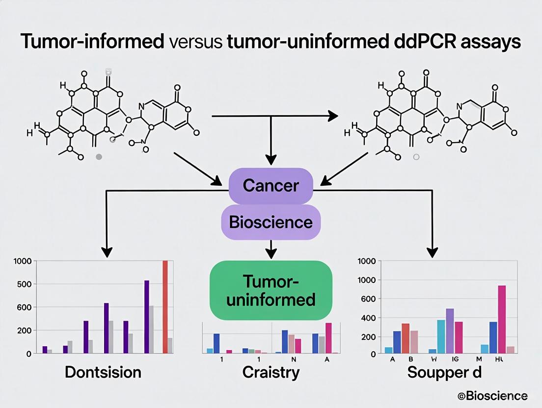
Tumor-Informed vs. Tumor-Uninformed ddPCR Assays
Tumor-Informed ddPCR Approach
Tumor-informed ddPCR assays require initial genetic analysis of tumor tissue to identify patient-specific mutations that can be tracked in liquid biopsies. This approach typically involves next-generation sequencing of tumor DNA to detect somatic alterations, followed by design of custom ddPCR assays targeting the specific mutations found in that individual's cancer [11]. In rectal cancer research, this method has demonstrated superior detection sensitivity compared to tumor-uninformed NGS panels, with ddPCR detecting ctDNA in 58.5% of baseline plasma samples versus 36.6% with NGS (p = 0.00075) [11].
The tumor-informed strategy offers several advantages for minimal residual disease detection and treatment monitoring. By focusing on mutations confirmed to be present in the primary tumor, this approach reduces false positives and increases specificity for detecting low levels of ctDNA [11]. Additionally, tumor-informed assays can be designed to target clonal mutations present in all cancer cells, providing a more reliable measure of overall tumor burden than heterogeneous mutations that may only be present in tumor subclones [13]. The main limitations of this approach include the need for tumor tissue availability, longer turnaround time due to required sequencing, and higher overall costs [11].
Tumor-Uninformed ddPCR Approach
Tumor-uninformed ddPCR assays utilize established cancer biomarkers without requiring prior knowledge of tumor-specific mutations. This approach typically targets recurrent mutations in known driver genes (e.g., KRAS, BRAF, EGFR) or cancer-specific methylation patterns [12] [15]. For lung cancer detection, multiplexed methylation-specific ddPCR assays have been developed targeting five tumor-specific methylation markers, achieving ctDNA-positive rates of 38.7-46.8% in non-metastatic disease and 70.2-83.0% in metastatic cases [12].
The tumor-uninformed strategy offers distinct advantages in clinical practice, including simpler workflow, faster turnaround time, and applicability when tumor tissue is unavailable [12]. Multiplexing capabilities further enhance this approach by enabling simultaneous detection of multiple biomarkers, increasing the overall detection sensitivity [12]. However, this method may have lower specificity compared to tumor-informed assays and could miss tumors that lack the targeted biomarkers [11]. The optimal approach depends on the specific clinical context and application, with tumor-informed assays generally preferred for minimal residual disease detection and tumor-uninformed assays suitable for initial screening or when tissue is limited.
Table 2: Comparison of Tumor-Informed vs. Tumor-Uninformed ddPCR Approaches
| Parameter | Tumor-Informed ddPCR | Tumor-Uninformed ddPCR |
|---|---|---|
| Requirement | Needs tumor tissue for sequencing | No tumor tissue required |
| Targets | Patient-specific mutations | Known cancer biomarkers (mutations, methylation) |
| Sensitivity in Localized Cancer | 58.5% (rectal cancer) [11] | 38.7-46.8% (lung cancer) [12] |
| Sensitivity in Metastatic Cancer | 80.8% (rectal cancer) [11] | 70.2-83.0% (lung cancer) [12] |
| Specificity | Higher (patient-specific targets) | Lower (population-level targets) |
| Turnaround Time | Longer (requires sequencing) | Shorter (direct analysis) |
| Cost | Higher (sequencing + custom assays) | Lower (standardized panels) |
| Ideal Application | MRD detection, therapy monitoring | Screening, initial diagnosis |
Research Reagent Solutions and Essential Materials
Successful implementation of ddPCR assays requires specific reagents and materials optimized for partitioning, amplification, and detection. The core component is the ddPCR supermix, which contains DNA polymerase, dNTPs, and buffer components formulated for droplet stability [14]. For mutation detection, hydrolysis probes (TaqMan-style) labeled with fluorescent dyes (FAM, HEX) provide specific signal generation with reduced background noise [14]. Primer sets targeting specific mutations or methylation sites must be carefully designed for specificity and efficiency, typically yielding amplicons of 65-150bp for optimal amplification in droplets [12] [14].
Specialized reagents for sample preparation include cell-free DNA extraction kits (e.g., DSP Circulating DNA Kit) designed to recover short fragments characteristic of ctDNA [12]. For methylation analysis, bisulfite conversion kits (e.g., EZ DNA Methylation-Lightning Kit) convert unmethylated cytosine to uracil while preserving methylated cytosines, enabling differentiation of tumor-derived DNA [12]. For ultra-rapid applications, detergent-free DNA extraction buffers (e.g., SwiftX Buffer ME) maintain droplet integrity while enabling rapid processing [13]. Droplet generation oil and surfactants create stable water-in-oil emulsions essential for consistent partitioning [10].
Table 3: Essential Research Reagents for ddPCR Assays
| Reagent Category | Specific Examples | Function in ddPCR Workflow |
|---|---|---|
| ddPCR Supermix | Bio-Rad ddPCR Master Mix | Provides enzymes, dNTPs, and optimized buffer for amplification in droplets |
| Hydrolysis Probes | FAM-labeled mutant probes, HEX-labeled wild-type probes | Sequence-specific detection with fluorescent signal upon amplification |
| Primer Sets | Mutation-specific primers, methylation-specific primers | Amplify target sequences with high specificity and efficiency |
| DNA Extraction Kits | DSP Circulating DNA Kit, TIANamp Genomic DNA Kit | Isolate high-quality nucleic acids from tissue or plasma samples |
| Bisulfite Conversion Kits | EZ DNA Methylation-Lightning Kit | Convert unmethylated cytosines to identify methylation status |
| Droplet Generation Oil | DG8 Cartridges for Droplet Generation | Create stable water-in-oil emulsions for sample partitioning |
| Rapid DNA Extraction Buffers | SwiftX Buffer ME | Enable ultra-rapid DNA preparation without inhibiting droplet formation |
Applications in Oncology Research and Protocol Implementation
ctDNA Monitoring in Treatment Response Assessment
ddPCR has emerged as a powerful tool for monitoring treatment response through serial assessment of ctDNA levels. The short half-life of ctDNA (approximately 16 minutes to several hours) enables real-time monitoring of tumor dynamics, providing earlier response assessment than conventional imaging [15]. In metastatic colorectal cancer, studies have demonstrated that patients with ctDNA detected after curative-intent therapy have significantly higher recurrence risk (up to 80-100%) compared to those with undetectable ctDNA [11]. Similar applications have been validated in lung cancer, where ctDNA clearance after initiating targeted therapy or immunotherapy correlates with improved progression-free survival [15].
The protocol for treatment monitoring involves collecting serial blood samples at predefined timepoints: before treatment (baseline), during therapy, and at follow-up intervals [11] [15]. For tumor-informed approaches, the same patient-specific mutations are tracked across all timepoints, while tumor-uninformed assays monitor consistent biomarker panels [11]. The quantitative nature of ddPCR enables calculation of molecular response based on the percentage change in ctDNA concentration from baseline, with early ctDNA reduction often predicting radiographic response [15]. This approach is particularly valuable for assessing minimal residual disease after surgery, where ctDNA detection can identify patients who might benefit from additional therapy despite no radiographic evidence of disease [11].
Multiplexed Methylation Detection in Lung Cancer
For tumor-uninformed applications, multiplexed methylation-specific ddPCR assays provide a robust approach for lung cancer detection and monitoring. The protocol begins with identifying lung cancer-specific methylation markers through bioinformatics analysis of public methylation databases (e.g., TCGA), selecting differentially methylated regions with maximal discrimination between tumor and normal samples [12]. The validated protocol involves bisulfite conversion of plasma-derived cell-free DNA, followed by multiplex ddPCR using primers and probes specific for the methylated sequences of five selected markers [12].
The analytical workflow includes rigorous quality control measures: assessing extraction efficiency using spike-in DNA fragments, evaluating potential lymphocyte contamination with immunoglobulin gene assays, and measuring total cfDNA concentration with reference gene assays [12]. Data analysis requires establishing clear cut-off values to determine ctDNA positivity, with studies comparing both fixed thresholds and statistical approaches based on background signals in control samples [12]. This multiplexed methylation approach demonstrates higher sensitivity for specific lung cancer subtypes, particularly small cell lung cancer and squamous cell carcinoma, highlighting how tumor-uninformed assays can be optimized for particular cancer types through careful biomarker selection [12].
Diagram 2: Comparison of Tumor-Informed and Tumor-Uninformed ddPCR Workflows. The tumor-informed pathway (green) requires initial tissue sequencing, while the tumor-uninformed pathway (red) utilizes known biomarkers for direct plasma analysis.
Droplet Digital PCR technology represents a significant advancement in molecular analysis, providing absolute quantification of nucleic acids through partitioning and end-point analysis. The applications in oncology research continue to expand, with both tumor-informed and tumor-uninformed approaches offering complementary strengths for different clinical scenarios. As the technology evolves with innovations such as ultra-rapid processing and enhanced multiplexing capabilities, ddPCR is poised to play an increasingly important role in precision oncology, from early detection and minimal residual disease monitoring to guiding targeted therapies and immunotherapies. The protocols and analytical frameworks presented herein provide researchers with comprehensive guidance for implementing these powerful approaches in cancer research and drug development programs.
Droplet Digital PCR (ddPCR) represents a paradigm shift in nucleic acid quantification, offering unparalleled sensitivity, precision, and reproducibility for molecular diagnostics and research. This article details the core technological advantages of ddPCR, with a specific focus on its application in tumor-informed versus tumor-uninformed circulating tumor DNA (ctDNA) assays. We provide structured comparative data, detailed experimental protocols for both assay types, and essential resource guides to facilitate implementation in research and clinical development settings.
Digital Droplet PCR (ddPCR) is a third-generation PCR technology that enables absolute quantification of nucleic acids without requiring a standard curve [16] [17]. The fundamental principle involves partitioning a single PCR reaction into thousands to millions of nanoliter-sized droplets, with each droplet acting as an independent PCR microreactor [16]. Following thermal cycling, droplets are analyzed via fluorescence to determine the presence or absence of the target sequence, and absolute quantification is calculated using Poisson statistics [17] [18]. This digital approach provides single-molecule resolution, making it exceptionally sensitive and accurate for detecting low-abundance targets—a critical capability in oncology for liquid biopsy applications [16] [18].
Core Technical Advantages: Quantitative Comparison
The unique partitioning methodology of ddPCR confers several distinct advantages over conventional PCR and qPCR techniques, particularly for ctDNA analysis in oncology.
Table 1: Analytical Performance Comparison Across PCR Platforms
| Parameter | Conventional PCR | qPCR (Real-Time PCR) | ddPCR (Digital Droplet PCR) |
|---|---|---|---|
| Sensitivity | Moderate | High | Very High (Single Copy Detection) [16] |
| Specificity | Moderate | High | Very High [16] |
| Tolerance to Inhibitors | Low | Moderate | High [16] [18] |
| Detection of Low DNA Input | Limited | Good | Excellent [16] |
| Quantification Capability | No | Relative Quantification | Absolute Quantification [16] [18] |
| Reproducibility | Variable | High | Very High [16] |
Table 2: Performance in Key Forensic Applications
| Feature/Use Case | Conventional PCR | qPCR | ddPCR |
|---|---|---|---|
| STR Profiling | ● | ○ | ○ |
| DNA Quantification | ○ | ● | ○ |
| Degraded DNA Analysis | Moderate | Moderate | Excellent [16] |
| Age Estimation via DNAm/miRNA | ○ | ○ | ● (MAD = 3.51 years) [16] |
| Body Fluid Identification | ○ | ● | ● [16] |
| Mixture Deconvolution | ○ | ○ | ● [16] |
| Microbial Forensics / PMI Estimation | ○ | ○ | ● [16] |
Abbreviations: MAD: Mean Absolute Deviation [16]; STR: Short Tandem Repeat; DNAm: DNA methylation; miRNA: MicroRNA; PMI: Postmortem Interval.
Absolute Quantification Without Standard Curves
Unlike qPCR, which relies on relative quantification against a standard curve, ddPCR provides absolute quantification of target nucleic acids [18]. This eliminates potential errors associated with standard curve preparation and interpolation, resulting in significantly lower inter-run and intra-run coefficients of variation [18]. This capability is particularly valuable for applications requiring precise copy number enumeration, such as CNV analysis and viral load quantification [19] [18].
Enhanced Sensitivity for Rare Event Detection
ddPCR's partitioning strategy enables detection of rare targets against a vast background of wild-type sequences. This makes it indispensable for detecting low-frequency alleles in liquid biopsy applications, where ctDNA can constitute less than 0.1% of total cell-free DNA [18]. Studies demonstrate ddPCR's superior sensitivity compared to next-generation sequencing (NGS) for ctDNA detection, with ddPCR detecting ctDNA in 58.5% of baseline plasma samples versus 36.6% for NGS (p = 0.00075) in localized rectal cancer [11] [20].
Superior Tolerance to PCR Inhibitors
The partitioning process in ddPCR effectively dilutes PCR inhibitors across thousands of droplets, minimizing their impact on amplification efficiency [16] [18]. Even if amplification is slightly delayed in affected droplets, the endpoint measurement remains reliable, unlike qPCR which depends on amplification kinetics [18]. This robustness simplifies sample preparation and enables accurate analysis of complex biological samples, including blood, stool, and environmentally compromised forensic specimens [16] [18].
Application Protocols: Tumor-Informed vs. Tumor-Uninformed ddPCR Assays
Tumor-Informed ddPCR Assay Protocol
Principle: Tumor-informed (personalized) assays first identify patient-specific mutations via tumor tissue sequencing, then design custom ddPCR assays to monitor these mutations in plasma ctDNA [11].
Workflow:
- Tumor Tissue Sequencing:
ddPCR Assay Design:
Plasma Processing and cfDNA Extraction:
- Collect blood in Streck Cell-Free DNA BCT tubes or EDTA tubes [11] [12].
- Process plasma within 4 hours by centrifuging at 2,000 × g for 10 minutes [12].
- Extract cfDNA from 4 mL plasma using specialized kits (e.g., DSP Circulating DNA Kit on QIAsymphony SP) [12].
- Elute cfDNA in 60 μL elution buffer [12].
ddPCR Reaction Setup:
- Concentrate extracted DNA to 20 μL using Amicon Ultra-0.5 Centrifugal Filter units [12].
- Prepare reaction mixture containing:
- 2-9 μL template DNA [11]
- ddPCR Supermix
- Custom-designed mutation-specific probes (FAM-labeled)
- Reference gene probes (HEX-labeled)
- Generate droplets using droplet generators (e.g., DG32) creating ~20,000 droplets [21].
PCR Amplification:
- Perform thermal cycling with optimized annealing temperatures.
- Typical protocol: 95°C for 10 min, 40 cycles of 94°C for 30 s and annealing temperature for 60 s, 98°C for 10 min [21].
Droplet Reading and Analysis:
Diagram Title: Tumor-Informed ddPCR Workflow
Tumor-Uninformed ddPCR Assay Protocol
Principle: Tumor-uninformed assays detect universal cancer biomarkers without prior knowledge of tumor genetics, using multiplex panels for methylation patterns or common mutations [12].
Workflow:
- Multiplex Panel Selection:
Sample Collection and Processing:
cfDNA Extraction and Bisulfite Conversion:
- Extract cfDNA from 4 mL plasma using the DSP Circulating DNA Kit [12].
- Add ~9,000 copies/mL of exogenous spike-in DNA (CPP1) to monitor extraction efficiency [12].
- Concentrate DNA using Amicon Ultra-0.5 Centrifugal Filter units [12].
- Perform bisulfite conversion using EZ DNA Methylation-Lightning Kit for methylation analysis [12].
Multiplex ddPCR Setup:
- Prepare reaction mixture containing:
- Bisulfite-converted DNA
- ddPCR Supermix for probes
- Multiple primer/probe sets for different targets
- Generate droplets as described in section 3.1.
- Prepare reaction mixture containing:
PCR Amplification and Analysis:
- Perform thermal cycling with optimized ramp rates.
- Analyze droplets using multi-channel detection.
- Determine ctDNA status using predefined cut-off methods [12].
Diagram Title: Tumor-Uninformed ddPCR Workflow
The Scientist's Toolkit: Essential Research Reagent Solutions
Table 3: Key Reagents and Materials for ddPCR Assays
| Reagent/Material | Function/Purpose | Example Products/Assays |
|---|---|---|
| Blood Collection Tubes | Preserves cell-free DNA for accurate liquid biopsy results | Streck Cell-Free DNA BCT tubes [11], EDTA tubes [12] |
| cfDNA Extraction Kits | Isolate high-quality cell-free DNA from plasma | DSP Circulating DNA Kit (Qiagen) [12], Maxwell RSC systems [12] |
| Bisulfite Conversion Kits | Convert unmethylated cytosines to uracils for methylation analysis | EZ DNA Methylation-Lightning Kit (Zymo Research) [12] |
| ddPCR Supermix | Optimized reaction mix for droplet-based digital PCR | Bio-Rad ddPCR Supermix for Probes [22] |
| Custom TaqMan Probes | Target-specific detection for tumor-informed assays | Bio-Rad ddPLEX ESR1 Mutation Detection Assay [22] |
| Multiplex Panels | Simultaneously detect multiple targets in tumor-uninformed approach | 5-plex methylation panels for lung cancer [12] |
| Droplet Generation Oil | Create stable water-in-oil emulsions for partitioning | DG32 Droplet Generation Oil [21] |
| Control Materials | Validate assay performance and extraction efficiency | Exogenous spike-in DNA (CPP1) [12], positive control templates [21] |
Comparative Performance Data in Clinical Applications
Table 4: ddPCR Performance Across Oncology Applications
| Application Context | Detection Rate/Sensitivity | Specificity | Key Findings |
|---|---|---|---|
| Rectal Cancer (Tumor-informed) | 58.5% (24/41) baseline detection [11] [20] | N/R | ddPCR significantly outperformed NGS (36.6% detection; p=0.00075) [11] [20] |
| Lung Cancer (Methylation-based) | Non-metastatic: 38.7-46.8% [12] Metastatic: 70.2-83.0% [12] | N/R | Higher sensitivity for SCLC and squamous cell carcinoma; potential for treatment monitoring [12] |
| Liquid Biopsy (MRD Detection) | Detects ctDNA at <0.1% VAF [18] | N/R | Enables minimal residual disease monitoring and early relapse detection [18] |
| Infectious Disease (BSI Detection) | 72.5% aggregate sensitivity [21] [23] | 63.1% aggregate specificity [21] [23] | Sensitivity increased to 84.9% when combined with clinical diagnosis [21] [23] |
| Copy Number Variation | 95% concordance with PFGE (gold standard) [19] | High precision for CNV resolution [19] | Superior to qPCR (60% concordance with PFGE) [19] |
Abbreviations: N/R: Not Reported; NGS: Next-Generation Sequencing; SCLC: Small Cell Lung Cancer; VAF: Variant Allele Frequency; BSI: Bloodstream Infection; PFGE: Pulsed-Field Gel Electrophoresis; MRD: Minimal Residual Disease.
Droplet Digital PCR technology provides significant advantages in sensitivity, specificity, and calibration-free quantification that make it particularly suitable for both tumor-informed and tumor-uninformed ctDNA assays. The absolute quantification capability, combined with exceptional tolerance to PCR inhibitors and sensitivity for rare targets, positions ddPCR as an essential tool for researchers and drug development professionals working in liquid biopsy applications, minimal residual disease detection, and precision oncology. As evidenced by the structured protocols and performance data presented herein, ddPCR offers a robust, reproducible platform that can be adapted to various research and clinical development needs.
Circulating tumor DNA (ctDNA) refers to fragmented DNA derived from tumor cells that is present in the bloodstream and other body fluids of cancer patients. As a component of liquid biopsy, ctDNA analysis provides a minimally invasive approach for cancer detection and monitoring, capturing tumor-specific genetic and epigenetic alterations. Understanding the fundamental biological properties of ctDNA—including its cellular origins, kinetics in circulation, and relationship with tumor burden—is essential for developing effective clinical assays. This knowledge forms the critical foundation for selecting appropriate methodological approaches, particularly in the context of tumor-informed versus tumor-uninformed ddPCR assays, which represent distinct pathways for ctDNA detection and quantification in research and clinical applications.
Biological Origin of ctDNA
Cellular Release Mechanisms
CtDNA originates from tumor cells through several distinct biological processes:
- Apoptosis (Programmed Cell Death): This is considered the primary source of ctDNA, producing short, fragmented DNA molecules typically ~166-200 base pairs in length, which correspond to nucleosomal DNA fragments.
- Necrosis: Uncontrolled cell death resulting in the release of longer, more randomly fragmented DNA molecules.
- Active Secretion: Tumor cells may actively release DNA through extracellular vesicles or other secretory mechanisms, though this pathway is less characterized [15] [24].
The ctDNA fragments carry tumor-specific characteristics including somatic mutations, copy number alterations, and epigenetic modifications such as abnormal methylation patterns, which differentiate them from normal cell-free DNA (cfDNA) derived from hematopoietic and other healthy cells [15] [25].
Factors Influencing ctDNA Release
The concentration and detectability of ctDNA in circulation are influenced by multiple biological factors:
- Tumor Vascularity: Highly vascularized tumors tend to release more ctDNA into the bloodstream.
- Tumor Location: Anatomical proximity to major blood vessels can enhance ctDNA shedding.
- Tumor Histology: Aggressive tumor subtypes often demonstrate higher rates of ctDNA release.
- Disease Stage: Advanced tumors typically shed more DNA than early-stage lesions [24].
Notably, ctDNA is more frequently detected in tumors with vascular invasion, and its release can be transiently stimulated by external factors such as radiotherapy, ultrasound, or mechanical stress applied to tumors [24].
Half-Life and Clearance Dynamics
Circulating Kinetics
CtDNA demonstrates rapid turnover in the bloodstream, with a remarkably short half-life estimated between 16 minutes to several hours [15]. This rapid clearance results from efficient elimination mechanisms:
- Hepatic Clearance: Liver macrophages (Kupffer cells) actively phagocytose circulating DNA fragments.
- Renal Excretion: Smaller DNA fragments are filtered and excreted through the kidneys.
- Enzymatic Degradation: Circulating nucleases in the blood degrade extracellular DNA [24].
This brief half-life enables ctDNA to serve as a real-time biomarker of tumor dynamics, reflecting changes in tumor burden much more rapidly than traditional imaging modalities [15].
Implications for Assay Timing
The rapid clearance kinetics have important implications for experimental design:
- Treatment Monitoring: The short half-life allows for detection of molecular responses to therapy within hours to days, far preceding radiographic changes.
- Minimal Residual Disease (MRD) Assessment: Timing of blood collection post-surgery is critical, as surgical trauma can cause transient increases in background cfDNA that may interfere with ctDNA detection for several weeks [24].
- Longitudinal Sampling: Frequent sampling is feasible and can provide dynamic assessment of tumor evolution during treatment [15].
Correlation with Tumor Burden
Quantitative Relationships
CtDNA levels demonstrate a strong correlation with tumor burden across multiple cancer types. The fraction of ctDNA in total cfDNA ranges from <0.01% in early-stage cancers to >90% in advanced metastatic disease [15]. This relationship forms the biological basis for using ctDNA as a quantitative biomarker for monitoring treatment response and disease progression.
Table 1: Prognostic Significance of ctDNA Detection at Different Treatment Time Points in Esophageal Cancer
| Time Point | Hazard Ratio for PFS | Hazard Ratio for OS | Clinical Implications |
|---|---|---|---|
| Baseline (before treatment) | 1.64 (95% CI: 1.30-2.07) | 2.02 (95% CI: 1.36-2.99) | Identifies high-risk patients who may benefit from treatment intensification |
| After Neoadjuvant Therapy | 3.97 (95% CI: 2.68-5.88) | 3.41 (95% CI: 2.08-5.59) | Predicts poor response to therapy; may guide adjuvant treatment decisions |
| During Follow-up | 5.42 (95% CI: 3.97-7.38) | 4.93 (95% CI: 3.31-7.34) | Enables early recurrence detection with ~4.5 months lead time versus imaging [26] |
Clinical Utility in Monitoring
The correlation between ctDNA levels and tumor burden enables several key clinical applications:
- Treatment Response Assessment: Decreasing ctDNA levels correlate with successful therapeutic response, while rising levels indicate progression or resistance.
- Early Recurrence Detection: ctDNA can identify molecular relapse months before clinical or radiographic recurrence, with studies showing an average lead time of 4.53 months (range: 0.98-11.6 months) compared to conventional imaging [26].
- Minimal Residual Disease (MRD) Detection: Post-treatment ctDNA positivity predicts subsequent clinical recurrence with high specificity, identifying patients who may benefit from additional therapy [15] [27].
Experimental Protocols for ctDNA Analysis
Blood Collection and Pre-analytical Processing
Proper pre-analytical handling is critical for reliable ctDNA detection:
Table 2: Blood Collection and Processing Protocols for ctDNA Analysis
| Step | Protocol Details | Rationale & Considerations |
|---|---|---|
| Blood Collection | - Use butterfly needles with 20-21G gauge- Collect 2×10 mL blood per tube (minimum 20mL total)- Avoid prolonged tourniquet use | Minimizes hemolysis and leukocyte activation that increases wild-type background DNA [24] |
| Collection Tubes | Option A: EDTA tubes (process within 2-6 hours at 4°C)Option B: Stabilizing tubes (Streck, PAXgene, Roche) - stable up to 7 days at room temperature | Stabilizing tubes prevent leukocyte lysis during storage/transport but may not be compatible with multi-analyte workflows [24] |
| Plasma Separation | - Double centrifugation: 2,000 × g for 10 min, then 10,000 × g for 10 min- Aliquot plasma to avoid freeze-thaw cycles | Removes cells and debris; reduces contamination with genomic DNA from blood cells [12] [24] |
| cfDNA Extraction | - Use validated kits (e.g., Maxwell RSC ccfDNA LV Plasma Kit, QIAsymphony DSP Circulating DNA Kit)- Elute in 60μL buffer | Ensures high yield and reproducibility; compatible with downstream applications [12] [28] |
Analytical Methods for ctDNA Detection
Droplet Digital PCR (ddPCR) Protocols
Tumor-Informed ddPCR Approach:
- Tumor Sequencing: First perform NGS (e.g., Ion AmpliSeq Cancer Hotspot Panel v2) on tumor tissue to identify patient-specific mutations.
- Assay Design: Design custom ddPCR probes targeting 1-2 mutations with highest variant allele frequency in the tumor.
- ddPCR Setup: Partition extracted cfDNA into ~20,000 droplets with mutation-specific probes.
- Amplification & Reading: Perform PCR amplification and count positive/negative droplets to absolutely quantify mutant DNA copies [11] [29].
Tumor-Uninformed ddPCR Approach:
- Panel Selection: Use pre-designed ddPCR assays targeting recurrent mutations in specific cancer types (e.g., BRAF V600E for melanoma, KRAS for colorectal cancer).
- Multiplexing: Simultaneously target multiple common mutations to increase detection sensitivity.
- Quantification: Calculate mutant copies per mL of plasma without prior tumor sequencing [29].
Methylation-Specific ddPCR Protocol
For detection of cancer-specific methylation patterns:
- Bisulfite Conversion: Treat extracted cfDNA with bisulfite reagents (e.g., EZ DNA Methylation-Lightning Kit) to convert unmethylated cytosines to uracils.
- Multiplex PCR Design: Design primers targeting differentially methylated regions identified through bioinformatic analysis (e.g., HOXA9 and other hypermethylated loci in cancer).
- ddPCR Analysis: Perform droplet-based PCR with methylation-specific probes and quantify methylated molecules [12].
Assay Selection Workflow: Tumor-Informed vs. Tumor-Uninformed Approaches
The following diagram illustrates the decision pathway for selecting between tumor-informed and tumor-uninformed ddPCR approaches in ctDNA research:
Research Reagent Solutions
Table 3: Essential Reagents and Kits for ctDNA Research
| Product Category | Specific Examples | Application & Purpose |
|---|---|---|
| Blood Collection Tubes with Stabilizers | Streck cfDNA BCT, PAXgene Blood ccfDNA (Qiagen), Roche cfDNA tubes | Preserve blood samples during storage/transport; prevent leukocyte DNA contamination [24] |
| cfDNA Extraction Kits | Maxwell RSC ccfDNA LV Plasma Kit (Promega), QIAsymphony DSP Circulating DNA Kit (Qiagen) | Isolate high-quality cfDNA from plasma with minimal fragmentation [12] [28] |
| ddPCR Systems & Reagents | Bio-Rad ddPCR System, Naica dPCR System (Stilla Technologies), mutation-specific ddPCR assays | Absolute quantification of mutant DNA copies without standard curves [11] [28] [29] |
| Bisulfite Conversion Kits | EZ DNA Methylation-Lightning Kit (Zymo Research) | Convert unmethylated cytosine to uracil for methylation-specific detection [12] |
| Targeted NGS Panels | Ion AmpliSeq Cancer Hotspot Panel v2, TruSight Oncology 500 ctDNA (Illumina) | Identify tumor-specific mutations for informed assay design; comprehensive profiling [11] [30] |
| Unique Molecular Identifiers (UMIs) | TruSight Oncology UMI Reagents (Illumina) | Reduce background noise in sequencing data; enable detection of low-frequency variants [30] [28] |
The biological properties of ctDNA—including its origin from tumor cells, short half-life in circulation, and strong correlation with tumor burden—provide the fundamental rationale for its application in cancer detection and monitoring. Understanding these characteristics is essential for selecting appropriate methodological approaches, particularly when deciding between tumor-informed and tumor-uninformed ddPCR strategies. Tumor-informed assays offer superior sensitivity for minimal residual disease detection by leveraging patient-specific mutation profiles, while tumor-uninformed approaches provide practical advantages for screening applications and situations where tumor tissue is unavailable. As ctDNA analysis continues to evolve, optimization of pre-analytical protocols and reagent systems will be crucial for enhancing assay performance and expanding clinical utility across diverse cancer types and disease stages.
Workflow and Implementation: From Sample Collection to Clinical Reporting
Circulating tumor DNA (ctDNA) analysis has emerged as a powerful, non-invasive tool for cancer monitoring in precision oncology. The tumor-informed approach represents a sophisticated methodology where a patient's unique tumor mutational profile, first identified via sequencing of tumor tissue, is used to create a highly personalized assay for tracking specific mutations in plasma cell-free DNA (cfDNA) [31] [32]. This strategy stands in contrast to tumor-uninformed assays, which use fixed, pre-determined panels of common cancer mutations without prior knowledge of an individual's tumor genetics [11] [4].
The clinical value of this approach lies in its enhanced sensitivity and specificity. By focusing on a set of mutations confirmed to be present in a patient's specific tumor, tumor-informed assays can achieve exceptionally low limits of detection, enabling applications such as Molecular Residual Disease (MRD) assessment after curative-intent therapy and early relapse detection [32] [15]. This application note details a standardized workflow from tumor tissue sequencing to the development and implementation of patient-specific droplet digital PCR (ddPCR) assays, providing researchers and drug development professionals with a robust protocol for precise ctDNA monitoring.
Performance Comparison: Tumor-Informed vs. Tumor-Uninformed Approaches
The selection between tumor-informed and tumor-uninformed methodologies involves critical trade-offs in sensitivity, specificity, workflow complexity, and cost. The tables below summarize the comparative performance and economic considerations of each approach.
Table 1: Analytical Performance Comparison
| Parameter | Tumor-Informed ddPCR | Tumor-Uninformed NGS Panel |
|---|---|---|
| Detection Sensitivity | High (VAF ~0.01%) [11] | Lower (VAF ~0.1-1%) [15] |
| Baseline Detection Rate (Rectal Cancer Study) | 58.5% (24/41) [11] | 36.6% (15/41) [11] |
| Assay Specificity | Very High (patient-specific) [32] | Moderate (panel-dependent) [4] |
| Number of Targets Tracked | Typically 1-2 per ddPCR assay [11] | Dozens to hundreds [11] [15] |
| Variant Detection Scope | Limited to pre-identified mutations | Can detect untargeted variants |
Table 2: Workflow and Economic Considerations
| Consideration | Tumor-Informed ddPCR | Tumor-Uninformed NGS Panel |
|---|---|---|
| Tissue Requirement | Mandatory (for initial sequencing) [31] [32] | Not required [11] |
| Assay Development Time | Longer (3-4 weeks for WES + probe design) [32] | Shorter (uses pre-existing panel) |
| Operational Cost per Sample | Lower (5–8.5-fold lower than NGS) [11] | Higher [11] |
| Assay Flexibility | High (adapts to each patient's tumor) [32] | Fixed (same panel for all patients) |
| Informatics Complexity | Moderate (requires somatic variant calling) [32] | Variable (can be high for large panels) |
Experimental Protocol: A Step-by-Step Guide
Stage 1: Tumor and Normal Tissue Sequencing and Analysis
Objective: To comprehensively identify somatic mutations present in a patient's tumor that are absent from their germline DNA.
Materials and Reagents:
- Tumor DNA Source: Formalin-Fixed Paraffin-Embedded (FFPE) tissue block or fresh-frozen tumor tissue [32].
- Normal DNA Source: Peripheral blood mononuclear cells (PBMCs) or matched whole blood [4] [32].
- DNA Extraction Kits: For FFPE (e.g., Qiagen DNeasy Blood & Tissue Kit) and high-molecular-weight DNA from blood [32].
- Library Prep Kit: e.g., Twist Library Preparation EF Kit 2.0 [32].
- Target Enrichment Panel: Whole-exome sequencing (WES) panel (e.g., Twist Human Core Exome) or comprehensive cancer hotspot panel (e.g., Ion AmpliSeq Cancer Hotspot Panel v2) [11] [32].
- Sequencing Platform: Illumina NovaSeq or similar for high-depth sequencing (e.g., 400 million reads for tumor, 140 million for normal) [32].
Methodology:
- Nucleic Acid Extraction: Extract genomic DNA from tumor and normal samples according to manufacturer protocols. Assess DNA concentration and quality using a spectrophotometer (e.g., NanoDrop) [33].
- Library Preparation and Enrichment: Prepare pre-capture libraries from 10-100 ng of tumor DNA and 50-100 ng of normal DNA. Perform target enrichment using the selected WES or cancer panel with the appropriate hybridization reagent kit [32].
- Sequencing: Sequence the libraries on an Illumina platform in paired-end mode (e.g., 2 × 150 bp) to achieve the recommended depth of coverage [32].
- Bioinformatic Analysis: Process the tumor-normal paired-end sequencing data through a somatic variant calling pipeline (e.g., megSAP pipeline, https://github.com/imgag/megSAP). The primary outputs are BAM and VCF files containing high-confidence somatic mutations [32].
Stage 2: Selection of Monitoring Targets and ddPCR Assay Design
Objective: To prioritize and select the most suitable somatic mutations from the NGS data for designing patient-specific ddPCR assays.
Methodology:
- Variant Prioritization: Import the somatic variant list (VCF file) into a clinical decision support system (e.g., GSvar) [32]. Apply the following selection criteria:
- Prioritize exonic variants (missense, nonsense) over intronic or intergenic variants [32].
- Select mutations with a high variant allele frequency (VAF) in the tumor tissue to ensure they are clonal [11].
- Avoid variants in low-complexity or repetitive genomic regions [32].
- Exclude variants that are clustered closely together [32].
- Final Target Selection: Typically, one to two mutations are selected for ddPCR assay design, chosen based on the highest VAF and technical suitability [11]. For research purposes, tracking more mutations is possible using multiple ddPCR reactions.
- Probe and Primer Design: Design TaqMan-style hydrolysis probes and primers for the selected mutations.
- The mutation should be located centrally within the probe sequence.
- Design both a mutant-specific probe (e.g., labeled with FAM) and a wild-type probe (e.g., labeled with HEX/VIC) for the same genomic locus to ensure specificity.
- Validate probe specificity in silico using tools like BLAST.
Stage 3: Plasma Collection, cfDNA Isolation, and ddPCR Setup
Objective: To isolate cfDNA from patient plasma and perform absolute quantification of the target mutations using a optimized ddPCR protocol.
Materials and Reagents:
- Blood Collection Tubes: Streck Cell-Free DNA BCT tubes [11] [4].
- cfDNA Extraction Kit: e.g., QIAamp Circulating Nucleic Acid Kit [31].
- ddPCR Supermix: e.g., ddPCR Supermix for Probes (Bio-Rad) [33].
- Custom Probes and Primers: Designed in Stage 2 [31].
- Droplet Generator and Reader: e.g., DropXpert S6 system or equivalent [33].
Methodology:
- Plasma Processing:
- Collect patient blood at baseline and serial timepoints (e.g., during treatment, follow-up) in Streck tubes [11] [32].
- Process tubes within the recommended timeframe (e.g., within 72 hours) with double centrifugation (e.g., 1,600 × g for 20 min, then 16,000 × g for 10 min) to isolate platelet-free plasma [31].
- Store plasma at -80°C until cfDNA extraction.
- cfDNA Isolation: Extract cfDNA from 2-10 mL of plasma using a specialized cfDNA isolation kit, following the manufacturer's instructions. Elute in a low TE buffer or nuclease-free water. Quantify cfDNA using a fluorometer (e.g., Qubit) [31].
- ddPCR Reaction Setup:
- Prepare a 20-22 µL reaction mixture as detailed in the table below.
- Include a no-template control (NTC) and, if available, a positive control for the mutation.
- Droplet Generation and PCR Amplification:
- Load the reaction mixture into a droplet generator cartridge along with droplet generation oil. Generate nanoliter-sized droplets according to the instrument's protocol [33].
- Transfer the emulsified sample to a 96-well PCR plate. Seal the plate and perform PCR amplification on a conventional thermal cycler using optimized conditions, for example:
- Droplet Reading and Data Analysis:
- Place the PCR plate in a droplet reader, which measures the fluorescence (FAM and HEX) in each droplet [17].
- Analyze the data using the instrument's accompanying software (e.g., QuantaSoft).
- Set thresholds to distinguish positive (mutant and wild-type) and negative droplets based on fluorescence amplitude.
- The software automatically calculates the concentration of mutant and wild-type DNA molecules (copies/µL) and the variant allele frequency (VAF) using Poisson statistics [17] [33].
The Scientist's Toolkit: Essential Research Reagents
Table 4: Key Reagents and Materials for Tumor-Informed ddPCR Workflow
| Item | Function/Application | Example Products/Catalog Numbers |
|---|---|---|
| Cell-Free DNA Blood Collection Tubes | Stabilizes nucleated blood cells to prevent background cfDNA release, enabling longer sample transport times. | Streck Cell-Free DNA BCT Tubes [11] |
| FFPE DNA Extraction Kit | Isolves DNA from archived formalin-fixed, paraffin-embedded (FFPE) tumor tissue blocks. | Qiagen DNeasy Blood & Tissue Kit, Qiagen FFPE DNA Kit (DP330) [32] [33] |
| Next-Generation Sequencing Panel | For target enrichment to identify tumor-specific somatic mutations from tumor DNA. | Ion AmpliSeq Cancer Hotspot Panel v2, Twist Human Core Exome [11] [32] |
| Droplet Digital PCR System | Partitions PCR reactions into droplets for absolute quantification of target DNA molecules. | DropXpert S6, Bio-Rad QX200 [33] |
| ddPCR Supermix | Optimized buffer, enzymes, and dNTPs for probe-based digital PCR in droplet systems. | ddPCR Supermix for Probes (Bio-Rad), Aplµs Digital PCR Mix [33] |
| Custom TaqMan Probes & Primers | Patient-specific oligonucleotides designed to detect the unique mutations identified in the tumor. | Designed using Primer 5.0 or similar software; synthesized by commercial providers [31] [33] |
The tumor-informed ddPCR workflow provides a highly sensitive and specific framework for monitoring tumor dynamics through liquid biopsy. This approach, which tailors the detection assay to the individual patient's tumor genetics, offers a significant advantage in sensitivity over tumor-uninformed methods, particularly for challenging applications like MRD detection [11] [32].
The integration of this workflow into clinical trial frameworks and ultimately routine practice holds the potential to transform patient management. It enables the early assessment of treatment efficacy, the detection of residual disease before it becomes radiologically apparent, and the early identification of relapse [32] [15]. As standardization improves and costs decrease, tumor-informed liquid biopsy is poised to become a cornerstone of precision oncology, allowing for more dynamic and personalized treatment strategies.
Digital droplet PCR (ddPCR) represents a transformative technology in molecular diagnostics, enabling absolute quantification of nucleic acid targets without standard curves by partitioning samples into thousands of nanoliter-sized droplets [34]. Within oncology applications, two distinct liquid biopsy approaches have emerged: tumor-informed assays that require prior sequencing of tumor tissue to identify patient-specific mutations, and tumor-uninformed assays that utilize predetermined panels of common cancer hotspot mutations without needing tumor tissue analysis [11]. This application note focuses on the latter approach, detailing methodologies for implementing fixed-panel hotspot mutations in tumor-uninformed ddPCR workflows.
Tumor-uninformed assays provide significant practical advantages in clinical settings where tumor tissue is unavailable, insufficient, or difficult to biopsy [15]. By targeting recurrent mutations in driver genes that are well-established in specific cancer types, these fixed panels enable rapid, cost-effective molecular profiling that is particularly valuable for treatment selection and disease monitoring [35]. The fundamental principle involves detecting and quantifying known mutant alleles present in circulating tumor DNA (ctDNA) against a background of wild-type DNA, leveraging ddPCR's exceptional sensitivity for rare variant detection down to 0.001% variant allele frequency (VAF) [36] [35].
The applications of fixed-panel ddPCR span multiple cancer types, including non-small cell lung cancer (NSCLC), colorectal cancer, breast cancer, and melanoma, among others [15]. Commonly targeted mutations include EGFR variants (L858R, T790M, exon 19 deletions), KRAS G12C/V, BRAF V600E, and PIK3CA hotspots, which have demonstrated clinical utility for therapy selection and response monitoring [35]. This document provides detailed protocols, performance characteristics, and implementation guidelines for deploying tumor-uninformed ddPCR assays in research and clinical settings.
Performance Characteristics and Technical Validation
Analytical Sensitivity and Specificity
Tumor-uninformed ddPCR assays demonstrate exceptional analytical sensitivity, consistently detecting mutant alleles at variant allele frequencies as low as 0.001% under optimized conditions [35]. This sensitivity surpasses most next-generation sequencing (NGS) platforms, which typically exhibit lower detection limits between 2-15% VAF depending on the specific workflow and mutation target [35]. The partitioning technology underlying ddPCR enables this high sensitivity by effectively concentrating rare mutant molecules into individual droplets where they can be amplified without competition from the abundant wild-type background [37].
The specificity of fixed-panel ddPCR assays is equally robust, with studies demonstrating clear discrimination between mutant and wild-type sequences even at minimal allele frequency differences [38]. This performance is maintained across various biological matrices, including plasma-derived cell-free DNA, formalin-fixed paraffin-embedded (FFPE) tissue DNA, and other clinical sample types [38]. The use of optimized allele-specific primers and probes contributes to this high specificity by ensuring preferential amplification of intended targets while minimizing cross-reactivity with similar sequences [38].
Comparison with Alternative Methodologies
When compared to other mutation detection platforms, tumor-uninformed ddPCR offers distinct advantages for targeted hotspot analysis. Table 1 summarizes the key performance characteristics and practical considerations relative to next-generation sequencing and tumor-informed approaches.
Table 1: Performance comparison of mutation detection methodologies
| Parameter | Tumor-Uninformed ddPCR | Tumor-Informed Assays | NGS Panels |
|---|---|---|---|
| Detection Sensitivity | 0.001% VAF [35] | 0.01% VAF [11] | 2-15% VAF [35] |
| Turnaround Time | 3-4 hours [35] | 7-14 days [11] | 5-10 days [11] |
| Cost per Sample | $50-100 [11] | $200-500 [11] | $500-1000 [11] |
| Multiplexing Capacity | 6-plex in single well [35] | Typically single-plex | Hundreds to thousands of targets |
| Tissue Requirement | None | Mandatory | Preferred but not always mandatory |
| Hands-on Time | <2 hours [35] | Variable | 4-8 hours |
The data reveal that tumor-uninformed ddPCR provides superior sensitivity and faster turnaround times at lower cost compared to both tumor-informed approaches and NGS, making it particularly suitable for applications requiring rapid results and high sensitivity for known mutations [11] [35]. The main limitation is the restricted number of targets simultaneously analyzed, though recent advances in multiplex ddPCR now enable detection of up to six mutations in a single reaction [35].
Clinical Performance Validation
In clinical validation studies, tumor-uninformed ddPCR has demonstrated robust performance across multiple cancer types. A 2025 study comparing ddPCR and NGS for ctDNA detection in localized rectal cancer found that ddPCR exhibited superior detection rates (58.5% vs. 36.6% in baseline plasma samples, p=0.00075) [11]. The study further established that positive ctDNA results correlated with higher clinical tumor stage and lymph node positivity, confirming the clinical relevance of ddPCR findings [11].
Harmonization trials have further validated the technical performance of ddPCR for hotspot mutation detection. A multi-institutional Italian study focusing on ESR1 mutations in breast cancer reported equivalent detection rates between ddPCR and NGS platforms across different mutant allele fractions (5.0%, 1.0%, and 0.5%), with successful mutation identification in 90% of samples at higher allele fractions and 80% at the lowest allele fraction [39]. This demonstrates that ddPCR maintains reliable performance even at low VAF levels commonly encountered in clinical samples.
Experimental Protocols and Workflows
Six-Plex Mutation Detection Protocol for NSCLC
The following protocol details a 6-plex ddPCR assay for simultaneous detection of key NSCLC mutations (EGFR exon 19 deletions, L858R, T790M, KRAS G12C, and BRAF V600E) plus wild-type EGFR control, adapted from validated methodologies [35].
Reagent Preparation and Reaction Setup
Materials Required:
- QX600 Droplet Digital PCR System (Bio-Rad)
- ddPCR Multiplex Supermix (catalog #12005909)
- Automated Droplet Generator (Bio-Rad, #1864101)
- DG32 Automated Droplet Generator Cartridges (#1864108)
- Automated Droplet Generation Oil for Probes (#1864110)
- C1000 Touch Thermal Cycler with 96-Deep Well Reaction Module (#1851197)
- Mutation detection assays (Bio-Rad Expert Design Assay #12008212)
- EGFR Exon 19 Deletions Screening Kit (#12002392)
Reaction Setup: Prepare 22μL reactions according to the following composition:
- 11μL ddPCR Multiplex Supermix (2X concentration)
- 1.1μL 20X mutation detection assay mix (final: 1X)
- 1.1μL 20X EGFR exon 19 deletions screening assay (final: 1X)
- 2-5μL DNA template (1-10 ng total input)
- Nuclease-free water to 22μL
Thermal Cycling Conditions: Utilize the following protocol on the C1000 Touch Thermal Cycler:
- Enzyme activation: 95°C for 10 minutes (1 cycle)
- Denaturation: 94°C for 30 seconds
- Annealing/Extension: 55°C for 1 minute (40 cycles)
- Enzyme deactivation: 98°C for 10 minutes (1 cycle)
- Hold: 4°C indefinitely
Droplet Generation and Reading:
- Transfer 20μL of the reaction mixture to DG32 cartridges
- Generate droplets using Automated Droplet Generator
- Transfer emulsified samples to 96-well PCR plate
- Perform thermal cycling as described
- Analyze droplets using QX600 Droplet Reader
Data Analysis and Interpretation
Following droplet reading, analyze fluorescence amplitude data using QuantaSoft software. The six-plex assay generates distinct clustering patterns across three two-dimensional plots:
- FAM/HEX channels: EGFR exon 19 deletions (mutant, blue) vs. EGFR wild-type (green)
- Cy5/Cy5.5 channels: KRAS G12C (light red) vs. EGFR L858R (purple)
- ROX/ATTO590 channels: BRAF V600E (dark red) vs. EGFR T790M (cyan)
Positive droplets are identified based on their fluorescence signature relative to negative controls and no-template controls. The software automatically calculates mutant allele concentration (copies/μL) and variant allele frequency based on Poisson statistics.
Ultra-Rapid Intraoperative ddPCR Protocol
For applications requiring extremely rapid turnaround, such as intraoperative surgical guidance, an optimized 15-minute ddPCR protocol has been developed and validated for IDH1 R132H and BRAF V600E mutations [40].
Ultra-Rapid DNA Extraction
- Transfer 2-4 mg tissue biopsy to 50μL rapid lysis buffer (20mM NaOH, 400μM EDTA, 0.1% Triton X-100)
- Homogenize using manual pestle (30 seconds)
- Incubate at 98°C for 3 minutes
- Centrifuge at 12,000 × g for 1 minute
- Transfer 5μL supernatant to ddPCR reaction
Accelerated ddPCR Protocol
- Prepare 20μL reactions containing:
- 10μL ddPCR Supermix
- 2μL mutation assay (final: 1X)
- 5μL ultra-rapid DNA extract
- 3μL nuclease-free water
- Generate droplets using automated droplet generator (2 minutes)
- Perform ultra-fast thermal cycling:
- 95°C for 30 seconds (enzyme activation)
- 45 cycles of: 94°C for 5 seconds, 55°C for 20 seconds
- 98°C for 30 seconds (enzyme deactivation)
- Read droplets using droplet reader (3 minutes)
This accelerated protocol maintains analytical performance equivalent to standard ddPCR (R² = 0.995) while reducing total processing time from >2 hours to just 15 minutes, enabling real-time surgical decision-making [40].
Research Reagent Solutions
Table 2: Essential research reagents for tumor-uninformed ddPCR
| Reagent/Catalog Number | Manufacturer | Application | Key Features |
|---|---|---|---|
| ddPCR Multiplex Supermix (#12005909) | Bio-Rad | Reaction master mix | Optimized for multiplexed probe-based assays, inhibitor-resistant |
| EGFR Exon 19 Deletions Screening Kit (#12002392) | Bio-Rad | NSCLC mutation detection | Detects 15 different EGFR exon 19 deletions with wild-type control |
| Expert Design Assays (Custom) | Bio-Rad | Target-specific detection | Pre-optimized assays for specific hotspot mutations |
| Automated Droplet Generation Oil (#1864110) | Bio-Rad | Droplet generation | Ensures stable droplet formation and thermal stability |
| DG32 Cartridges (#1864108) | Bio-Rad | Droplet generation | Automated microfluidic droplet generation |
| Cell-Free DNA Blood Collection Tubes | Streck | Sample collection | Preserves blood cfDNA for up to 14 days at room temperature |
| Maxwell RSC ccfDNA Plasma Kit | Promega | cfDNA extraction | High-efficiency recovery of low-abundance ctDNA |
| QX600 Droplet Reader (#12013328) | Bio-Rad | Droplet reading | Six-color detection enabling advanced multiplexing |
Workflow Diagram
Tumor-Uninformed ddPCR Workflow: The diagram illustrates the complete workflow from sample collection to clinical decision-making, highlighting the streamlined nature of fixed-panel ddPCR assays that do not require prior tumor sequencing.
Applications in Precision Oncology
Therapy Selection and Guidance
Tumor-uninformed ddPCR assays employing fixed hotspot panels enable rapid therapy selection by detecting actionable mutations in driver oncogenes. For NSCLC, the simultaneous detection of EGFR sensitizing mutations (L858R, exon 19 deletions) informs eligibility for EGFR tyrosine kinase inhibitors, while EGFR T790M detection identifies resistance mechanisms requiring next-generation inhibitors [35]. Similarly, BRAF V600E identification in melanoma, colorectal cancer, and other malignancies directs patients toward BRAF/MEK inhibitor combinations, demonstrating the clinical utility of these targeted approaches [40] [15].
The high sensitivity of ddPCR is particularly valuable for analyzing liquid biopsy samples where ctDNA abundance may be low. Studies have demonstrated that ddPCR can detect ESR1 mutations in breast cancer patients at allele frequencies as low as 0.1%, enabling identification of hormonal therapy resistance and guiding transition to alternative treatments including novel SERDs (Selective Estrogen Receptor Degraders) [39]. This capability for detecting emerging resistance mutations during treatment monitoring represents a key application of fixed-panel ddPCR assays in managing advanced cancers.
Minimal Residual Disease Monitoring
The exceptional sensitivity of ddPCR makes it suitable for minimal residual disease (MRD) detection following curative-intent treatment. While tumor-informed approaches currently dominate MRD applications, fixed-panel ddPCR assays offer a practical alternative for monitoring common driver mutations in malignancies with recurrent genetic alterations [15]. In acute myeloid leukemia, ddPCR has demonstrated capability to detect residual mutant alleles at frequencies as low as 0.002%, enabling early relapse prediction and guiding preemptive therapeutic interventions [36].
In solid tumors, studies have validated the prognostic significance of post-treatment ctDNA detection using fixed ddPCR panels. For colorectal cancer patients, postoperative ctDNA positivity identified through KRAS and BRAF mutation tracking correlates with significantly higher recurrence risk (up to 80-100%), enabling stratification for adjuvant therapy intensification [11] [15]. Similar applications have been established in breast cancer (ESR1, PIK3CA), prostate cancer (AR alterations), and other malignancies, supporting the clinical utility of tumor-uninformed ddPCR for residual disease assessment.
Intraoperative Surgical Guidance
The development of ultra-rapid ddPCR protocols has enabled novel applications in intraoperative settings, where molecular data can directly influence surgical decision-making [40]. By implementing 15-minute ddPCR assays for IDH1 R132H in gliomas and BRAF V600E in melanomas, surgeons can obtain genetic characterization of tumor margins during procedures, guiding extent of resection based on molecular rather than solely histological assessment.
This approach has been successfully implemented in 22 brain tumor cases, with ddPCR measurements demonstrating virtually identical performance to standard protocols (R² = 0.995) despite dramatically reduced processing time [40]. The technology enables quantification of tumor cell densities ranging from >1,300 tumor cells/mm³ within tumor cores to <5 tumor cells/mm³ at marginal regions, providing unprecedented resolution for defining surgical boundaries. This application represents a significant advancement in cancer surgery, enabling molecularly-guided resections that may improve patient outcomes.
The analysis of circulating tumor DNA (ctDNA) using droplet digital PCR (ddPCR) has emerged as a powerful tool for cancer management, enabling non-invasive molecular stratification, monitoring of treatment response, and identification of resistance mutations [41] [15]. The pre-analytical phase—encompassing blood collection, plasma processing, and cell-free DNA (cfDNA) extraction—represents a critical vulnerability in liquid biopsy workflows, as variations in these initial procedures can significantly impact the integrity and quantitation of analytes. This is particularly crucial for ctDNA analysis, where tumor-derived DNA often constitutes only a minor fraction (<0.01% to <10%) of the total cfDNA in early-stage cancers and minimal residual disease settings [41] [11]. Effective pre-analytical protocols must minimize background wild-type DNA contamination from in vitro leukocyte lysis while preserving the fragile ctDNA signal [41]. This document outlines standardized protocols and provides evidence-based guidance for pre-analytical procedures tailored to ddPCR-based ctDNA analysis, with specific considerations for both tumor-informed and tumor-uninformed assay approaches.
Blood Collection Tubes: Selection and Comparison
The choice of blood collection tube is a primary determinant for the stability of cfDNA and the success of subsequent ddPCR analysis. Tubes are categorized based on their additives and their ability to stabilize nucleated blood cells to prevent genomic DNA contamination of the plasma cfDNA fraction.
Table 1: Characteristics and Applications of Common Blood Collection Tubes for Liquid Biopsy
| Tube Type (Top Color) | Additive | Primary Mechanism | Stability for cfDNA | Key Applications / Notes |
|---|---|---|---|---|
| K₃EDTA (Lavender) [42] | K₃EDTA (Liquid) | Chelates calcium to inhibit clotting | Limited; requires rapid processing (≤6h) [41] | Standard hematology; requires cold storage and processing within 6h to prevent gDNA release [41]. |
| Cell-Free DNA BCT (Streck) [42] [41] | K₃EDTA + Preservative | Formaldehyde-free preservative stabilizes nucleated cells | High; stable for up to 7-14 days at RT [41] [43] | Ideal for multi-center trials; enables delayed processing and shipping at ambient temperatures [41]. |
| Sodium Citrate (Light Blue) [42] | Buffered 3.2% Sodium Citrate | Binds calcium to inhibit clotting | Limited; requires rapid processing | Coagulation studies; must be filled completely due to critical blood-to-anticoagulant ratio [42]. |
| ACD (Yellow) [42] | Acid Citrate Dextrose | Binds calcium and provides nutrients for cells | Moderate for specific applications | Special immunohematology, HLA typing, DNA studies [42]. |
| Serum Separator Tube (SST/Gold) [42] | Clot activator + gel separator | Activates clotting, gel separates serum | Not recommended for cfDNA/ctDNA | Common chemistry panels; not suitable for drug or nucleic acid analysis due to potential binding to gel [42]. |
Protocol: Blood Collection and Handling
- Tube Selection: For ddPCR ctDNA assays, Streck Cell-Free DNA BCT tubes are recommended for their superior stability, which allows for flexibility in processing timelines and reduces background wild-type DNA [41] [43]. K₃EDTA tubes are acceptable only if processing can be guaranteed within 6 hours of collection [41].
- Collection: Draw blood using a standard venipuncture technique and evacuated tube system.
- Mixing: Immediately after collection, invert tubes gently but thoroughly 8-10 times to ensure proper mixing with additives. Do not shake violently, as this can cause hemolysis [42] [44].
- Storage before Processing:
Plasma Processing: Centrifugation Protocols
A double-centrifugation protocol is critical to obtain platelet-poor plasma and remove cellular contaminants that could contribute genomic DNA, thereby confounding the detection of low-frequency variants in ddPCR.
Protocol: Double-Centrifugation for Plasma Separation
- First Centrifugation (Cell Separation):
- Centrifuge filled blood collection tubes at 800-1600 × g for 10 minutes at room temperature [41].
- This step separates plasma from blood cells, resulting in a supernatant containing plasma, platelets, and some residual cells.
- Plasma Transfer: Carefully transfer the supernatant (plasma) into a new conical centrifuge tube using a sterile pipette, taking great care not to disturb the buffy coat (white cell layer) or the red blood cell pellet.
- Second Centrifugation (Platelet/Residual Cell Removal):
- Centrifuge the transferred plasma at a higher speed of 14,000-16,000 × g for 10 minutes at room temperature [41].
- This step clears remaining platelets and cellular debris.
- Final Plasma Aliquot: Transfer the resulting platelet-poor supernatant into cryovials. If not proceeding immediately to extraction, store plasma at -80°C [41].
Table 2: Impact of Pre-analytical Variables on cfDNA and ctDNA Analysis [41]
| Pre-analytical Variable | Condition | Effect on cfDNA Levels | Recommendation for ddPCR Assays |
|---|---|---|---|
| Collection Tube | K₃EDTA (Processed immediately) | Baseline (Reference) | Acceptable only with immediate processing. |
| K₃EDTA (96h delay) | Significant increase | Not recommended for delayed processing. | |
| Cell-Free DNA BCT (96h delay) | Remains stable | Recommended for clinical workflows involving storage or shipment. | |
| Storage Temperature | K₃EDTA at Room Temperature | Gradual increase over time | Avoid; cold storage is better if using EDTA. |
| K₃EDTA at 4°C | Less variation than RT, but still elevated | Best practice for K₃EDTA tubes if processing is delayed up to 24h. | |
| Centrifugation Protocol | 820 × g then 14,000 × g | Standard for cell-free plasma | Effective for most applications. |
| 1600 × g then 3000 × g | Similar cfDNA yields | A viable alternative; may be gentler on equipment. |
cfDNA Extraction and Quantification
The extraction method must efficiently recover short, fragmented cfDNA while removing PCR inhibitors. The following protocol is based on the widely used QIAamp Circulating Nucleic Acid Kit.
Protocol: cfDNA Extraction using Silica-Membrane Technology
- Lysis: Add a provided buffer to 1-5 mL of plasma to denature proteins and release nucleic acids.
- Binding: In the presence of a chaotropic salt, apply the lysate to a silica-membrane column. cfDNA binds to the membrane under specific pH and salt conditions.
- Washing: Perform two wash steps with different wash buffers to remove salts, proteins, and other contaminants without eluting the bound DNA.
- Elution: Elute the purified cfDNA in a small volume (e.g., 20-60 µL) of a low-salt elution buffer or nuclease-free water. For maximum yield, perform two elutions through the same column [41].
- Carrier RNA: Consider adding carrier RNA (e.g., 6.2 µg per sample as used in one study [41]) during lysis to improve the yield of low-concentration cfDNA fragments, though this may not be compatible with all downstream applications.
Considerations for Tumor-Informed vs. Tumor-Uninformed ddPCR
- Tumor-Informed ddPCR: This approach relies on prior knowledge of somatic mutations from tumor tissue sequencing to design custom probes [11] [15]. The paramount pre-analytical goal is to maximize the integrity and detectability of a specific mutant allele. This involves minimizing all sources of wild-type DNA background to achieve the lowest possible variant allele frequency (VAF) detection limit. The use of stabilizing BCTs is highly advantageous here.
- Tumor-Uninformed ddPCR: This approach uses pre-designed probes for common mutations in a cancer type (e.g., KRAS, PIK3CA, BRAF) without requiring prior tumor sequencing [15]. While still sensitive to pre-analytical variables, it may be slightly more robust as it does not depend on a single, patient-specific marker. However, the same stringent protocols for preventing wild-type DNA contamination apply.
The Scientist's Toolkit: Essential Reagents and Materials
Table 3: Key Research Reagent Solutions for Pre-analytical Workflow
| Item | Function | Example Product(s) |
|---|---|---|
| Cell-Free DNA BCT | Stabilizes blood cells to prevent gDNA release during storage/transport, preserving the native cfDNA profile. | Streck Cell-Free DNA BCT [42] [41] |
| cfDNA Extraction Kit | Isolves and purifies short, fragmented cfDNA from plasma; critical for removing PCR inhibitors. | QIAamp Circulating Nucleic Acid Kit [41] |
| Carrier RNA | Improves recovery yield of low-abundance cfDNA during silica-membrane-based extraction. | Included in some QIAamp kits [41] |
| Droplet Digital PCR Supermix | Optimized reaction mix for probe-based ddPCR, enabling precise partitioning and amplification. | ddPCR Supermix for Probes (Bio-Rad) [45] [46] |
| Nuclease-Free Water | Serves as a diluent in PCR reactions and elution of nucleic acids, free of RNases and DNases. | Various manufacturers |
Workflow Visualization
The following diagram illustrates the complete pre-analytical workflow for cfDNA analysis, highlighting critical decision points and the parallel paths for tumor-informed and tumor-uninformed ddPCR assays.
Diagram 1: Comprehensive pre-analytical workflow for plasma cfDNA processing, detailing the paths for different blood collection tubes and their convergence toward downstream ddPCR applications. BCT, Blood Collection Tube; RT, Room Temperature; ddPCR, Droplet Digital PCR.
Digital Droplet PCR (ddPCR) represents a transformative technology in molecular diagnostics, enabling the absolute quantification of nucleic acids with exceptional precision and sensitivity. In oncology, this technology is revolutionizing three critical areas: Minimal Residual Disease (MRD) detection, therapy monitoring for advanced treatments like Chimeric Antigen Receptor T-cell (CAR-T) therapy, and cancer recurrence surveillance through liquid biopsy. This application note details protocols and data within the overarching research context comparing tumor-informed versus tumor-uninformed ddPCR assays, providing researchers and drug development professionals with actionable methodologies for implementation in translational and clinical research.
The table below summarizes key performance characteristics of ddPCR across various oncological applications, demonstrating its versatility and robustness.
Table 1: Performance Metrics of ddPCR in Clinical Oncology Applications
| Application Area | Detection Sensitivity | Quantitative Range | Key Advantages | Representative Cancer Types |
|---|---|---|---|---|
| MRD Detection | Up to 1x10⁻⁴ [47] [48] | Linear from 0.01% to 100% [49] | Absolute quantification without standard curves; superior sensitivity to qPCR [48] | Acute Lymphoblastic Leukemia (ALL) [47] [48], Acute Myeloid Leukemia (AML) [49] |
| CAR-T Therapy Monitoring | 20 copies/µg DNA [50] | Up to 10⁵ copies/µg DNA [50] | Robust quantification in blood, bone marrow, and tissue; correlates with clinical outcomes [50] | B-cell Lymphoma, Acute Lymphoblastic Leukemia (ALL) [50] |
| Recurrence Surveillance (ctDNA) | 0.01% Variant Allele Frequency (VAF) [11] | Varies with tumor burden | Early detection of recurrence prior to biochemical or clinical relapse [51] | Colorectal Cancer [52] [11], Gynecological Cancers [51] |
| Infectious Disease in Immunocompromised | 4.5 DNA copies/reaction [46] | Wide dynamic range | Higher sensitivity and specificity than culture and qPCR [46] | Candidemia in cancer patients [46] |
Experimental Protocols
Protocol 1: Tumor-Informed ddPCR for MRD Detection in Leukemia
This protocol leverages prior tumor sequencing to create a patient-specific assay for highly sensitive MRD tracking [47].
- Step 1: Identification of Somatic Mutations. Perform Whole Exome Sequencing (WES) on paired tumor (e.g., bone marrow aspirate) and germline (e.g., remission blood) DNA samples to identify tumor-specific single nucleotide variants (SNVs) with high clonality [47].
- Step 2: Assay Design. Design and validate ddPCR primers and TaqMan probes (FAM/HEX-labeled) targeting the selected somatic SNV and a reference wild-type sequence.
- Step 3: DNA Extraction and Digestion. Extract genomic DNA from follow-up patient samples (e.g., bone marrow) using a kit such as the QIAamp DNA Mini kit. Digest ~150 ng of the DNA with a restriction enzyme (e.g., Hind III) to reduce viscosity and improve droplet generation [47].
- Step 4: ddPCR Reaction Assembly. Prepare a 22 µL reaction mixture containing:
- 11 µL of ddPCR Supermix for Probes (No dUTP)
- Primers (final concentration 0.25 µM each)
- Probes (final concentration 1 µM each)
- 10 Units of restriction enzyme (e.g., Hind III)
- 150 ng of digested genomic DNA [47]
- Step 5: Droplet Generation and PCR Amplification. Generate droplets using a QX200 Droplet Generator. Perform PCR amplification in a thermal cycler using optimized conditions. A representative two-step protocol is: 5 cycles of 60°C annealing/extension, followed by 35 cycles at 64°C [49].
- Step 6: Droplet Reading and Data Analysis. Read the plate on a QX200 Droplet Reader. Analyze using Poisson statistics to determine the variant allele frequency (VAF), which represents the MRD level [47].
Protocol 2: Tumor-Uninformed ddPCR for ctDNA in Recurrence Surveillance
This "liquid biopsy" protocol detects circulating tumor DNA without prior knowledge of the patient's tumor genetics, using fixed panels of known mutations or methylation markers [52] [51].
- Step 1: Blood Collection and Plasma Processing. Collect patient blood into cell-free DNA blood collection tubes (e.g., Streck Cell Free DNA BCT). Process within 2 hours using double centrifugation (e.g., 3000 g for 10 min) to isolate platelet-poor plasma [52] [11].
- Step 2: Cell-free DNA (cfDNA) Extraction. Extract cfDNA from 2-8 mL of plasma using a circulating nucleic acid kit (e.g., QIAamp Circulating Nucleic Acid Kit from Qiagen) [52].
- Step 3: Assay Selection.
- Methylation-based (Tumor-Agnostic): Use sodium bisulfite conversion on cfDNA, followed by multiplex ddPCR assays targeting cancer-specific methylation markers (e.g., C9orf50, CLIP4, KCNQ5 for colorectal cancer) [52].
- Mutation Panel-based: Use predefined ddPCR assays for common oncogenic mutations in genes like TP53 or PIK3CA [51].
- Step 4: ddPCR Setup and Analysis. Assemble the ddPCR reaction similar to Protocol 1, using the converted DNA or cfDNA as a template. For methylation-based detection, a sample is typically called positive if at least two of three marker assays show a positive signal [52].
Comparative Workflow: Tumor-Informed vs. Tumor-Uninformed Assays
The diagram below illustrates the key procedural differences between the two primary ddPCR strategies.
The Scientist's Toolkit: Research Reagent Solutions
Successful implementation of ddPCR in oncology research relies on a core set of reagents and tools. The following table details essential components and their functions.
Table 2: Key Research Reagents and Materials for Oncology ddPCR Assays
| Reagent / Material | Function / Application | Examples / Notes |
|---|---|---|
| Nucleic Acid Extraction Kits | Isolation of high-quality gDNA from tissue/cells or cfDNA from plasma. | QIAamp DNA Mini kit (tissue) [47], QIAamp Circulating Nucleic Acid Kit (plasma) [52] |
| ddPCR Supermix | Provides optimized buffer, dNTPs, and polymerase for probe-based digital PCR. | Bio-Rad ddPCR Supermix for Probes (No dUTP) is commonly used [47]. |
| Restriction Enzymes | Digest genomic DNA to reduce viscosity and prevent clogging during droplet generation. | Hind III [47] or EcoRI-HF [49] are frequently used. |
| Assay Probes & Primers | Target-specific detection of mutations, fusions, or methylation markers. | Custom TaqMan assays for patient-specific SNVs [47] or pre-designed panels for common mutations/methylation sites [52] [51]. |
| Droplet Generation Oil | Creates the water-in-oil emulsion necessary for partitioning the PCR reaction. | QX200 Droplet Generation Oil for Probes [46]. |
| Reference Gene Assay | Normalizes for DNA input quantity and quality, crucial for absolute quantification. | RPP30 is a widely used reference gene [50]. |
ddPCR technology provides a powerful and versatile platform for sensitive molecular analysis in oncology. The choice between a tumor-informed strategy, which offers maximum specificity and sensitivity for tracking known mutations in MRD and therapy monitoring, and a tumor-uninformed approach, which provides a broader, more rapid tool for recurrence surveillance, depends on the specific clinical or research question, available resources, and sample type. The protocols and data presented herein offer a foundation for researchers to implement these robust assays, ultimately contributing to refined patient monitoring, personalized treatment strategies, and accelerated drug development.
Navigating Challenges and Enhancing ddPCR Assay Performance
Circulating tumor DNA (ctDNA) analysis represents a transformative approach in oncology, enabling non-invasive assessment of tumor genetics through liquid biopsy. However, a paramount challenge constraining its clinical utility is the inherently low abundance of ctDNA in blood, particularly in early-stage cancers or minimal residual disease (MRD), where it can constitute less than 0.1% of total cell-free DNA (cfDNA) [53] [54] [55]. This limitation is compounded by both biological factors, such as low tumor DNA shedding and rapid clearance from circulation, and technical hurdles in detection assays. The choice between tumor-informed and tumor-uninformed (or tumor-agnostic) approaches for droplet digital PCR (ddPCR) assays is central to navigating these challenges, as each strategy presents distinct trade-offs between sensitivity, specificity, and practical feasibility [11] [54]. This application note delineates the core limitations in low-abundance ctDNA analysis and provides detailed protocols to optimize assay performance within the context of ddPCR-based detection strategies.
Core Limitations in Low-Abundance ctDNA Analysis
The reliable detection of low-abundance ctDNA is impeded by a confluence of biological and technical barriers. Understanding these is critical for developing robust assays.
Biological Limitations
The fundamental biology of ctDNA generation and clearance dictates a low signal-to-noise ratio that assays must overcome.
- Low Tumor Shedding and Fraction: The ctDNA fraction in total cfDNA is highly variable, influenced by tumor type, stage, volume, and location. In early-stage disease, the ctDNA fraction is frequently below 0.1%, and the absolute number of mutant molecules can be vanishingly small. For example, a 10 mL blood draw from a lung cancer patient might yield only ~8,000 haploid genome equivalents. A mere 0.1% ctDNA fraction translates to just eight mutant genome equivalents available for detection in the entire sample [53].
- Rapid In Vivo Clearance: ctDNA has a short half-life, estimated between 16 minutes to 2.5 hours, as it is rapidly cleared by the liver and, to a lesser extent, the kidneys [54]. This allows for real-time monitoring but necessitates careful timing of blood collection, especially post-treatment.
- Pre-analytical Confounders: Physiological and pathological conditions can elevate background wild-type cfDNA, masking the ctDNA signal. These include inflammation, autoimmune diseases, physical exercise, and recent surgical trauma, which can increase total cfDNA for up to several weeks [24].
Technical Limitations
Assay sensitivity and specificity are bounded by several technical constraints.
- Limits of Detection (LoD) and Input DNA: Achieving a 99% probability of detecting a variant at a 0.1% Variant Allele Frequency (VAF) requires a sequencing depth of approximately 10,000x coverage [53]. For ddPCR, the input DNA quantity is critical; low cfDNA mass directly limits the number of mutant molecules analyzed.
- False Positives and Background Noise: In ddPCR, false positive signals can arise from polymerase errors during early PCR cycles or from off-target amplification. This background noise sets a practical LoD, as distinguishing true low-frequency mutations from artifactual signals becomes statistically challenging [56].
- Assay Design Multiplexing Challenges: Combining multiple assays in a single reaction (multiplexing) to screen several mutations from a limited sample volume is technically demanding. Challenges include varying amplification efficiencies between assays, primer-dimer formation, and spectral overlap of fluorescent probes [56].
Table 1: Key Comparative Features of Tumor-Informed vs. Tumor-Uninformed ddPCR Assays
| Feature | Tumor-Informed ddPCR | Tumor-Uninformed ddPCR |
|---|---|---|
| Principle | Custom probes designed against patient-specific mutations identified from prior tumor sequencing [54] | Uses fixed panels targeting common hotspot mutations (e.g., KRAS, BRAF, EGFR) [57] [54] |
| Sensitivity | High; can detect VAFs as low as 0.01% [54] | Lower; limited by the prevalence of the targeted mutation in the patient's ctDNA [11] |
| Specificity | Very high; low false-positive rate as assays are tailored to known variants [54] | Moderate; vulnerable to false positives from Clonal Hematopoiesis (CHIP) [54] |
| Tissue Requirement | Requires tumor tissue (FFPE or biopsy) for initial sequencing, a key limiting factor [54] | No tumor tissue required [54] |
| Turnaround Time | Longer (weeks); includes tumor sequencing and custom assay design [54] | Short (days); ready-to-use assays [54] |
| Best Application | MRD detection, recurrence monitoring, therapy response in known tumors [11] [54] | Initial screening, patient stratification, tumors where tissue is unavailable [57] [54] |
Recommended Protocols and Methodologies
To address the limitations above, the following protocols outline a systematic approach for optimizing robust singleplex and multiplex ddPCR assays, with a focus on minimizing false positives.
Pre-Analytical Phase: Blood Collection and Plasma Processing
The pre-analytical phase is critical for preserving ctDNA integrity and maximizing yield [24].
- Blood Collection: Draw a minimum of 2 × 10 mL of blood using butterfly needles to minimize cell lysis. Collect into specialized blood collection tubes (BCTs) containing cfDNA preservatives, such as Streck Cell-Free DNA BCT or PAXgene Blood ccfDNA tubes. These tubes stabilize nucleated blood cells, preventing the release of genomic DNA and allowing sample storage at room temperature for up to 7 days [11] [58] [24].
- Plasma Processing: Process tubes within the manufacturer's stipulated time. Perform two-step centrifugation: first at 1,600 × g for 10 minutes at 4°C to separate plasma from cells, then transfer the supernatant to a new tube and centrifuge at 16,000 × g for 10 minutes to remove remaining cellular debris [58].
- cfDNA Extraction: Extract cfDNA from the clarified plasma (up to 4 mL recommended) using commercially available kits (e.g., QIAamp Circulating Nucleic Acid Kit, Maxwell RSC ccfDNA Plasma Kit, or MagBind cfDNA Kit). Elute in a small volume (e.g., 20-50 µL) of low-EDTA TE buffer or the kit's elution buffer to maximize DNA concentration. Quantify yield using fluorescence-based methods (e.g., Qubit dsDNA HS Assay) [56] [58].
Analytical Phase: Optimized ddPCR Assay Workflow
This protocol, adapted from the systematic optimization in [56], ensures high-confidence mutation detection.
Assay Design and Optimization
- Probe Design: Utilize locked nucleic acid (LNA) or minor groove binder (MGB) chemistry in hydrolysis probes (e.g., TaqMan) to enhance allele discrimination and binding specificity. For tumor-informed assays, design probes against the specific mutation identified in the patient's tumor. For tumor-uninformed assays, select probes for region-specific high-frequency hotspots [56].
- Reaction Optimization: Titrate primer and probe concentrations (typical range: 200-900 nM for primers, 100-250 nM for probes) to find the combination that yields the highest fluorescence amplitude separation between positive and negative droplets and minimal rain (intermediate fluorescence). A sample reaction mixture for the Bio-Rad QX200 system is below:
- Droplet Generation and PCR: Generate droplets using the QX200 Droplet Generator. Transfer 40 µL of the droplet emulsion to a 96-well PCR plate, seal, and perform PCR on a thermal cycler. Use a touchdown thermal cycling protocol to improve specificity:
- 95°C for 10 minutes (enzyme activation)
- 45 cycles of:
- 94°C for 30 seconds (denaturation)
- Touchdown: Start 2°C above the probe's Tm, decreasing 0.5°C every cycle for the first 4-6 cycles, then continue at the final annealing temperature for the remaining cycles (e.g., 60°C for 60 seconds).
- 98°C for 10 minutes (enzyme deactivation)
- Hold at 12°C [56].
- Droplet Reading and Analysis: Read the plate on the QX200 Droplet Reader. Set the threshold for positive/negative droplets based on the fluorescence amplitude of no-template controls (NTCs) and positive template controls (PTCs) containing known mutant and wild-type DNA.
False Positive Management and Validation
- Establish a False Positive Rate (FPR): Run multiple NTCs (water, elution buffer) to determine the assay-specific FPR. A well-optimized assay should have an FPR of < 0.001% [56].
- Define the Limit of Blank (LoB): The LoB is the highest apparent mutant count expected from a negative sample. It is calculated statistically from the NTC results (e.g., 99th percentile). A sample is only considered positive if the mutant count exceeds the LoB [56].
- Use of Unique Molecular Identifiers (UMIs): While more common in NGS, UMI-based strategies can be integrated into ddPCR workflows to tag and distinguish original DNA molecules from PCR errors, though this adds complexity [53].
Diagram 1: ctDNA Analysis Workflow from Blood to Result.
The Scientist's Toolkit: Essential Reagents and Materials
Table 2: Key Research Reagent Solutions for ctDNA ddPCR Assays
| Item | Function/Application | Example Products & Notes |
|---|---|---|
| Blood Collection Tubes (BCTs) | Stabilizes blood cells during storage/transport, prevents background gDNA release. | Streck Cell-Free DNA BCT; PAXgene Blood ccfDNA Tubes; Roche cfDNA Tubes [24]. |
| cfDNA Extraction Kits | Isolate and purify fragmented cfDNA from plasma. | QIAamp Circulating Nucleic Acid Kit (Qiagen); Maxwell RSC ccfDNA Plasma Kit (Promega); MagBind cfDNA Kit (Omega Bio-Tek) [56] [58]. |
| ddPCR Supermix | Provides optimized buffer, enzymes, and dNTPs for probe-based digital PCR. | Bio-Rad ddPCR Supermix for Probes (no dUTP) [56]. |
| LNA/MGB Probes | Hydrolysis probes with enhanced binding affinity and specificity for superior allele discrimination. | PrimeTime LNA Probes (IDT); TaqMan MGB Probes (Thermo Fisher) [56]. |
| gDNA / Mutation Reference Standards | Essential controls for assay validation, sensitivity, and specificity determination. | Horizon Discovery gDNA Reference Standards; Synthetic gBlocks (IDT) [56]. |
| Primers & Probes (Custom) | Target-specific oligonucleotides for tumor-informed or tumor-uninformed assays. | Designed using tools from IDT, Bio-Rad, Thermo Fisher; HPLC-purified probes recommended [56]. |
Performance Data and Comparison
Direct comparative studies highlight the performance characteristics of different approaches in real-world scenarios.
Table 3: Quantitative Performance Comparison of Detection Methods
| Study Context | Detection Method | Key Performance Metric | Findings & Implications |
|---|---|---|---|
| Localized Rectal Cancer [11] | Tumor-informed ddPCR vs. Tumor-uninformed NGS Panel | Baseline Detection Rate | ddPCR: 58.5% (24/41)\nNGS Panel: 36.6% (15/41)\n(p = 0.00075). Demonstrates superior sensitivity of tumor-informed ddPCR. |
| Early-Stage Breast Cancer [55] | QX200 ddPCR vs. Absolute Q pdPCR | Concordance in ctDNA positivity | Concordance > 90%. Both platforms are highly comparable for ctDNA analysis in early-stage disease. |
| Pan-Cancer Observational [59] | ctDNA NGS Panel vs. Tissue Testing | Turnaround Time & Actionable Variants | ctDNA results were available 21 days quicker on average. Concurrent testing found 19% of patients had actionable variants only in ctDNA. |
The challenge of low ctDNA abundance demands a meticulous, end-to-end optimized approach from sample collection to data analysis. The strategic choice between tumor-informed and tumor-uninformed ddPCR is fundamental; the former offers superior sensitivity and specificity for longitudinal monitoring of known mutations in MRD and response assessment, while the latter provides a rapid, viable option for initial screening when tissue is unavailable. By adhering to rigorous pre-analytical protocols, systematically optimizing assays to minimize false positives, and understanding the performance characteristics of each method, researchers and drug developers can reliably detect low-frequency ctDNA signals. This enables the robust application of liquid biopsy in critical areas like early cancer detection, therapy guidance, and monitoring treatment resistance.
Clonal hematopoiesis of indeterminate potential is an age-related condition in which hematopoietic stem cells acquire somatic mutations, leading to their clonal expansion in the blood. While CHIP itself is a premalignant state, it represents a significant challenge for liquid biopsy analysis because these mutations can be detected in cell-free DNA and mistakenly interpreted as tumor-derived. The genes most frequently mutated in CH include DNMT3A, TET2, ASXL1, and JAK2. Critically for cancer diagnostics, CH mutations can also affect cancer-associated genes such as TP53, KRAS, PIK3CA, and EGFR, creating a substantial risk of false-positive results in circulating tumor DNA analysis. This biological noise can directly impact patient management by leading to incorrect therapy selection, particularly when mutations are misclassified as tumor-derived and used to guide targeted treatment decisions.
Tumor-Informed Versus Tumor-Uninformed Approaches for CHIP Mitigation
The fundamental difference between tumor-informed and tumor-uninformed (tumor-naïve) approaches lies in the use of prior knowledge about the patient's tumor mutational profile to guide ctDNA analysis.
Table 1: Comparison of Tumor-Informed and Tumor-Uninformed Approaches
| Feature | Tumor-Informed Approach | Tumor-Uninformed Approach |
|---|---|---|
| Basis for Assay Design | Personalized based on mutations identified in patient's tumor tissue | Fixed, predefined panel applied to all patients |
| Tissue Requirement | Requires tumor sample (from resection or biopsy) | No tumor tissue required |
| CHIP Mitigation Capability | High (known CH mutations can be filtered during panel design) | Low (limited ability to distinguish CH from tumor mutations) |
| Sensitivity | High (detection limits as low as 0.01% VAF reported) | Moderate (typically 0.1-0.2% VAF detection limit) |
| Initial Turnaround Time | Longer (requires tumor sequencing and custom panel design) | Shorter (immediate application of standardized panel) |
| Best Application | Minimal residual disease detection, recurrence monitoring | Situations where tumor tissue is unavailable |
Table 2: Clinical Performance Comparison in Colorectal Cancer
| Performance Metric | Tumor-Informed Assay | Tumor-Uninformed Assay |
|---|---|---|
| Hazard Ratio for Recurrence | 8.66 (95% CI: 6.38-11.75) | 3.76 (95% CI: 2.58-5.48) |
| Detection Rate in Stage 0-IV Pancreatic Cancer | 56% | 39% |
| Ability to Filter CHIP Mutations | Excellent | Limited |
Impact of CHIP on Liquid Biopsy Interpretation
Studies have demonstrated that a significant proportion of variants detected in plasma cfDNA originate from clonal hematopoiesis rather than tumors. Research in gastrointestinal cancers revealed that approximately 17% of mutations detected in pre-operative cfDNA from colorectal cancer patients were CH-related. Without paired white blood cell sequencing, these CH-derived mutations can persist in post-operative monitoring and be mistaken for minimal residual disease or recurrence. In some cases, up to 10% of CH mutations detected in plasma are classified as potentially actionable oncogenic mutations, creating risk for inappropriate treatment selection if misclassified.
Experimental Protocols for CHIP Mitigation in ddPCR Assays
Sample Collection and Processing Protocol
Materials Required:
- Streck Cell-Free DNA BCT blood collection tubes
- Centrifuge with swinging bucket rotor
- Low DNA-binding pipette tips and tubes
- Plasma separation equipment
- DNA extraction kit (Promega Maxwell RSC, Omega Bio-Tek MagBind, or QIAamp CNA)
Procedure:
- Collect peripheral blood using Streck Cell-Free DNA BCT tubes (3 × 9 mL tubes recommended)
- Process samples within 6 hours of collection (or according to tube manufacturer specifications)
- Centrifuge at 800-1600 × g for 10-20 minutes at room temperature to separate plasma
- Transfer supernatant to new tubes and perform a second centrifugation at 16,000 × g for 10 minutes
- Aliquot plasma and store at -80°C until cfDNA extraction
- Extract cfDNA from 1-4 mL plasma using validated extraction kits according to manufacturer instructions
- Elute cfDNA in low-EDTA TE buffer or manufacturer-recommended elution buffer
- Quantify cfDNA using fluorometric methods (Qubit) and fragment analysis (Bioanalyzer, TapeStation)
Tumor-Informed ddPCR Assay Design and Optimization
Materials Required:
- Bio-Rad QX200 Droplet Digital PCR System
- ddPCR Supermix for Probes (no dUTP)
- Custom PrimeTime probes with LNA bases
- DNA from matched tumor tissue
- White blood cell DNA from same patient
Procedure: Step 1: Tumor Mutation Identification
- Sequence tumor DNA using NGS panel (≥50 genes) or whole exome/genome sequencing
- Identify 1-16 somatic mutations with highest variant allele frequency for monitoring
- Cross-reference mutations with common CH genes (DNMT3A, TET2, ASXL1, JAK2, TP53)
Step 2: White Blood Cell Filtering
- Sequence matched white blood cell DNA to identify CH mutations
- Exclude any tumor mutations also present in WBC DNA from monitoring panel
- Design ddPCR assays only for tumor-specific mutations not found in WBC
Step 3: ddPCR Assay Optimization
- Design primers and probes targeting selected tumor-specific mutations
- Incorporate locked nucleic acid bases into probes to enhance discrimination
- Test annealing temperature gradients (55-65°C) to optimize signal separation
- Evaluate primer (100-900 nM) and probe (50-250 nM) concentrations
- Validate assay specificity using synthetic controls and non-template controls
Diagram Title: Tumor-informed ddPCR Workflow with CHIP Filtering
Multiplex ddPCR Validation for MRD Monitoring
Materials Required:
- QX200 Droplet Reader and Droplet Generator
- ddPCR 96-well plates and foil seals
- Positive template controls (synthetic DNA fragments)
- Negative template controls (water, TE buffer, elution buffer)
Procedure:
- Prepare 22 µL ddPCR reactions with 11 µL of 2× ddPCR Supermix
- Add optimized primer and probe concentrations for 1-2 target mutations
- Add 5-10 µL of extracted cfDNA (2-20 ng total)
- Generate droplets using AutoDG instrument (approximately 20,000 droplets/reaction)
- Perform PCR amplification with optimized thermal cycling conditions
- Read plate on QX200 Droplet Reader after 4-hour incubation at 12°C
- Analyze data using Quantasoft software with predetermined threshold settings
- Calculate mutant copies/mL plasma using Poisson statistics and extraction controls
Research Reagent Solutions for CHIP Mitigation Studies
Table 3: Essential Research Reagents for CHIP Mitigation
| Reagent/Category | Specific Examples | Function/Application |
|---|---|---|
| Blood Collection Tubes | Streck Cell-Free DNA BCT | Preserves cfDNA, prevents WBC lysis and gDNA release |
| cfDNA Extraction Kits | Promega Maxwell RSC, QIAamp CNA, MagBind cfDNA | Isolation of high-quality cfDNA from plasma |
| Digital PCR Systems | Bio-Rad QX200 | Absolute quantification of mutant allele frequency |
| NGS Panels | Ion AmpliSeq Cancer Hotspot Panel, Custom panels | Tumor mutation identification for informed approaches |
| Specialized Probes | PrimeTime LNA Probes | Enhanced discrimination for single nucleotide variants |
| Control Materials | Horizon Discovery gDNA, IDT gBlocks | Assay validation and quantification standards |
| DNA Quantitation | Qubit dsDNA HS Assay, Bioanalyzer HS DNA | Quality control of input material |
Tumor-informed ddPCR assays represent a superior approach for mitigating false positives caused by clonal hematopoiesis in liquid biopsy applications. By leveraging prior knowledge of both tumor mutations and white blood cell-derived CH mutations, these personalized assays can achieve higher specificity and sensitivity for true tumor-derived signals. The implementation of systematic protocols for sample processing, mutation selection, and assay optimization ensures reliable detection of minimal residual disease and recurrence monitoring while effectively filtering biological noise from clonal hematopoiesis. As the liquid biopsy field evolves, tumor-informed approaches are increasingly recognized as the standard for high-stakes applications where false-positive results could significantly impact clinical decision-making.
Diagram Title: CHIP-Tumor Mutation Overlap Impact
Droplet digital PCR (ddPCR) has emerged as a cornerstone technology for the precise detection and absolute quantification of nucleic acids, particularly in the challenging context of liquid biopsy. Its application in oncology, for analyzing circulating tumor DNA (ctDNA) within the total cell-free DNA (cfDNA) pool, necessitates continuous refinement to achieve maximal sensitivity and specificity. This is especially critical for detecting minimal residual disease (MRD) and resistance mutations, where ctDNA fractions can be exceptionally low. The performance of ddPCR assays hinges on several interdependent factors. This application note details key strategies for enhancing ddPCR sensitivity, focusing on the crucial roles of cfDNA input, probe design with rigorous optimization, and advanced multiplexing. These principles are framed within the ongoing methodological comparison of tumor-informed versus tumor-uninformed assay approaches, providing a practical guide for researchers and drug development professionals.
The Critical Role of cfDNA Input in Assay Sensitivity
The absolute quantity of cfDNA used in a ddPCR reaction is a fundamental determinant of assay sensitivity. This is because the total number of mutant DNA molecules available for detection is directly proportional to the input mass. Insufficient input material increases the risk of stochastic sampling error, where low-abundance mutant targets are not partitioned into droplets for amplification.
Quantitative Evidence: Input Volume and Sensitivity
A pivotal study investigating plasma cfDNA for EGFR mutations in advanced non-small cell lung cancer (NSCLC) patients provided clear evidence of this relationship. The sensitivity of ddPCR for detecting TKI-sensitizing EGFR mutations was directly correlated with the amount of cfDNA input, declining significantly as the input decreased [60].
Table 1: Impact of cfDNA Input on Detection Sensitivity
| cfDNA Input Level | Sensitivity of ddPCR | Statistical Significance |
|---|---|---|
| Higher Input | 82.6% | p = 0.028 |
| Lower Input | 46.7% |
This study underscores that low cfDNA input is a major limiting factor for sensitivity. The concentration of cfDNA in plasma can itself be a variable, influenced by patient-specific factors such as gender, disease stage, and tumor burden [60]. Therefore, maximizing input within the technical limits of the ddPCR system is a primary strategy for improving the detection of rare variants.
Probe Design and Assay Optimization for Robust Performance
The design and optimization of primers and probes are critical for achieving high sensitivity and specificity. A well-optimized assay minimizes technical artifacts like "rain" (droplets with intermediate fluorescence) and ensures clear separation between positive and negative droplet populations.
Key Principles for Assay Configuration
- Probe and Primer Concentrations: Optimization of oligonucleotide concentrations is essential. While standard qPCR concentrations can be a starting point, ddPCR often benefits from adjusted concentrations. One optimized protocol for GMO analysis, which is analogous to mutation detection, uses 900 nM for primers and 250 nM for probes (a "high" concentration) to improve assay performance and droplet separation [61].
- Thermal Cycling Conditions: The annealing/extension temperature is a key parameter. Utilizing a thermal cycler with a gradient function to test a range of temperatures (e.g., from 55°C to 65°C) is recommended to identify the optimal temperature that provides the strongest fluorescence signal and clearest separation between clusters [61].
- Droplet Separation Value: For objective optimization, a droplet separation value can be calculated. This metric combines the absolute fluorescence signal distance between positive and negative droplet populations and the variation within these populations. A higher separation value indicates a more robust and reliable assay [61].
Table 2: Research Reagent Solutions for ddPCR Assay Development
| Reagent / Tool | Function / Application | Key Considerations |
|---|---|---|
| ddPCR Supermix for Probes | Master mix for probe-based ddPCR reactions | Provides optimized buffer, enzymes, and dNTPs for partitioned amplification. |
| Hydrolysis Probes (e.g., TaqMan) | Sequence-specific detection of target alleles. | Fluorophore (FAM, HEX, Cy5) and quencher (e.g., BBQ) combinations must match ddPCR system channels. |
| gBlocks Gene Fragments | Synthetic DNA oligonucleotides for assay validation. | Used as positive controls for probe validation and determining limits of detection (LOD). |
| One-Step RT-ddPCR Advanced Kit | Enables reverse transcription and ddPCR in a single reaction. | Essential for RNA virus detection or gene expression analysis from limited samples. |
Experimental Protocol: Assay Optimization Using an Experience Matrix
The following protocol, adapted from the "experience matrix" approach, provides a systematic method for optimizing a ddPCR assay [61].
- Assay Setup: Prepare a series of ddPCR reactions using a positive control (e.g., synthetic DNA with the target mutation or a reference material).
- Parameter Variation: Systematically vary key parameters:
- Oligonucleotide Concentrations: Test at least two sets: a "normal" concentration (e.g., from a validated qPCR assay) and a "high" concentration (e.g., 900 nM primers/250 nM probes).
- Annealing Temperature: Perform a thermal gradient experiment across a plausible range (e.g., 58°C to 62°C).
- ddPCR Run and Data Acquisition: Run the reactions on a droplet generator and thermal cycler. Read the plate on the droplet reader.
- Data Analysis and Separation Value Calculation:
- For each tested condition, export the fluorescence amplitude data for the relevant channels.
- Calculate the mean fluorescence signal for the positive (
MeanPositive) and negative (MeanNegative) droplet populations, along with their standard deviations (SDPositiveandSDNegative). - Compute the Droplet Separation Value (S) using the formula:
S = ( | MeanPositive - MeanNegative | ) / ( SDPositive + SDNegative )
- Matrix Population and Assay Selection: Populate an "experience matrix" with the calculated separation values for each combination of parameters. The condition yielding the highest separation value (S) represents the best-suited assay parameters, ensuring minimal rain and robust, quantifiable results.
Advancing Multiplexing Capabilities in ddPCR
Multiplexing, or the simultaneous detection of multiple targets in a single reaction, conserves precious sample, reduces reagent costs, and increases throughput. Advances in ddPCR technology have significantly expanded multiplexing capabilities.
Strategies for High-Plex Detection
- Concentration-Based Multiplexing: In 2-color ddPCR systems, multiplexing beyond two targets is possible by using different primer/probe concentrations to create "high" and "low" fluorescence intensity clusters for different targets within the same color channel. A 9-plex RT-ddPCR assay for viral surveillance successfully used this strategy, creating two distinct primer/probe mixes (ppmix A and ppmix B) with different final concentrations to differentiate between targets in the same fluorescence channel on a 2D plot [62].
- Multi-Color ddPCR Systems: Next-generation ddPCR systems with 4-6 fluorescence channels dramatically expand multiplexing potential without relying on complex concentration adjustments. For instance, the QX600 system enables the development of highly multiplexed assays, such as one capable of simultaneously detecting up to 37 key NSCLC variants in a single well [63].
Experimental Protocol: Setting Up a High-Plex ddPCR Assay
The following outlines the workflow for a complex multiplex assay as demonstrated in a 9-plex viral detection assay [62].
- Assay Design: Design all primer and probe sets in silico, ensuring they target conserved regions and are specific. Label probes with distinct fluorophores (FAM, HEX, ROX, Cy5, ATTO590) that are compatible with the ddPCR system's channels.
- Reaction Assembly:
- Use a One-Step RT-ddPCR Advanced Kit for Probes.
- For a 20 µL reaction, combine:
- 5.0 µL of Supermix
- 2.0 µL of Reverse Transcriptase (for RNA targets)
- 1.0 µL of 300 mM DTT (for RNA targets)
- Primers and probes at their pre-optimized final concentrations.
- 5 µL of nucleic acid template (cfDNA or RNA)
- Nuclease-free H2O to the final volume.
- Droplet Generation and PCR Amplification:
- Generate droplets using a commercial droplet generator.
- Transfer the emulsified samples to a 96-well PCR plate and seal.
- Perform PCR amplification with cycling conditions optimized for the assay (e.g., 50°C for 1h (RT step), 95°C for 10 min, 40 cycles of 94°C for 30s and 61°C for 1 min, 98°C for 10 min).
- Droplet Reading and Analysis:
- Read the plate on a droplet reader capable of detecting the multiple fluorophores used.
- Analyze the absolute copy numbers of each target using the instrument's software, which applies Poisson statistics to the count of positive and negative droplets.
Tumor-Informed vs. Tumor-Uninformed Assay Context
The strategies discussed are applied within two primary ddPCR frameworks for liquid biopsy.
- Tumor-Uninformed (Agnostic) Assays: These use fixed, broad panels to detect common hotspot mutations. They offer a quicker turnaround and do not require tumor tissue. However, they can have lower sensitivity for MRD detection and may be susceptible to false positives from clonal hematopoiesis (CHIP) [54].
- Tumor-Informed Assays: These are custom-designed based on the unique mutation profile of a patient's tumor, typically obtained via next-generation sequencing (NGS). This approach allows for the design of highly specific probes for the patient's mutations, enabling exceptional sensitivity down to 0.01% variant allele frequency (VAF) and very low false-positive rates, making it the preferred method for MRD detection [54]. The optimization and multiplexing strategies detailed above are paramount for developing these sensitive, patient-specific assays.
The following workflow diagram illustrates the strategic decision-making process and experimental workflow for applying these ddPCR strategies in liquid biopsy analysis.
The choice between tumor-informed and tumor-uninformed (tumor-agnostic) approaches for droplet digital PCR (ddPCR) assays presents researchers and clinicians with significant logistical considerations. These approaches differ fundamentally in their workflow requirements, particularly regarding tissue availability, turnaround time, and operational costs. Tumor-informed assays require prior sequencing of tumor tissue to identify patient-specific mutations for tracking in plasma, while tumor-uninformed assays use fixed, pre-established panels targeting mutations or methylation patterns common to a specific cancer type [54]. Understanding these logistical hurdles is critical for designing efficient and feasible studies, especially within the fast-paced environment of drug development. This application note provides a detailed quantitative comparison and standardized protocols to guide decision-making.
Quantitative Logistical Comparison
The operational differences between the two approaches significantly impact project planning and resource allocation. The following table summarizes key quantitative metrics based on recent studies.
Table 1: Logistical Comparison of Tumor-Informed vs. Tumor-Uninformed ddPCR Assays
| Logistical Factor | Tumor-Informed ddPCR | Tumor-Uninformed ddPCR |
|---|---|---|
| Typical Turnaround Time | Several days to weeks (requires tissue sequencing & custom probe design) [54] | ~3 hours for ddPCR run after DNA extraction; significantly faster overall [64] |
| Cost Analysis | 5–8.5-fold higher operational costs than NGS; additional costs for tumor sequencing [11] | Lower overall cost; no tumor sequencing or custom design required [17] [64] |
| Tissue Availability | Mandatory; requires high-quality tumor tissue sample (e.g., FFPE block) [54] | Not required; can be performed without tumor tissue [54] |
| Assay Development | Custom probes designed for patient-specific mutations [11] | Fixed panels for common mutations or methylation markers [12] [54] |
| Key Logistical Advantage | High sensitivity (down to 0.01% VAF) for tracking specific mutations [11] [54] | Rapid, cost-effective, and feasible when tissue is unavailable [64] [54] |
Detailed Experimental Protocols
The following protocols outline the core workflows for both ddPCR approaches, highlighting the steps that contribute to their respective logistical profiles.
Protocol for Tumor-Informed ddPCR Assay
This protocol is adapted from studies investigating ctDNA in rectal cancer and is designed for maximum sensitivity in minimal residual disease (MRD) detection [11] [54].
1. Tumor DNA Sequencing and Analysis:
- Input Material: Obtain formalin-fixed paraffin-embedded (FFPE) tumor tissue section or fresh-frozen tissue.
- DNA Extraction: Isolate tumor DNA using a commercial kit (e.g., Qiagen DNeasy Blood & Tissue Kit). Quantify DNA using a fluorometer (e.g., Qubit) [64].
- Sequencing: Perform Whole Exome Sequencing (WES) or targeted Next-Generation Sequencing (NGS) (e.g., using an Ion AmpliSeq Cancer Hotspot Panel v2) on the tumor DNA to identify somatic mutations [11] [4].
- Bioinformatic Analysis: Analyze sequencing data to select 1-2 target mutations with the highest variant allele frequency (VAF) for ddPCR assay design [11].
2. Plasma Collection and cfDNA Isolation:
- Blood Collection: Draw patient blood into cell-free DNA blood collection tubes (e.g., Streck Cell Free DNA BCT). Process within 4-6 hours [11] [12].
- Plasma Isolation: Centrifuge tubes at 2,000 × g for 10 minutes to separate plasma. Transfer supernatant to a new tube and centrifuge at 10,000 × g for 10 minutes to remove residual cells [12].
- cfDNA Extraction: Extract cfDNA from 4 mL plasma using a commercial kit (e.g., DSP Circulating DNA Kit on QIAsymphony SP). Elute in a 60 µL volume [12].
3. Tumor-Informed ddPCR Setup:
- Assay Design: Design and order custom TaqMan probe assays for the patient-specific mutations identified in Step 1.
- Reaction Preparation: Prepare a 20-22 µL ddPCR reaction mix containing:
- 10 µL of ddPCR Supermix for Probes (no dUTP)
- 1 µL of custom mutation-specific FAM-labeled assay
- 1 µL of reference gene (e.g., wild-type) HEX-labeled assay
- 2-9 µL of extracted cfDNA template (up to 90 µL total reaction volume can be used to increase input)
- Nuclease-free water to volume [11]
- Droplet Generation: Load the reaction mix into a DG8 cartridge with droplet generation oil for probes. Generate 20,000 droplets using a QX200 Droplet Generator.
- PCR Amplification: Transfer generated droplets to a 96-well PCR plate. Seal the plate and run amplification on a conventional thermal cycler using manufacturer-recommended cycling conditions.
- Droplet Reading and Analysis: Read the plate on a QX200 Droplet Reader. Analyze data using QuantaSoft software to determine the absolute concentration (copies/µL) of mutant and wild-type DNA fragments. A sample is deemed ctDNA-positive if any mutant DNA is detected above a pre-defined threshold [11].
Protocol for Tumor-Uninformed Methylation-Specific ddPCR Multiplex Assay
This protocol, based on a validated assay for lung cancer, utilizes a pre-designed panel of methylation markers, eliminating the need for tumor tissue and custom design [12].
1. Plasma Collection and cfDNA Isolation:
- This step is identical to Steps 2.1-2.3 in the tumor-informed protocol. Include a spike-in DNA fragment (e.g., CPP1) before extraction to monitor extraction efficiency [12].
2. Bisulfite Conversion and ddPCR Setup:
- Concentration and Conversion: Concentrate the eluted cfDNA to 20 µL using a centrifugal filter unit (e.g., Amicon Ultra-0.5). Perform bisulfite conversion using a commercial kit (e.g., EZ DNA Methylation-Lightning Kit, Zymo Research) according to the manufacturer's instructions. Elute converted DNA in 15 µL M-Elution Buffer [12].
- Multiplex ddPCR Reaction: Prepare a 20-22 µL ddPCR reaction mix containing:
- 10 µL of ddPCR Supermix for Probes (no dUTP)
- 1.1 µL of a multiplexed assay (e.g., containing five pre-validated lung cancer-specific methylation probes, such as HOXA9)
- 5 µL of bisulfite-converted cfDNA
- Nuclease-free water to volume [12]
- Droplet Generation, Amplification, and Reading: Follow the same droplet generation, PCR amplification, and reading steps as in the tumor-informed protocol (Steps 3.3-3.5).
3. Data Analysis:
- Use QuantaSoft software to quantify the number of methylation-positive droplets for each marker.
- Apply a pre-validated cut-off method (e.g., based on the mean + 3 standard deviations of signals from healthy donor plasma) to determine ctDNA positivity. A sample is positive if the signal for any marker exceeds the cut-off [12].
Workflow Visualization
The core logistical difference is encapsulated in the initial steps of each workflow, as shown in the following diagram.
The Scientist's Toolkit: Essential Research Reagents
Successful implementation of these protocols relies on specific, high-quality reagents and tools.
Table 2: Key Research Reagent Solutions for ddPCR ctDNA Assays
| Reagent/Material | Function/Application | Example Products & Specifications |
|---|---|---|
| cfDNA Blood Collection Tubes | Preserves blood cell integrity and prevents genomic DNA contamination for up to 7 days, critical for reproducible cfDNA yields. | Streck Cell-Free DNA BCT tubes [11] [4] |
| Nucleic Acid Extraction Kits | Isolate high-purity, short-fragment cfDNA from plasma; some allow concurrent extraction of DNA and RNA from FFPE tissue. | Qiagen DSP Circulating DNA Kit [12], EZ2 AllPrep DNA/RNA FFPE Kit [64] |
| Bisulfite Conversion Kits | Chemically converts unmethylated cytosine to uracil, enabling methylation-specific ddPCR assays. | EZ DNA Methylation-Lightning Kit (Zymo Research) [12] |
| ddPCR Supermix & Chemistries | Optimized reaction mix for partitioning and endpoint fluorescence detection in droplet-based digital PCR. | Bio-Rad ddPCR Supermix for Probes (no dUTP) [11] [12] |
| Custom TaqMan Assays | Fluorescently labeled probes and primers designed to detect a single, specific mutation (for tumor-informed). | Thermo Fisher Scientific TaqMan Custom DNA Assays [11] |
| Methylation-Specific Panels | Pre-validated, multiplexed assays targeting differentially methylated regions (DMRs) common in specific cancer types. | Custom 5-plex panels (e.g., for lung cancer) [12] |
| Droplet Generation & Reading Oil | Specialized oils and surfactants for stable water-in-oil emulsion formation and consistent droplet flow during reading. | Bio-Rad Droplet Generation Oil for Probes [17] |
The decision between tumor-informed and tumor-uninformed ddPCR assays is fundamentally a logistical trade-off. The tumor-informed approach offers high sensitivity for MRD detection but is contingent on tissue availability and involves longer turnaround times and higher costs. In contrast, the tumor-uninformed approach provides a rapid, cost-effective, and tissue-independent alternative, making it suitable for large-scale screening and rapid treatment monitoring, though potentially with lower sensitivity for very low tumor burdens [11] [12] [54]. The choice should be guided by the specific research question, available resources, and clinical context.
Performance Benchmarking: ddPCR vs. NGS and Across Assay Types
The evolution of liquid biopsy, particularly the analysis of circulating tumor DNA (ctDNA), has introduced a paradigm shift in cancer management. Two predominant methodological approaches have emerged: tumor-informed and tumor-uninformed (or tumor-agnostic) assays. The tumor-informed approach, which involves initial sequencing of tumor tissue to identify patient-specific mutations for subsequent tracking in plasma, is often implemented using highly sensitive techniques like droplet digital PCR (ddPCR). In contrast, tumor-uninformed assays use fixed, broad panels to detect common cancer alterations without prior tissue analysis. This application note provides a detailed, evidence-based comparison of these approaches, focusing on direct head-to-head assessments of detection rates and concordance, to guide researchers and drug development professionals in assay selection and implementation.
Tabulated Performance Comparison of Detection Methodologies
Data synthesized from recent peer-reviewed studies and validation trials provide a quantitative foundation for comparing the performance of various ctDNA detection methodologies. The tables below summarize key performance metrics across different cancer types and technological platforms.
Table 1: Head-to-Head Comparison of Detection Methodologies in Clinical Studies
| Cancer Type | Comparison | Key Performance Metric | Tumor-Informed / Targeted | Tumor-Uninformed / NGS Panel | Reference |
|---|---|---|---|---|---|
| Rectal Cancer (Localized) | ddPCR vs. NGS Panel (tumor-uninformed) | Detection Rate in Baseline Plasma | 58.5% (24/41 patients) | 36.6% (15/41 patients; p=0.00075) | [11] |
| Advanced Solid Tumors (17+ types) | Northstar Select (smNGS) vs. 6 Commercial LB assays | Additional Pathogenic SNV/Indels Detected | 51% more detected | - (Comparator baseline) | [65] [66] |
| Additional Copy Number Variants (CNVs) Detected | 109% more detected | - (Comparator baseline) | [65] [66] | ||
| Detection in CNS Cancers | 87% alteration detection rate | 27-55% reported rate (incl. VUS) | [65] | ||
| ER+ Breast Cancer (Initial Staging) | 18F-FES PET/CT vs. 18F-FDG PET/CT | Specificity | 100% | 30.77% | [67] |
| Positive Predictive Value (PPV) | 100% | 94.74% | [67] |
Table 2: Analytical Performance of Advanced Liquid Biopsy Assays
| Assay / Technology | Variant Type | Limit of Detection (LOD95) | Specificity | Key Technological Differentiator |
|---|---|---|---|---|
| Northstar Select (smNGS) | SNV/Indels | 0.15% VAF | >99.9% | Single-molecule NGS with QCT technology [65] [66] |
| CNV (Amplifications) | 2.1 copies | >99.9% | [65] | |
| CNV (Losses) | 1.8 copies | >99.9% | [65] | |
| ddPCR (Tumor-informed) | SNV/Indels | 0.01% VAF [54] | High (post-NGS confirmation) | Requires prior NGS on tumor tissue [68] |
| First-Generation LB Assays (Comparator) | CNV (Amplifications) | 2.46-3.83 copies [65] | - | - |
| CNV (Losses) | ≥20-30.4% tumor fraction [65] | - | - |
Experimental Protocols for Key Cited Studies
Protocol: Tumor-Informed ddPCR for ctDNA Monitoring in Epithelial Ovarian Cancer
This protocol, adapted from Kang et al. (2025), outlines a robust workflow for personalized ctDNA monitoring in heterogeneous cancers like Epithelial Ovarian Cancer (EOC), which often lacks hotspot driver mutations [68].
1. Tumor Tissue Sequencing and Target Selection
- Tissue Source: Obtain FFPE tumor tissue blocks from diagnostic or surgical specimens.
- DNA Extraction: Isect tumor genomic DNA using a commercial kit, ensuring a minimum DNA input of 10-50 ng.
- Targeted NGS: Sequence the tumor DNA using a targeted NGS panel (e.g., covering genes like TP53, PIK3CA, PTEN, KRAS, RB1).
- Bioinformatic Analysis: Identify and select one or two pathogenic somatic mutations (e.g., single nucleotide variants, indels) with the highest variant allele frequency (VAF) in the tumor tissue for ctDNA tracking.
2. Plasma Collection and Cell-free DNA (cfDNA) Isolation
- Blood Collection: Draw peripheral blood (e.g., 3 x 9 mL) into Streck cfDNA BCT or similar cell-stabilizing blood collection tubes to prevent genomic DNA contamination.
- Plasma Processing: Perform double-centrifugation (e.g., 1600 x g for 20 min, then 16,000 x g for 10 min at 4°C) within the tube's specified stability window (e.g., up to 7 days).
- cfDNA Extraction: Isolate cfDNA from 2-4 mL of plasma using a commercial cfDNA extraction kit. Elute in a low-volume buffer (e.g., 20-50 µL) and quantify by fluorometry.
3. ddPCR Assay Design and Validation
- Probe Design: Design and order custom FAM/HEX-labeled ddPCR assays (probes and primers) specific to the patient's selected mutations and the corresponding wild-type sequence.
- Assay Optimization: Optimize annealing temperature and primer/probe concentrations using control samples.
- Analytical Validation: Validate each custom assay for specificity and sensitivity using synthetic mutants or patient tumor DNA serially diluted into wild-type cfDNA.
4. ddPCR Run and Data Analysis
- Reaction Setup: Prepare 20-40 µL ddPCR reactions containing ddPCR Supermix, the custom assay, and 5-10 µL of extracted cfDNA.
- Droplet Generation: Generate 20,000 droplets per sample using a droplet generator.
- PCR Amplification: Run endpoint PCR on a thermal cycler with a optimized protocol (e.g., 95°C for 10 min, 40 cycles of 94°C for 30 sec and 55-60°C for 1 min, 98°C for 10 min; ramp rate 2°C/sec).
- Droplet Reading and Quantification: Read the plate on a droplet reader. Use analysis software to count mutant-positive, wild-type-positive, and negative droplets. Calculate the mutant ctDNA concentration (copies/µL) and variant allele frequency (VAF).
5. Longitudinal Monitoring and Data Interpretation
- Collect serial blood samples at key clinical timepoints (e.g., pre-treatment, during therapy, at suspected recurrence).
- Plot ctDNA levels over time and correlate with other biomarkers (e.g., CA-125) and radiographic findings to assess treatment response and disease recurrence [68].
Protocol: Head-to-Head Comparison of ddPCR and NGS for ctDNA Detection
This protocol summarizes the methodology from a 2025 study comparing ddPCR and a tumor-uninformed NGS panel for detecting ctDNA in localized rectal cancer [11].
1. Patient Cohort and Sample Collection
- Cohort: Enroll patients with non-metastatic, sporadic rectal cancer planned for curative-intent surgery. A development cohort (n=41) and a validation cohort (n=26) were used [11].
- Baseline Plasma: Collect pre-therapy plasma samples before any neoadjuvant treatment on the day of the first clinical visit. Use cell-stabilizing blood collection tubes.
2. Tumor Mutation Identification (for ddPCR)
- Tumor DNA: Isolate DNA from pre-treatment tumor biopsies or post-neoadjuvant surgical resection specimens.
- NGS Panel Sequencing: Sequence tumor DNA using a targeted panel (e.g., Ion AmpliSeq Cancer Hotspot Panel v2) to identify somatic mutations.
- Informatics: Select 1-2 mutations with the highest VAF in the tumor for designing patient-specific ddPCR assays.
3. Parallel ctDNA Detection
- cfDNA Isolation: Extract cfDNA from baseline plasma samples.
- ddPCR (Tumor-Informed): For each patient, perform ddPCR using the custom-designed probes targeting the identified tumor mutations. A sample is deemed ctDNA-positive if any targeted mutation is detected.
- NGS Panel (Tumor-Uninformed): Subject the same cfDNA sample to a targeted NGS panel sequencing (the same HS1 panel, optimized for ctDNA). The variant calling threshold was lowered to 0.01% VAF. The sample is positive if any oncogenic mutation is found.
4. Statistical Analysis
- Compare the detection rates (ctDNA-positivity) between ddPCR and NGS using Fisher's exact test (e.g., p=0.00075) [11].
- Correlate ctDNA status with clinical variables like tumor stage and lymph node status.
Workflow and Pathway Diagrams
Tumor-Informed vs. Tumor-Uninformed ctDNA Analysis Workflow
Decision Pathway for ctDNA Assay Selection
The Scientist's Toolkit: Essential Research Reagent Solutions
Table 3: Key Reagents and Materials for ctDNA Workflows
| Item | Function/Benefit | Application Notes |
|---|---|---|
| Cell-Free DNA BCT (Streck) | Blood collection tube with preservatives to stabilize nucleated blood cells, preventing lysis and release of wild-type genomic DNA during transport/storage. | Enables room temperature storage of blood samples for up to 7 days, critical for multi-center trials [24]. |
| PAXgene Blood ccfDNA Tube (Qiagen) | Alternative cell-stabilizing blood collection tube for cfDNA preservation. | Allows for extended sample integrity comparable to Streck tubes; choice may depend on vendor agreements and validation [24]. |
| Custom ddPCR Assay Probes | FAM/HEX-labeled TaqMan-style probes and primers designed against a patient-specific mutation identified from prior tumor NGS. | Essential for the tumor-informed pathway. Requires careful in-silico design and wet-lab validation for optimal performance [68]. |
| Ion AmpliSeq Cancer Hotspot Panel v2 | Targeted NGS panel covering hotspot mutations in 50 genes. Used for initial tumor genotyping in a tumor-informed workflow or directly for tumor-uninformed plasma screening. | Provides broad coverage of common oncogenic drivers; suitable for tumors with known mutational hotspots [11]. |
| cfDNA Extraction Kits | Silica-membrane or magnetic bead-based kits optimized for low-concentration, short-fragment DNA from large-volume plasma inputs (e.g., 4-10 mL). | Maximizing cfDNA yield is critical for sensitivity. Ensure protocols are optimized for the specific plasma volume and sample type [24]. |
| Droplet Generation Oil & ddPCR Supermix | Reagents for partitioning aqueous PCR reactions into ~20,000 nanodroplets, enabling absolute quantification of target DNA molecules. | Use supermix and oil from the same manufacturer as the droplet generator to ensure consistent droplet formation and stability. |
Sensitivity and Specificity Metrics in Different Cancer Types and Stages
Circulating tumor DNA (ctDNA) analysis has emerged as a transformative tool in oncology, enabling non-invasive assessment of tumor dynamics through liquid biopsy. The accurate detection of ctDNA is paramount for applications in minimal residual disease (MRD) monitoring, treatment response assessment, and early cancer detection. Two principal methodological paradigms have been developed: tumor-informed assays, which require prior sequencing of tumor tissue to identify patient-specific alterations, and tumor-uninformed assays, which use established, general markers such as recurrent mutations or methylation patterns. Droplet digital PCR (ddPCR) serves as a key detection platform in both approaches due to its high sensitivity, absolute quantification capability, and cost-effectiveness [11] [24]. This application note provides a comprehensive comparison of the sensitivity and specificity metrics achieved by these approaches across various cancer types and disease stages, supported by detailed experimental protocols for implementation.
Performance Metrics Across Cancer Types
The sensitivity and specificity of ctDNA detection assays vary significantly depending on cancer type, stage, and the specific technological approach used. The following tables summarize key performance metrics from recent studies.
Table 1: Sensitivity and Specificity of Tumor-Informed ddPCR Assays
| Cancer Type | Clinical Context | Sensitivity | Specificity | Reference |
|---|---|---|---|---|
| Rectal Cancer | Pre-therapy, Localized | 58.5% (24/41) | Not Specified | [11] |
| Melanoma (Stage III) | Baseline (Post-resection) | 100% for Prediction | 100% for Prediction | [29] |
| Colorectal Cancer | MRD Detection (Signatera) | 94% Longitudinal | 100% Longitudinal | [69] |
| Breast Cancer | MRD Detection (Signatera) | 100% Longitudinal | 100% Longitudinal | [69] |
| Renal Cell Carcinoma | MRD Detection (Signatera) | 100% Longitudinal | 100% Longitudinal | [69] |
Table 2: Sensitivity and Specificity of Tumor-Uninformed ddPCR Assays
| Cancer Type | Assay Target | Clinical Context | Sensitivity | Specificity | Reference |
|---|---|---|---|---|---|
| Lung Cancer | 5-Marker Methylation Multiplex | Non-Metastatic | 38.7% - 46.8% | Not Specified | [12] |
| Lung Cancer | 5-Marker Methylation Multiplex | Metastatic | 70.2% - 83.0% | Not Specified | [12] |
| Melanoma | BRAFV600E/K Mutations | Baseline (Post-resection) | 13% (79/597) | Not Specified | [29] |
| Ovarian Cancer | Tumor-Type Informed Methylation | Baseline | 91.7% (11/12) | Not Specified | [70] |
Key Observations
- Cancer Stage Impact: Sensitivity consistently improves with more advanced disease stages due to higher tumor burden and consequent increased ctDNA shed. In lung cancer, sensitivity nearly doubled from non-metastatic to metastatic disease using a tumor-uninformed methylation multiplex [12].
- Tumor-Informed Advantage: Tumor-informed assays generally achieve superior sensitivity, particularly in the MRD setting, as they are customized to track a patient's unique mutational signature, thereby reducing background noise [11] [69].
- Pan-Cancer Performance: Validated tumor-informed assays like Signatera demonstrate that high sensitivity (>94%) and specificity (100%) can be achieved across diverse solid tumors, including breast, lung, and colorectal cancers [69].
Experimental Protocols
Protocol 1: Tumor-Informed ddPCR for MRD Detection
This protocol is adapted from studies in rectal cancer and pan-cancer MRD detection [11] [69].
1. Sample Collection and Processing
- Blood Collection: Draw a minimum of 2x10 mL of venous blood into Streck Cell-Free DNA BCT tubes or equivalent EDTA tubes with cell-stabilizing preservatives.
- Plasma Isolation: Process tubes within 2-6 hours (EDTA) or up to 7 days (Streck) at room temperature. Centrifuge at 2,000 × g for 10 minutes to separate plasma. Transfer the supernatant and perform a second centrifugation at 10,000 × g for 10 minutes to remove residual cells.
- Storage: Store isolated plasma at -80°C if not used immediately.
2. Tumor and Germline DNA Sequencing
- DNA Extraction: Extract high-molecular-weight DNA from formalin-fixed paraffin-embedded (FFPE) tumor tissue and matched peripheral blood mononuclear cells (PBMCs) using a commercial kit (e.g., Qiagen DNeasy Blood & Tissue Kit).
- Sequencing: Perform Whole Exome Sequencing (WES) or a comprehensive targeted panel (e.g., Ion AmpliSeq Cancer Hotspot Panel v2) on tumor and germline DNA.
- Bioinformatic Analysis: Identify 1-16 somatic single nucleotide variants (SNVs) present in the tumor but absent in the germline, prioritizing variants with high allele frequency.
3. Cell-free DNA (cfDNA) Extraction
- Extract cfDNA from 4-10 mL of plasma using a commercial kit (e.g., QIAsymphony DSP Circulating DNA Kit). Elute in a low-volume buffer (e.g., 60 µL).
4. ddPCR Assay Setup
- Probe Design: Design specific ddPCR assays (FAM/HEX probes) for the selected patient-specific mutations.
- Reaction Setup: Prepare a 20-22 µL reaction mix containing:
- 10 µL of ddPCR Supermix for Probes (no dUTP)
- 1 µL of each designed assay (FAM-labeled mutant probe, HEX-labeled wild-type probe)
- 2-9 µL of extracted cfDNA template (up to 20,000 genome equivalents)
- Droplet Generation: Generate approximately 20,000 droplets using an Automated Droplet Generator or QX200 Droplet Generator.
- PCR Amplification: Perform thermal cycling on a C1000 Touch Thermal Cycler using a standard profile: 95°C for 10 min (enzyme activation), followed by 40 cycles of 94°C for 30 sec (denaturation) and 55-60°C for 1 min (annealing/extension), and a final 98°C for 10 min (enzyme deactivation). Hold at 4°C.
5. Data Acquisition and Analysis
- Read the plate on a QX200 Droplet Reader or equivalent.
- Analyze using companion software (QuantaSoft). Set a threshold to distinguish positive (FAM+) from negative droplets. A sample is considered ctDNA-positive if at least one droplet contains a mutant allele, confirmed by a certified geneticist [11].
Protocol 2: Tumor-Uninformed Methylation-Specific ddPCR for Lung Cancer
This protocol is adapted from the development and validation of a 5-marker methylation multiplex for lung cancer detection [12].
1. Sample Collection and cfDNA Extraction
- Follow the same steps as in Protocol 1, sections 1 and 3, for blood collection, plasma isolation, and cfDNA extraction.
2. Bisulfite Conversion
- Concentrate extracted cfDNA to 20 µL using a centrifugal filter unit (e.g., Amicon Ultra-0.5).
- Treat the concentrated DNA with bisulfite using a commercial kit (e.g., EZ DNA Methylation-Lightning Kit, Zymo Research) to convert unmethylated cytosines to uracils. Elute in 15 µL of M-Elution Buffer.
3. Multiplex ddPCR Assay
- Marker Selection: The assay simultaneously targets five lung cancer-specific differentially methylated regions (e.g., including HOXA9).
- Reaction Setup: Prepare a 20-22 µL reaction mix containing:
- 10 µL of ddPCR Supermix for Probes (no dUTP)
- A predetermined optimal concentration of each methylation-specific assay (FAM-labeled for methylated sequences)
- 5 µL of bisulfite-converted cfDNA
- Droplet Generation and PCR: Generate droplets and perform PCR amplification as described in Protocol 1, section 4.
4. Data Analysis and Cut-off Determination
- Analyze the droplet data using QuantaSoft or similar software.
- Determine ctDNA status using two alternative methods:
- Method A (Fixed Threshold): A sample is positive if the concentration of any single methylation marker is above a predefined, validated threshold.
- Method B (Composite Score): A sample is positive if a combined score, integrating signals from all five markers, exceeds a validated threshold. This method generally yields higher sensitivity [12].
Workflow Visualization
The following diagram illustrates the key decision points and procedural differences between tumor-informed and tumor-uninformed ddPCR approaches.
The Scientist's Toolkit: Essential Research Reagents
Table 3: Key Reagents and Materials for ctDNA ddPCR Assays
| Item | Function/Application | Example Products & Specifications |
|---|---|---|
| Blood Collection Tubes | Preserves cell-free DNA integrity post-phlebotomy, prevents genomic DNA contamination from white blood cell lysis. | Streck Cell-Free DNA BCT; PAXgene Blood ccfDNA Tubes (Qiagen) [24]. |
| cfDNA Extraction Kit | Isolates short-fragment, circulating DNA from plasma with high efficiency and purity. | QIAsymphony DSP Circulating DNA Kit (Qiagen); Maxwell RSC ccfDNA Plasma Kit (Promega) [12]. |
| ddPCR Supermix | Provides optimized reagents for probe-based PCR reactions in a water-oil emulsion droplet system. | ddPCR Supermix for Probes (No dUTP) (Bio-Rad) [11] [12]. |
| Bisulfite Conversion Kit | Chemically converts unmethylated cytosine to uracil for discrimination in methylation-specific assays. | EZ DNA Methylation-Lightning Kit (Zymo Research) [12]. |
| Droplet Generator & Reader | Instrumentation for generating thousands of nanoliter-sized droplets and reading fluorescence post-PCR. | QX200 Droplet Digital PCR System (Bio-Rad) [11] [12]. |
| Custom ddPCR Assays | TaqMan-style probe and primer sets designed to detect specific mutations or methylation markers. | Bio-Rad PrimePCR Custom Assays; Thermofisher Scientific Custom TaqMan Assays [11] [12]. |
Tumor-informed ddPCR assays currently provide superior sensitivity and specificity for ctDNA detection, especially in challenging low-disease-burden scenarios like MRD. However, tumor-uninformed approaches, particularly those leveraging multi-marker methylation panels, offer a practical and cost-effective alternative with respectable performance, especially in advanced cancers. The choice between these approaches depends on the specific clinical or research question, required sensitivity, tissue availability, and logistical considerations. The protocols and data summarized herein provide a framework for the implementation and critical evaluation of these powerful liquid biopsy techniques.
The analysis of circulating tumor DNA (ctDNA) has become a cornerstone of liquid biopsy applications in oncology, enabling non-invasive tumor genotyping, monitoring of treatment response, and detection of minimal residual disease (MRD). Two primary technologies have emerged for ctDNA analysis: droplet digital PCR (ddPCR) and next-generation sequencing (NGS). This application note provides a structured comparison of these platforms, focusing on analytical sensitivity, cost-effectiveness, and operational workflow, with specific consideration of their implementation in both tumor-informed and tumor-uninformed contexts for drug development research.
The fundamental distinction in ctDNA testing approaches lies in assay design. Tumor-informed (or patient-specific) assays require prior knowledge of mutations present in a patient's tumor tissue, often obtained via sequencing of tumor biopsies. These mutations are then tracked in plasma using highly sensitive detection methods. In contrast, tumor-uninformed (or tumor-agnostic) assays utilize predetermined panels of common cancer-associated mutations without requiring initial tumor tissue analysis, offering faster turnaround but potentially lower sensitivity for patient-specific alterations [4].
Performance Comparison: Sensitivity and Specificity
Analytical Sensitivity in ctDNA Detection
The analytical sensitivity of ddPCR and NGS varies significantly based on assay design, with tumor-informed approaches generally achieving superior performance. A direct comparative study in localized rectal cancer demonstrated that ddPCR detected ctDNA in 58.5% (24/41) of baseline plasma samples, significantly outperforming a targeted NGS panel which detected ctDNA in only 36.6% (15/41) of the same samples (p = 0.00075) [11] [20]. This sensitivity advantage is particularly pronounced in tumor-informed applications where ddPCR probes are designed against patient-specific mutations identified through prior tumor sequencing.
NGS sensitivity is highly dependent on sequencing depth and bioinformatic processing. To detect variants with 99% probability at variant allele frequencies (VAF) of 0.1%, a coverage depth of approximately 10,000x is required [53]. However, the practical limit of detection (LoD) for many commercial NGS panels is around 0.5% VAF, which can be improved to 0.1% with specialized error-correction methods and unique molecular identifiers (UMIs), increasing alteration detection rates from 50% to approximately 80% [53].
Table 1: Direct Comparison of ddPCR vs. NGS Performance Characteristics
| Parameter | Droplet Digital PCR (ddPCR) | Next-Generation Sequencing (NGS) |
|---|---|---|
| Detection Sensitivity | VAF as low as 0.01% [11] | Typically 0.1%-0.5% VAF with standard panels; lower with enhanced methods [53] |
| Baseline Detection Rate (Rectal Cancer Study) | 58.5% (24/41 patients) [11] [20] | 36.6% (15/41 patients) [11] [20] |
| Multiplexing Capability | Limited (typically 1-4 targets per reaction) | High (dozens to hundreds of targets simultaneously) |
| Variant Types Detected | Known point mutations, CNVs (with specific assays) | Point mutations, CNVs, fusions, indels, rearrangements |
| Absolute Quantification | Yes, without standard curves | Relative quantification, requires bioinformatic normalization |
| Typical Time to Result | 4-8 hours (after assay design) | 3-7 days (including library prep and data analysis) |
Emerging Approaches: Tumor-Type Informed Methylation Analysis
Beyond mutation tracking, epigenetic alterations offer promising avenues for ctDNA detection. A tumor-type informed approach utilizing DNA methylation patterns has demonstrated performance comparable to tumor-informed mutation tracking in epithelial ovarian cancer. This method identified 52,173 differentially methylated loci (DMLs) as tumor-specific markers and achieved 70.2% concordance with tumor-informed mutation detection in plasma samples [4]. This hybrid approach maintains the practicality of a standardized assay while approaching the sensitivity of patient-specific methods, requiring fewer sequencing data than comprehensive tumor-informed approaches.
Economic and Operational Considerations
Cost Analysis and Resource Requirements
The economic implications of technology selection significantly impact research feasibility and clinical translation. Operational cost assessments indicate that ddPCR provides a 5–8.5-fold reduction in operational expenses compared to NGS [11]. A detailed cost analysis for spinal muscular atrophy diagnosis (a CNV detection application) found the cost per test for ddPCR was INR 1,646 ($20), compared to INR 5,970 ($70) for multiplex ligation-dependent probe amplification (MLPA), with ddPCR-based diagnosis being 83.6% cost-effective based on probabilistic sensitivity analysis [71].
Table 2: Economic and Workflow Comparison Between ddPCR and NGS
| Consideration | Droplet Digital PCR (ddPCR) | Next-Generation Sequencing (NGS) |
|---|---|---|
| Cost Per Test | ~$20 (operational cost) [71] | ~5-8.5x higher than ddPCR [11] |
| Equipment Complexity | Moderate (dedicated instrument, microfluidics) | High (sequencing platform, computing infrastructure) |
| Personnel Expertise | Standard molecular biology skills | Specialized bioinformatics and NGS expertise required |
| Assay Development | Required for each target (probe design) | Fixed panels with limited customization |
| Throughput | Medium (dozens of samples, limited targets) | High (hundreds of samples, multiple targets simultaneously) |
| Data Analysis Complexity | Low (automated target counting) | High (alignment, variant calling, annotation) |
Workflow Integration and Practical Implementation
The ddPCR workflow involves four key steps: (1) partitioning of PCR mixture into thousands of nanoliter-sized droplets, (2) PCR amplification to endpoint, (3) fluorescence analysis of each droplet, and (4) absolute quantification based on Poisson statistics [17]. This process provides a calibration-free absolute quantification of target molecules, enabling high precision and reproducibility without standard curves.
NGS workflows are substantially more complex, requiring: (1) library preparation with fragmentation and adapter ligation, (2) optional target enrichment (hybridization or amplicon-based), (3) cluster generation and sequencing, (4) primary data analysis (base calling), (5) secondary analysis (alignment, variant calling), and (6) tertiary analysis (annotation, interpretation) [53]. The requirement for UMI incorporation and specialized bioinformatic pipelines for error correction adds further complexity but is essential for distinguishing true low-frequency variants from sequencing artifacts [53].
Experimental Protocols for Comparative Studies
Tumor-Informed ddPCR Protocol for ctDNA Detection
Step 1: Tumor Mutation Identification
- Perform NGS (e.g., Ion AmpliSeq Cancer Hotspot Panel v2) on tumor DNA to identify somatic mutations [11].
- Select 1-2 mutations with the highest variant allele frequencies (VAF) for ddPCR assay design.
Step 2: Custom ddPCR Assay Design
- Design TaqMan probes specific to the identified mutations.
- Include a reference assay for a wild-type sequence or reference gene.
Step 3: Plasma Collection and cfDNA Extraction
- Collect blood in Streck Cell-Free DNA BCT tubes (3 × 9 mL tubes recommended) [11].
- Process within 2-6 hours if using EDTA tubes, or within 7 days at room temperature with specialized BCTs [24].
- Isolate cfDNA using validated extraction kits (QIAamp Circulating Nucleic Acid Kit or similar).
- Quantify cfDNA using fluorometry (Qubit dsDNA HS Assay).
Step 4: ddPCR Reaction Setup
- Prepare 20μL reaction mix: 10μL ddPCR Supermix, 1μL each of mutation-specific and reference assays, 2-9μL template cfDNA, nuclease-free water to volume.
- Partition into 20,000 droplets using QX200 Droplet Generator.
- Transfer droplets to 96-well PCR plate and seal.
Step 5: PCR Amplification
- Amplify using thermal cycling conditions: 95°C for 10 min (enzyme activation), 40 cycles of 94°C for 30 sec and 55-60°C for 60 sec (annealing/extension), 98°C for 10 min (enzyme deactivation), 4°C hold [11].
Step 6: Droplet Reading and Analysis
- Read plate on QX200 Droplet Reader.
- Analyze using QuantaSoft software to determine target concentration (copies/μL).
- Calculate variant allele frequency: VAF = [mutant copies/(mutant copies + wild-type copies)] × 100.
Targeted NGS Protocol for ctDNA Detection
Step 1: Library Preparation
- Use 10-60 ng of cfDNA as input [53].
- Perform library preparation with UMI incorporation (e.g., NEBNext Ultra II DNA Library Prep).
- Use targeted hybridization capture (e.g., Twist Human Methylome Panel) or amplicon-based approach.
Step 2: Sequencing
- Sequence on Illumina platform (NovaSeq 6000) to achieve minimum 10,000x raw coverage [4] [53].
- Use paired-end sequencing (2 × 100 bp) for optimal alignment.
Step 3: Bioinformatic Processing
- Demultiplex raw sequencing data.
- Align to reference genome (BWAmeth for methylation data) [4].
- Perform UMI-based deduplication to remove PCR duplicates.
- Use error-correction algorithms (MuTect, VarScan) for variant calling.
- Apply variant filtering (VAF ≥0.1% with supporting reads ≥3) [53].
The Scientist's Toolkit: Essential Research Reagents and Solutions
Table 3: Key Research Reagent Solutions for ddPCR and NGS Workflows
| Reagent Category | Specific Examples | Function & Importance |
|---|---|---|
| Blood Collection Tubes | Streck Cell-Free DNA BCT, PAXgene Blood ccfDNA Tube | Preserves cfDNA quality by preventing white blood cell lysis during transport/storage [24] |
| Nucleic Acid Extraction | QIAamp Circulating Nucleic Acid Kit | Isulates high-quality cfDNA from plasma while removing PCR inhibitors |
| ddPCR Master Mix | ddPCR Supermix for Probes | Provides optimized reagents for PCR amplification in droplet format |
| Targeted Capture Panels | Twist Human Methylome Panel, Ion AmpliSeq Cancer Hotspot Panel | Enriches for genomic regions of interest prior to NGS sequencing [4] |
| Unique Molecular Identifiers (UMIs) | NEBNext Unique Dual Index UMI Sets | Tags individual DNA molecules to distinguish true variants from PCR/sequencing errors [53] |
| Bioinformatic Tools | MethylDackel, DMRseq, QuantaSoft | Specialized software for analyzing methylation data, identifying DMRs, and quantifying ddPCR results [4] |
The choice between ddPCR and NGS for ctDNA analysis depends on research objectives, resource availability, and required data comprehensiveness. The following decision pathway provides guidance for method selection:
ddPCR represents the optimal choice when research demands ultra-sensitive detection of known mutations, requires absolute quantification, operates with limited budget, or needs rapid turnaround times. Its superior sensitivity at low VAFs makes it particularly valuable for tumor-informed MRD detection and therapy response monitoring [11] [17].
NGS is indispensable when research objectives include comprehensive genomic profiling, discovery of novel alterations, detection of diverse variant types (fusions, indels), or analyzing DNA methylation patterns. Despite higher costs and complexity, its breadth of detection provides invaluable data for exploratory studies and biomarker discovery [4] [53].
A hybrid approach leveraging both technologies offers a powerful strategy for comprehensive ctDNA analysis—using NGS for initial tumor characterization and ddPCR for subsequent high-sensitivity monitoring of identified mutations. This approach balances the discovery power of NGS with the quantitative precision and sensitivity of ddPCR, making it particularly valuable for longitudinal studies in drug development.
The detection of circulating tumor DNA (ctDNA) has emerged as a cornerstone of precision oncology, enabling non-invasive monitoring of tumor burden, treatment response, and minimal residual disease (MRD) [15]. Within this field, a fundamental dichotomy exists between tumor-informed approaches, which require prior sequencing of a patient's tumor tissue to identify patient-specific mutations for tracking, and tumor-uninformed (or tumor-agnostic) approaches, which utilize fixed panels of known cancer-associated alterations without requiring tumor tissue [70] [72]. While tumor-informed methods offer high sensitivity, they are hampered by logistical complexity, turnaround time, and cost. Tumor-uninformed methods, though more practical, often sacrifice sensitivity [12].
Tumor-type informed methylation panels represent a novel hybrid category that strategically bridges this gap. These approaches leverage recurrent epigenetic alterations—specifically, DNA methylation patterns—that are conserved across a specific cancer type but are absent in normal tissues [70] [73]. By targeting a sufficient number of these consistently altered methylation loci, this strategy achieves a sensitivity comparable to advanced tumor-informed methods while retaining the practical versatility of a standardized, "one-size-fits-all" assay tailored to a given tumor type [70]. This application note details the experimental protocols, analytical performance, and implementation guidelines for these emerging assays.
Experimental Protocols and Workflows
Core Protocol: Developing a Tumor-Type Informed Methylation Panel
The development of a tumor-type informed methylation panel is a multi-stage process, from initial biomarker discovery to clinical validation. The workflow for developing a classifier for Epithelial Ovarian Cancer (EOC) provides a robust template [70] [74].
Step 1: Discovery of Differentially Methylated Loci (DMLs)
- Sample Selection: Identify appropriate sample sets for comparison. This typically includes:
- Tumor Tissues: Fresh-frozen or FFPE samples from the cancer type of interest (e.g., EOC).
- Normal Control Tissues: Healthy tissue from the same organ (e.g., normal ovarian tissue).
- Peripheral Blood Mononuclear Cells (PBMCs): To control for background signal from hematopoietic cells in liquid biopsies.
- Methylation Profiling: Perform genome-wide methylation analysis on all sample types. Enzymatic Methyl-seq (EM-seq) using a panel like the Twist Human Methylome Panel or Whole Genome Bisulfite Sequencing (WGBS) can be employed [70] [73].
- Bioinformatic Analysis: Process sequencing reads (using tools like Trim Galore, BWAmeth, and MethylDackel) and identify DMLs with a defined methylation difference threshold (e.g., ≥ 30%) and statistical significance (e.g., q-value < 0.001) using R packages such as DSS and MethylKit [70].
Step 2: Classifier Training and Panel Design
- Marker Selection: From the thousands of identified DMLs, select a refined set that best distinguishes cancer from normal samples in both tissue and plasma.
- Classifier Training: Train a machine learning model (e.g., a Support Vector Machine classifier) using methylation data from plasma cfDNA of cancer patients and healthy donors. This classifier will output a probability score for the presence of tumor-derived cfDNA [70] [74].
Step 3: Assay Validation in Clinical Cohorts
- Analytical Validation: Determine the Limit of Detection (LoD) and specificity using serially diluted reference materials [72] [75].
- Clinical Validation: Apply the validated assay to longitudinal plasma samples from patient cohorts (e.g., advanced-stage EOC patients receiving chemotherapy). Compare its performance against established methods like tumor-informed mutation tracking and clinical standards like CA-125 and imaging [70] [68].
The following workflow diagram summarizes this development process:
Application Protocol: Detecting MRD in Patient Plasma
Once validated, the application of the tumor-type informed methylation panel for MRD detection in a clinical setting is straightforward.
Sample Preparation and Processing
- Blood Collection: Collect peripheral blood (e.g., 10-20 mL) in cell-free DNA blood collection tubes (e.g., Streck BCT).
- Plasma Isolation: Centrifuge tubes within 4-6 hours of collection to isolate plasma. A second high-speed centrifugation step is recommended to remove residual cells.
- cfDNA Extraction: Extract cfDNA from 2-5 mL of plasma using commercial kits (e.g., Qiagen DSP Circulating DNA Kit). Elute in a low-volume buffer (e.g., 60 μL) [12] [11].
- Bisulfite Conversion: Treat extracted cfDNA with bisulfite using a commercial kit (e.g., Zymo Research EZ DNA Methylation-Lightning Kit) to convert unmethylated cytosines to uracils, while methylated cytosines remain unchanged [12] [75].
Methylation Analysis via ddPCR
- Assay Setup: For each patient sample, run the bisulfite-converted DNA in the pre-optimized multiplex ddPCR assay. The assay should include:
- Targets: Probes for the panel of tumor-type specific methylated loci.
- Reference: A control assay for a reference gene to quantify total cfDNA.
- Droplet Generation and PCR: Generate droplets using a droplet generator (e.g., Bio-Rad QX200) and perform PCR amplification.
- Droplet Reading and Analysis: Read the droplets on a droplet reader and analyze the data using associated software. The result is a quantitative measure of methylated alleles per volume of plasma [12] [75].
Data Interpretation
- A positive MRD signal is determined by the presence of methylated targets above a predefined threshold, which is established during assay validation to ensure high specificity [12]. The classifier score from an NGS-based approach can be used to predict relapse risk [70].
Performance Data and Comparative Analysis
Quantitative Performance of Methylation-Based Approaches
Extensive validation studies demonstrate the robust performance of tumor-type informed methylation panels across various cancer types. The table below summarizes key performance metrics from recent studies.
Table 1: Performance Metrics of Tumor-Type Informed Methylation Assays
| Cancer Type | Technology Platform | Sensitivity / Detection Rate | Specificity | Key Clinical Correlation | Source |
|---|---|---|---|---|---|
| Epithelial Ovarian Cancer (EOC) | Tumor-type informed methylation (NGS + SVM Classifier) | Baseline: 91.7% (11/12); End-of-treatment: 72.7% (16/22) | Not explicitly stated | Detection post-treatment associated with relapse (HR=9.44) and poorer OS (p=0.041) | [70] [74] |
| Eight Cancers* | Multiplex ddPCR (3 targets) | Varies by type: 53.8% to 100% in tissue | 80% to 100% in tissue | Overall cvAUC: 0.948 | [75] |
| Lung Cancer | Methylation-specific ddPCR (5-plex) | Non-metastatic: 38.7-46.8%; Metastatic: 70.2-83.0% | High (specific cut-offs examined) | Potential for prognostication and treatment guidance | [12] |
| Colorectal Cancer | ctDNA methylation (ColonSecure study) | 86.4% (89/103) for detecting CRC | 90.7% | Superior to conventional serum markers (CEA, CA19-9) | [76] |
*Lung, breast, colorectal, prostate, pancreatic, head and neck, liver, and esophageal cancer.
Head-to-Head Comparison with Other ctDNA Approaches
The value of the tumor-type informed approach is most evident when compared directly with other ctDNA strategies. A 2025 study in EOC provided a direct comparison, revealing significant advantages.
Table 2: Comparative Analysis of ctDNA Detection Strategies in Epithelial Ovarian Cancer
| Parameter | Tumor-Informed (Mutation Tracking) | Tumor-Type Informed (Methylation) | Implications |
|---|---|---|---|
| Prerequisite | Tumor tissue for WES/WGS (avg. 72 mutations/patient) | No tumor tissue needed; uses predefined methylation panel | Simpler logistics, faster turnaround for methylation approach [70] |
| Baseline Detection | 95.5% (21/22 patients) | 91.7% (11/12 patients) | High concordance (70.2%); both effective for initial detection [70] [74] |
| End-of-Treatment MRD Detection | Lower than methylation approach | 72.7% (16/22 patients) | Superior sensitivity for MRD by methylation [70] |
| Prognostic Power | Not as strong for relapse | Significantly associated with relapse (HR=9.44) and poorer OS | Enhanced risk stratification by methylation classifier [70] [74] |
| Practicality & Cost | High cost and data burden from WES | Lower sequencing data requirement; more practical | More efficient and scalable clinical solution [70] |
The following diagram visualizes the performance and logistical trade-offs between these approaches, highlighting the niche of the tumor-type informed strategy.
The Scientist's Toolkit: Essential Research Reagents
Implementing tumor-type informed methylation panels requires a suite of specialized reagents and tools. The following table details key components for the experimental workflow.
Table 3: Essential Research Reagents and Materials for Methylation-Based ctDNA Analysis
| Reagent / Tool | Function / Purpose | Example Products / Kits |
|---|---|---|
| cfDNA Blood Collection Tubes | Preserves cfDNA and prevents genomic DNA contamination from white blood cell lysis during transport and storage. | Streck Cell-Free DNA BCT tubes [70] [11] |
| cfDNA Extraction Kit | Isolves short-fragment cfDNA from plasma with high efficiency and purity. | Qiagen DSP Circulating DNA Kit [12] [11] |
| Bisulfite Conversion Kit | Chemically converts unmethylated cytosine to uracil, enabling differentiation of methylated and unmethylated alleles during PCR. | Zymo Research EZ DNA Methylation-Lightning Kit [12] [75] |
| Targeted Methylation Panel | A predefined set of probes to capture and sequence cancer-specific methylated regions. | Twist Human Methylome Panel [70] |
| ddPCR Supermix for Probes | A PCR mix optimized for probe-based hydrolysis assays in a droplet format, providing high sensitivity and precision for absolute quantification. | Bio-Rad ddPCR Supermix for Probes (may require optimization for bisulfite-converted DNA) |
| NGS Library Prep Kit | Prepares bisulfite-converted or enzymatically treated DNA for high-throughput sequencing on platforms like Illumina. | NEBNext Enzymatic Methyl-seq Kit [70] |
Tumor-type informed methylation panels represent a significant advancement in the ctDNA landscape, effectively balancing the high sensitivity of tumor-informed approaches with the practicality of tumor-agnostic assays. The robust clinical data, particularly in cancers like EOC, demonstrate their superior capability in detecting MRD and predicting patient outcomes, which is paramount for guiding adjuvant therapy decisions and improving survival [70] [74].
Future development will focus on expanding these panels to cover broader cancer types and standardizing them for routine clinical use. The integration of multi-omics data and the application of more sophisticated machine learning models will further enhance the accuracy of these assays. For researchers and drug developers, adopting these hybrid methylation-based approaches offers a powerful, scalable tool for monitoring treatment efficacy in clinical trials and advancing the field of precision oncology.
Conclusion
The choice between tumor-informed and tumor-uninformed ddPCR assays is not a matter of superiority but of strategic application. Tumor-informed assays offer superior sensitivity and specificity for minimal residual disease detection and longitudinal monitoring in clinical trials, making them ideal when tumor tissue is available. In contrast, tumor-uninformed assays provide a rapid, practical solution for profiling and scenarios where tissue is inaccessible. Future directions should focus on standardizing protocols, reducing costs and turnaround times for personalized assays, and validating these approaches in large-scale, prospective clinical trials. The integration of multi-omic data, such as DNA methylation, with ddPCR technology holds significant promise for advancing precision oncology and solidifying the role of liquid biopsy in clinical decision-making.
References
- 1. Tumor-Informed vs Tumor-Agnostic ctDNA: A Deep Dive ... [https://www.targetedonc.com/view/tumor-informed-vs-tumor-agnostic-ctdna-a-deep-dive-into-mrd-assay-selection]
- 2. Tumor-agnostic minimal residual disease [https://en.wikipedia.org/wiki/Tumor-agnostic_minimal_residual_disease]
- 3. Tumor Informed vs Agnostic MRD [https://canaryonco.com/blog/tumor-informwed-vs-agnostic-mrd/]
- 4. Improved tumor-type informed compared to tumor-informed ... [https://jeccr.biomedcentral.com/articles/10.1186/s13046-025-03433-4]
- 5. Utility of tumor-informed circulating tumor DNA in the ... [https://pmc.ncbi.nlm.nih.gov/articles/PMC8748058/]
- 6. Tumor-informed or tumor-agnostic circulating tumor DNA ... [https://pmc.ncbi.nlm.nih.gov/articles/PMC9909342/]
- 7. Tumor-Agnostic Therapies in Practice - PubMed Central - NIH [https://pmc.ncbi.nlm.nih.gov/articles/PMC11899253/]
- 8. Understanding MRD in Clinical Trials [https://www.bloodpac.org/bloodpac-blog/mrd-clinical-trials]
- 9. dPCR: A Technology Review - PMC - PubMed Central [https://pmc.ncbi.nlm.nih.gov/articles/PMC5948698/]
- 10. How does ddPCR work? [https://mogene.com/how-does-ddpcr-work/]
- 11. Performance Comparison of Droplet Digital PCR and Next ... [https://pmc.ncbi.nlm.nih.gov/articles/PMC12062871/]
- 12. Development and validation of a methylation-specific ... [https://www.nature.com/articles/s41598-025-15228-w]
- 13. Ultra-Rapid Droplet Digital PCR Enables Intraoperative Tumor ... [https://pmc.ncbi.nlm.nih.gov/articles/PMC11160868/]
- 14. A Droplet Digital PCR-Based Approach for Quantitative ... [https://pmc.ncbi.nlm.nih.gov/articles/PMC10706834/]
- 15. Circulating tumor DNA to monitor treatment response in ... [https://www.nature.com/articles/s41698-025-00876-y]
- 16. Can a blood drop tell your age? Exploring digital droplet PCR in forensics [https://timesofindia.indiatimes.com/blogs/forensicsciencetech/can-a-blood-drop-tell-your-age-exploring-digital-droplet-pcr-in-forensics/]
- 17. Digital PCR: from early developments to its future application ... [https://pubs.rsc.org/en/content/articlehtml/2025/lc/d5lc00055f]
- 18. Droplet Digital PCR: Advantages and Applications in Molecular... [https://www.labx.com/resources/droplet-digital-pcr-advantages-and-applications-in-molecular-diagnostics/5581]
- 19. Digital droplet PCR is an accurate and precise method to ... [https://www.nature.com/articles/s41598-025-20944-4]
- 20. Performance Comparison of Droplet Digital PCR and Next- ... [https://pubmed.ncbi.nlm.nih.gov/40346007/]
- 21. Clinical validation of a multiplex droplet digital PCR for diagnosing... [https://ccforum.biomedcentral.com/articles/10.1186/s13054-022-04116-8]
- 22. Biodesix Announces Expanded Bio-Rad Laboratories ... [https://www.biospace.com/press-releases/biodesix-announces-expanded-bio-rad-laboratories-partnership-agreement-to-develop-droplet-digital-pcr-high-complexity-assays]
- 23. Clinical validation of a multiplex droplet digital PCR for diagnosing... [https://pubmed.ncbi.nlm.nih.gov/35941654/]
- 24. Improvement of the sensitivity of circulating tumor DNA- ... [https://www.explorationpub.com/Journals/etat/Article/1002333]
- 25. Molecular counting enables accurate and precise ... [https://www.nature.com/articles/s41598-025-90013-3]
- 26. Circulating tumor DNA predicts prognosis at different time ... [https://pmc.ncbi.nlm.nih.gov/articles/PMC12507613/]
- 27. Clinical applications of cell-free DNA-based liquid biopsy ... [https://www.sciencedirect.com/science/article/pii/S1936523325002505]
- 28. Absolute Quantification of Nucleotide Variants in Cell-Free ... [https://pmc.ncbi.nlm.nih.gov/articles/PMC11898635/]
- 29. Clinical validation of droplet digital PCR assays in ... [https://pubmed.ncbi.nlm.nih.gov/40250457/]
- 30. ctDNA Sequencing | NGS for ctDNA analysis [https://www.illumina.com/areas-of-interest/cancer/clinical-cancer-research/somatic-mutations/ctdna-sequencing.html]
- 31. Tumor-informed circulating tumor DNA detection for ... [https://www.sciencedirect.com/science/article/pii/S2352551725000538]
- 32. GeneBits: ultra-sensitive tumour-informed ctDNA ... [https://pmc.ncbi.nlm.nih.gov/articles/PMC12382282/]
- 33. Droplet digital PCR assay for precise determination of ... [https://pmc.ncbi.nlm.nih.gov/articles/PMC12291395/]
- 34. Digital PCR: from early developments to its future application ... [https://pmc.ncbi.nlm.nih.gov/articles/PMC12278255/]
- 35. Rapid Detection of Mutations Using ddPCR [https://www.technologynetworks.com/diagnostics/application-notes/rapid-detection-of-mutations-using-ddpcr-388863]
- 36. Digital PCR: A Reliable Tool for Analyzing and Monitoring ... [https://pmc.ncbi.nlm.nih.gov/articles/PMC7247671/]
- 37. Droplet-Based Digital PCR: Application in Cancer Research [https://www.sciencedirect.com/science/article/abs/pii/S0065242316300956]
- 38. Single-Color Digital PCR Provides High-Performance ... [https://pmc.ncbi.nlm.nih.gov/articles/PMC6593258/]
- 39. Harmonization trial on ESR1 testing strategies in ER+/HER2 [https://www.sciencedirect.com/science/article/pii/S295019542500030X]
- 40. Ultra-rapid droplet digital PCR enables intraoperative ... [https://www.sciencedirect.com/science/article/abs/pii/S2666634025000315]
- 41. Effects of Collection and Processing Procedures on Plasma ... [https://pmc.ncbi.nlm.nih.gov/articles/PMC6197164/]
- 42. Blood Collection Tubes | Laboratory Test Guide [https://dlmp.uw.edu/test-guide/view/blood_collection_tubes]
- 43. Evaluation of Storage Tubes for Combined Analysis ... [https://pmc.ncbi.nlm.nih.gov/articles/PMC6387045/]
- 44. Blood Collection Tubes [https://clevelandcliniclabs.com/laboratory-resources/specimen-collection-transport/collection/blood-collection-tubes/]
- 45. Development and evaluation of a droplet digital PCR assay ... [https://pmc.ncbi.nlm.nih.gov/articles/PMC10237378/]
- 46. Evaluation of Droplet Digital PCR Assay for the Diagnosis ... [https://www.frontiersin.org/journals/microbiology/articles/10.3389/fmicb.2021.700008/full]
- 47. Minimal residual disease detection by mutation-specific ... [https://pmc.ncbi.nlm.nih.gov/articles/PMC10225364/]
- 48. A New View on Minimal Residual Disease Quantification in ... [https://www.sciencedirect.com/science/article/pii/S1525157822001453]
- 49. Application of droplet digital PCR in minimal residual ... [https://www.nature.com/articles/s41598-024-57016-y]
- 50. A Comprehensive ddPCR Strategy for Sensitive and Reliable ... [https://pmc.ncbi.nlm.nih.gov/articles/PMC11354041/]
- 51. Sensitive Detection of Gynecological Cancer Recurrence ... [https://www.mdpi.com/1422-0067/25/22/11997]
- 52. Circulating tumour DNA and risk of recurrence in patients ... [https://pmc.ncbi.nlm.nih.gov/articles/PMC11555384/]
- 53. Real-World Technical Hurdles of ctDNA NGS Analysis [https://pmc.ncbi.nlm.nih.gov/articles/PMC12564474/]
- 54. Circulating Tumor DNA Testing for Minimal Residual ... [https://www.mdpi.com/2073-4409/14/3/161]
- 55. Comparative study of droplet-digital PCR and absolute Q ... [https://www.sciencedirect.com/science/article/pii/S0009898123004758]
- 56. Optimisation of robust singleplex and multiplex droplet ... [https://www.nature.com/articles/s41598-019-49043-x]
- 57. Circulating Tumor DNA Detection by Digital-Droplet PCR in ... [https://pmc.ncbi.nlm.nih.gov/articles/PMC7956845/]
- 58. Droplet digital PCR-based detection of circulating tumor DNA ... [https://pmc.ncbi.nlm.nih.gov/articles/PMC8218704/]
- 59. Validation and Clinical Utility of a Pan-Cancer Circulating ... [https://www.sciencedirect.com/science/article/pii/S1525157825002211]
- 60. Total DNA input is a crucial determinant of the sensitivity ... [https://pmc.ncbi.nlm.nih.gov/articles/PMC5351596/]
- 61. Optimization of digital droplet polymerase chain reaction for ... [https://pmc.ncbi.nlm.nih.gov/articles/PMC4827695/]
- 62. One Assay, Nine Targets: Advancing Viral Surveillance ... [https://pmc.ncbi.nlm.nih.gov/articles/PMC12529474/]
- 63. VIDEO | Highly Sensitive and Specific Multiplexed Detection of ... [https://magazine.eacr.org/biorad-webinar-october-2025/]
- 64. A Novel Digital PCR Assay for Accurate Detection and ... [https://www.mdpi.com/2072-6694/17/5/811]
- 65. BillionToOne's Northstar Select® Demonstrates Superior ... [https://www.northstaronc.com/news/billiontoones-northstar-select-r-demonstrates-superior-sensitivity-in-prospective-head-to-head-validation-study-publication]
- 66. Liquid Biopsy Assay Shows Superior Sensitivity in Head-to ... [https://clpmag.com/diagnostic-technologies/molecular-diagnostics/sequencing-systems/liquid-biopsy-assay-shows-superior-sensitivity-head-head-study/]
- 67. Head-to-head comparison of 18F-FDG and 18F-FES PET/CT ... [https://pmc.ncbi.nlm.nih.gov/articles/PMC10725859/]
- 68. Tumor-informed circulating tumor DNA detection for ... [https://pubmed.ncbi.nlm.nih.gov/40994830/]
- 69. Signatera™ Genome Clinical Performance Highlighted at ... [https://www.natera.com/company/news/signateratm-genome-clinical-performance-highlighted-at-asco-2025/]
- 70. Improved tumor-type informed compared to ... [https://pmc.ncbi.nlm.nih.gov/articles/PMC12160408/]
- 71. Cost estimation for spinal muscular atrophy diagnosis ... [https://ruralneuropractice.com/cost-estimation-for-spinal-muscular-atrophy-diagnosis-using-multiplex-ligation-dependent-probe-amplification-and-droplet-digital-polymerase-chain-reaction/]
- 72. Analytical validation of a hybrid-approach combining tumor ... [https://journals.plos.org/plosone/article?id=10.1371/journal.pone.0334282]
- 73. a roadmap for DNA methylation biomarkers in liquid biopsies [https://www.nature.com/articles/s41388-025-03624-5]
- 74. Improved tumor-type informed compared to ... [https://pubmed.ncbi.nlm.nih.gov/40506726/]
- 75. Simultaneous detection of eight cancer types using a ... [https://pmc.ncbi.nlm.nih.gov/articles/PMC11705734/]
- 76. Advancements in DNA methylation technologies and their ... [https://pmc.ncbi.nlm.nih.gov/articles/PMC12309559/]