Unraveling the Hierarchical Model: How Cancer Stem Cells Drive Tumor Initiation and Progression
This article provides a comprehensive analysis of the cancer stem cell (CSC) hierarchical model, a cornerstone theory explaining tumor initiation and heterogeneity.
Unraveling the Hierarchical Model: How Cancer Stem Cells Drive Tumor Initiation and Progression
Abstract
This article provides a comprehensive analysis of the cancer stem cell (CSC) hierarchical model, a cornerstone theory explaining tumor initiation and heterogeneity. Targeted at researchers and drug development professionals, it explores the foundational biology of CSCs, details state-of-the-art methodologies for their isolation and study, addresses common experimental challenges and optimization strategies, and critically evaluates evidence validating and comparing the hierarchical model against alternative theories. The synthesis aims to inform both fundamental research and the development of novel, targeted therapeutic interventions aimed at eradicating the tumor-initiating cell population.
Deconstructing the Hierarchy: The Foundational Biology of Cancer Stem Cells in Tumor Genesis
Cancer stem cell (CSC) theory posits that tumor growth and heterogeneity are driven by a subpopulation of cells with stem-like properties: self-renewal, differentiation, and tumor initiation capacity. Two primary models exist to explain tumor cell hierarchy and dynamics:
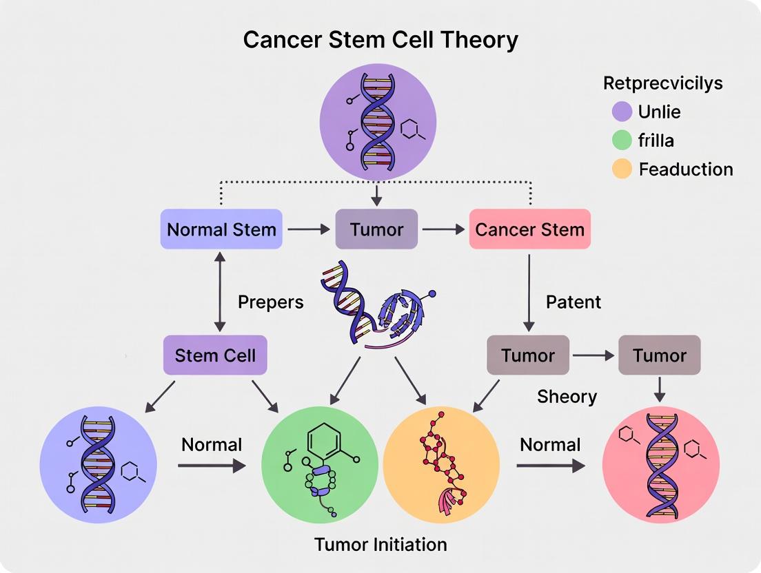
- The Hierarchical (CSC) Model: Tumors are organized akin to normal tissues, with a unidirectional hierarchy. A small, distinct subset of CSCs sits at the apex, exclusively capable of self-renewal and generating the bulk, non-tumorigenic progeny that constitute the tumor mass. Tumor propagation is deterministic and reliant on this CSC pool.
- The Stochastic Model: Tumor cells are biologically similar, and any cell within the tumor mass has a low, random probability of acquiring tumorigenic potential. Tumor behavior is driven by probabilistic events, and there is no fixed hierarchical organization.
This whitepaper delineates the core principles, experimental evidence, and methodologies that define and distinguish these competing paradigms within the context of tumor initiation research.
Core Principles & Comparative Analysis
| Principle | Hierarchical (CSC) Model | Stochastic Model |
|---|---|---|
| Tumor Organization | Rigid, unidirectional hierarchy. | Fluid, non-hierarchical, or reversible states. |
| Tumorigenic Potential | Restricted to a rare, phenotypically distinct CSC population. | Potentially present in any tumor cell, stochastically activated. |
| Self-Renewal | An intrinsic, defining property of CSCs. | A transient state that can be entered/exited by many cells. |
| Differentiation | Unidirectional, from CSC to non-tumorigenic progeny. | Plastic and bidirectional; non-stem cells can dedifferentiate. |
| Primary Driver | Deterministic (based on cell phenotype). | Probabilistic (based on random intracellular events). |
| Therapeutic Implication | Must target and eradicate CSCs for cure. | Must target a broad population to reduce the probability of tumorigenic conversion. |
| Key Evidence | FACS isolation of CSC-enriched populations via surface markers (e.g., CD44+/CD24- in breast) showing exclusive tumor-initiating capacity in limiting dilution assays. | Lineage tracing and single-cell clonal studies showing that non-CSC populations can stochastically regenerate the original tumor heterogeneity. |
Key Experimental Paradigms & Protocols
The Gold Standard:In VivoLimiting Dilution Transplantation Assay (LDA)
Purpose: To functionally assess tumor-initiating cell frequency and self-renewal capacity. Protocol:
- Tumor Cell Preparation: Generate single-cell suspension from primary tumor or xenograft.
- Cell Sorting: Use FACS to isolate putative CSC (e.g., CD44+CD24-) and non-CSC (e.g., CD44-CD24+) populations based on marker expression.
- Serial Dilution: Inject sorted cells orthotopically or subcutaneously into immunocompromised mice (NOD/SCID/IL2Rγ-null) at serially decreasing cell doses (e.g., 10,000, 1000, 100, 10 cells).
- Observation & Analysis: Monitor mice for tumor formation over several months. The frequency of tumor-initiating cells (TIC) is calculated using extreme limiting dilution analysis (ELDA) software, which compares the Poisson-based probability of tumor take between different populations.
Lineage Tracing and Clonal Analysis
Purpose: To track the fate of single cells and their progeny in situ over time, testing hierarchy vs. stochasticity. Protocol:
- Genetic Labeling: Introduce a heritable, indelible marker (e.g., Confetti fluorescent reporter, DNA barcode) into a subset of tumor cells in an autochthonous mouse model.
- Tumor Initiation & Growth: Allow tumors to develop and progress from the labeled founder cells.
- Multicolor Imaging & Sequencing: At endpoints, analyze tumors via intravital or whole-mount microscopy and single-cell sequencing to map clonal architectures.
- Interpretation: A fixed hierarchy shows predictable lineage patterns; stochastic models show dynamic, interconverting clonal contributions.
Visualizing Signaling Pathways and Cellular States
Core Signaling in CSC Maintenance
CSC Maintenance Signaling Network
Model Comparison: Tumor Cell Hierarchy
Hierarchical vs Stochastic Tumor Organization
Experimental Workflow: Limiting Dilution Assay
LDA Workflow for CSC Validation
The Scientist's Toolkit: Key Research Reagents & Materials
| Reagent/Material | Function in CSC Research | Application Example |
|---|---|---|
| Fluorescent-Activated Cell Sorter (FACS) | High-throughput isolation of live cell populations based on specific surface marker expression. | Sorting CD44+/CD24- cells from breast cancer cell lines for transplantation. |
| Anti-human CD44 (APC conjugate) | Antibody to label and isolate cells expressing CD44, a common CSC marker in multiple cancers. | Used in combination with other markers for CSC enrichment prior to LDA. |
| Anti-human CD24 (PE conjugate) | Antibody to label cells expressing CD24; often used as a negative selection marker in breast CSC assays. | Defining the CD44+CD24- phenotype in breast cancer. |
| Matrigel Basement Membrane Matrix | Provides a 3D, physiologically relevant extracellular matrix to support tumor cell growth and engraftment. | Mixed with tumor cells for subcutaneous or orthotopic injections in mice. |
| NOD/SCID/IL2Rγ-null (NSG) Mice | Immunodeficient mouse strain with minimal innate immunity, allowing efficient engraftment of human tumor cells. | The host for in vivo limiting dilution tumor initiation assays. |
| Extreme Limiting Dilution Analysis (ELDA) Software | Open-source statistical tool for calculating tumor-initiating cell frequency from limiting dilution data. | Quantifying and comparing TIC frequency between sorted populations. |
| Lentiviral barcode library | Introduces unique genetic barcodes into cells to enable high-resolution clonal tracking. | Studying stochastic clonal dynamics and tumor evolution in lineage tracing experiments. |
The Cancer Stem Cell (CSC) theory posits that tumors are organized hierarchically, analogous to normal tissues, with a subpopulation of cells at the apex possessing stem-like properties. This model is central to understanding tumor initiation, therapeutic resistance, and relapse. Within this thesis framework, the definitive identification and functional characterization of CSCs rely on three interdependent pillars: the expression of specific key markers, the capacity for self-renewal, and the potential for differentiation. This guide provides a technical dissection of these hallmarks, offering current methodologies and data critical for research and drug development targeting this foundational population.
Key Markers: Surface and Functional Identifiers
CSC markers are often context-dependent, varying by tumor type. They typically include cell surface proteins, transcription factors, and enzymes that facilitate identification and isolation via techniques like Fluorescence-Activated Cell Sorting (FACS). The table below summarizes key markers across major cancer types.
Table 1: Key CSC Markers Across Tumor Types
| Tumor Type | Common CSC Markers | Associated Signaling Pathways | Notes/Function |
|---|---|---|---|
| Breast Cancer | CD44+/CD24-/low, ALDH1 (high activity) | Wnt, Notch, Hedgehog | CD44+/CD24- phenotype enriched in tumor-initiating capacity. |
| Colorectal Cancer | CD133 (PROM1), LGR5, CD44, EpCAM | Wnt/β-catenin | LGR5 is a direct Wnt target and marker of stem cells in crypt. |
| Glioblastoma | CD133, CD44, Integrin α6, A2B5 | PI3K/AKT, STAT3 | CD133+ cells demonstrate radio/chemo-resistance. |
| Pancreatic Cancer | CD133, CD44, CD24, ESA (EpCAM+) | Hedgehog, TGF-β | Often used in combination (e.g., CD44+CD24+ESA+). |
| Acute Myeloid Leukemia | CD34+/CD38- | NF-κB, PI3K/AKT | Phenotype mirrors normal hematopoietic stem cells. |
| Lung Cancer | CD133, CD44, ALDH1 (high activity) | Wnt, Notch | ALDH1 activity is a functional marker of stemness. |
Experimental Protocol: Isolation of Breast CSCs via FACS for CD44/CD24 Phenotype
- Tissue Processing: Generate a single-cell suspension from patient-derived xenograft (PDX) tumors or primary samples using enzymatic digestion (Collagenase/Hyaluronidase mix).
- Antibody Staining: Resuspend ~1x10^7 cells in FACS buffer (PBS + 2% FBS). Incubate with fluorophore-conjugated anti-human CD44 (e.g., FITC) and anti-human CD24 (e.g., PE) antibodies (or respective isotype controls) for 30 min at 4°C in the dark.
- Viability Staining: Add a viability dye (e.g., DAPI or 7-AAD) prior to sorting to exclude dead cells.
- FACS Sorting: Use a high-speed cell sorter. Gate on viable, single cells. The CSC-enriched population is typically sorted as CD44high/CD24low/negative. The non-CSC population (CD44low/CD24high) serves as a control.
- Validation: Sorted populations are immediately used for functional assays (detailed below).
Self-Renewal Capacity: The Definitive Functional Assay
The ability to generate identical copies of themselves upon division is the core functional hallmark of CSCs. This is quantitatively measured in vitro and in vivo.
Table 2: Quantitative Metrics for Self-Renewal Assays
| Assay | Key Readout | Typical Data Range (CSC-enriched vs. Non-CSC) | Interpretation |
|---|---|---|---|
| Extreme Limiting Dilution Analysis (ELDA) | Tumor-Initiating Frequency | 1 in 10^3 to 1 in 10^4 (enriched) vs. 1 in 10^5 to no tumors (non-CSC) | Statistical measure of stem cell frequency in vivo. |
| Sphere-Forming Assay (Serum-Free) | Number & Size of Primary/Secondary Spheres | >10-fold increase in sphere # for enriched population. Secondary sphere formation >30% of plated cells. | In vitro surrogate for self-renewal and anchorage-independent growth. |
| Serial Transplantation | Number of Successful Transplant Generations | CSC-enriched: ≥3 generations; Non-CSC: 0-1 generations. | Gold-standard proof of long-term self-renewal in vivo. |
Experimental Protocol: In Vitro Sphere-Forming Assay
- Substrate Coating: Coat ultra-low attachment plates with a thin layer of synthetic hydrogel (e.g., Poly-HEMA) to prevent cell adhesion.
- Cell Plating: Plate FACS-sorted single cells (e.g., 500-1000 cells/well) in serum-free medium supplemented with growth factors (20 ng/mL EGF, 10 ng/mL bFGF), B27 supplement, and antibiotics.
- Culture: Maintain cells at 37°C, 5% CO2 for 7-14 days. Do not disturb the plates. Replace half of the medium with fresh pre-warmed medium every 3-4 days.
- Quantification: After 7-14 days, image wells using an inverted microscope. Count spheres >50 µm in diameter using automated or manual counting software.
- Secondary Sphere Formation: Collect primary spheres by gentle centrifugation, dissociate into single cells using Accutase, and replate at the same density in fresh sphere-forming medium. The formation of new spheres confirms self-renewal capacity.
Differentiation Capacity: Recapitulating Tumor Heterogeneity
CSCs must be able to differentiate into the non-tumorigenic, bulk tumor cells that constitute the tumor mass, thereby recapitulating the original tumor's heterogeneity.
Experimental Protocol: In Vitro Differentiation and Lineage Tracing
- Induction of Differentiation: Isolate CSCs (e.g., via FACS). Plate these cells in standard serum-containing (10% FBS) adherent tissue culture conditions on Matrigel-coated plates.
- Culture Duration: Maintain cells for 10-14 days, allowing them to adhere and proliferate.
- Analysis of Differentiation: Harvest cells and analyze for loss of CSC marker expression (e.g., downregulation of CD133, ALDH1 activity) and gain of lineage-specific differentiation markers via:
- Flow Cytometry: For surface differentiation antigens.
- qRT-PCR: For lineage-specific gene expression profiles.
- Immunofluorescence/Histology: For morphological changes and protein expression (e.g., cytokeratins for epithelial differentiation, GFAP for glial differentiation).
- Functional Validation: Differentiated cells should show a marked reduction (or loss) of tumor-initiating capacity in in vivo limiting dilution assays compared to the parental CSC population.
Visualization of Core Signaling Pathways
Diagram Title: Core Signaling Pathways Governing CSC Hallmarks
Diagram Title: Experimental Workflow for Validating CSC Hallmarks
The Scientist's Toolkit: Essential Research Reagents
Table 3: Key Research Reagent Solutions for CSC Studies
| Reagent/Material | Function in CSC Research | Example Application |
|---|---|---|
| Ultra-Low Attachment Plates | Prevents cell adhesion, enabling 3D sphere growth in serum-free conditions. | In vitro sphere-forming assays for self-renewal. |
| Recombinant EGF & bFGF | Essential growth factors for maintaining stem cell proliferation in serum-free culture. | Component of sphere-forming/ stem cell medium. |
| B27 Serum Supplement | Provides hormones, vitamins, and antioxidants to support neural and other stem cell survival. | Serum-free medium formulation for CSC culture. |
| Matrigel / Basement Membrane Extract | Provides a 3D extracellular matrix for organoid culture or differentiation studies. | 3D organoid assays, induced differentiation protocols. |
| Fluorophore-Conjugated Antibodies (CD44, CD24, CD133) | Primary tool for identifying and isolating CSC populations via flow cytometry. | FACS-based isolation and characterization. |
| ALDEFLUOR Assay Kit | Measures Aldehyde Dehydrogenase (ALDH) enzyme activity, a functional CSC marker. | Functional identification of CSCs independent of surface markers. |
| In Vivo Luciferase Reporter System | Enables bioluminescent tracking of tumor cell growth and metastasis in animal models. | Longitudinal monitoring of tumor initiation and growth from implanted CSCs. |
| RHO/ROCK Pathway Inhibitor (Y-27632) | Enhances survival of dissociated single stem cells, preventing anoikis. | Used during initial plating after cell sorting for sphere assays. |
Within the hierarchical model of cancer stem cell (CSC) theory, tumor initiation and recurrence are driven by a rare subpopulation of cells with self-renewal and pluripotent capacities. A critical determinant of CSC fate is the specialized microenvironment, or niche. This whitepaper provides a technical examination of the niche hypothesis, detailing the bi-directional crosstalk between CSCs and their microenvironment that maintains stemness, promotes tumor initiation, and confers therapy resistance. We synthesize current molecular mechanisms, experimental methodologies, and quantitative data to guide therapeutic strategies targeting the CSC-niche unit.
The hierarchical model posits that tumors are organized akin to normal tissues, with CSCs at the apex. The niche hypothesis extends this model by asserting that extrinsic signals from the local microenvironment are indispensable for maintaining CSC properties. The niche is a dynamic, anatomically distinct compartment composed of cellular components (e.g., cancer-associated fibroblasts (CAFs), mesenchymal stem cells (MSCs), endothelial cells, immune cells) and acellular factors (e.g., extracellular matrix (ECM), hypoxia, cytokines, metabolic substrates). This unit creates a permissive ecosystem for CSC quiescence, self-renewal, and protection.
Core Signaling Pathways in the CSC Niche
The following pathways represent the principal axes of communication within the niche.
Diagram Title: Core Signaling Pathways Linking the Niche to CSC Stemness
Quantitative Data: Niche Components and Their Functional Impact
Table 1: Impact of Specific Niche Components on CSC Properties Across Cancer Types
| Niche Component | Cancer Type | Key Effector Molecule(s) | Effect on CSC Frequency | Reported Change | Experimental Model |
|---|---|---|---|---|---|
| Hypoxia | Glioblastoma | HIF-1α, HIF-2α | Increases | Up to 5-fold increase in CD133+ cells | Patient-derived xenografts (PDX) |
| CAFs | Pancreatic Ductal Adenocarcinoma | IL-6, LIF | Increases | ~3-fold increase in tumor-initiating capacity | Co-injection in vivo (mouse) |
| Tumor-Associated Macrophages (M2) | Breast Cancer | CCL2, TGF-β | Increases | 2.5-fold increase in ALDH+ cells | 3D co-culture in vitro |
| Endothelial Cells | Colorectal Cancer | Notch Ligand (DLL4) | Increases | Promotes chemoresistance; 4-fold higher serial transplantation efficiency | Organoid co-culture |
| ECM Stiffness | Hepatocellular Carcinoma | Integrin β1, YAP/TAZ | Increases | Drives dedifferentiation; 10-fold increase in tumor initiation | Hydrogels with tunable stiffness |
| Bone Marrow Mesenchymal Cells | Acute Myeloid Leukemia | CXCL12 | Increases | Protects CSCs from chemotherapy; maintains quiescence | Human-mouse xenograft |
Table 2: Clinical Correlations of Niche Marker Expression
| Niche Marker | Cancer Type | High Expression Correlates With | Hazard Ratio (HR) for Poor Prognosis | Study (Sample Size) |
|---|---|---|---|---|
| CAF Signature (α-SMA, FAP) | Pancreatic | Shorter overall survival, metastasis | HR: 2.1 (95% CI: 1.5-3.0) | Meta-analysis (n=850) |
| HIF-1α | Head and Neck | Locoregional failure, resistance to radiation | HR: 1.8 (95% CI: 1.3-2.5) | Prospective cohort (n=298) |
| CD163+ M2 Macrophages | Gastric | Advanced stage, lymph node invasion | HR: 2.4 (95% CI: 1.7-3.4) | Immunohistochemistry (n=512) |
| LOX (ECM Crosslinker) | Breast | Bone metastasis, reduced relapse-free survival | HR: 1.9 (95% CI: 1.4-2.6) | TCGA analysis (n=1100) |
Experimental Protocols for Niche-CSC Research
Protocol: In Vivo Lineage Tracing and Niche Labeling
Objective: To trace the fate of CSCs and their interaction with labeled niche cells over time. Materials: See "The Scientist's Toolkit" (Section 6). Procedure:
- Model Generation: Cross a CSC-specific driver mouse (e.g., Lgr5-CreERT2 for intestinal cancer) with a fluorescent reporter (Rosa26-tdTomato). For niche labeling, cross a niche-specific driver (e.g., Col1a1-CreER for fibroblasts) with a separate reporter (Rosa26-eGFP).
- Tumor Induction: Administer a carcinogen (e.g., AOM) or conditionally activate an oncogene (e.g., KrasG12D) in the compound transgenic mice.
- Pulse Labeling: Administer tamoxifen to induce Cre recombination, permanently labeling CSCs (tdTomato+) and niche cells (eGFP+) at a defined timepoint.
- Time-Course Analysis: Sacrifice cohorts of mice at serial timepoints (e.g., 1, 4, 8 weeks). Harvest tumors and process for imaging.
- Multiplex Imaging: Generate frozen or paraffin sections. Perform immunofluorescence for differentiation markers (e.g., Cytokeratin) and stemness markers (e.g., SOX9). Image using confocal microscopy.
- Quantification: Use image analysis software to quantify: a) The percentage of tdTomato+ CSCs that are in direct contact with eGFP+ niche cells. b) The clonal expansion of single labeled CSCs over time. c) The spatial distribution of CSCs relative to blood vessels (CD31+) or hypoxic areas (pimonidazole staining).
Protocol: 3D Biomimetic Co-culture for Functional Assays
Objective: To functionally test the necessity and sufficiency of specific niche components on CSC self-renewal. Materials: Tunable stiffness hydrogels (e.g., PEG-based or collagen-Matrigel), recombinant cytokines, neutralizing antibodies. Procedure:
- Cell Isolation: Isolate primary human CSCs via fluorescence-activated cell sorting (FACS) using validated surface markers (e.g., CD44+/CD24- for breast cancer). Isolate primary human stromal cells (e.g., CAFs from patient tissue).
- Hydrogel Preparation: Prepare hydrogel precursor solution at a physiologically relevant stiffness (e.g., 0.5-5 kPa). Mix with stromal cells at a defined density (e.g., 10^4 cells/mL). Polymerize in a 96-well plate.
- CSC Seeding: Seed fluorescently labeled CSCs on top of or embedded within the hydrogel.
- Condition Modulation: Add small molecule pathway inhibitors (e.g., DAPT for Notch), neutralizing antibodies (e.g., anti-IL-6), or recombinant proteins (e.g., Wnt3a).
- Endpoint Analysis (7-14 days):
- Self-Renewal: Dissociate spheres, re-plate at clonal density, and count secondary sphere formation.
- Differentiation: Fix and stain for lineage-specific markers.
- Viability/Proliferation: Perform live-dead staining or EdU incorporation assays.
- Gene Expression: Recover cells for qRT-PCR of stemness genes (NANOG, OCT4, SOX2).
Diagram Title: Integrated Experimental Workflow for Niche-CSC Studies
Therapeutic Implications: Targeting the Niche
Strategies to disrupt the niche include:
- Disrupting physical interactions: Anti-integrin antibodies (e.g., against αvβ3), FAK inhibitors.
- Neutralizing paracrine signals: Anti-IL-6R (tocilizumab), CXCR4 antagonists (plerixafor), TGF-β traps.
- Depleting pro-tumorigenic stromal cells: FAP-targeting CAR-T cells, CSF1R inhibitors to deplete macrophages.
- Normalizing aberrant ECM: LOXL2 inhibitors, hyaluronidase.
- Alleviating hypoxia: HIF inhibitors, vascular normalizing agents (anti-VEGF).
The Scientist's Toolkit
Table 3: Essential Research Reagents for Niche-CSC Investigations
| Reagent/Category | Example Product/Model | Primary Function in Niche Research |
|---|---|---|
| In Vivo Lineage Tracing System | ROSA26-loxP-Stop-loxP-tdTomato mice (Ai14), Lgr5-CreERT2 mice | Enables indelible, cell-type-specific fluorescent labeling and fate mapping of CSCs or niche cells upon tamoxifen induction. |
| Tunable 3D Hydrogels | PEG-based (e.g., CytoSoft plates), Collagen I/Matrigel mixes | Provides a biomimetic, stiffness-controlled 3D matrix to model physical niche properties and embed co-cultures. |
| Cytokine/Niche Factor Panel | Recombinant human Wnt3a, DLL4, IL-6; Recombinant mouse SDF-1α | Used to supplement cultures to test sufficiency of specific niche signals on CSC behavior. |
| Neutralizing Antibodies | Anti-human/mouse IL-6R, Anti-TGF-β, Anti-DLL4 | Used in vitro and in vivo to block specific paracrine signaling axes to test necessity. |
| Hypoxia Mimetics & Reporters | Pimonidazole HCl, HIF-1α Stabilizer (CoCl2), HRE-GFP reporter cells | Labels hypoxic regions in vivo (pimonidazole) or mimics/reads out hypoxia signaling in vitro. |
| Stromal Cell Isolation Kits | Human CAF Isolation Kit (FACS-based), Mouse Endothelial Cell Isolation Kit | For purification of specific niche cell populations from primary tumors for functional co-culture studies. |
| Small Molecule Pathway Inhibitors | DAPT (γ-secretase/Notch), LGK974 (Porcupine/Wnt), Vismodegib (Smo/Hh) | Pharmacologically disrupts key stemness pathways within the niche-CSC unit. |
Epigenetic and Metabolic Drivers of the CSC State
Within the hierarchical model of cancer stem cell (CSC) theory, a subpopulation of tumor cells with stem-like properties is responsible for tumor initiation, therapeutic resistance, and metastasis. The CSC state is not fixed but is dynamically regulated by intrinsic epigenetic reprogramming and extrinsic metabolic adaptations within the tumor microenvironment. This whitepaper synthesizes current research on the core epigenetic and metabolic mechanisms that establish and maintain the CSC state, providing a technical guide for researchers targeting these drivers.
Epigenetic Regulation of the CSC State
Epigenetic modifications provide a plastic, heritable layer of control over gene expression programs that define CSCs, enabling rapid adaptation without genetic mutation.
DNA Methylation and Hydroxymethylation
Global hypomethylation coupled with promoter-specific hypermethylation is a hallmark of CSCs. Key tumor suppressor genes (e.g., p16INK4a, PTEN) are often silenced by polycomb repressive complex 2 (PRC2)-mediated H3K27me3 marks, followed by DNA methyltransferase (DNMT) activity for stable repression.
Table 1: Key DNA Methylation Changes in CSCs
| Gene/Region | Modification in CSCs | Functional Consequence | Experimental Model |
|---|---|---|---|
| CDH1 (E-cadherin) promoter | Hypermethylation | Loss of cell adhesion, increased invasion | Breast CSCs (MCF-7) |
| SOX2 enhancer | Hypomethylation | Activation of stemness program | Glioblastoma CSCs |
| OCT4 promoter | Hydroxymethylation (5hmC) | Pluripotency gene activation | Colon CSCs |
| LINE-1 repetitive elements | Global hypomethylation | Genomic instability | Pancreatic CSCs |
Histone Modifications
Post-translational modifications of histones directly modulate chromatin accessibility. Bivalent domains (co-existing H3K4me3 activation and H3K27me3 repression marks) at developmental gene promoters are a key feature, priming CSCs for fate transitions.
Experimental Protocol: ChIP-seq for Bivalent Domain Mapping in CSCs
- Crosslinking & Cell Lysis: Fix 10^7 CSCs with 1% formaldehyde for 10 min. Quench with 125mM glycine. Lyse cells in SDS lysis buffer.
- Chromatin Shearing: Sonicate lysate to achieve 200-500 bp DNA fragments. Verify fragment size by agarose gel electrophoresis.
- Immunoprecipitation: Incubate chromatin with 5 µg of specific antibody (anti-H3K4me3, anti-H3K27me3) or IgG control overnight at 4°C with rotation.
- Bead Capture & Washing: Add Protein A/G magnetic beads, incubate 2 hours. Wash sequentially with Low Salt, High Salt, LiCl, and TE buffers.
- Elution & Decrosslinking: Elute chromatin in elution buffer (1% SDS, 0.1M NaHCO3). Add NaCl to 200mM and reverse crosslinks at 65°C overnight.
- DNA Purification: Treat with RNase A and Proteinase K. Purify DNA using phenol-chloroform extraction and ethanol precipitation.
- Library Prep & Sequencing: Prepare sequencing library using commercial kit (e.g., NEBNext Ultra II). Sequence on Illumina platform (≥50M reads, paired-end).
- Data Analysis: Align reads to reference genome (e.g., Bowtie2). Call peaks (e.g., MACS2). Identify bivalent domains as genomic regions with both H3K4me3 and H3K27me3 peaks.
Chromatin Remodelers
ATP-dependent complexes like SWI/SNF (BAF) facilitate lineage-specific gene expression. Subunit switching (e.g., ARID1A to ARID1B) confers CSC-specific chromatin remodeling.
Metabolic Drivers of the CSC State
CSCs exhibit metabolic flexibility, often shifting between glycolysis and oxidative phosphorylation (OXPHOS) to meet biosynthetic demands and survive stress.
Glycolytic Plasticity
While many CSCs rely on OXPHOS, a subset utilizes high glycolysis, a phenomenon linked to hypoxic niches. Pyruvate dehydrogenase kinase (PDK) activity diverts pyruvate from mitochondria, promoting lactate production.
Table 2: Metabolic Enzyme Expression in CSCs vs. Non-CSCs
| Metabolic Pathway | Key Enzyme | Expression in CSCs (Fold Change) | Assay Used | Cancer Type |
|---|---|---|---|---|
| Glycolysis | HK2 | +3.5 to +5.2 | qRT-PCR, Western | Glioblastoma |
| PPP (Biosynthesis) | G6PD | +4.1 | Metabolomics (LC-MS) | AML |
| Fatty Acid Oxidation | CPT1A | +6.8 | Seahorse XF Analyzer | Breast Cancer |
| Glutamine Metabolism | GLS1 | +2.9 | Stable Isotope Tracing | Lung Cancer |
Mitochondrial Metabolism
Many CSCs maintain high mitochondrial membrane potential and efficient OXPHOS. This is coupled with low ROS production via upregulated antioxidant systems (e.g., NRF2, SOD2). Fatty acid oxidation (FAO) is a critical energy source in quiescent CSCs.
Experimental Protocol: Metabolic Flux Analysis using Seahorse XF Analyzer
- Cell Seeding: Seed 20,000 CSCs per well in a Seahorse XF96 cell culture microplate. Include control non-CSCs. Incubate overnight.
- Media Exchange: 1 hour before assay, replace media with Seahorse XF Base Medium (pH 7.4) supplemented with 10mM glucose, 1mM pyruvate, and 2mM L-glutamine. Incubate at 37°C, non-CO2.
- Sensor Cartridge Loading: Load Seahorse XFp Sensor Cartridge ports with modulators:
- Port A: 10µM Oligomycin (ATP synthase inhibitor).
- Port B: 10µM FCCP (mitochondrial uncoupler).
- Port C: 10µM Rotenone + 10µM Antimycin A (Complex I & III inhibitors).
- Port D: 50mM 2-DG (glycolysis inhibitor).
- Assay Run: Calibrate cartridge. Run the Mito Stress Test program (3 baseline measurements, 3 measurements after each injection).
- Data Calculation: Using Wave software, calculate:
- Basal OCR = (Last baseline measurement) - (Non-mitochondrial OCR).
- ATP-linked OCR = (Last baseline) - (Oligomycin measurement).
- Maximal OCR = (Max FCCP measurement) - (Non-mitochondrial OCR).
- Glycolysis (ECAR) analyzed similarly.
Nutrient Sensing and Signaling
Metabolic pathways are intertwined with key signaling cascades (e.g., PI3K/AKT/mTOR, AMPK). mTORC1 activity promotes anabolic processes but can be suppressed in quiescent CSCs, which instead activate AMPK and autophagy.
Epigenetic-Metabolic Crosstalk
A bidirectional relationship exists where metabolites serve as substrates or co-factors for epigenetic enzymes, and epigenetic changes regulate metabolic gene expression.
Metabolites as Epigenetic Modulators
- α-Ketoglutarate (α-KG): A co-factor for Ten-eleven translocation (TET) DNA demethylases and Jumonji-domain histone demethylases (KDMs). High α-KG promotes a stem-like state.
- S-adenosylmethionine (SAM): The universal methyl donor for DNMTs and histone methyltransferases (HMTs). SAM levels, influenced by methionine cycle and one-carbon metabolism, dictate global methylation capacity.
- Acetyl-CoA: Substrate for histone acetyltransferases (HATs). Generated from citrate via ACLY or from acetate via ACSS2, linking glycolysis and lipid metabolism to chromatin acetylation.
Diagram Title: Metabolic-Epigenetic Crosstalk in CSC Regulation
Key Signaling Pathways Integrating Epigenetics and Metabolism
The Hippo-YAP/TAZ and Wnt/β-catenin pathways are central hubs, receiving inputs from cell density, mechanics, and nutrients to regulate CSC transcriptional and epigenetic programs.
Diagram Title: Hippo and Wnt Pathway Integration in CSCs
The Scientist's Toolkit: Research Reagent Solutions
Table 3: Essential Reagents for CSC Epigenetic & Metabolic Research
| Reagent/Category | Specific Example(s) | Function & Application | Key Provider(s) |
|---|---|---|---|
| CSC Enrichment | Anti-CD44 / CD133 Magnetic Beads | Immunomagnetic separation of CSC surface markers. | Miltenyi Biotec, STEMCELL Tech |
| DNMT Inhibitors | 5-Azacytidine, Decitabine | Nucleoside analogs causing DNA hypomethylation; used to test gene re-expression. | Sigma-Aldrich, Cayman Chemical |
| HDAC Inhibitors | Vorinostat (SAHA), Trichostatin A (TSA) | Block histone deacetylases, increase histone acetylation, alter CSC phenotype. | Selleckchem, MedChemExpress |
| Metabolic Inhibitors | 2-DG, Etomoxir, CB-839 | Target glycolysis (2-DG), FAO (Etomoxir), glutaminase (CB-839) for functional assays. | Tocris, Sigma-Aldrich |
| Epigenetic Probes | JIB-04 (KDM inhibitor), GSK-J4 (KDM6A/B inhibitor) | Small molecule inhibitors of specific histone demethylases. | Abcam, Cayman Chemical |
| Metabolic Tracers | U-13C-Glucose, 13C15N-Glutamine | Stable isotope-labeled nutrients for tracing metabolic flux via GC/LC-MS. | Cambridge Isotope Labs |
| ChIP-grade Antibodies | Anti-H3K27me3, Anti-H3K4me3, Anti-H3K9ac | High-specificity antibodies for chromatin immunoprecipitation assays. | Cell Signaling Tech, Active Motif |
| Seahorse XF Assay Kits | XFp Mito Stress Test Kit, XF Glycolysis Stress Test Kit | Pre-optimized reagent kits for real-time metabolic flux analysis. | Agilent Technologies |
| In Vivo CSC Models | NOG/NSG mice, Matrigel | Immunodeficient mice for tumor initiation assays; basement membrane matrix for sphere culture. | Jackson Lab, Corning |
Targeting the synergistic interplay between epigenetic and metabolic drivers presents a promising strategy to eliminate the therapy-resistant CSC compartment. Future drug development should focus on dual-action agents (e.g., inhibitors of both IDH1 and DNMTs) and context-specific combinations that consider the dynamic plasticity of the CSC state within the hierarchical tumor model. Advanced models, including patient-derived organoids and engineered niches, are essential for translating these insights into effective therapies.
1. Introduction: Framing within the Cancer Stem Cell (CSC) Thesis
The hierarchical model of tumor initiation posits that tumor growth and propagation are driven by a subpopulation of cells with stem-like properties: Cancer Stem Cells (CSCs). These cells self-renew, differentiate, and are often therapy-resistant. "Intra-tumoral hierarchy" describes the organized lineage relationships from CSCs to more differentiated progeny. "Plasticity" refers to the dynamic ability of non-CSC tumor cells to re-acquire stem-like states in response to microenvironmental cues or therapeutic insult. This whitepaper details the experimental frameworks for investigating these core concepts, integrating recent findings on the molecular regulators of plasticity.
2. Quantitative Data Summary: Key Metrics in CSC & Plasticity Research
Table 1: Common Functional & Molecular Metrics for CSCs
| Metric Category | Specific Assay/Measurement | Typical Quantitative Output (Representative Ranges) | Implication for Hierarchy/Plasticity |
|---|---|---|---|
| Functional Capacity | In Vivo Limiting Dilution Assay | Tumor-Initiating Cell Frequency (e.g., 1/10,000 to 1/100 cells) | Gold standard for defining hierarchical potential. |
| Sphere-Formation Assay | Number & Diameter of Primary/Secondary Spheres (e.g., 5-50 spheres per 10^3 cells) | Measures self-renewal and clonogenicity in vitro. | |
| Surface Phenotype | Flow Cytometry for CSC Markers | % of Marker+ Population (e.g., CD44+/CD24- in breast: 1-10%; CD133+ in glioma: 5-30%) | Enables prospective isolation for functional study. |
| Transcriptional State | qPCR for Stemness Factors | Fold-Change in OCT4, SOX2, NANOG, MYC mRNA (e.g., 2- to 100-fold increase in CSCs) | Indicates activation of core regulatory programs. |
| Epigenetic State | ATAC-seq/ChIP-seq | Chromatin Accessibility or H3K27ac Peaks at Pluripotency Loci | Reveals epigenetic priming for plasticity. |
Table 2: Therapeutic Challenges Linked to Plasticity
| Therapy Type | Observed Plasticity Response | Key Mediators (Examples) | Experimental Evidence Increase in CSC Marker+ Cells Post-Therapy |
|---|---|---|---|
| Chemotherapy (e.g., Paclitaxel) | Dedifferentiation of surviving cells | IL-6/STAT3, TGF-β, YAP/TAZ | 2- to 5-fold increase in tumor sphere formation. |
| Radiation Therapy | Enhanced stem-like phenotype | NF-κB, WNT/β-catenin, ROS signaling | 3- to 8-fold increase in ALDH+ population. |
| Targeted Therapy (e.g., EGFRi) | Phenotypic switching & drug tolerance | AXL, JAK/STAT, Hedgehog signaling | Up to 10-fold expansion of drug-tolerant persister cells. |
3. Experimental Protocols for Key Investigations
Protocol 1: Lineage Tracing In Vivo to Map Hierarchy and Plasticity Objective: To fate-map tumor cell populations and track transitions between states. Materials: Cre/Lox or similar genetically engineered mouse model (GEMM); tumor cells expressing inducible Cre recombinase and fluorescent reporter (e.g., Confetti); Tamoxifen for induction. Method:
- Induce stochastic labeling of a defined cell population (e.g., differentiated cells expressing Krt14) in established GEMM tumors with tamoxifen.
- Allow tumor progression or apply therapeutic intervention (e.g., chemotherapy).
- Harvest tumors at multiple timepoints (e.g., 1, 2, 4 weeks post-induction/therapy).
- Perform multiplex immunofluorescence or flow cytometry on dissociated tumors to analyze reporter expression in different phenotypic compartments (CSC vs. non-CSC markers).
- Quantify the appearance of labeled cells in the CSC compartment as evidence of plasticity (dedifferentiation).
Protocol 2: Assessing Plasticity via Single-Cell RNA Sequencing (scRNA-seq) Objective: To characterize transcriptional states and identify transitional trajectories. Materials: Fresh tumor tissue, Single-cell suspension kit, Chromium Controller (10x Genomics), scRNA-seq library prep kit, Bioinformatic pipelines (Seurat, Monocle3). Method:
- Generate single-cell suspension from untreated and therapy-exposed tumors (viability >80%).
- Capture cells, prepare barcoded libraries following manufacturer protocol.
- Sequence libraries to a target depth of >50,000 reads per cell.
- Perform quality control, normalization, and integration of datasets.
- Cluster cells and annotate clusters using known marker genes (stemness, differentiation, stress).
- Perform trajectory inference (pseudotime) and RNA velocity analysis to predict state transitions and directionality, identifying genes driving plasticity.
4. Signaling Pathways Governing Plasticity
Title: Signaling Network Driving Tumor Cell Plasticity
5. Integrated Experimental Workflow for Plasticity Studies
Title: Core Workflow for Investigating Plasticity
6. The Scientist's Toolkit: Key Research Reagent Solutions
Table 3: Essential Reagents for CSC & Plasticity Research
| Reagent Category | Specific Example(s) | Function & Application |
|---|---|---|
| CSC Phenotypic Isolation | Anti-human CD44-APC, Anti-human CD24-PE, Anti-human CD133/1-PE-Vio615 | Antibody conjugates for fluorescence-activated cell sorting (FACS) to prospectively isolate live CSC and non-CSC populations for downstream assays. |
| Functional Assay Media | Serum-Free DMEM/F12, B-27 Supplement, Recombinant EGF, Recombinant bFGF | Essential components for non-adherent tumor sphere culture, which enriches for and tests self-renewing cell capacity. |
| Lineage Tracing | Tamoxifen, Doxycycline, CreERT2 or Tet-On/Off Lentiviral Constructs | Inducible systems for temporal control of genetic labeling (e.g., fluorescent reporter activation) to track cell fate in vitro and in vivo. |
| Plasticity Induction | Recombinant Human TGF-β1, CHIR99021 (WNT agonist), Interleukin-6 | Cytokines and small molecules used to stimulate signaling pathways known to induce dedifferentiation in controlled experiments. |
| scRNA-seq Kits | Chromium Next GEM Single Cell 3' Kit (10x Genomics), BD Rhapsody Cartridge Kit | Commercial kits for generating barcoded single-cell RNA sequencing libraries from suspended cells. |
| Pathway Inhibitors | SB431542 (TGF-βRI inhibitor), XAV-939 (Tankyrase/WNT inhibitor), Stattic (STAT3 inhibitor) | Small molecule tools to block specific plasticity-driving pathways for mechanistic validation and target exploration. |
From Theory to Bench: Advanced Methods for Isolating, Characterizing, and Targeting CSCs
Within the framework of the cancer stem cell (CSC) hierarchical model, the isolation of tumor-initiating cells is a foundational step. This model posits that tumor growth and heterogeneity are driven by a subpopulation of cells with stem-like properties: self-renewal, differentiation potential, and enhanced resistance. The precise identification and isolation of these CSCs are therefore critical for investigating tumor initiation, progression, and therapy resistance. This technical guide details the primary methodologies—Fluorescence-Activated Cell Sorting (FACS) and Magnetic-Activated Cell Sorting (MACS)—utilizing canonical surface marker panels (CD44, CD133) and enzymatic activity (ALDH) for the isolation of putative CSCs from solid and hematological malignancies.
Core Principles of CSC Isolation
Surface Marker-Based Isolation
The selection of surface markers is tissue and cancer-type specific, informed by extensive research linking them to poor prognosis and tumorigenicity in vivo.
- CD44: A transmembrane glycoprotein involved in cell-cell interaction, adhesion, and migration. The CD44+ population is enriched for CSCs in breast, colorectal, pancreatic, and head and neck cancers.
- CD133 (Prominin-1): A pentaspan transmembrane glycoprotein. CD133+ cells demonstrate tumor-initiating capacity in brain, prostate, colon, and liver cancers.
- Combined Panels: Often, a combination of markers (e.g., CD44+CD24- for breast cancer, CD44+CD133+ for colorectal cancer) provides greater specificity and enrichment for tumor-initiating cells.
Functional Marker-Based Isolation: ALDH
Aldehyde dehydrogenase (ALDH) is a detoxifying enzyme responsible for oxidizing intracellular aldehydes. High ALDH activity (ALDHbright) is a functional marker of stem/progenitor cells in both normal and malignant tissues, including leukemia, breast, and lung cancers. It is assayed via a fluorogenic substrate (BODIPY-aminoacetaldehyde, DEAB as inhibitor control).
Quantitative Comparison of FACS vs. MACS
Table 1: Comparative analysis of FACS and MACS for CSC isolation.
| Parameter | Fluorescence-Activated Cell Sorting (FACS) | Magnetic-Activated Cell Sorting (MACS) |
|---|---|---|
| Principle | Detection of fluorescently-labeled antibodies or substrates via lasers. | Binding of magnetic bead-conjugated antibodies, separation via magnetic field. |
| Resolution | High (multi-parameter, single-cell). | Moderate (primarily positive/negative selection). |
| Sorting Speed | ~10,000-50,000 cells/sec (varies by sorter). | ~108 cells in ~30 minutes. |
| Purity | Very High (>95-99%). | High (90-99%, depends on protocol). |
| Cell Viability | High (maintained with proper conditions). | High. |
| Throughput | Lower (analytical and preparative). | Very High (preparative). |
| Cost | High (instrument, maintenance). | Lower. |
| Multi-Marker Panels | Excellent (4+ colors standard). | Limited (typically 1-2 markers sequentially). |
| Key Application | High-purity isolation for functional assays, multi-parameter analysis. | Rapid bulk enrichment for downstream culture or molecular analysis. |
Detailed Experimental Protocols
Protocol 1: FACS Isolation of CD44+CD133+ Cells from Dissociated Tumor
This protocol is for isolating a dual-positive CSC population from a single-cell suspension of human colorectal carcinoma tissue.
- Sample Preparation: Generate a single-cell suspension from fresh tumor tissue using enzymatic digestion (e.g., collagenase/hyaluronidase mix) and mechanical dissociation. Pass through a 70µm cell strainer. Perform RBC lysis if necessary.
- Viability Staining: Resuspend cells in PBS with a viability dye (e.g., Fixable Viability Dye eFluor 780, 1:1000) for 30 min on ice in the dark. Wash.
- Fc Receptor Blocking: Incubate cells with human Fc receptor blocking reagent (10 min, 4°C).
- Surface Antibody Staining: Incubate with titrated, fluorochrome-conjugated antibodies against CD44 (e.g., APC, clone BJ18) and CD133/1 (e.g., PE, clone AC133) for 30 min on ice in the dark. Include fluorescence-minus-one (FMO) controls.
- Wash & Resuspend: Wash cells twice with FACS buffer (PBS + 2% FBS). Resuspend in buffer with DAPI (1 µg/mL) for live/dead gating. Keep on ice.
- FACS Sorting: Use a high-speed sorter (e.g., BD FACSAria III). Gate sequentially on: single cells (FSC-A vs. FSC-H), viable cells (DAPI- / Viability dye-), then CD44+CD133+ population. Sort into collection tubes containing complete culture medium. Validate purity by re-analyzing a fraction of sorted cells.
Protocol 2: ALDH Activity Assay Combined with FACS (ALDEFLUOR)
This protocol details the identification of cells with high ALDH enzymatic activity.
- Prepare Cell Suspension: As in Protocol 1.
- ALDEFLUOR Incubation: Aliquot cells into two tubes: Test and Control (DEAB). Resuspend both in ALDEFLUOR assay buffer. Add the activated ALDEFLUOR substrate (BODIPY-aminoacetaldehyde) to each tube. Immediately add the ALDH inhibitor diethylaminobenzaldehyde (DEAB) to the Control tube. Mix well.
- Incubation: Incubate both tubes at 37°C for 30-60 minutes. Protect from light.
- Centrifuge & Resuspend: Pellet cells, resuspend in ice-cold ALDEFLUOR buffer. Keep on ice.
- FACS Analysis/Sorting: Analyze immediately. The ALDHbright population is defined as the fluorescent population present in the Test sample but absent in the DEAB-inhibited control. Sort this population for downstream assays.
Signaling Pathways in Cancer Stem Cell Maintenance
The markers used for isolation are not merely identifiers; they are functional components of signaling networks that sustain CSC properties.
Experimental Workflow for CSC Isolation & Validation
A complete research pipeline from tumor processing to functional validation of isolated CSCs.
The Scientist's Toolkit: Key Research Reagent Solutions
Table 2: Essential materials and reagents for CSC isolation experiments.
| Reagent/Material | Function/Description | Example Product/Catalog |
|---|---|---|
| Tissue Dissociation Kit | Enzymatic blend for gentle tissue disaggregation into single cells while preserving epitopes. | Miltenyi Biotec Tumor Dissociation Kit; STEMCELL Technologies GentleMACS Dissociator. |
| Fluorochrome-Conjugated Antibodies | Antibodies specific to human/mouse CD44, CD133, CD24, etc., for FACS detection. | BioLegend (e.g., anti-human CD44-APC); Miltenyi (anti-human CD133/1-PE). |
| ALDEFLUOR Kit | Complete kit containing BODIPY- aminoacetaldehyde substrate and DEAB inhibitor for ALDH activity assay. | STEMCELL Technologies, Catalog #01700. |
| MACS MicroBeads & Columns | Magnetic beads conjugated to antibodies and columns for positive/negative selection via MACS. | Miltenyi Biotec CD133 MicroBead Kit, LS Columns. |
| Viability Dye | Fixable or non-fixable dye to exclude dead cells during sorting (critical for purity). | Thermo Fisher LIVE/DEAD Fixable Viability Dyes; DAPI. |
| Fc Receptor Blocking Reagent | Human or mouse IgG to block non-specific antibody binding via Fc receptors. | TruStain FcX (BioLegend); Human FcR Blocking Reagent (Miltenyi). |
| FACS Collection Medium | Serum-rich or specialized medium to maintain cell viability during and after sorting. | RPMI/F12 with 20% FBS; mTeSR Plus for stem cells. |
| Ultra-Low Attachment Plates | For 3D sphere formation assays (mammosphere assay) to assess self-renewal. | Corning Costar Ultra-Low Attachment Multiwell Plates. |
| Immunocompromised Mice | For in vivo tumorigenicity assays via subcutaneous or orthotopic injection of sorted cells. | NOD/SCID; NSG (NOD-scid IL2Rγnull) mice. |
The isolation of CSCs via FACS and MACS using defined marker panels is a cornerstone of experimental oncology research grounded in the hierarchical model. FACS offers high-purity, multi-parametric resolution essential for definitive functional studies, while MACS provides rapid, high-throughput enrichment. The choice of technique and marker panel must be empirically validated for each cancer type, and findings must be corroborated by rigorous in vitro and in vivo functional assays. These isolation techniques remain indispensable for deconvoluting tumor heterogeneity, understanding the mechanisms of tumor initiation and relapse, and developing targeted therapeutic strategies against the resilient CSC compartment.
The hierarchical model of tumorigenesis posits that a subpopulation of cells, cancer stem cells (CSCs), possesses the exclusive ability to initiate and sustain tumor growth, self-renew, and generate heterogeneous progeny. Validating this model requires functional proof of these cardinal stem cell properties. Two assays have emerged as indispensable, complementary gold standards: the in vitro sphere formation assay and the in vivo limiting dilution transplantation (LDT) assay. This guide details their execution, interpretation, and integration within CSC research and therapeutic development.
In Vitro Sphere Formation Assay
Core Principle & Rationale
This assay tests the capacity of single cells to survive in non-adherent, serum-free conditions and form clonal, non-adherent spherical colonies ("spheres" or "tumorspheres"). It is a surrogate for self-renewal and proliferative potential in vitro, enriching for cells with stem-like properties.
Detailed Protocol
A. Reagent Preparation:
- Basal Medium: DMEM/F12.
- Essential Supplements (StemPro NSC SFM or equivalent):
- B27 Supplement (50x): Provides hormones, vitamins, and antioxidants.
- N2 Supplement (100x): Supplies insulin, transferrin, selenite, and other proteins.
- Recombinant Human EGF (20 ng/mL final): Critical for proliferation of progenitor cells.
- Recombinant Human bFGF (20 ng/mL final): Supports self-renewal; must be replenished every 2-3 days.
- Heparin (2-4 µg/mL): Stabilizes bFGF.
- Antibiotics: Penicillin/Streptomycin (1%).
- Dissociation Enzyme: Accutase or StemPro Accutase for gentle single-cell dissociation.
B. Procedure:
- Tumor Dissociation: Generate a single-cell suspension from primary tumor or xenograft using enzymatic (Collagenase IV/DNase I) and mechanical dissociation. Filter through a 40µm cell strainer.
- Viability & Counting: Determine viable cell count using Trypan Blue exclusion.
- Plating: Serially dilute cells in complete sphere medium. Plate in ultra-low attachment (ULA) multi-well plates (e.g., Corning Costar) at densities ranging from 1-100 cells/µL (e.g., 1000 cells/well in a 96-well plate for LDA analysis).
- Culture: Incubate at 37°C, 5% CO2. Do not disturb for 5-7 days to allow initial cluster formation.
- Feeding: Every 2-3 days, carefully add a small volume (e.g., 20% of well volume) of fresh, pre-warmed medium containing EGF and bFGF.
- Analysis: After 7-21 days (protocol-dependent), score spheres under an inverted microscope. Only spheres with a diameter >50-100 µm (user-defined threshold) are counted.
Data Analysis & Interpretation
Results are typically analyzed as sphere-forming efficiency (SFE) or sphere-forming unit (SFU).
SFE (%) = (Number of spheres formed / Number of single cells plated) × 100
For quantitating frequency of sphere-initiating cells, a Limiting Dilution Analysis (LDA) in vitro is performed (see Table 1 and Section 4).
Signaling Pathways in Sphere Formation
Sphere formation is regulated by core stemness pathways. Inhibition of these pathways often reduces SFE.
Diagram Title: Core Signaling Pathways Driving Tumor Sphere Formation
In Vivo Limiting Dilution Transplantation (LDT) Assay
Core Principle & Rationale
This is the definitive in vivo functional assay for CSCs. It quantitatively measures the frequency of tumor-initiating cells (TICs) capable of regenerating a tumor upon serial transplantation into immunocompromised host animals (e.g., NOD/SCID, NSG mice). It directly tests self-renewal, differentiation, and recapitulation of tumor heterogeneity in vivo.
Detailed Protocol
A. Pre-Transplantation:
- Cell Preparation: Generate a pure, viable single-cell suspension as in 2.2.B.1. Consider pre-enrichment (e.g., FACS for putative CSC surface markers like CD44+/CD24-, CD133+).
- Serial Dilution: Prepare a series of cell doses (e.g., 10, 100, 1000, 10,000, 100,000 cells) in an appropriate, cold, non-serum medium (e.g., PBS with 0.1% BSA). Keep on ice.
- Matrix: For solid tumors, mix cells 1:1 with Basement Membrane Extract (e.g., Corning Matrigel) to enhance engraftment.
B. Transplantation:
- Site: Inject cells subcutaneously (flank), orthotopically (organ-matched), or intravenously (for metastasis assays).
- Replicates: A minimum of 6-12 mice per cell dose is required for robust LDA statistics.
- Controls: Include vehicle-only (Matrigel/PBS) injections.
C. Post-Transplantation Monitoring:
- Tumor Formation: Palpate weekly. A tumor is considered positive upon reaching a predefined volume (e.g., >50-100 mm³).
- Latency: Record time-to-tumor for each positive mouse.
- Endpoint: Terminate at maximal tumor size (per IACUC protocol). Excise, measure, and process tumors for histology or serial passaging.
Data Analysis: Limiting Dilution Analysis (LDA)
The frequency of TICs is calculated using Poisson statistics, fitting the data to the equation:
P(x=0) = e^(-φ*d), where P(x=0) is the probability of no tumor growth, φ is the TIC frequency, and d is the number of cells transplanted.
Analysis is performed using specialized software (e.g., ELDA: Extreme Limiting Dilution Analysis webtool or StatMod package in R).
Table 1: Representative Data from CSC Functional Assays
| Tumor Type / Cell Line | Enriched Population | In Vitro SFE (%) | In Vivo TIC Frequency (LDA) | Key Host Model | Reference (Example) |
|---|---|---|---|---|---|
| Breast Cancer (Primary) | CD44+CD24- | 0.5 - 5.0 | 1 in 100 - 1,000 | NOD/SCID | Al-Hajj et al., 2003 |
| Glioblastoma | CD133+ | 1.0 - 20.0 | 1 in 100 - 10,000 | NOD/SCID/IL2Rγnull (NSG) | Singh et al., 2004 |
| Colon Cancer | CD133+EpCAM+ | 0.1 - 3.0 | 1 in 250 - 5,000 | NSG | O'Brien et al., 2007 |
| Pancreatic Cancer | CD44+CD24+ESA+ | 0.2 - 1.5 | 1 in 500 - 10,000 | NOD/SCID | Li et al., 2007 |
| Melanoma | ABCB5+ | 0.05 - 1.0 | 1 in 1,000,000* | NSG | Schatton et al., 2008 |
| Lung Cancer | Side Population | 0.3 - 2.0 | 1 in 1,000 - 30,000 | NOD/SCID | Ho et al., 2007 |
* Note: Melanoma TIC frequency can be very low in standard models, highlighting model dependency.
Table 2: Comparative Analysis of the Two Gold Standard Assays
| Parameter | In Vitro Sphere Formation | In Vivo Limiting Dilution Transplant |
|---|---|---|
| Primary Property Measured | Clonogenic survival & self-renewal in defined conditions. | Tumor initiation & self-renewal in vivo. |
| Throughput & Cost | Higher throughput, lower cost. | Low throughput, very high cost (animals, time). |
| Time to Result | 1-3 weeks. | 2-6+ months. |
| Microenvironment | Lacks physiologic niche, cytokines, vasculature, immune cells. | Provides complete, physiologic in vivo niche. |
| Key Outcome Metric | Sphere-Forming Efficiency (SFE). | Tumor-Initiating Cell (TIC) Frequency (from LDA). |
| Serial Propagation | Possible (sphere passaging). | Definitive (serial transplantation is gold standard for self-renewal). |
| Therapeutic Predictive Value | Moderate; identifies targets affecting stemness in vitro. | High; definitive for identifying agents that eradicate TICs in vivo. |
Integrated Experimental Workflow
Diagram Title: Integrated CSC Validation Workflow from In Vitro to In Vivo
The Scientist's Toolkit: Essential Research Reagent Solutions
Table 3: Key Reagents for CSC Functional Assays
| Reagent / Material | Supplier Examples | Function in Assay |
|---|---|---|
| Ultra-Low Attachment (ULA) Plates | Corning Costar, Nunclon Sphera | Prevents cell adhesion, forcing anchorage-independent growth critical for sphere formation. |
| Basement Membrane Extract (Matrigel) | Corning, Cultrex | Provides extracellular matrix support for 3D culture and in vivo transplantation, improving engraftment. |
| StemPro NSC SFM / MammoCult | Thermo Fisher, STEMCELL Tech. | Defined, serum-free media kits optimized for stem/progenitor cell growth, containing B27, N2, EGF, bFGF. |
| Recombinant Human EGF & bFGF | PeproTech, R&D Systems | Essential growth factors for maintaining stemness and proliferation in sphere cultures. |
| Accutase / StemPro Accutase | Thermo Fisher | Gentle, enzyme-based cell dissociation solution for generating single cells without damaging surface markers. |
| B27 & N2 Supplements | Thermo Fisher | Chemically defined supplements providing hormones, proteins, and lipids essential for neural and stem cell survival. |
| Cell Strainers (40µm) | Falcon, pluriSelect | Removal of cell clumps and debris to ensure a true single-cell suspension for accurate plating/injection. |
| Fluorescence-Activated Cell Sorter (FACS) | BD, Beckman Coulter | Isolation of highly pure subpopulations based on CSC surface markers prior to functional assays. |
| Immunocompromised Mice (NSG, NOD/SCID) | Jackson Laboratory, Charles River | Host models with impaired innate and adaptive immunity, allowing engraftment of human tumor cells. |
| ELDA Software / StatMod R Package | (Bioinformatics Tools) | Statistical tools for calculating stem cell frequencies and confidence intervals from limiting dilution data. |
Within the framework of the cancer stem cell (CSC) theory and the hierarchical model of tumorigenesis, understanding clonal dynamics and cellular ancestry is paramount. Lineage tracing and barcoding are foundational techniques that enable the reconstruction of cellular pedigrees, mapping the fate of individual cells and their progeny over time. This technical guide details the core methodologies, applications, and analytical frameworks for employing these tools to dissect tumor initiation, progression, and therapeutic resistance.
Core Principles and Techniques
Lineage Tracing
Lineage tracing involves the heritable labeling of a progenitor cell to track all its descendant cells. In cancer research, this is used to test the CSC hypothesis by determining if a single cell can give rise to a heterogeneous tumor.
Key Experimental Protocol: Cre-lox-Based Lineage Tracing in Mouse Models
- Objective: To irreversibly label a defined cell population and its progeny in vivo.
- Materials: Transgenic mouse with a Cre-inducible reporter (e.g., Rosa26-loxP-STOP-loxP-tdTomato), a cell-type-specific Cre-driver mouse (e.g., Lgr5-CreERT2 for intestinal stem cells).
- Methodology:
- Crossbreeding: Generate compound transgenic mice harboring both the Cre-driver and the reporter allele.
- Induction: Administer tamoxifen (via intraperitoneal injection or oral gavage) to adult mice. Tamoxifen activates the CreERT2 fusion protein, inducing nuclear translocation.
- Recombination: Cre mediates recombination at loxP sites, excising the STOP cassette and permanently activating tdTomato expression in the target cell (e.g., Lgr5+ cell).
- Tumor Induction: Apply a carcinogen or utilize a genetic model to initiate tumorigenesis.
- Analysis: At serial time points, harvest tumors and analyze via flow cytometry and immunohistochemistry. The presence of heterogeneous tdTomato+ lineages within a tumor demonstrates clonal origin from the labeled stem cell.
Cellular Barcoding
Cellular barcoding utilizes unique, heritable DNA sequences to label individual progenitor cells, allowing for the simultaneous tracking of thousands of clones.
Key Experimental Protocol: Lentiviral Barcode Library Generation and Transplantation
- Objective: To quantify clonal output and dynamics in a population of transplanted cells (e.g., putative CSCs).
- Materials: A diverse plasmid library of random DNA barcodes (e.g., 10-30bp randommers), lentiviral packaging system, target cells (e.g., primary tumor cells).
- Methodology:
- Library Production: Clone the diverse barcode pool into a lentiviral vector upstream of a constant PCR-amplifiable region and a fluorescent reporter.
- Virus Production: Generate high-titer, replication-incompetent lentivirus in HEK293T cells.
- Cell Labeling: Infect the target cell population at a low Multiplicity of Infection (MOI <0.3) to ensure most cells receive a single, unique barcode.
- Transplantation: Inject barcoded cells into immunodeficient recipient mice (e.g., NSG).
- Harvest & Sequencing: After tumor formation, dissociate tumors, isolate genomic DNA, amplify barcodes via PCR, and perform high-throughput sequencing.
- Analysis: Bioinformatic pipelines map sequencing reads to the reference barcode library to count the frequency of each barcode, representing the size of each clone.
Data Synthesis and Analysis
Table 1: Comparative Output of Lineage Tracing vs. Barcoding in CSC Studies
| Feature | Genetic Lineage Tracing (Cre-lox) | Cellular Barcoding (Lentiviral) |
|---|---|---|
| Labeling Resolution | Defined cell population (by promoter) | Single cell (stochastic infection) |
| Clonal Tracking Capacity | Low (typically 1-3 colors) | Very High (10^5 - 10^6 unique barcodes) |
| Temporal Control | Yes (via inducible CreERT2) | No (labeling occurs at infection) |
| Primary Readout | Spatial fate mapping, histology | Quantitative clonal abundance, dynamics |
| Key Application | Validating CSC of origin in situ | Measuring clonal competition & evolution |
Table 2: Key Findings from Barcoding Studies in Human AML
| Study (Representative) | Model System | Key Quantitative Finding | Implication for CSC Hierarchy |
|---|---|---|---|
| Leukemia Stem Cell (LSC) Dynamics | Patient-derived xenograft (PDX) in NSG mice | ~1 in 10^4 AML cells can initiate leukemia; clonal output is highly variable. | Confirms functional hierarchy and LSC rarity. |
| Chemotherapy Response | AML PDX treated with Cytarabine | Pre-treatment dominant clones are often replaced by minor, resistant clones. | Therapy reshapes the clonal architecture, revealing latent resistance. |
The Scientist's Toolkit: Research Reagent Solutions
| Item | Function in Lineage Tracing/Barcoding |
|---|---|
| Cre-lox Reporter Mouse Lines | Provide the genetically encoded, heritable label activated upon Cre-mediated recombination (e.g., Rosa26-LSL-tdTomato). |
| Inducible CreERT2 Drivers | Enable temporal control over labeling initiation in specific cell types upon tamoxifen administration. |
| Lentiviral Barcode Library | Delivers a diverse pool of unique DNA sequences into the genome of target cells for high-resolution clonal tracking. |
| Tamoxifen | Synthetic ligand that binds to and activates the CreERT2 fusion protein, allowing controlled induction of labeling. |
| Next-Generation Sequencing (NGS) Platform | Essential for decoding and quantifying the abundance of thousands of cellular barcodes from complex tissue samples. |
| Single-Cell RNA-Seq with Barcoding | Allows simultaneous readout of clonal identity (barcode) and transcriptional state (gene expression) of individual cells. |
Visualizing Workflows and Pathways
Diagram 1: Cre-lox Inducible Lineage Tracing Workflow
Diagram 2: Cellular Barcoding and Clonal Evolution
Diagram 3: CSC Hierarchy & Clonal Expansion Model
Within the framework of the hierarchical model of tumorigenesis, Cancer Stem Cells (CSCs) represent a distinct, often rare, subpopulation with the capacity for self-renewal, differentiation, and tumor initiation. Their inherent resistance to conventional therapies and role in metastasis underscore the critical need to define their molecular signatures. This whitepaper details contemporary omics methodologies—specifically single-cell RNA sequencing (scRNA-seq) and advanced proteomics—that enable the precise dissection of these signatures, offering unprecedented resolution for CSC research and therapeutic target discovery.
Single-Cell RNA Sequencing for CSC Transcriptomic Profiling
scRNA-seq dissects transcriptional heterogeneity within tumors, enabling the de novo identification of CSC states without reliance on pre-defined surface markers. It captures gene expression profiles of individual cells, allowing for the reconstruction of cellular hierarchies and differentiation trajectories.
Detailed Experimental Protocol: 10x Genomics Chromium Platform
A. Single-Cell Suspension Preparation & Viability
- Dissociate fresh tumor tissue or patient-derived xenografts (PDXs) using a gentleMACS Dissociator with a validated enzyme cocktail (e.g., Miltenyi Biotec Tumor Dissociation Kit).
- Pass the cell suspension through a 40μm Flowmi cell strainer.
- Perform RBC lysis if necessary (e.g., using ACK Lysing Buffer).
- Assess viability via Trypan Blue or AO/PI staining on an automated cell counter. Target viability >80%.
- Resuspend cells at 700-1,200 cells/μL in PBS + 0.04% BSA.
B. Single-Cell Partitioning, Barcoding, and Library Prep
- Load cells, Gel Beads, and Partitioning Oil onto a 10x Genomics Chromium Chip B.
- Aim for 5,000-10,000 cells recovered per lane to minimize doublets.
- Perform GEM-RT (Gel Bead-in-emulsion Reverse Transcription) in a Veriti 96-Well Thermal Cycler to generate barcoded, full-length cDNA.
- Break emulsions, purify cDNA with DynaBeads MyOne SILANE beads.
- Amplify cDNA via PCR (12 cycles).
- Fragment, A-tail, and index ligate cDNA to construct sequencing libraries using the Chromium Next GEM Single Cell 3’ Kit v3.1.
- Assess library quality on an Agilent 4200 TapeStation (HS D1000 tape).
C. Sequencing & Data Processing
- Sequence on an Illumina NovaSeq 6000 (S4 Flow Cell) to a minimum depth of 50,000 reads per cell.
- Use Cell Ranger (v7.1.0) pipeline for demultiplexing, barcode processing, alignment (to GRCh38/GRCm38), and UMI counting.
- Downstream analysis in R (Seurat v5.0): QC filtering (mitochondrial % <20, detected genes >500), normalization (SCTransform), PCA, UMAP/t-SNE, graph-based clustering (FindNeighbors, FindClusters), and marker gene identification (FindAllMarkers).
Key Outputs and CSC Identification
- Cluster Analysis: Identification of rare subclusters expressing canonical CSC markers (e.g., PROM1 (CD133), ALDH1A1, CD44).
- Stemness Scoring: Calculation of stemness indices using gene signatures (e.g., from MSigDB) on a per-cell basis.
- Trajectory Inference: Use of Monocle3 or Slingshot to model differentiation trajectories and pinpoint putative CSC states at branching points or trajectory origins.
Table 1: Representative Quantitative Findings from Recent scRNA-seq Studies of CSCs
| Cancer Type | Key CSC Marker(s) Identified | Prevalence in Tumor | Associated Pathways (from GSEA) | Publication Year | Reference (PMID) |
|---|---|---|---|---|---|
| Glioblastoma | CD44, PROM1, ITGB8 | 1.5% - 4.2% | Hypoxia, EMT, PI3K-AKT-mTOR | 2023 | 36513092 |
| Colorectal Cancer | LGR5, EPHB2, SMOC2 | 2.8% - 7.1% | Wnt/β-catenin, BMP/TGF-β | 2022 | 35859285 |
| Breast Cancer | ALDH1A3, CD49f, PROCR | 0.8% - 3.5% | Notch, Hedgehog, ROS Signaling | 2024 | 38297124 |
| Pancreatic Cancer | CD133, CXCR4, ALDH1 | 1.2% - 5.0% | IL-6/JAK/STAT3, NF-κB | 2023 | 36774578 |
Workflow for Single-Cell RNA-Seq Analysis of CSCs (Max Width: 760px)
Proteomic Approaches for CSC Functional Signatures
Mass Spectrometry-Based Proteomics
While scRNA-seq defines transcriptional potential, proteomics characterizes the functional executants. Bulk and single-cell proteomics quantify protein expression, post-translational modifications (PTMs), and signaling network activity critical to CSC function.
Detailed Experimental Protocol: CSC Phosphoproteomics via LC-MS/MS
A. CSC Enrichment and Lysis
- Enrich CSCs from cell lines (e.g., MCF-7) via FACS sorting for CD44+/CD24- or side population assay using Hoechst 33342.
- Lyse 1x10^6 sorted cells in 200μL of Urea Lysis Buffer (8M Urea, 50mM Tris-HCl pH 8.0, 1x PhosSTOP phosphatase inhibitor, 1x cOmplete protease inhibitor).
- Sonicate on ice (10 cycles of 30s ON/30s OFF, Bioruptor Pico).
- Centrifuge at 16,000g for 15min at 4°C. Collect supernatant.
B. Protein Digestion and Phosphopeptide Enrichment
- Reduce with 5mM DTT (30min, RT), alkylate with 15mM IAA (30min, RT in dark).
- Dilute urea to <2M with 50mM Tris-HCl. Digest with Lys-C (1:100 w/w, 2h, RT) followed by Trypsin (1:50 w/w, overnight, 37°C).
- Acidify with 1% TFA, desalt using Sep-Pak C18 cartridges.
- Resuspend peptides in 100μL Binding/Wash Buffer (80% ACN, 0.1% TFA).
- Enrich phosphorylated peptides using TiO2 Mag Sepharose beads (Cytiva). Incubate with 5mg beads for 30min with rotation.
- Wash sequentially with 200μL Wash Buffer I (80% ACN, 1% TFA) and Wash Buffer II (10% ACN, 0.2% TFA).
- Elute phosphopeptides with 50μL Elution Buffer (1% NH4OH).
C. LC-MS/MS Analysis and Data Processing
- Separate peptides on a 50cm EASY-Spray column using a Dionex UltiMate 3000 RSLCnano system with a 120-min gradient (2-30% ACN).
- Analyze on an Orbitrap Eclipse Tribrid Mass Spectrometer in DDA mode: MS1 (Orbitrap, 120k resolution), MS2 (Ion Trap, top-speed for 3s cycle).
- Process raw files using MaxQuant (v2.4.0) against the UniProt human database.
- Phosphosite localization probability >0.75. Normalize label-free quantification (LFQ) intensities.
- Perform pathway analysis (KEGG, Reactome) using Perseus or PhosR.
Table 2: Proteomic and Phosphoproteomic Signatures of CSCs
| Analytic Focus | Technique | Key Finding in CSCs vs. Non-CSCs | Implication for CSC Function |
|---|---|---|---|
| Global Proteome | TMT-LC-MS/MS | Upregulation of Aldehyde Dehydrogenase (ALDH1A1), EpCAM, Integrins | Drug detoxification, adhesion |
| Phosphoproteome | TiO2-LC-MS/MS | Hyperphosphorylation of STAT3 (Y705), FAK (Y397), β-Catenin (S552) | Enhanced survival, migration, and stemness signaling |
| Surfaceome | Cell Surface Capture (CSC) MS | Elevated CD133, CD47, EGFRvIII | Immune evasion, targeted therapy resistance |
| PTM Crosstalk | Acetylome & Ubiquitinome MS | Deacetylation of SOX2 (K75), Enhanced K63-linked ubiquitination of TRAF6 | Pluripotency maintenance, NF-κB activation |
Key Signaling Pathways Activated in Cancer Stem Cells (Max Width: 760px)
The Scientist's Toolkit: Essential Research Reagents & Solutions
Table 3: Key Reagents for CSC Omics Research
| Category | Item/Kit | Vendor Example | Primary Function in CSC Research |
|---|---|---|---|
| Cell Preparation | Tumor Dissociation Kit, human | Miltenyi Biotec | Gentle enzymatic dissociation of solid tumors for viable single-cell suspension. |
| Anti-human CD44-APC, CD24-FITC | BioLegend | Antibody conjugates for FACS-based isolation of putative breast CSCs (CD44+/CD24-). | |
| Hoechst 33342 | Thermo Fisher | DNA dye for Side Population (SP) assay via flow cytometry, identifying dye-effluxing CSCs. | |
| Single-Cell Genomics | Chromium Next GEM Single Cell 3' Kit v3.1 | 10x Genomics | Integrated solution for partitioning, barcoding, and preparing scRNA-seq libraries. |
| DMEM/F-12, B-27 Supplement | Thermo Fisher | Basal medium and supplement for culturing and expanding primary tumor spheres. | |
| Proteomics & Signaling | PhosSTOP Phosphatase Inhibitor Cocktail | Roche/Sigma | Preserves the native phosphorylation state of proteins during cell lysis. |
| TiO2 Mag Sepharose | Cytiva | Magnetic beads for specific, high-yield enrichment of phosphorylated peptides for MS. | |
| Luminex Assay (Phospho-STAT3) | R&D Systems | Multiplexed bead-based immunoassay to quantify pathway activation in small cell numbers. | |
| Data Analysis | Seurat Toolkit | CRAN/Bioconductor | Comprehensive R package for the analysis and interpretation of scRNA-seq data. |
| MaxQuant Software | Max Planck Institute | Free, versatile platform for MS-based proteomics data processing and LFQ analysis. |
Integrated Multi-Omic Analysis and Future Perspectives
The true power of omics lies in integration. Computational frameworks like MOFA+ and Seurat Weighted Nearest Neighbor (WNN) analysis can harmonize scRNA-seq and proteomic datasets from the same tumor sample, correlating transcript abundance with protein expression or PTM status at a cellular-resolution level. This identifies master regulators (e.g., a transcription factor with both high mRNA and nuclear protein expression specifically in the CSC cluster) and validates functional signaling nodes.
For the thesis on the hierarchical model, these approaches move the field from a static, marker-based definition of CSCs to a dynamic, state-aware understanding. They reveal:
- Plasticity: How non-CSCs re-acquire stemness under therapeutic pressure.
- Niche Interactions: How CSC-specific ligand-receptor pairs mediate communication with the tumor microenvironment.
- Actionable Targets: Prioritization of targets (e.g., a hyperactive kinase with a specific phosphosite) present exclusively in the therapy-resistant CSC compartment.
Future directions include spatial transcriptomics/proteomics to preserve architectural context, live-cell imaging coupled with omics (image-omics), and the application of single-cell multi-omics (e.g., CITE-seq, which measures RNA and surface proteins simultaneously) to build definitive atlases of CSC signatures across malignancies, directly testing and refining the hierarchical model of tumor initiation and propagation.
Within the framework of the cancer stem cell (CSC) theory hierarchical model, a subpopulation of tumor cells with stem-like properties is responsible for tumor initiation, therapeutic resistance, metastasis, and relapse. This paradigm shift necessitates a drug discovery approach targeting these CSCs specifically. High-throughput screening (HTS) represents a primary engine for identifying novel chemical or biological agents that selectively eliminate CSCs or disrupt their self-renewal pathways, while sparing normal somatic and stem cells.
Core HTS Strategies for CSC-Specific Agents
HTS campaigns for CSC-targeting compounds employ two principal strategies:
- Phenotypic Screening: Uses complex, biologically relevant assays (e.g., 3D spheroid formation) to identify compounds that modulate the CSC phenotype without pre-defined molecular targets.
- Target-Based Screening: Focuses on specific molecular pathways critical for CSC maintenance (e.g., Wnt/β-catenin, Hedgehog, Notch). Assays are designed to measure inhibition of a specific target protein or pathway activity.
Quantitative Data from Recent CSC-HTS Campaigns
Table 1: Representative High-Throughput Screening Campaigns for CSC-Specific Agents (2020-2023)
| Screening Type | Primary Assay Readout | Library Size | Hit Rate | Key Validated Target/Pathway | Reference (Example) |
|---|---|---|---|---|---|
| Phenotypic (3D) | Mammosphere Formation Inhibition | ~50,000 compounds | 0.12% | PLK1 / STAT3 | Nat Commun. 2021 |
| Target-Based | β-Catenin/TCF Transcriptional Reporter | ~200,000 compounds | 0.05% | TNKS1/2 (Wnt pathway) | Cell Stem Cell. 2022 |
| Phenotypic (Co-culture) | ALDH+ Cell Depletion (Flow Cytometry) | ~10,000 natural extracts | 0.8% | NRF2-KEAP1 axis | Cancer Res. 2023 |
| Target-Based | NanoBRET for Protein-Protein Interaction (Hedgehog) | ~20,000 small molecules | 0.02% | SMO/GLI interaction | J Med Chem. 2022 |
Detailed Experimental Protocols
Protocol 1: Primary HTS – Mammosphere Formation Assay
- Objective: Identify compounds that inhibit the self-renewal capacity of putative CSCs.
- Materials: Ultra-low attachment 384-well plates, serum-free mammary epithelial growth medium (MEGM) supplemented with B27, EGF (20 ng/mL), bFGF (10 ng/mL), and heparin (4 µg/mL).
- Procedure:
- Cell Preparation: Dissociate patient-derived xenograft (PDX) cells or established cancer cell lines (e.g., MCF-7, SUM159) to single-cell suspension using enzymatic and mechanical methods.
- Plating: Seed cells at a low density (500-1000 cells/well) in 50 µL of sphere medium into ultra-low attachment plates.
- Compound Addition: Using an acoustic liquid handler, pin-transfer compounds from a pre-dispensed library (final concentration typically 10 µM). Include DMSO-only wells as negative controls and Salinomycin (5 µM) wells as a positive control.
- Incubation: Culture plates for 5-7 days at 37°C, 5% CO₂.
- Endpoint Analysis: Add 20 µL of CellTiter-Glo 3D reagent per well. Shake for 5 minutes, incubate for 25 minutes in the dark, and measure luminescence. A >50% reduction in luminescence vs. DMSO control defines a primary hit.
Protocol 2: Secondary Validation – Aldefluor Assay & Flow Cytometry
- Objective: Confirm that primary hits selectively target the ALDH-high CSC subpopulation.
- Materials: Aldefluor kit (STEMCELL Technologies), flow cytometry tubes, inhibitor of ALDH (DEAB), flow cytometer.
- Procedure:
- Cell Treatment: Treat dissociated tumor cells with primary hit compounds (at IC₅₀ determined in Protocol 1) or vehicle for 72 hours.
- Staining: Harvest 1 x 10⁶ cells per sample. Resuspend in Aldefluor assay buffer containing the BODIPY-aminoacetaldehyde (BAAA) substrate. For each sample, prepare a parallel control tube containing substrate + the ALDH inhibitor DEAB.
- Incubation: Incubate all tubes for 45 minutes at 37°C.
- Analysis: Wash cells, resuspend in cold buffer, and analyze immediately on a flow cytometer using a 488 nm laser. Gate the ALDH-high population based on the fluorescent signal in the FITC channel, using the DEAB-treated control to set the negative boundary. A compound causing a significant reduction in the ALDH-high percentage indicates CSC-specific activity.
Visualization of Key Concepts
The Scientist's Toolkit: Essential Research Reagent Solutions
Table 2: Key Reagents for CSC-Specific HTS
| Reagent Category | Specific Product/Example | Function in CSC-HTS |
|---|---|---|
| Specialized Media | MammoCult or StemPro Serum-Free Media | Provides defined, serum-free conditions to support CSC survival and sphere growth while minimizing differentiation. |
| Extracellular Matrix | Cultrex Basement Membrane Extract (BME) or Matrigel | Enables 3D organoid or embedded spheroid cultures that better mimic the CSC niche for phenotypic screening. |
| CSC Marker Detection | Aldefluor Kit | Fluorescent-based flow cytometry assay to identify and quantify cells with high ALDH activity, a functional CSC marker. |
| Viability Assay (3D) | CellTiter-Glo 3D | Optimized luminescent ATP assay for quantifying cell viability in 3D microtissue formats, critical for HTS endpoint reading. |
| Reporters | Cignal TCF/LEF or GLI Reporter Lentivirus | Stable cell lines with pathway-specific luciferase reporters for target-based HTS of Wnt or Hedgehog inhibitors. |
| Dissociation Enzymes | StemPro Accutase | Gentle cell dissociation reagent for generating single-cell suspensions from spheres or tumors without harming CSCs. |
| Selective Inhibitors (Controls) | Salinomycin, Napabucasin (STAT3 inhibitor) | Well-characterized CSC-active compounds used as positive controls in primary screening and validation assays. |
Navigating Experimental Pitfalls: Optimization Strategies for Robust CSC Research
The hierarchical model of tumor initiation posits that a subpopulation of cells, cancer stem cells (CSCs), drives tumorigenesis, progression, and therapy resistance. A central challenge in validating and exploiting this model is the identification of definitive CSC markers. This guide addresses the critical issue of marker variability and context-dependence across cancer types, a significant hurdle in CSC research and the development of targeted therapies. Markers identified as CSC-specific in one tissue or cancer type often fail to consistently identify the tumor-initiating population in others, complicating universal therapeutic targeting and biomarker development.
Quantitative Analysis of Marker Expression Variability
The table below summarizes the expression and functional significance of commonly cited CSC markers across major cancer types, based on recent literature and clinical data.
Table 1: Variability of Key CSC Markers Across Cancer Types
| Marker (Gene Symbol) | Primary Function | Breast Cancer (BC) | Colorectal Cancer (CRC) | Glioblastoma (GBM) | Lung Adenocarcinoma (LUAD) | Acute Myeloid Leukemia (AML) |
|---|---|---|---|---|---|---|
| CD44 | Hyaluronan receptor, cell adhesion & signaling | High in Basal-like; associated with EMT & metastasis. | Expressed in subset; often co-expressed with EPHA2 or CD166. | Widely expressed; CSC subset defined by CD44+/CD133- or CD44+. | Heterogeneous; associated with invasive potential. | Common marker; often paired with CD34- or CD38-. |
| CD133 (PROM1) | Cholesterol transporter, membrane organization | Controversial; low frequency in some subtypes. | Robust functional CSC marker in numerous studies. | Classical subtype hallmark; strong CSC association. | Expressed in subset; functional role context-dependent. | Not a standard marker in AML. |
| ALDH1 | Aldehyde dehydrogenase, detoxification & differentiation | High ALDH activity defines aggressive CSC pool. | Activity present; may mark different subset than CD133. | Expressed; prognostic value varies by study. | Associated with chemotherapy resistance. | High activity in functional leukemic stem cells (LSCs). |
| LGR5 | Wnt target gene, receptor for R-spondins | Expressed in rare subset; role in metastasis. | Definitive normal and neoplastic stem cell marker in intestine. | Not typically expressed. | Expressed in subset; potential role in tumorigenesis. | Not expressed in hematologic malignancies. |
| EpCAM | Epithelial cell adhesion molecule | Expressed on most carcinoma cells, not CSC-specific. | High expression; used for CTC isolation but ubiquitous. | Not applicable (non-epithelial cancer). | Ubiquitous in adenocarcinomas. | Not expressed. |
Experimental Protocols for Functional CSC Validation
Given marker variability, functional validation is non-negotiable. Below are detailed protocols for key assays.
In VivoLimiting Dilution Transplantation Assay (Gold Standard)
Purpose: Quantitatively measure tumor-initiating cell frequency. Protocol:
- Cell Preparation: Sort target cell populations (Marker+ vs. Marker-) via FACS using validated antibodies.
- Serial Dilution: Prepare a series of decreasing cell doses (e.g., 10,000, 1,000, 100, 10 cells) in a 1:1 mixture of cold Matrigel/PBS.
- Transplantation: Inject each dose subcutaneously or orthotopically into immunodeficient mice (NOD/SCID/IL2Rγ-null recommended). Use at least 5 mice per dose.
- Monitoring: Palpate weekly for tumor formation over 4-6 months.
- Analysis: Calculate tumor-initiating cell frequency using extreme limiting dilution analysis (ELDA) software. A significantly higher frequency in the Marker+ population validates functional enrichment.
Sphere-Formation Assay (In VitroSurrogate)
Purpose: Assess self-renewal and clonogenic potential under non-adherent conditions. Protocol:
- Culture Setup: Plate single-cell suspensions in ultra-low attachment plates.
- Serum-Free Media: Use DMEM/F12 supplemented with B27 (1:50), 20ng/mL EGF, 20ng/mL bFGF, and 4μg/mL heparin.
- Seeding Density: Plate sorted cells at clonal density (e.g., 500-1000 cells/mL).
- Incubation: Culture for 7-14 days at 37°C, 5% CO₂, with weekly half-media changes.
- Quantification: Count spheres >50μm diameter. Self-renewal can be tested by dissociating primary spheres and replating for secondary sphere formation.
Signaling Pathways Governing Marker Expression and Plasticity
Marker expression is dynamically regulated by core developmental signaling pathways, contributing to context-dependence.
Diagram 1: Core Pathways Regulating CSC Marker Expression.
Integrated Experimental Workflow for Context-Specific Marker Discovery
Diagram 2: Workflow for Defining Context-Specific CSC Signatures.
The Scientist's Toolkit: Key Reagent Solutions
Table 2: Essential Research Reagents for CSC Marker Studies
| Reagent Category | Specific Example(s) | Function & Application in CSC Research |
|---|---|---|
| Flow Cytometry Antibodies | Anti-human CD44 (APC), Anti-CD133/1 (PE), Anti-EPCAM (FITC) | Identification and fluorescence-activated cell sorting (FACS) of putative CSC populations based on surface marker expression. |
| ALDH Activity Assay | ALDEFLUOR Kit (StemCell Technologies) | Functional identification of cells with high aldehyde dehydrogenase activity, a conserved CSC property. |
| In Vivo Model Systems | NOD.Cg-Prkdcscid Il2rgtm1Wjl/SzJ (NSG) Mice | Immunodeficient host for xenotransplantation assays; gold standard for measuring tumor-initiating cell frequency. |
| Extracellular Matrix | Corning Matrigel Matrix, GFR | Provides a physiological 3D environment for in vivo tumor cell injection and in vitro 3D organoid culture. |
| CSC Culture Media | StemMACS CSC Medium, Human; Serum-Free Organoid Media Kits | Chemically defined, serum-free media formulations supporting the growth and maintenance of CSCs in vitro. |
| Single-Cell Genomics Kits | 10x Genomics Chromium Single Cell Gene Expression, ATAC Solutions | Enables high-throughput profiling of transcriptomes and epigenomes from single sorted cells to define heterogeneous CSC states. |
Advancing the Cancer Stem Cell (CSC) theory and the hierarchical model of tumorigenesis requires robust in vivo validation. The core tenet—that a rare subpopulation of cells with self-renewal capacity drives tumor initiation, progression, and therapy resistance—must be tested through serial transplantation. The fidelity of these experiments is critically dependent on two factors: the immunodeficient host, which permits engraftment, and the transplantation site, which provides the necessary microenvironmental (niche) signals. This guide details the optimization of these components for CSC-driven tumor initiation research.
Evolution and Selection of Immunodeficient Hosts
The choice of host model dictates the permissible degree of human cell engraftment and the complexity of the human immune components that can be studied alongside tumor cells.
Table 1: Comparison of Key Immunodeficient Mouse Strains for CSC Research
| Mouse Strain | Key Genetic Defect(s) | Key Features for CSC Research | Typical Engraftment Latency | Primary Limitations |
|---|---|---|---|---|
| NOD-scid | Prkdcscid on NOD background | Reduced NK cell activity; Lack T/B cells. | Moderate to Long (8-20 weeks) | High sensitivity to irradiation; Residual innate immunity. |
| NSG (NOD-scid IL2Rγnull) | Prkdcscid, Il2rgnull | Lack T, B, NK cells; Defective macrophage/dendritic function. | Short (4-12 weeks) | Very low background immunity; Enables high-grade engraftment. |
| NRG (NOD-Rag1null IL2Rγnull) | Rag1null, Il2rgnull | Similar to NSG; Rag1 mutation is more stable than scid. | Short (4-12 weeks) | Similar to NSG; preferred for genetic stability in some studies. |
| NOG (NOD-scid IL2Rγnull) | Prkdcscid, Il2rgnull (Taconic) | Functionally equivalent to NSG. | Short (4-12 weeks) | Commercial strain variant of NSG. |
| BRGS (BALB/c-Rag2null IL2Rγnull* SirpαNOD) | Rag2null, Il2rgnull, Sirpα transgene | Human myeloid engraftment support; Reduced mouse macrophage phagocytosis of human cells. | Short (4-12 weeks) | Enhanced human hematopoietic support for niche studies. |
| Humanized (e.g., NSG-HIS) | Prkdcscid, Il2rgnull + Human Hematopoietic Stem Cells | Possess a functional human immune system (HIS). | Long (12-20 weeks for HIS) | Enables study of human CSCs within an autologous immune context. High variability. |
Transplantation Site Selection and Rationale
The site of injection provides critical niche signals that can dramatically alter CSC behavior, tumor take rate, and metastatic propensity.
Table 2: Comparison of Common Transplantation Sites for CSC Studies
| Transplantation Site | Methodology Synopsis | Advantages for CSC Research | Disadvantages/Limitations |
|---|---|---|---|
| Subcutaneous (S.Q.) | Injection into the flank or dorsal region. Simple, tumor growth easily monitored by caliper. | Technically simple; Allows direct measurement; Low morbidity. | Non-orthotopic; Lacks true tissue-specific niche; Poor for metastasis studies. |
| Orthotopic | Injection into the native organ/tissue of the tumor's origin (e.g., mammary fat pad for breast cancer). | Provides correct microenvironment; Better models tumor-stroma interactions, invasion, metastasis. | Technically challenging; Requires imaging (IVIS, MRI) for monitoring; Higher morbidity. |
| Intravenous (I.V.) / Intracardiac | Tail vein or left ventricle injection for systemic dissemination. | Models hematogenous spread; Assesses circulating tumor cell (CTC) and metastatic stem cell potential. | Primarily assesses later metastatic steps; Low tumor take rate at primary site. |
| Renal Capsule | Surgical implantation under the kidney capsule, a highly vascularized site. | Excellent vascular supply promotes high take rate; Good for studying early angiogenesis. | Surgical procedure required; Not orthotopic for most cancers. |
| Intratibial | Direct injection into the bone marrow cavity of the tibia. | Essential for studying bone-metastatic cancers (e.g., prostate, breast) and leukemia stem cell niches. | Requires precision; Can cause bone fracture; Monitored by X-ray/µCT. |
Key Experimental Protocols
Protocol 1: Orthotopic Transplantation of Breast CSCs into the Mammary Fat Pad of NSG Mice
- Cell Preparation: Sort or enrich for putative breast CSCs (e.g., CD44+/CD24- or ALDH+ populations) from dissociated patient-derived xenografts (PDXs) or cell lines.
- Mouse Preparation: Anesthetize 6-8 week old female NSG mice using isoflurane.
- Surgery: Make a small midline incision in the skin of the ventral side. Gently expose the 4th inguinal mammary fat pad.
- Injection: Using an insulin syringe with a 29-gauge needle, inject 10,000 - 50,000 cells in a 50:50 mix of Matrigel: PBS directly into the fat pad. Volume should not exceed 50 µL.
- Closure: Reposition the fat pad and close the skin incision with surgical glue or clips.
- Monitoring: Monitor weekly by palpation and bioluminescent imaging (if cells are luciferase-tagged). Tumors are harvested at a predetermined endpoint (e.g., 1-1.5 cm diameter) for serial passage or analysis.
Protocol 2: Limiting Dilution Analysis (LDA) for CSC Frequency Determination
- Cell Sorting: Prepare a single-cell suspension of your tumor sample. Sort cells into different putative CSC (e.g., Marker+) and non-CSC (Marker-) populations via FACS.
- Serial Dilution: Prepare a series of cell doses (e.g., 10, 100, 1000, 10000 cells) for each population in injection medium (often with Matrigel).
- Transplantation: Inject each cell dose into a cohort of immunodeficient mice (n=5-8 per dose) via your chosen route (e.g., subcutaneous or orthotopic).
- Endpoint Monitoring: Monitor mice for tumor formation over 3-6 months. A mouse is scored as "positive" if a tumor forms.
- Data Analysis: Use statistical software (e.g., ELDA: Extreme Limiting Dilution Analysis) to calculate the frequency of tumor-initiating cells (T-IC) and their confidence intervals for each population. A significantly higher T-IC frequency in the putative CSC fraction validates enrichment.
Visualizing Key Concepts
Title: Workflow for CSC Validation In Vivo
Title: Evolution of Immunodeficient Mouse Models
The Scientist's Toolkit: Essential Research Reagents & Materials
Table 3: Key Reagent Solutions for In Vivo CSC Studies
| Reagent/Material | Function/Purpose | Application Notes |
|---|---|---|
| Matrigel / Cultrex BME | Basement membrane extract providing extracellular matrix support. | Enhances tumor cell engraftment and take rate when mixed 1:1 with cells for injection. Kept on ice. |
| Recombinant Human Cytokines (e.g., SCF, IL-6, EGF) | Support survival and proliferation of human stem/progenitor cells in vivo. | Often co-injected with cells or delivered via slow-release pellets to support niche factors. |
| Lentiviral Vectors for Luciferase/GFP | Genetically labels cells for in vivo tracking (IVIS imaging) and ex vivo identification. | Enables longitudinal monitoring of tumor growth and metastasis without sacrificing animals. |
| Anti-CD122 Antibody (TM-β1) | Depletes mouse NK cells by blocking IL-2/IL-15 receptor. | Used in less immunodeficient models (e.g., NOD-scid) to further reduce innate immunity pre-transplant. |
| Fluorochrome-conjugated Antibodies for FACS | Identifies and isolates putative CSC populations based on surface markers (e.g., CD44, CD24, CD133, EpCAM). | Critical for pre-transplant enrichment. Validation with functional assays is essential. |
| ELDA Software (Web Tool) | Statistical analysis of limiting dilution assay data to calculate tumor-initiating cell frequency. | Determines statistical significance and confidence intervals between different cell populations. |
| Isoflurane Anesthesia System | Provides safe and reversible inhalation anesthesia for surgical procedures (orthotopic, renal capsule). | Standard for survival surgeries, allowing precise cell placement. |
Within the framework of cancer stem cell (CSC) theory and the hierarchical model of tumorigenesis, a central challenge is the functional validation of CSCs through ex vivo assays. These assays—including tumorsphere formation, drug resistance profiling, and in vivo serial transplantation—are predicated on the stable maintenance of the CSC state. However, CSCs exhibit profound phenotypic and functional plasticity, readily transitioning between stem-like and non-stem-like states in response to microenvironmental cues lost during in vitro culture. This plasticity introduces significant variability, confounding assay reproducibility and the interpretation of results in tumor initiation research. This guide details technical strategies to mitigate plasticity and stabilize the CSC phenotype ex vivo, ensuring consistent and reliable experimental data.
Core Signaling Pathways Governing CSC Plasticity
CSC state is dynamically regulated by key developmental and stemness pathways. Their activation must be preserved or modulated ex vivo.
Diagram 1: Core Signaling Networks Regulating CSC State
Table 1: Impact of Culture Conditions on CSC Marker Expression and Functional Output
| Culture Condition Variable | Effect on CSC Marker (e.g., CD44+, ALDH+) | Impact on Tumorsphere Formation Efficiency | Key Supporting References (Example) |
|---|---|---|---|
| Standard Serum-Containing Media | Marked decrease (>70% reduction) | Reduced by 60-90% | Gupta et al., Cell, 2011 |
| Serum-Free, Defined Media + Growth Factors | Preservation or modest increase (10-30%) | Maintained or increased 2-5 fold | Lee et al., Nat. Protocols, 2016 |
| Hypoxia (1-3% O₂) | Significant increase (2-4 fold) | Increased by 3-8 fold | Mohyeldin et al., Cell Stem Cell, 2010 |
| 3D ECM Scaffold (Matrigel/Collagen) | Upregulation (1.5-3 fold) | Improved size and clonality | Benton et al., Nat. Methods, 2014 |
| Small Molecule Inhibitors (e.g., TGF-βi, ROCKi) | Context-dependent stabilization | Variable; can prevent anoikis | Liu et al., Cancer Res., 2020 |
Table 2: Efficacy of Pharmacological Stabilizers in Ex Vivo Assays
| Stabilizing Agent/Target | Concentration Range | Reported Outcome on CSC Frequency | Assay Context |
|---|---|---|---|
| CHIR99021 (GSK-3β inhibitor) | 3-6 µM | 2-4 fold increase in ALDH+ cells | Breast Cancer Tumorsphere |
| Valproic Acid (HDAC inhibitor) | 0.5-1 mM | Prolongs quiescent state, enriches transplantability | Glioblastoma Neurosphere |
| B27 Supplement (Serum-Free) | 1X-2X | Essential for baseline stemness marker retention | Various Tumorsphere Cultures |
| Y-27632 (ROCK inhibitor) | 5-10 µM | Prevents anoikis, improves single-cell survival | Primary CSC Dissociation/Passage |
| Recombinant Human LIF | 10-20 ng/mL | Supports pluripotency gene expression in certain CSCs | Colorectal Cancer Organoid |
Detailed Experimental Protocols
Protocol 1: Establishing a Stabilized CSC Culture from Primary Dissociates
Objective: To isolate and culture CSCs while minimizing differentiation-induced plasticity.
Materials: See "The Scientist's Toolkit" below.
Procedure:
- Tumor Dissociation: Mechanically mince and enzymatically digest tumor tissue (e.g., using Collagenase IV/DNase I cocktail) at 37°C for 30-60 min. Quench with serum-containing medium. Filter through a 40µm strainer.
- CSC Enrichment (Optional): Perform magnetic-activated cell sorting (MACS) or fluorescence-activated cell sorting (FACS) for validated surface markers (e.g., CD44, CD133, EpCAM) in a buffer containing 10µM Y-27632.
- Plating in Stabilizing Conditions:
- Resuspend cells in serum-free, defined medium (e.g., DMEM/F12 supplemented with B27, N2, 20ng/mL EGF, 20ng/mL bFGF).
- Add small molecule stabilizers (e.g., 3µM CHIR99021, 5µM Y-27632).
- Option A (Suspension Culture): Plate cells at low density (500-1000 cells/cm²) on ultra-low attachment plates for tumorsphere formation.
- Option B (3D-ECM Culture): Embed cells in growth factor-reduced Matrigel droplets (70% v/v) and culture with defined medium overlay.
- Culture Maintenance: Incubate at 37°C in a hypoxic chamber (2% O₂). Refresh 50% of the medium containing fresh factors every 2-3 days. Passage spheres/organoids using gentle enzymatic (Accutase) dissociation when they reach 100-150µm diameter.
Protocol 2: Functional Validation viaIn VivoLimiting Dilution Assay (LDA)
Objective: To quantitatively measure tumor-initiating cell frequency from stabilized vs. control cultures.
Procedure:
- Cell Preparation: Harvest cells from stabilized and standard culture conditions. Dissociate to single cells, confirm viability (>90% via Trypan Blue).
- Serial Dilution: Prepare a series of cell doses (e.g., 10, 100, 1000, 10000 cells) in a 1:1 PBS:Matrigel mixture. Keep on ice.
- Transplantation: Inject each cell dose subcutaneously or orthotopically into immunocompromised mice (e.g., NSG), with at least 5 mice per dose.
- Monitoring: Palpate weekly for tumor formation over 4-6 months. Record tumor latency and incidence.
- Data Analysis: Calculate tumor-initiating cell frequency using extreme limiting dilution analysis (ELDA) software. Compare 95% confidence intervals between stabilized and control groups.
Experimental Workflow Visualization
Diagram 2: Integrated Workflow for Ex Vivo CSC Stabilization & Assay
The Scientist's Toolkit: Essential Reagents and Materials
Table 3: Key Research Reagent Solutions for CSC Stabilization
| Item | Function in CSC Stabilization | Example Product/Catalog # (Representative) |
|---|---|---|
| Ultra-Low Attachment Plates | Prevents adherent differentiation, enforces anchorage-independent growth of spheres. | Corning Costar Ultra-Low Attachment Multiple Well Plates |
| Growth Factor-Reduced Matrigel | Provides a 3D extracellular matrix mimicking the niche, supporting signaling and structure. | Corning Matrigel Growth Factor Reduced Basement Membrane Matrix |
| B-27 & N-2 Supplements | Serum-free, defined formulations of hormones, proteins, and lipids essential for stem cell survival. | Gibco B-27 Supplement & N-2 Supplement |
| Recombinant Human EGF & bFGF | Core mitogens for epithelial and neural stem-like cell proliferation ex vivo. | PeproTech Recombinant Human EGF & bFGF |
| ROCK Inhibitor (Y-27632) | Reduces anoikis and improves single-cell survival post-dissociation by inhibiting Rho kinase. | Tocris Y-27632 (dihydrochloride) |
| GSK-3β Inhibitor (CHIR99021) | Potent activator of Wnt/β-catenin signaling, promoting self-renewal programs. | Stemgent CHIR99021 |
| Hypoxia Chamber/Workstation | Maintains low oxygen tension (1-3% O₂), a critical physiological cue for CSCs. | Baker Ruskinn InvivO₂ 400 Workstation |
| TruStain FcX Block | Minimizes non-specific antibody binding in FACS-based sorting of cell surface markers. | BioLegend TruStain FcX (anti-mouse CD16/32) |
Within the framework of the hierarchical model of cancer stem cell (CSC) theory, the functional definition of a true CSC is a cell capable of initiating a tumor upon transplantation and recapitulating the heterogeneity of the original malignancy. A significant challenge in tumor initiation research is distinguishing these bona fide CSCs from transiently proliferating or enriched progenitor populations that possess limited self-renewal potential. This guide details the methodological and interpretative strategies essential for making this critical distinction.
Core Functional Assays & Data Interpretation
The gold-standard in vivo assay for CSC validation is the limiting dilution xenotransplantation assay. Interpretation hinges on quantitating tumor-initiating cell (TIC) frequency. Simple enrichment percentages from surface markers (e.g., CD44+/CD24-) are insufficient; rigorous statistical analysis of the transplantation data is required.
Table 1: Key Metrics for Interpreting Limiting Dilution Assays
| Metric | Calculation/Description | Interpretation in CSC Context |
|---|---|---|
| Tumor-Initiating Cell (TIC) Frequency | Calculated using extreme limiting dilution analysis (ELDA) software or Poisson statistics. | Lower frequency indicates a rarer, more potent population. True CSCs show significantly higher frequency in the putative CSC fraction vs. non-CSC. |
| p-Value (Comparison) | Statistical significance of difference in TIC frequency between sorted populations (e.g., via ELDA's chi-square test). | p < 0.05 indicates a significant enrichment of TICs in the test population. |
| Sphere-Forming Efficiency (SFE) | (Number of spheres formed / Number of cells seeded) * 100%. A complementary in vitro assay. | Higher SFE in serially passaged spheres suggests self-renewal. Correlates with, but does not replace, in vivo data. |
| Lineage Tracing In Vivo | Using genetic markers (e.g., Cre-Lox) to track progeny of a single cell within a tumor. | Direct evidence of a single cell's ability to generate heterogeneous progeny, the definitive hallmark of a CSC. |
Critical Experimental Protocols
Serial TransplantationIn Vivo
Purpose: To assess self-renewal, the defining property of CSCs. Protocol:
- Tumors generated from primary transplanted cells are harvested, dissociated, and sorted again based on the same putative CSC markers.
- These cells are then re-injected into secondary, and preferably tertiary, recipient mice.
- Key Control: Include a cohort where non-CSC marked cells are serially transplanted. True CSCs will sustain tumor formation over multiple rounds, while progenitors will fail after 1-2 rounds.
Lineage Tracing and Clonal Tracking
Purpose: To provide definitive evidence of clonal origin and differentiation capacity. Protocol:
- Introduce a heritable, unique genetic barcode (e.g., using lentiviral libraries or CRISPR-Cas9 scar systems) into the putative CSC population before transplantation.
- Upon tumor formation, use single-cell sequencing or barcode retrieval to analyze the clonal composition.
- Data Interpretation: A true CSC-derived tumor will show a polyclonal architecture descended from multiple barcoded CSCs, with the barcode present in both stem and differentiated cell fractions.
Single-Cell RNA-Seq (scRNA-seq) Analysis
Purpose: To deconvolute heterogeneity within an "enriched" population and identify distinct transcriptional states. Protocol:
- Perform scRNA-seq on FACS-sorted putative CSCs and non-CSCs.
- Use clustering algorithms (e.g., Seurat, Scanpy) to identify subpopulations.
- Perform trajectory inference (pseudotime analysis) using tools like Monocle3 or PAGA to map differentiation trajectories.
- Data Interpretation: True CSCs should occupy a root state in the differentiation trajectory. Progenitor populations will be intermediate states with high proliferative gene signatures but lacking core stemness regulator expression.
The Scientist's Toolkit: Research Reagent Solutions
Table 2: Essential Reagents for CSC/Progenitor Distinction Studies
| Item | Function | Example/Note |
|---|---|---|
| Fluorescent-Antibody Panels | For FACS sorting of surface marker-defined populations. | Anti-human CD44, CD24, CD133, ESA. Include lineage depletion markers. |
| Matrigel / Basement Membrane Matrix | Provides a 3D scaffold for in vivo injections and in vitro sphere assays. | Essential for supporting tumor initiation and growth. |
| ELDA Software | Open-source web tool for statistical analysis of limiting dilution assays. | Calculates TIC frequency, confidence intervals, and p-values. |
| Lentiviral Barcoding Library | For clonal tracking and lineage tracing experiments. | Systems like ClonTracer or home-made libraries with diverse barcodes. |
| In Vivo Luciferase Reporter | Enables non-invasive tracking of tumor initiation and growth. | Lentiviral constructs (e.g., EF1a-FLuc-P2A-GFP) for bioluminescence imaging. |
| Stemness Pathway Inhibitors | Functional validation of pathway dependency. | Small molecules targeting Wnt (e.g., IWP-2), Notch (DAPT), Hedgehog (Cyclopamine). |
| Viability Dye (e.g., PI, 7-AAD) | Critical for excluding dead cells during FACS, preventing assay artifacts. | Must be used in all sorting protocols for functional assays. |
Visualization of Key Concepts
Title: CSC Hierarchical Differentiation Model
Title: Serial Transplantation Workflow
Title: Canonical Wnt/β-Catenin Signaling in CSCs
The Cancer Stem Cell (CSC) theory posits a hierarchical organization within tumors, where a subset of cells with stem-like properties drives tumor initiation, progression, and therapy resistance. A core challenge in validating this model lies in the functional identification and isolation of CSCs. This whitepaper addresses the critical standardization issues that impede reproducible definition of functional CSC criteria, directly impacting experimental reproducibility and translational drug development within this foundational research paradigm.
Core Standardization Challenges in Functional Assays
Functional assays remain the gold standard for proving CSC properties: in vivo tumorigenicity, in vitro sphere formation, and therapy resistance. However, methodological variability severely compromises cross-study comparison.
Table 1: Key Sources of Variability in Limiting Dilution Transplantation (LDA)
| Variable Factor | Common Range/Options | Impact on Calculated CSC Frequency |
|---|---|---|
| Host Mouse Strain | NOD/SCID, NSG, NOG, BRG | NSG/NOG show higher engraftment vs. NOD/SCID, lowering estimated frequency. |
| Cell Preparation Time | Immediate post-isolation vs. overnight rest | Longer ex vivo time can decrease engraftment potential. |
| Injection Site | Subcutaneous, Renal Capsule, Mammary Fat Pad, Orthotopic | Orthotopic sites often increase take rate. |
| Matrix Used | Matrigel (Lot-variable), PBS, Cultured Medium | Matrigel generally enhances engraftment. |
| Injection Volume | 50 µL - 200 µL | Can affect local microenvironment and cell survival. |
| Time to Tumor Onset | 8 weeks - >6 months | Shorter endpoint underestimates latent CSCs. |
Detailed Experimental Protocols for Core Assays
Standardized Protocol forIn VivoLimiting Dilution Assay (LDA)
Objective: Quantitatively measure tumor-initiating cell frequency. Materials: See "Scientist's Toolkit" below. Method:
- Cell Preparation: Generate single-cell suspension from primary tumor or cell line using a defined enzyme cocktail (e.g., GentleMACS). Pass through 40µm strainer. Viability >90% (Trypan Blue).
- Dose Preparation: Serially dilute cells (e.g., 10^5, 10^4, 10^3, 10^2) in 1:1 PBS:Growth Factor Reduced Matrigel (50µL total/injection). Keep on ice.
- Animal Preparation: Use 8-10 week-old female NSG mice. Anesthetize. Swipe injection site (e.g., 4th mammary fat pad) with ethanol.
- Transplantation: Using a 0.5mL insulin syringe, inject 50µL cell/matrix mix per site. Minimum n=8 mice per dilution. Include "Matrigel-only" controls.
- Monitoring: Palpate weekly. Tumor formation (>1mm^3) is endpoint. Observe for up to 24 weeks. Record exact latency.
- Analysis: Calculate CSC frequency using extreme limiting dilution analysis (ELDA) software (https://bioinf.wehi.edu.au/software/elda/). Report with 95% confidence intervals.
Standardized Protocol forIn VitroSphere Formation Assay
Objective: Assess self-renewal and clonogenic potential under non-adherent conditions. Method:
- Plate Coating: Coat ultra-low attachment plates with 1% pluronic F-127 for 2 hrs at 37°C to further inhibit adhesion.
- Medium Formulation: Use serum-free DMEM/F12 supplemented with:
- 20ng/mL recombinant human EGF
- 10ng/mL recombinant human bFGF
- 1x B-27 supplement (minus vitamin A)
- 0.5% methylcellulose to prevent aggregate fusion.
- Seeding: Seed cells as a true single-cell suspension (verified by microscopy) at clonal density (e.g., 1-10 cells/µL). Seed in 100µL droplets in a 96-well plate or larger volumes in 24-well plates.
- Culture & Feeding: Do not disturb for first 7 days. On day 7, add 50µL of fresh pre-warmed medium per 100µL well.
- Quantification: Image at day 10-14. Count only spheres >50µm diameter. Calculate sphere-forming efficiency: (number of spheres / number of cells seeded) * 100%.
Signaling Pathways Governing CSC Function
CSC maintenance is regulated by core conserved pathways. Reproducible identification requires assessment of pathway activation status.
Proposed Workflow for Integrated CSC Identification
A reproducible framework requires combining orthogonal methods.
The Scientist's Toolkit: Research Reagent Solutions
Table 2: Essential Reagents for Reproducible CSC Research
| Item | Function & Rationale for Standardization | Example Product/Cat. # (for reference) |
|---|---|---|
| Ultra-Low Attachment Plates | Prevents cell adhesion, forcing growth in suspension for sphere assays. Lot-to-lot consistency is critical. | Corning Costar CLS3471 |
| Growth Factor-Reduced (GFR) Matrigel | Defined, basement membrane matrix for in vivo injections and 3D culture. High lot variability requires batch testing and reporting. | Corning 356231 |
| Defined Enzyme Dissociation Kits | Gentle, reproducible tissue dissociation to single cells while preserving surface epitopes. | Miltenyi Biotec Human Tumor Dissociation Kit |
| Recombinant Human EGF & bFGF | Essential growth factors for serum-free CSC medium. Use carrier-free, [LAL]-tested to ensure consistency. | PeproTech AF-100-15 & 100-18B |
| B-27 Supplement (Minus Vitamin A) | Serum-free neural/CSC supplement. Vitamin A omission standardizes differentiation cues. | Gibco 12587010 |
| Validated Flow Cytometry Antibodies | Direct conjugates for live-cell sorting of CSC surface markers (CD44, CD133, EpCAM). Require consistent clone, fluorophore, and titer. | BioLegend 103008 (CD44-APC) |
| Hoechst 33342 | DNA-binding dye for Side Population (SP) analysis via efflux by ABC transporters like ABCG2. Concentration and incubation time must be strictly controlled. | Thermo Fisher H3570 |
| In Vivo NSG Mice | Immunodeficient NOD.Cg-Prkdcscid Il2rgtm1Wjl/SzJ mice. The gold standard host for human xenografts. Engraftment rates define CSC frequency. | The Jackson Laboratory 005557 |
| ELDA Software | Open-source web tool for statistical analysis of limiting dilution data. Mandatory for calculating CSC frequency and confidence intervals. | https://bioinf.wehi.edu.au/software/elda/ |
Evidence and Debate: Validating the Hierarchical Model and Comparing Competing Theories
Within the broader thesis on the Cancer Stem Cell (CSC) theory hierarchical model for tumor initiation research, seminal experimental evidence has been crucial in establishing the paradigm that tumors are organized hierarchically, with a subpopulation of CSCs at the apex driving tumorigenesis, self-renewal, and therapeutic resistance. This guide details the key landmark studies that provided the foundational evidence for this model.
The Seminal Proof: Acute Myeloid Leukemia (AML)
The first definitive evidence for the CSC hierarchy came from studies of human AML transplanted into immunodeficient mice.
Experimental Protocol
- Source: Human AML patient bone marrow or peripheral blood samples.
- Separation: Fractionation of cells based on surface marker expression (primarily CD34⁺ CD38⁻) using Fluorescence-Activated Cell Sorting (FACS).
- Transplantation: Serial transplantation of sorted cell populations into sublethally irradiated Non-Obese Diabetic/Severe Combined Immunodeficient (NOD/SCID) mice.
- Analysis: Assessment of leukemia engraftment, differentiation patterns (via flow cytometry), and serial transplantability to confirm self-renewal.
Key Quantitative Data
Table 1: Tumor-Initiating Capacity of AML Cell Fractions in NOD/SCID Mice
| Cell Population (AML) | Phenotype | Mice Injected (n) | Mice Engrafted (n) | Tumor-Initiating Cell Frequency (Estimate) |
|---|---|---|---|---|
| Unfractionated | Mixed | Variable | Variable | 1 in 10⁴ – 10⁵ |
| Primitive Fraction | CD34⁺ CD38⁻ | Various | High | As high as 1 in 10² |
| Differentiated Fraction | CD34⁺ CD38⁺ | Various | None/Low | Extremely low (<1 in 10⁶) |
| Differentiated Fraction | CD34⁻ | Various | None | 0 |
The Scientist's Toolkit: Key Reagents for CSC Isolation & Validation
Table 2: Essential Research Reagent Solutions for CSC Studies
| Item/Category | Example Specifics | Function in Experiment |
|---|---|---|
| Immunodeficient Mouse Model | NOD/SCID, NSG (NOD-scid IL2Rγnull) | Provides in vivo environment for human cell engraftment and tumor initiation assays. |
| FACS Antibody Panels | Anti-human CD34, CD38, lineage-specific markers | Isolates phenotypically distinct cell populations for functional testing. |
| In Vivo Imaging System | Bioluminescence (Luciferase), Fluorescence | Non-invasive tracking of tumor engraftment, growth, and metastasis. |
| Sphere-Formation Media | Serum-free, defined growth factors (EGF, bFGF, B27) | Assesses clonal self-renewal and proliferation in vitro (mammosphere/neurosphere assay). |
| Clonogenic Assay Media | Methylcellulose-based media with cytokines | Quantifies proliferative potential of single cells in vitro. |
Solid Tumor Validation: Breast Cancer
The CSC model was extended to solid tumors with the identification of tumorigenic breast cancer cells.
Experimental Protocol
- Source: Primary breast tumors or pleural effusions.
- Marker Identification: Used flow cytometry to profile cells and identified a CD44⁺ CD24⁻/low LINEAGE⁻ phenotype enriched for tumorigenic cells.
- Functional Assays:
- In vitro: Mammosphere formation assay in non-adherent, serum-free conditions.
- In vivo: Limiting dilution transplantation of sorted populations into the mammary fat pad of NOD/SCID mice.
- Lineage Tracing: Tumors from engrafted CD44⁺ CD24⁻/low cells were analyzed to show they could recapitulate the original tumor's heterogeneity.
Key Quantitative Data
Table 3: Tumorigenic Potential of Sorted Breast Cancer Cell Populations
| Cell Population (Breast Cancer) | Phenotype | Cells Injected (Range) | Tumor Incidence | Tumor-Initiating Cell Frequency (Calculated) |
|---|---|---|---|---|
| Unfractionated / Unsorted | Mixed | 10³ – 10⁶ | Variable | ~1 in 10⁶ |
| Tumorigenic-Enriched | CD44⁺ CD24⁻/low LINEAGE⁻ | 10² – 10⁵ | High | As high as 1 in 10² |
| Non-Tumorigenic | CD44⁻ CD24⁺ | Up to 5 x 10⁵ | 0% | Not detectable |
Lineage Tracing: The Definitive In Vivo Evidence
Genetic lineage tracing within unperturbed tumors in native microenvironments provided the most rigorous proof of the CSC hierarchy.
Experimental Protocol (e.g., Intestinal Cancer)
- Model: Lgr5-EGFP-IRES-CreERT2 mouse crossed with Cre-inducible reporter (Rosa26-LacZ or Rosa26-Confetti).
- Induction: Tamoxifen administration stochastically labels a single Lgr5⁺ stem cell (and all its progeny) with a heritable marker.
- Tumor Initiation: Cross with Apcᶠˡ/ᶠˡ (or other oncogene) model to drive intestinal adenoma formation.
- Analysis: Tumor crypts are analyzed longitudinally. A single colored "monoclonal" crypt demonstrates that it arose from one labeled Lgr5⁺ cell. The persistence of labeled cells and generation of all differentiated cell types within the tumor proves functional hierarchy.
Key Quantitative Data
Table 4: Lineage Tracing Evidence for CSC-Driven Tumor Growth
| Tracing Model (Cancer Type) | Labeled CSC Marker | Key Quantitative Finding | Implication |
|---|---|---|---|
| Intestinal Adenoma | Lgr5 | >90% of tumor crypts were monoclonal, derived from a single Lgr5⁺ cell. | Tumor maintenance is fueled by CSCs with self-renewal. |
| Glioblastoma (GBM) | Olig2, Nes, Glast | A subset of lineage-traced cells persisted long-term and generated diverse, differentiated progeny. | Hierarchical organization exists in unperturbed GBM. |
| Skin Papilloma | Lgr6, CD34 | Clonal units maintained by a single stem cell generating all tumor cell types. | Solid tumor growth follows a stem cell-driven pattern. |
Therapeutic Resistance: A Functional Hallmark
CSCs often employ enhanced DNA repair, quiescence, and ABC transporter expression to evade therapy.
Experimental Protocol for Assessing Resistance
- Treatment: In vitro exposure of tumor cell populations to chemotherapeutics (e.g., Temozolomide for GBM, Paclitaxel for breast cancer) or radiation.
- Post-Treatment Analysis:
- Viability Assays: Compare IC₅₀ between sorted CSC and non-CSC populations.
- FACS Re-analysis: Measure the relative enrichment of the CSC marker-positive fraction post-treatment.
- Functional Assays: Compare sphere-forming or tumor-initiating capacity of pre- vs. post-treatment cells.
Key Signaling Pathways in CSC Maintenance & Resistance
The convergence of evidence from xenotransplantation assays, in vitro self-renewal studies, definitive genetic lineage tracing, and analyses of therapeutic resistance forms the robust experimental foundation for the CSC hierarchical model. This paradigm continues to guide oncology research towards developing therapies targeting the root of tumor growth and recurrence.
This whitepaper examines the central paradox in contemporary cancer stem cell (CSC) theory: the reconciliation of the hierarchical CSC model with the clonal evolution model through the lens of cellular plasticity. Within the broader thesis on the hierarchical model's role in tumor initiation, dynamic reprogramming emerges as a critical mechanism enabling non-CSCs to re-acquire stem-like properties, fueling tumor heterogeneity, therapy resistance, and relapse. This document provides a technical guide to the core concepts, experimental evidence, and methodologies driving this field.
Conceptual Framework: Bridging Hierarchical and Stochastic Models
The classical Hierarchical Model posits a rigid, unidirectional differentiation cascade from CSCs to non-tumorigenic progeny. In contrast, the Clonal Evolution Model emphasizes genetic diversity and Darwinian selection. Plasticity, particularly dynamic CSC reprogramming, serves as the nexus, where environmental pressures (e.g., hypoxia, chemotherapy) induce epigenetic and transcriptional shifts in non-CSCs, leading to de-differentiation or trans-differentiation. This creates a fluid continuum where CSC frequency and identity are not fixed but context-dependent.
Key Signaling Pathways Governing Plasticity
The induction of stemness is regulated by core developmental pathways and stress-response signals.
Diagram Title: Signaling Pathways Driving Non-CSC to CSC Reprogramming
Quantitative Evidence for Plasticity
Recent studies provide quantitative support for the plasticity model, measuring reprogramming rates and functional consequences.
Table 1: Key Quantitative Findings on CSC Plasticity
| Cancer Type | Reprogramming Stimulus | Measured Increase in CSC Frequency | Key Functional Outcome | Citation (Example) |
|---|---|---|---|---|
| Breast Cancer | Chemotherapy (Paclitaxel) | ALDH+ cells increased from 2.1% to 12.8% | Increased sphere formation & metastasis | Liu et al., 2023 |
| Glioblastoma | Radiation (2Gy x 5) | CD133+ cells increased from 5.4% to 21.7% | Enhanced in vivo tumor initiation | Chen et al., 2022 |
| Pancreatic Cancer | Hypoxia (1% O2) | CD24+CD44+ESA+ cells increased 3.5-fold | Gemcitabine resistance | Sharma et al., 2023 |
| Colorectal Cancer | TGF-β treatment | LGR5+ cells increased from 1.5% to 9.2% | Chemoresistance in xenografts | Park et al., 2024 |
| Lung Adenocarcinoma | Co-culture with M2 Macrophages | Sphere-forming efficiency increased from 0.5% to 4.1% | Upregulation of OCT4 & SOX2 | Rodriguez et al., 2023 |
Experimental Protocols for Investigating Plasticity
Protocol: Lineage Tracing and Fate MappingIn Vivo
This protocol is the gold standard for demonstrating plasticity within the native tumor microenvironment.
- Genetic Engineering: Generate tumor cells (e.g., from GEMMs or patient-derived xenografts) where a constitutive promoter (e.g., Rosa26) drives a fluorescent reporter (e.g., tdTomato). A second, inducible CSC-specific promoter (e.g., Lgr5, Prom1) drives Cre-ERT2 and a distinct fluorescent reporter (e.g., GFP).
- Pulse Phase: Administer tamoxifen to induce Cre activity, permanently labeling CSCs (tdTomato+ GFP+). Non-CSCs are tdTomato+ GFP-.
- Challenge Phase: Apply a selective pressure: chemotherapy (e.g., Temozolomide i.p.), radiation (focal, 2Gy/day), or induce hypoxia.
- Chase & Analysis: Monitor tumors over time via imaging and terminal analysis by flow cytometry. The appearance of tdTomato+ GFP+ cells from a GFP- population provides direct evidence of non-CSC to CSC reprogramming.
- Validation: Sort the newly emerged GFP+ cells and functionally validate CSC properties via extreme limiting dilution transplantation (ELDA) and secondary sphere formation assays.
Protocol: Single-Cell RNA-Seq (scRNA-seq) for Plasticity Trajectories
To capture the transcriptional continuum during reprogramming.
- Sample Preparation: Treat a bulk population of sorted non-CSCs (e.g., CD44low for breast cancer) with the stimulus (e.g., TGF-β 10ng/mL for 72h). Include an untreated control.
- Cell Processing: At multiple timepoints (0h, 24h, 48h, 72h), create a single-cell suspension. Use a live-cell dye for viability staining.
- Library Generation: Load cells onto a platform (10x Genomics Chromium). Follow manufacturer protocol for GEM generation, reverse transcription, cDNA amplification, and library construction. Pool samples with different cell hashes.
- Bioinformatic Analysis: Align reads (Cell Ranger). Use R/Python packages (Seurat, Scanpy) for QC, normalization, and integration. Perform dimensionality reduction (PCA, UMAP). Apply trajectory inference and pseudo-time ordering algorithms (Monocle3, PAGA, Slingshot) to reconstruct the path from non-CSC to CSC state. Identify key transitional gene modules.
The Scientist's Toolkit: Essential Research Reagents
Table 2: Key Research Reagent Solutions for Plasticity Studies
| Reagent/Material | Function/Application | Example Product/Catalog # |
|---|---|---|
| ALDEFLUOR Assay Kit | Functional identification of CSCs via ALDH enzyme activity. | StemCell Technologies, #01700 |
| Recombinant Human TGF-β1 | Induces EMT and CSC reprogramming in multiple carcinomas. | PeproTech, #100-21 |
| Doxycycline-inducible Lentiviral Vectors | For controlled expression of reprogramming factors (OCT4, SOX2, MYC). | Addgene, #Plasmid 122064 (pInducer20) |
| CellTrace Violet/CFSE | Cell proliferation dye to track divisions and correlate with stemness loss/gain. | Thermo Fisher, C34557 / C34554 |
| MACS CSC Surface Marker Kits | Magnetic separation of CSCs based on markers (CD44, CD133, EpCAM). | Miltenyi Biotec, various |
| HIF-1α Stabilizer (DMOG) | Mimics hypoxic conditions to activate hypoxia-induced reprogramming. | Cayman Chemical, #71210 |
| 3D Tumor Sphere Culture Media | Serum-free media for clonal expansion of CSCs in vitro. | StemCell Technologies, #05751 |
| In Vivo Imaging System (IVIS) | Luciferase-based tracking of tumor initiation & growth from limited cell numbers. | PerkinElmer, IVIS Spectrum |
| Chromium Next GEM Chip K | For high-throughput single-cell partitioning for scRNA-seq. | 10x Genomics, #1000286 |
| Tamoxifen (for in vivo) | Induces Cre-ERT2 activity in lineage tracing mouse models. | Sigma, T5648 |
Integrated Model and Therapeutic Implications
The synthesis of hierarchy and evolution yields an Integrated Plasticity Model. Tumor progression is driven by genetically distinct subclones, within which a dynamically regulated CSC compartment exists. Therapy acts as a potent selective pressure, eliminating sensitive CSCs but inducing reprogramming in resilient non-CSCs, leading to relapse.
Diagram Title: Integrated Model of Clonal Evolution and CSC Plasticity
This model mandates a shift in therapeutic strategy from static CSC eradication to plasticity targeting. Promising approaches include:
- Differentiation Therapy: Locking CSCs in a non-proliferative state.
- Niche Disruption: Targeting microenvironmental signals (e.g., Wnt, IL-6) that support reprogramming.
- Epigenetic Modulators: Using inhibitors of histone demethylases (e.g., KDM5) or DNA methyltransferases to freeze the epigenetic landscape.
- Stress Pathway Inhibition: Co-targeting therapy-induced survival pathways (e.g., HIF-1α, NF-κB) alongside standard cytotoxic agents.
Resolving "The Plasticity Challenge" is fundamental to advancing the CSC hierarchical model within tumor initiation research. Acknowledging the dynamic interplay between genetic clonal evolution and phenotypic cellular reprogramming provides a more accurate, albeit complex, framework for understanding tumor progression and therapeutic failure. Future research must prioritize the development of robust, standardized assays to quantify plasticity in situ and translate these insights into combination therapies that concurrently target the CSC state and its capacity for regeneration.
Within the broader thesis on cancer stem cell (CSC) theory and hierarchical models of tumor initiation, a central challenge is reconciling the classic hierarchical view with emerging evidence of cellular plasticity and the tumor microenvironment's (TME) influence. This whitepaper presents integrative models that unify these paradigms, providing a framework for advanced research and therapeutic targeting.
Current Paradigms in Tumor Initiation
- Hierarchical Model: Proposes a unidirectional, stem-cell-like hierarchy where a rare subset of CSCs with self-renewal capacity generates the heterogeneous tumor bulk.
- Plasticity Model: Suggests non-CSC tumor cells can dedifferentiate or reprogram into CSCs in response to intrinsic/extrinsic cues, creating a bidirectional, dynamic system.
- Ecosystem View: Emphasizes the TME (immune cells, fibroblasts, vasculature, extracellular matrix) as a critical determinant of CSC fitness, phenotype, and therapeutic resistance.
Quantitative Data Synthesis
Table 1: Key Quantitative Evidence Shaping Integrative Models
| Phenomenon | Experimental System | Key Metric | Reported Value/Range | Implication for Integration |
|---|---|---|---|---|
| CSC Frequency | Primary AML xenografts (Limiting dilution) | CSC frequency (LCI) | 0.0001% - 1% of tumor | Supports rarity in hierarchy, but varies widely. |
| Non-CSC to CSC Conversion | Breast Cancer (IL-6/JAK/STAT3 induction) | Conversion Rate | ~0.5% - 4% of cells | Demonstrates measurable plasticity. |
| EMT-Induced Stemness | Pancreatic Cancer (TGF-β exposure) | CD44+CD133+ CSC increase | 3 to 8-fold increase | Links plasticity program to CSC marker expression. |
| Microenvironment-Driven Resistance | Melanoma (Co-culture with CAFs) | Drug (Vemurafenib) IC50 Shift | 5 to 15-fold increase | Quantifies ecosystem impact on therapy. |
| Clonal Evolution Rate | Colorectal Cancer (Lineage tracing) | New clone emergence/month | Variable, context-dependent | Highlights dynamic competition within hierarchy. |
Core Integrative Model: A Bidirectional, Niche-Responsive Hierarchy
The proposed model posits that tumor organization is a hierarchy whose boundaries are fluid, regulated by cell-intrinsic plasticity programs and extrinsic ecosystem signals. The TME acts as a permissive or instructive "niche" that modulates the equilibrium between stem-like and differentiated states.
Experimental Protocols for Validation
Protocol 1: Lineage Tracing with Inducible Plasticity Drivers
- Aim: Visualize non-CSC to CSC conversion in real-time in vivo.
- Methodology:
- Generate tumor cells expressing a CreER-driven fluorescent reporter (e.g., tdTomato) under a differentiated cell-specific promoter (e.g., Krt8 for epithelium).
- Introduce an inducible construct for a plasticity driver (e.g., SOX2, OCT4) or ecosystem-derived cytokine (e.g., IL-6) via a separate, Doxycycline-inducible system.
- Implant cells into immunodeficient or immunocompetent (syngeneic) mice. Allow primary tumors to form.
- Administer Tamoxifen to permanently label differentiated cells (tdTomato+). Subsequently, administer Doxycycline to induce the plasticity driver.
- Analyze tumors by flow cytometry and immunofluorescence for the co-expression of the lineage label (tdTomato) and CSC markers (e.g., CD44, CD133). Confirm functional CSC capacity via serial transplantation of sorted tdTomato+ cells.
Protocol 2: Dissecting Niche-CSC Crosstalk via Organotypic Co-culture
- Aim: Quantify the impact of specific TME components on CSC phenotype and plasticity.
- Methodology:
- Isolate Components: Extract primary Cancer-Associated Fibroblasts (CAFs) and tumor-infiltrating myeloid cells from patient-derived xenografts or surgical samples.
- Establish 3D Co-culture: Seed CSCs (sorted via surface markers or ALDH activity) in a Matrigel/collagen I matrix. Embed CAFs and myeloid cells in the same gel at defined ratios (e.g., 1:5 CSC:stroma).
- Modulate Pathways: Add small-molecule inhibitors or neutralizing antibodies targeting candidate crosstalk pathways (e.g., Anti-IL-6R, CXCR4 inhibitor AMD3100, TGF-β receptor inhibitor).
- Endpoint Analysis:
- Sphere Formation: Dissociate gels after 7-10 days, re-plate single cells in ultra-low attachment plates. Count primary and secondary spheres.
- Molecular Profiling: Perform single-cell RNA-Seq on cells retrieved from co-culture to assess heterogeneity and stemness signatures.
- Drug Challenge: Treat co-cultures with standard chemotherapy and measure apoptosis via Caspase-3/7 assay.
Key Signaling Pathways in Integration
The Scientist's Toolkit: Essential Research Reagents
Table 2: Key Reagents for Investigating Integrative Models
| Reagent Category | Specific Example(s) | Function in Experimentation |
|---|---|---|
| CSC Isolation & Detection | Anti-human CD44-APC, Anti-CD133-PE, ALDEFLUOR Assay Kit | Flow cytometry-based identification and sorting of CSC populations based on surface markers or enzymatic activity (ALDH). |
| Lineage Tracing | pKrt8-CreER; Rosa26-LSL-tdTomato plasmid, 4-Hydroxytamoxifen | Genetically tags differentiated cell lineages upon Tamoxifen administration for fate-mapping plasticity. |
| Inducible Gene Expression | Tet-On 3G System, Doxycycline hydate | Allows controlled, temporal overexpression or knockdown of plasticity drivers (e.g., SOX2) in vitro and in vivo. |
| Pathway Modulation | Recombinant human IL-6, TGF-β1; Small-molecule inhibitors (e.g., S3I-201 for STAT3, LDN-193189 for BMP) | Activates or inhibits specific niche signaling pathways to test their role in modulating hierarchy and plasticity. |
| 3D Culture & Niche Modeling | Growth Factor Reduced Matrigel, Collagen Type I, Primary Human CAFs | Recreates a physiologically relevant tumor-stroma ecosystem for organotypic co-culture experiments. |
| Functional Validation In Vivo | NOD/SCID/IL2Rγnull (NSG) mice, Matrigel for orthotopic injection | Gold-standard host for xenotransplantation assays to test tumor initiation capacity and self-renewal via serial transplantation. |
| High-Resolution Phenotyping | 10x Genomics Single Cell 3' Reagent Kit, Antibodies for CITE-Seq | Enables simultaneous analysis of transcriptomic and proteomic states at single-cell resolution within heterogeneous tumors. |
The cancer stem cell (CSC) theory posits a hierarchical organization within tumors, wherein a subset of cells with stem-like properties (self-renewal, differentiation, tumor initiation) drives tumorigenesis, progression, and therapy resistance. This model fundamentally challenges the traditional "bulk tumor-debulking" paradigm, which treats all tumor cells as equipotent. This whitepaper provides a technical comparison of therapeutic strategies targeting CSCs versus those aimed at reducing overall tumor burden, analyzing their mechanistic basis, experimental validation, and therapeutic impact within the context of advancing oncology research and drug development.
Core Mechanistic Divergence
Bulk Tumor-Debulking Strategies: These conventional approaches (cytotoxic chemotherapy, radiation, most targeted therapies) aim to shrink tumor volume by inducing apoptosis or necrosis in proliferating bulk tumor cells. They primarily target rapidly dividing cells but often spare quiescent CSCs, potentially leading to tumor regrowth and relapse.
CSC-Targeted Strategies: These emerging approaches aim to eradicate the root of tumorigenesis by targeting the unique biological properties of CSCs, including their self-renewal pathways, detoxification mechanisms, and interactions with the niche microenvironment. The goal is to achieve long-term remission by preventing tumor regeneration.
Key Signaling Pathways and Therapeutic Targets
Diagram Title: Core Pathways Targeted by Bulk vs. CSC Strategies
Quantitative Comparison of Therapeutic Impact
Table 1: Comparative Outcomes in Preclinical Models
| Parameter | Bulk-Targeted Therapy (e.g., Paclitaxel) | CSC-Targeted Therapy (e.g., Hedgehog Inhibitor) | Measurement Method |
|---|---|---|---|
| Primary Tumor Volume Reduction | High (70-90%) | Low to Moderate (20-50%) | Caliper measurement / IVIS imaging |
| Tumor Regrowth Post-Therapy | Rapid (within 14-28 days) | Delayed or absent (>60 days) | Longitudinal monitoring |
| Tumor Initiating Cell Frequency | Increased (2-5 fold) | Drastically Reduced (10-100 fold) | Limiting dilution assay (LDA) in vivo |
| Metastatic Burden | May be unchanged or increased | Often significantly reduced | Ex vivo bioluminescence of organs |
| Therapy-Resistant Pop. Enrichment | Yes (CD44+/CD24-, ALDH+ cells) | No | Flow cytometry for CSC markers |
Table 2: Clinical Trial Insights (Selected Examples)
| Strategy/Therapeutic Class | Example Agent | Key Efficacy Findings & Limitation | Phase (Status) |
|---|---|---|---|
| Bulk-Debulking (Cytotoxic) | Gemcitabine (Pancreatic) | Improves progression-free survival (PFS) but median overall survival (OS) remains <12 mos; high relapse. | III (Standard of Care) |
| Bulk-Debulking (Targeted) | Erlotinib (EGFR inhibitor) | Shrinks bulk tumor; efficacy limited by rapid emergence of resistant clones. | III (Standard of Care) |
| CSC-Targeted (Hedgehog) | Vismodegib (Basal Cell Carcinoma) | High response in advanced BCC, validating pathway; limited efficacy in pancreatic/colorectal as monotherapy. | II/III (Approved) |
| CSC-Targeted (CD47) | Magrolimab (Anti-CD47) | "Don't eat me" blockade; promotes phagocytosis of CSCs; shows promise in AML/MDS combinations. | III (Ongoing) |
| CSC-Targeted (ALDH) | Disulfiram (Repurposed) | ALDH inhibition reduces CSC frequency in preclinical models; clinical efficacy in combination under investigation. | II (Ongoing) |
Experimental Protocols for Key Assessments
Protocol 1: Limiting Dilution Assay (LDA) for Tumor-Initiating Cell Frequency
- Purpose: Quantitatively compare the frequency of tumor-initiating cells (T-ICs) in residual tumors post-bulk vs. post-CSC-targeted therapy.
- Method:
- Tumor Processing: Harvest treated and control tumors, dissociate into single-cell suspensions.
- Cell Sorting: Optionally sort subpopulations (e.g., CSC marker+ vs. marker-) via FACS.
- Serial Dilution & Transplantation: Inoculate immunocompromised mice (NSG) with a serial dilution of cells (e.g., from 10,000 down to 10 cells). Use at least 3-5 mice per dilution.
- Observation: Monitor for tumor formation over 4-6 months.
- Analysis: Calculate T-IC frequency using extreme limiting dilution analysis (ELDA) software. Compare 95% confidence intervals between treatment groups.
Protocol 2: In Vivo Lineage Tracing via Genetic Barcoding
- Purpose: Track clonal dynamics and stem cell output in response to different therapies.
- Method:
- Barcode Library Lentiviral Transduction: Introduce a diverse genetic barcode library into primary tumor cells in vitro.
- Tumor Establishment: Transplant barcoded cells to form a polyclonal tumor in vivo.
- Treatment: Administer bulk or CSC-targeted therapy.
- Sampling & Sequencing: Harvest tumors pre- and post-treatment, and at relapse. Extract genomic DNA, amplify barcodes via PCR, and perform high-throughput sequencing.
- Analysis: Identify barcode diversity and abundance. CSC-targeting should reduce clonal diversity (eliminate founder clones), while bulk therapy may lead to clonal selection and expansion of resistant, CSC-rich clones.
The Scientist's Toolkit: Key Research Reagent Solutions
Table 3: Essential Materials for CSC vs. Bulk Therapy Research
| Reagent/Material | Function in Research | Example Vendor/Cat. (Illustrative) |
|---|---|---|
| ALDEFLUOR Assay Kit | Functional identification of CSCs via ALDH enzyme activity. | StemCell Technologies, #01700 |
| Annexin V / PI Apoptosis Kit | Distinguish apoptotic (bulk-targeted effect) vs. live/dead cells. | BD Biosciences, #556547 |
| CSC Marker Antibody Panel | FACS sorting/isolation of CSC subsets (e.g., CD44, CD133, EpCAM). | BioLegend, various |
| Wnt/β-catenin Reporter Cell Line | Monitor activity of key CSC self-renewal pathway in response to drugs. | ATCC, #CRL-3275 (SW480) |
| Ultra-Low Attachment Plates | Culture tumorspheres to assess self-renewal capacity in vitro. | Corning, #3471 |
| Patient-Derived Xenograft (PDX) Models | Preclinically test therapies on tumors retaining original heterogeneity. | Jackson Laboratory, Therapeutically Relevant PDXs |
| Cytotoxicity/Cell Viability Assay (MTT/CCK-8) | Measure bulk cell kill in 2D cultures. | Sigma-Aldrich, #M5655 / Dojindo, #CK04 |
| Pathway-Specific Small Molecule Inhibitors | Tool compounds for in vitro and in vivo targeting (e.g., LGK974 (Wnt), GANT61 (Hh)). | Selleckchem, various |
Integrated Therapeutic Workflow
Diagram Title: Logical Workflow of Therapy-Induced Tumor Evolution
The comparative analysis underscores that bulk-debulking and CSC-targeted strategies yield fundamentally different biological and clinical impacts. The future of curative oncology lies in rationally designed combination therapies: using debulking agents to reduce tumor burden and alleviate symptoms, while concurrently employing CSC-targeted agents to eliminate the regenerative reservoir, delay resistance, and improve long-term survival. Validating predictive biomarkers for CSC dependency and developing agents targeting CSC plasticity are critical next steps for translating this hierarchical model into improved patient outcomes.
Within the framework of the hierarchical model of cancer stem cell (CSC) theory, the intrinsic tumor-initiating and self-renewing capacity of CSCs is posited to be the principal driver of therapeutic resistance, disease progression, and relapse. Consequently, quantifying the "CSC burden" within tumors has emerged as a critical focus for prognostication and relapse prediction. This technical guide synthesizes current methodologies, correlative data, and experimental protocols central to this paradigm.
Quantitative Correlations of CSC Burden with Clinical Outcomes
Table 1: CSC Marker Expression and Correlation with Prognosis in Solid Tumors
| Cancer Type | Primary CSC Marker(s) | Measurement Method | Clinical Correlation (Hazard Ratio for Relapse/Death) | Key Study (Year) |
|---|---|---|---|---|
| Breast Cancer | CD44+/CD24-/low, ALDH1 | IHC, Flow Cytometry | HR: 2.5 (95% CI: 1.8-3.4) for distant metastasis | Liu et al. (2024) |
| Colorectal Cancer | LGR5, CD133, CD44 | IHC, mRNA-seq | High LGR5: HR: 3.1 (95% CI: 2.2-4.3) for recurrence | Zhang et al. (2023) |
| Glioblastoma | CD133, SOX2, Integrin α6 | IHC, Single-Cell RNA-seq | CD133+ >10%: Median OS reduced by 8.2 months | Patel et al. (2023) |
| Pancreatic Ductal Adenocarcinoma | CD24+/CD44+/ESA+, ALDH1 | IHC, Organoid Assay | Triple-positive: HR: 4.0 (95% CI: 2.5-6.2) for progression | Smith et al. (2024) |
| Lung Adenocarcinoma | CD166, ALDH1, OCT4 | CyTOF, Multiplex IHC | High ALDH1/CD166 co-expression: 3.8x increased relapse risk | Chen et al. (2023) |
Table 2: Functional CSC Metrics as Predictors of Treatment Failure
| Functional Assay | Readout | Correlation with Outcome | Experimental Context |
|---|---|---|---|
| In Vivo Limiting Dilution | Tumor-Initiating Cell Frequency (TICF) | Pre-treatment TICF >1/10,000: 92% PPV for post-chemotherapy relapse | Patient-derived xenograft (PDX) models |
| Sphere-Forming Assay | Primary & Secondary Sphere Formation Efficiency (SFE) | ΔSFE (Post-Pre-treatment) >15%: Correlated with radiographic progression within 6 mos | Circulating tumor cell cultures |
| Therapeutic Stress Assay | % ALDH+ cells post-therapy (ex vivo) | Increase >5% post-exposure: Associated with 4.2x risk of recurrence in adjuvant setting | Neoadjuvant chemotherapy tumor samples |
Key Experimental Protocols
Protocol 1: Multiplex Immunofluorescence (mIF) for CSC Burden Quantification in FFPE Tissue
Objective: Simultaneous detection and spatial analysis of multiple CSC markers and the tumor microenvironment.
- Deparaffinization & Antigen Retrieval: Cut 4-5µm FFPE sections. Perform heat-induced epitope retrieval (HIER) in Tris-EDTA buffer (pH 9.0) at 97°C for 30 mins.
- Primary Antibody Incubation: Apply first primary antibody (e.g., anti-CD44, rabbit monoclonal). Incubate overnight at 4°C.
- Tyramide Signal Amplification (TSA): Use a compatible TSA-based multiplex kit (e.g., Opal, Akoya). Apply HRP-conjugated secondary antibody, followed by fluorophore-conjugated tyramide reagent.
- Antibody Stripping: Remove antibody complex via microwave HIER in retrieval buffer to strip antibodies, preserving tissue integrity.
- Iterative Staining: Repeat steps 2-4 for subsequent markers (e.g., CD24, ALDH1, cytokeratin, CD3).
- Counterstaining & Imaging: Stain nuclei with DAPI. Acquire whole-slide images using a multispectral imaging system. Use spectral unmixing software for quantitative analysis of co-expression phenotypes.
Protocol 2: In Vivo Limiting Dilution Transplantation Assay
Objective: Quantitatively determine the frequency of tumor-initiating cells (TICF) in a cell population.
- Cell Preparation: Generate a single-cell suspension from primary tumor or PDX model. Determine viability (>95% via trypan blue).
- Serial Dilution: Prepare a series of cell doses (e.g., 10,000, 3,000, 1,000, 300, 100, 30 cells) in a 1:1 mix of Matrigel and serum-free media. Keep on ice.
- Transplantation: Using an insulin syringe, inject 50-100 µL of each cell dilution subcutaneously or orthotopically into immunocompromised mice (e.g., NSG). Use at least 5 mice per dilution.
- Tumor Monitoring: Palpate weekly for tumor formation over 12-24 weeks. A positive "take" is defined as a palpable tumor >2mm persisting for 2 consecutive weeks.
- Data Analysis: Calculate TICF using extreme limiting dilution analysis (ELDA) software. Input number of positive and total injection sites for each dose. The software outputs the frequency with 95% confidence intervals and statistical significance (p-value) between groups.
Pathway and Workflow Visualizations
Title: Core Wnt/β-Catenin Pathway in CSC Self-Renewal
Title: Integrated Workflow for Assessing CSC Burden
The Scientist's Toolkit: Essential Research Reagents
Table 3: Key Research Reagent Solutions for CSC Burden Analysis
| Reagent / Material | Function / Application | Example Product / Target |
|---|---|---|
| Fluorophore-Conjugated Tyramide (TSA) | Signal amplification for low-abundance CSC markers in mIF. | Opal 7-Color Kit (Akoya) |
| Spectrally Matched Antibodies | Primary antibodies validated for sequential mIF protocols. | Anti-human CD44, CD24, ALDH1A1 (CST) |
| Matrigel Basement Membrane Matrix | Provides 3D environment for in vitro sphere assays and in vivo transplants. | Corning Matrigel Growth Factor Reduced |
| Extreme Limiting Dilution Analysis (ELDA) Software | Open-source web tool for calculating TICF from limiting dilution data. | ELDA: bioinf.wehi.edu.au/software/elda |
| Viability Dye for FACS | Excludes dead cells during fluorescence-activated cell sorting for functional assays. | Zombie NIR Fixable Viability Kit (BioLegend) |
| Selective Pathway Inhibitors | Tool compounds for probing CSC pathway dependency (in vitro/in vivo). | LGK974 (Porcupine/Wnt), GANT61 (GLI). |
| NSG (NOD.Cg-Prkdcscid Il2rgtm1Wjl/SzJ) Mice | Gold-standard immunodeficient host for human CSC xenograft studies. | The Jackson Laboratory, Stock 005557 |
Conclusion
The cancer stem cell hierarchical model provides a powerful, though evolving, framework for understanding tumor initiation, therapeutic resistance, and relapse. While robust methodologies have solidified its foundations, challenges around plasticity and model standardization persist. The convergence of evidence increasingly supports an integrative view, where a cellular hierarchy coexists with dynamic plasticity influenced by the tumor microenvironment. For biomedical and clinical research, the imperative is to leverage advanced single-cell and spatial technologies to map these dynamics in patient samples precisely. The future of oncology lies in developing combinatorial therapies that simultaneously target the resilient CSC core, the more differentiated bulk tumor, and the supportive niche, moving us closer to durable cures.